Dr Stephen Chay, Critical Care Services, on behalf of NIV Working Group, South Tees Hospitals Foundation Trust, September 2017. Review date: 24 months. Clinical guidelines for non-invasive ventilation in acute respiratory failure. Summary: Non-invasive ventilation (NIV) is increasingly being considered as a treatment option in acute respiratory failure. This guideline reviews some of the common clinical conditions where NIV may be considered. The evidence (or lack of) for the use of NIV under such circumstances is discussed. Some of the contraindications to NIV are discussed. A series of simple flow diagrams are introduced to guide clinical carers in the use of NIV. A similar series of flow diagrams are also introduced to help carers wean their patients from NIV.
Transcript
Dr Stephen Chay, Critical Care Services, on behalf of NIV Working Group, South Tees Hospitals Foundation Trust, September 2017. Review date: 24 months.
Clinical guidelines for non-invasive ventilation in
acute respiratory failure.
Summary:
Non-invasive ventilation (NIV) is increasingly being considered as a treatment
option in acute respiratory failure. This guideline reviews some of the common
clinical conditions where NIV may be considered. The evidence (or lack of) for
the use of NIV under such circumstances is discussed. Some of the
contraindications to NIV are discussed. A series of simple flow diagrams are
introduced to guide clinical carers in the use of NIV. A similar series of flow
diagrams are also introduced to help carers wean their patients from NIV.
Clinical guidelines for non-invasive ventilation in acute respiratory failure
Introduction
Non-invasive ventilation (NIV) is an increasingly popular treatment option in
varied clinical situations in patients presenting with acute respiratory failure
(Caples & Gay 2005). NIV differentiates itself from other techniques which
bypass the upper airway with a tracheal tube, laryngeal mask or tracheostomy
– the invasive forms of ventilation. The advantages of NIV relate to the
disadvantages of invasive ventilation – the potential for upper airway trauma,
ventilator-associated pneumonia, impaired speech and swallowing and the
relatively high costs and resource utilisation. NIV should complement the use
of invasive ventilation and should not be regarded as its replacement. Careful
patient selection and regular, well-timed bedside clinical assessments are keys
to success. Failure of NIV should be recognised early as it can only delay more
definitive therapy with invasive ventilation.
Definitions Respiratory failure is defined as a failure to maintain adequate gas exchange.
This manifest itself as abnormalities in arterial blood gas tensions. Type 1 failure
is defined by a PaO2 <8 kPa and a normal or low PaCO2 (≤5 kPa). Type 2 failure
is defined by a PaO2 <8 kPa and a PaCO2 >6.5 kPa (BTS 2016).
In acute hypercapnic respiratory failure, the arterial blood gas tensions will
show a high PaCO2, low pH (pH < 7.35) and normal bicarbonate levels.
In chronic hypercapnic respiratory failure, the arterial blood gas tensions will
show a high PaCO2, normal pH (pH 7.35 – 7.45) and high bicarbonate levels.
In acute-on-chronic hypercapnic respiratory failure, the arterial blood gas
tensions will show a high PaCO2, low pH (pH < 7.35) and high bicarbonate
levels.
Therefore, in clinical use, it is usually the low pH and not an elevated PaCO2 that determines the presence of acute respiratory failure and the need to
consider NIV. An understanding of arterial blood gases is vital to the provision
and management of patients with respect to NIV.
Clinical guidelines for non-invasive ventilation in acute respiratory failure
NIV is defined as respiratory support delivered via a non-invasive interface –
this is typically a face mask or nasal mask. However, other less common
interfaces may be deployed e.g. nasal plugs/pillows, oral mouthpieces and full
head helmets. Respiratory support may be delivered using continuous positive
airway pressure (CPAP) devices or those that deliver bi-level positive airway
pressure (BiPAP§). For the purposes of this document, NIV includes both CPAP
and BiPAP.
CPAP is often likened to breathing with your head stuck out of a moving car. It
aims to deliver a continuous, single positive pressure throughout both the
inspiratory and expiratory phases of breathing. It improves oxygenation by
opening up collapsed airways, improving functional residual capacity (FRC) and
improving preload and afterload in cardiogenic pulmonary oedema (Bersten et
al 1991, Lin et al 1995). CPAP may also help by reducing the efforts required
for breathing by improving lung compliance by preventing alveolar collapse
(liken to the fact that to blow up a balloon that is already partially inflated is
easier than a balloon that is collapsed) and by counteracting against the
excessive intrinsic PEEP (positive end expiratory pressure) seen in obstructive
lung conditions such as chronic obstructive pulmonary disease (COPD) (BTS
2016).
BiPAP (BTS 2016) aims to deliver two levels of positive airway pressure
support. The lower level is similar to CPAP although it is more commonly called
expiratory positive airway pressure (EPAP) as it is present only at the expiratory
phase of breathing. The higher level of pressure is present at inspiration and is
called the inspiratory positive airway pressure (IPAP). This higher level of
pressure is triggered when the machine senses the patient’s inspiratory effort
and aims to assist inspiration. The size of the breath (tidal volume) generated
in a particular patient is dependent on the difference between EPAP and IPAP
settings – the larger the difference between EPAP and IPAP settings, the larger
the pressure difference between expiration and inspiration, resulting in a larger
breath. In spontaneous mode (S Mode), the cycling from IPAP to EPAP and
back to IPAP is under total patient control and is synchronised with the patient’s
own inspiratory and expiratory cycles. In timed mode (T Mode), the cycling from
§ BiPAP is a registered trademark of Respironics, Inc. As all current bi-level ventilators in use at South Tees Hospitals NHS Trust are from Respironics, the term BiPAP is used in this document to describe bi-level positive pressure ventilation. This is not an endorsement of Respironics bi-level ventilators over similar products made by other manufacturers.
Clinical guidelines for non-invasive ventilation in acute respiratory failure
the different pressure levels is independent of the patient and is dependent on
the number of breaths set. The two modes may be combined (S/T Mode) where
the patient’s breathing is ventilator-assisted (S Mode) – if the patient does not
breath a set minimum number of breaths per minute, the ventilator will supply
those additional breaths (T Mode). It is envisaged that the S/T Mode will be the
appropriate mode of ventilation under most clinical circumstances.
Indications The indications for NIV depend upon the goals of therapy in the patients
presenting with acute respiratory failure. These may be improving gas
exchange resulting in enhanced oxygenation, better carbon dioxide elimination
and normalising acidaemia; reducing cardiac workload and improving
of success include those with mild to moderate hypercapnia and acidaemia (pH
7.25 – 7.35) with mild hypoxaemia, a good level of consciousness and rapid
improvements (<2 hours) to physiological parameters following the start of NIV
(BTS 2002, Mehta & Hill 2001). Factors associated with a poorer response to
NIV includes the more severely sick patient, the presence of pneumonia on
Clinical guidelines for non-invasive ventilation in acute respiratory failure
chest radiograph, poor conscious level (Glasgow coma score <11), respiratory
rate >30 breaths per minute, copious respiratory secretions, poor nutritional
status and poor fitting of the face mask (Ambrosino et al 1995, BTS 2016,
Confalonieri et al 2005). Defining the goals of NIV at the start can help target
treatment and can also identify the patient in whom NIV is failing so that more
definitive treatment such as invasive ventilation can be considered (Caples &
Gay 2005).
Contraindications to NIV The clinical conditions for which NIV is being used continue to increase. There
are no absolute contraindications to the use of NIV (BTS 2016) but there are
some conditions where the use of invasive ventilation may be preferable. Invasive respiratory support may be preferable in:
• Recent facial trauma/burns including recent extensive facial surgery
where the fitting of the interface may be compromised, painful or
compromise the underlying surgery.
• Recent base of skull fracture with continuing CSF rhinorrhoea.
Although not contraindicated, careful consideration of the risk-benefit ratio must
be taken in the following conditions prior to starting NIV. In patients for whom
invasive intubation are deemed inappropriate, then NIV may be suitable in spite
of the presence of these problems.
• Recent upper airway or upper gastrointestinal tract surgery.
• Fixed obstruction of the upper airway.
• Inability to protect the airway.
• Life threatening hypoxaemia.
• Haemodynamic instability.
• Severe co-morbidities.
• Impaired conscious levels.
• Confusion or agitation.
• Vomiting.
• Bowel obstruction.
Clinical guidelines for non-invasive ventilation in acute respiratory failure
• Copious secretions.
In the patient with recent upper airway or upper gastrointestinal (particularly
oesophageal) surgery, the use of NIV must be discussed with the surgical team
(upper gastrointestinal, ENT, oro-maxillofacial or neurosurgery) involved prior
to its commencement.
In patients with a severe respiratory acidosis (pH < 7.2) associated with type 2
respiratory failure, BiPAP may be started in conjunction with the appropriate
medical therapies. If further escalation of support is deemed appropriate in the
event of failure of NIV, then it is recommended that the Intensive Care Unit be
contacted early. Close monitoring and frequent analyses of blood gases are
recommended in the early stages as a failure to show rapid improvements may
be an early sign that NIV may not be ultimately successful.
An undrained pneumothorax by itself is not a contraindication to NIV. However,
under most circumstances, it would be expected that an intercostal pleural drain
will be inserted before the start of NIV (BTS 2016). In the event that NIV is
started without the prior insertion of an intercostal drain, the patient must be
monitored closely for an expanding pneumothorax and personnel and
equipment must be immediately available should it become necessary to do so.
Monitoring Clinical monitoring (BTS 2016) is essential and is not replaced by physiological
monitoring. Clinical monitoring should include looking at the coordination of the
patient’s respiratory efforts with the ventilator, the degree of chest expansion,
respiratory rate and its trend, heart rate and its trend, patient comfort and mental
state and a clinical examination of the chest. The fitting of the chosen interface
and the degree of air leak should also be noted.
All patients on NIV should have continuous ECG, automated BP set to
appropriate intervals and continuous pulse oximetry monitoring as a minimum.
The alarm limits for the monitors should be appropriately set. Arterial blood gas
analysis should be performed an hour after the start of NIV – earlier, if clinically
indicated. Arterialised capillary blood gas measurements (Pitkin et al 1994) may
be used as a surrogate to arterial blood gas analysis, particularly if it has been
Clinical guidelines for non-invasive ventilation in acute respiratory failure
referenced to an earlier arterial blood gas measurement. The use of an
indwelling arterial cannula is not essential but may be more likely in the
HDU/ICU setting.
Treatment failure The conclusion that NIV has failed depends on the objectives set at the start of
the treatment. Some factors to consider when arriving at such a conclusion may
be a general deterioration in the patient’s condition, inability improve the
respiratory failure and normalise oxygenation and carbon dioxide elimination,
intolerance and inability to coordinate respiration with that of the ventilator, the
development of new symptoms or complications and a wish to withdraw
treatment. Care must be taken that all the NIV settings and the fitting of the
interface are optimised (BTS 2016). A management plan in the event of NIV
failure will usually have already been made and this should be commenced.
Withdrawal/weaning of NIV One of the advantages of NIV is that breaks can be instituted for meals,
physiotherapy and other activities. In the first 24 hours of treatment, the patient
should be ventilated for as long as possible and for as long as tolerated (Kramer
et al 1995). Thereafter, the decision to wean NIV will be made based upon the
assessment of the general improvements and stability of the patient’s condition.
A good sign is when a patient independently decides to stop the use of NIV.
Generally, weaning of NIV should be in a stepwise fashion, reducing daytime
ventilation before night-time ventilation (Brown et al 1998), with the rate of
withdrawal based upon the preservation of favourable clinical and physiological
parameters such as the patient’s general condition, respiratory rate, heart rate,
mental state and indices of gas exchange.
In the event that NIV is being withdrawn due to failure to improve and the patient
is deemed unsuitable for further escalation of treatment, then the stepwise
method may be omitted. An assessment of palliative needs must be made prior
to stopping NIV. The decision to start end-of-life care does not require the
Clinical guidelines for non-invasive ventilation in acute respiratory failure
withdrawal of NIV – NIV may still be a reasonable component within the end-
of-life care package (Shee & Green 2003). Under such circumstances, all
relevant and involved parties must understand that the continuation of NIV may
prolong the terminal illness. However, usual practice would be to withdraw NIV
and to manage any symptoms of breathlessness with opioids and
benzodiazepines.
Clinical guidelines for non-invasive ventilation in acute respiratory failure
NIV initiation pathway
Optimise/continue medical support. • Controlled oxygen. • Regular and frequent nebulised
bronchodilators – prescribed and administered.
• Steroids if indicated. • Antibiotics if indicated. • Full treatment of cardiac failure
(diuretics, nitrates). • Effective analgesia if indicated.
Assessed by/discussed with experienced/senior doctor or ICU. No contraindications to NIV. Agreed management plan in case of NIV failure.
Acute type 2 respiratory failure or
at risk of acute type 2 respiratory failure.
(pH < 7.35, PaO2 < 8 kPa, PaCO2 > 6.5 kPa)
Acute type 1 respiratory failure or
at risk of acute type 1 respiratory failure
(pH > 7.35, PaO2 < 8 kPa, PaCO2 < 6.5 kPa)
BiPAP CPAP
Respiratory failure despite optimisation of medical support?
No
Yes
Clinical guidelines for non-invasive ventilation in acute respiratory failure
CPAP pathway Start CPAP using body weight as a guide:
• < 60 Kg: 5 cm H2O pressure/CPAP valve • 60 – 90 Kg: 7.5 cm H2O pressure/CPAP valve • > 90 Kg: 10 cm H2O pressure/CPAP valve
Appropriate oxygen to maintain SpO2 > 92% (88% - 92% in patients known to be sensitive to oxygen).
Minimum monitoring: • Continuous ECG and pulse oximetry • Automated non-invasive BP • Full EWS observations
Blood gas analysis in 1 hour or earlier if indicated.
Improvements in clinical parameters (e.g. respiratory rate, heart rate, mental state) and/or physiological parameters (blood gas analysis)?
Continue and repeat blood gas
analysis in 2 hours or earlier if indicated.
Type 2 respiratory failure or
at risk of Type 2 respiratory failure (PaO2 < 8 kPa, PaCO2 > 6 kPa)
or patient tiring
Consider BiPAP.
Increase oxygen by 10% and/or CPAP by 2.5 cm H2O increments. • Perform blood gas analysis 1 hour after each
intervention or earlier if indicated. • Inform ICU if CPAP > 10 cm H2O; maximum CPAP 15
cm H2O. • Ask for ICU/experienced help early if requiring
increasing levels of support without improvements.
• Senior help must be sought when maximum support levels are reached.
Yes No
Yes No
Clinical guidelines for non-invasive ventilation in acute respiratory failure
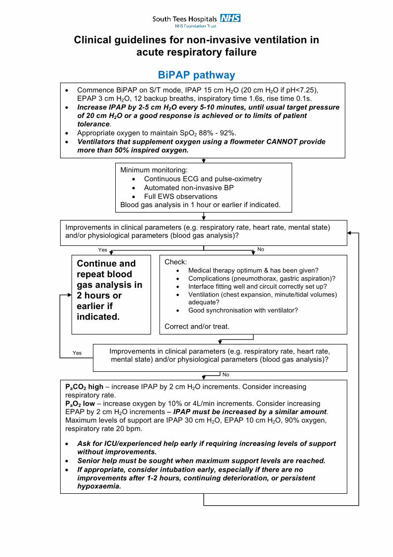
BiPAP pathway
• Commence BiPAP on S/T mode, IPAP 15 cm H2O (20 cm H2O if pH<7.25), EPAP 3 cm H2O, 12 backup breaths, inspiratory time 1.6s, rise time 0.1s.
• Increase IPAP by 2-5 cm H2O every 5-10 minutes, until usual target pressure of 20 cm H2O or a good response is achieved or to limits of patient tolerance.
• Appropriate oxygen to maintain SpO2 88% - 92%. • Ventilators that supplement oxygen using a flowmeter CANNOT provide
more than 50% inspired oxygen.
Minimum monitoring: • Continuous ECG and pulse-oximetry • Automated non-invasive BP • Full EWS observations
Blood gas analysis in 1 hour or earlier if indicated.
Improvements in clinical parameters (e.g. respiratory rate, heart rate, mental state) and/or physiological parameters (blood gas analysis)?
Continue and repeat blood gas analysis in 2 hours or earlier if indicated.
Check: • Medical therapy optimum & has been given? • Complications (pneumothorax, gastric aspiration)? • Interface fitting well and circuit correctly set up? • Ventilation (chest expansion, minute/tidal volumes)
adequate? • Good synchronisation with ventilator?
Correct and/or treat.
Improvements in clinical parameters (e.g. respiratory rate, heart rate, mental state) and/or physiological parameters (blood gas analysis)?
PaCO2 high – increase IPAP by 2 cm H2O increments. Consider increasing respiratory rate. PaO2 low – increase oxygen by 10% or 4L/min increments. Consider increasing EPAP by 2 cm H2O increments – IPAP must be increased by a similar amount. Maximum levels of support are IPAP 30 cm H2O, EPAP 10 cm H2O, 90% oxygen, respiratory rate 20 bpm. • Ask for ICU/experienced help early if requiring increasing levels of support
without improvements. • Senior help must be sought when maximum support levels are reached. • If appropriate, consider intubation early, especially if there are no
improvements after 1-2 hours, continuing deterioration, or persistent hypoxaemia.
Yes No
Yes
No
Clinical guidelines for non-invasive ventilation in acute respiratory failure
CPAP Weaning Pathway
Pre-conditions: • Primary illness treated or under medical control. • Targets in clinical and physiological parameters (respiratory rate, heart rate,
mental state, blood gas parameters) achieved and stable for ≥ 12 hours. • CPAP ≤ 10 cm H2O pressure/CPAP valve.
Reduce CPAP by 2.5 cm H2O every 4-6 hourly. Minimum CPAP = 5 cm H2O pressure/CPAP valve.
Stable clinical and physiological signs/parameters?
CPAP = 5 cm H2O pressure/CPAP valve for 4-6 hours?
Stable clinical and physiological signs/parameters?
Controlled oxygen via face mask to maintain SpO2 > 92% (88% - 92% in patients known to be sensitive to oxygen).
Stable clinical and physiological signs/parameters for 4-6 hours?
Continue with medical treatment and controlled oxygen.
Increase CPAP by 2.5 – 5 cm H2O pressure/CPAP valve and review pre-conditions.
Restart NIV (see NIV pathway) and review pre-conditions.
Yes
No
Yes
Yes
Yes
No
No
No
Notes: • Some patients may be suitable for weaning even when CPAP > 10 cm H2O – discuss
with experienced doctor/ICU. • The rate of weaning in this pathway is a guide only – some patients can be weaned
faster, others more slowly. • The weaning process may be interrupted overnight in order to promote rest. • Some patients (e.g. with post-operative lung collapse) may continue to require CPAP
support overnight in order to overcome nocturnal hypoventilation. • The PaO2 should not be used as the sole weaning parameter with the Boussignac
system as the delivered oxygen is uncontrolled and typically in excess of 60%. • This pathway is not suitable for patients for whom NIV is being withdrawn and
palliative-only management started.
Clinical guidelines for non-invasive ventilation in acute respiratory failure
BiPAP Weaning Pathway
Pre-conditions: • Primary illness treated or under medical control. • Targets in clinical and physiological parameters (respiratory rate, heart rate,
mental state, blood gas parameters) achieved and stable for ≥ 12 hours. • IPAP ≤ 18 cm H2O, EPAP ≤ 8 cm H2O, total respiratory rate ≤ 24 bpm.
Reduce IPAP by 2 cm H2O and EPAP by 2 cm H2O every 4–6 hourly. Reduce rate to 12 bpm. Minimum IPAP = 12 cm H2O, EPAP = 4 cm H2O.
Stable clinical and physiological signs/parameters?
IPAP = 12 cm H2O and EPAP = 4 cm H2O for 4-6 hours?
Stable clinical and physiological signs/parameters?
Controlled oxygen via face mask to maintain SpO2 > 92% (88% - 92% in patients known to be sensitive to oxygen).
Stable clinical and physiological signs/parameters for 4-6 hours?
Continue with medical treatment and controlled oxygen.
Restart NIV (see NIV pathway) and review pre-conditions.
Increase IPAP by 4 cm H2O and/or EPAP by 2 cm H2O and review pre-conditions. Consider CPAP.
Yes
Yes
Yes
Yes
No
No
No
No
Notes: • The rate of weaning in this pathway is a guide only – some patients can be weaned
faster, others more slowly. • The weaning process may be interrupted overnight in order to promote rest. • Some patients may be weaned from BiPAP onto CPAP, before being weaned
completely off NIV. • Some patients (e.g. with post-operative lung collapse) may continue to require CPAP
support overnight in order to overcome nocturnal hypoventilation. • This pathway is not suitable for patients for whom NIV is being withdrawn and
palliative-only management started.
Clinical guidelines for non-invasive ventilation in acute respiratory failure
References.
1. Ambrosino N, et al (1995). Non-invasive mechanical ventilation in acute
respiratory failure due to chronic obstructive airways disease: correlates
for success. Thorax 50:755-757.
2. Antonelli M, et al (1998). A comparison of non-invasive positive pressure
ventilation and conventional mechanical ventilation in patients with acute
respiratory failure. N Engl J Med 339:429-435.
3. Auriant I, et al (2001). Noninvasive ventilation reduces mortality in acute
respiratory failure following lung resection. Am J Respir Crit Care Med
164:1231-1235.
4. Bersten AD, et al (1991). Treatment of severe cardiogenic pulmonary
edema with continuous positive airway pressure delivered by face mask.
N Engl J Med 325:1825-1830.
5. Bollinger CT, Van Eeden SF (1990). Treatment of multiple rib fractures.
Randomized controlled trial comparing ventilatory with nonventilatory
management. Chest 104:943-948.
6. Bott J et al (1993). Randomised controlled trial of nasal ventilation in
acute ventilatory failure due to chronic obstructive airways disease.
Lancet 341:1555-1557.
7. Bourke SC, Tomlinson M, Williams TL, et al (2006). Effects of non-
invasive ventilation on survival and quality of life in patients with
amyotrophic lateral sclerosis: a randomised controlled trial. Lancet
Neurol 5:140-7.
8. British Thoracic Society/Intensive Care Society (BTS/ICS) Acute
Hypercapnic Respiratory Failure Guideline Development Group (2016).
BTS/ICS Guidelines for the Ventilatory Management of Acute
Hypercapnic Respiratory Failure in Adults. Thorax 71:ii1-ii35.
9. British Thoracic Society/Scottish Intercollegiate Guidelines Network
(BTS/SIGN) (2005). British guideline on the management of asthma: a
national clinical guideline. Revised edition November 2005.
Clinical guidelines for non-invasive ventilation in acute respiratory failure
and standard treatment in emergency department patients with acute
cardiogenic pulmonary oedema. Emerg Med J 21:155-161.
17. Elliott MW, et al (1990). Non-invasive mechanical ventilation for acute
respiratory failure. BMJ 300:358-360.
18. Esteban A, et al (2004). Noninvasive positive pressure ventilation for
respiratory failure after extubation. N Engl J Med 350:2452-2460.
19. Ferrer M, et al (2003). Noninvasive ventilation in severe hypoxaemic
respiratory failure: a randomized clinical trial. Am J Respir Crit Care Med
168:1438-1444.
20. Garpestad E, Hill N (2005). Noninvasive ventilation for acute respiratory
failure. But how severe? Chest 128:3790-3791.
Clinical guidelines for non-invasive ventilation in acute respiratory failure
21. Gray A, Goodacre S, et al, on behalf of the 3CPO Trialist (2008). Non-
invasive ventilation in acute pulmonary edema. NEJM 359:142-151.
22. Hilbert G, et al (1998). Noninvasive pressure support ventilation in
COPD patients with postextubation hypercapnic respiratory
insufficiency. Eur Resp J 11:1349-1353.
23. Hilbert G, et al (2001). Noninvasive ventilation in immunosuppressed
patients with pulmonary infiltrates, fever and acute respiratory failure. N
Engl J Med 344:481-487.
24. Honrubia T et al (2005). Noninvasive vs conventional mechanical
ventilation in acute respiratory failure. A multicenter, randomized
controlled trial. Chest 128:3916-3924.
25. Hurst JM, et al (1985). Use of CPAP mask as the sole mode of ventilatory
support I trauma patients with mild to moderate respiratory insufficiency.
J Trauma 25:1065-1068.
26. Keenan SP, et al (2002). Noninvasive positive-pressure ventilation for
postextubation respiratory distress: A randomized controlled trial. JAMA
287:3238-3244.
27. Keenan SP, et al (2004). Does non-invasive positive pressure ventilation
improve outcome in acute hypoxaemic respiratory failure? A systematic
review. Crit Care Med 32:2516-2523.
28. Kilger E, et al (1999). Effects of non-invasive positive pressure
ventilatory support in non-COPD patients with acute respiratory
insufficiency after early extubation. Intensive Care Med 25:1374-1380.
29. Kramer N, et al (1995). Randomized, prospective trial of non-invasive
positive pressure ventilation in acute respiratory failure. Am J Respir Crit
Care Med 151:1799-1806.
30. Lin M, et al (1995). Reappraisal of CPAP therapy in acute pulmonary
edema: short-term results and long-term follow up. Chest 107:1379-
1386.
31. Meduri GU, et al (1996). Noninvasive positive pressure ventilation in
status asthmaticus. Chest 110:767-774.
Clinical guidelines for non-invasive ventilation in acute respiratory failure
32. Mehta S, et al (1997). Randomized prospective trial of bilevel versus
continuous positive airway pressure in acute pulmonary oedema. Crit
Care Med 25:620-628).
33. Mehta S, Hill NS (2001). Noninvasive ventilation. Am J Respir Crit Care
Med 163:540-577.
34. Miro AM, Shivaram U, Hertig I (1993). Continuous positive airway
pressure in COPD patients in acute hypercapnic respiratory failure.
Chest 103:266-268.
35. Nava S, et al (1998). Noninvasive mechanical ventilation in the weaning
of patients with respiratory failure due to chronic obstructive pulmonary
disease. A randomized controlled trial. Ann Intern Med 128:721-728.
36. Peter JV, et al (2006). Effect of non-invasive positive pressure ventilation
(NIPPV) on mortality in patients with acute cardiogenic pulmonary
oedema: a meta-analysis. Lancet 367:1155-1163.
37. Pitkin AD, et al (1994). Arterialised earlobe blood gas analysis: an
underused technique. Thorax 49:364-366.
38. Plant PK, Owen JL, Elliot MW (2000). Early use of non-invasive
ventilation for acute exacerbations of chronic obstructive pulmonary
disease on general respiratory wards: a multicentre randomised
controlled trial. Lancet 355:1931-1935.
39. Ram FSF, Picot J, et al (2004). Nin-invasive positive pressure ventilation
for treatment of respiratory failure due to exacerbations of chronic
obstructive pulmonary disease. Cochrane database of systematic
reviews 2004 issue 3.
40. Rusterholtz T, et al (1999). Non-invasive pressure support ventilation
(NIPSV) with face mask in patients with acute cardiogenic pulmonary
edema (ACPE). Intensive Care Med 25:15-20.
41. Soroksky A, et al (2003). A pilot prospective, randomized, placebo-
controlled trial of bilevel positive airway pressure in acute asthma attack.
Chest 123:1018-1025.
Clinical guidelines for non-invasive ventilation in acute respiratory failure
42. Shee CD & Green M (2003). Non-invasive ventilation and palliation:
experience in a district general hospital and a review. Palliative Medicine
17(1):21-26.
43. Squadrone V, et al (2005). Continuous positive airway pressure for
treatment of postoperative hypoxaemia: a randomized controlled trial.
JAMA 293:589-595.
44. Wysocki M, et al (1995). Noninvasive pressure support ventilation in
patients with acute respiratory failure. Chest 107:761-768.
The NIV group comprises: Dr. I Gonzalez, Consultant, Critical Care Services Dr. Sathyamurthy, Consultant, Respiratory Medicine Dr. A Gemmill, Consultant, Accident & Emergencies Dr. S Chay, Associate Specialist, Critical Care Services Sister D Hodgson, Senior Sister, Neurosurgical HDU, JCUH Sister M Tiernan, Senior Clinical Educator, Institute of Learning, Research & Innovation Mr P Howard, Senior Physiotherapist, Critical Care Services