AUDIOLOGY SERVICES A HANDBOOK OF CLINICAL PRACTICE EASTERN VIRGINIA MEDICAL SCHOOL Department of Otolaryngology-Head and Neck Surgery Division of Audiology Sentara Norfolk General Hospital, River Pavilion 600 Gresham Drive, Suite 1100 Norfolk, Virginia 23507 (757) 388-6200 (757) 388-6201 (fax) Nathan Michalak, Au.D., CCC-A Director of Audiology EVMS Hearing and Balance Center
Transcript
AUDIOLOGY SERVICES
A HANDBOOK OF CLINICAL PRACTICE
EASTERN VIRGINIA MEDICAL SCHOOL Department of Otolaryngology-Head and Neck Surgery
Division of Audiology Sentara Norfolk General Hospital, River Pavilion
600 Gresham Drive, Suite 1100 Norfolk, Virginia 23507
(757) 388-6200 (757) 388-6201 (fax)
Nathan Michalak, Au.D., CCC-A Director of Audiology EVMS Hearing and Balance Center
HEARING AND BALANCE CENTER – DIVISION OF AUDIOLOGY
Nathan Michalak, Au.D., CCC-A (Director of Audiology) Ashley Wampler, Ph.D., CCC-A Melissa Pola, M.S., CCC-A Sara Gill, Au.D., CCC-A Anne Davis, Au.D., CCC-A Michael LeMay, M.A., CCC-A George Whitaker, Au.D. CCC-A Bridget Block, Au.D. CCC-A
The Division of Audiology provides comprehensive diagnostic and rehabilitative services for adult and pediatric patients with hearing loss, tinnitus, and balance abnormalities. A full array of auditory communication, tinnitus, and balance assessment is provided, along with rehabilitative treatment methods to include: hearing aids, cochlear implants, baha, tinnitus maskers, and aural rehabilitation.
The basic hearing test or audiogram tests one's ability to hear pure tones in each ear. Best results are obtained by a trained audiologist in a special soundproof testing booth. Simple tests, such as ones done in many schools, may be useful for screening, but a careful audiogram is necessary for accurate diagnosis of most hearing problems.
A complete audiogram will test both the bone conduction (the ability to hear a sound when it transmitted through bone) and the air conduction (the ability to hear a sound when it transmitted through air). A comparison between these two type of conduction can be very useful in localizing which part of the hearing mechanism is responsible for the loss. In particular, the test is useful in determining if the loss is due to a problems with the portion of the middle ear that conducts sound from the ear canal to the inner ear (in which case it would be called a "conductive" hearing loss) or if it is due to the inner ear or the nerve that conducts the sound signals to the brain (in which case it would be called a "sensorineural" hearing loss).
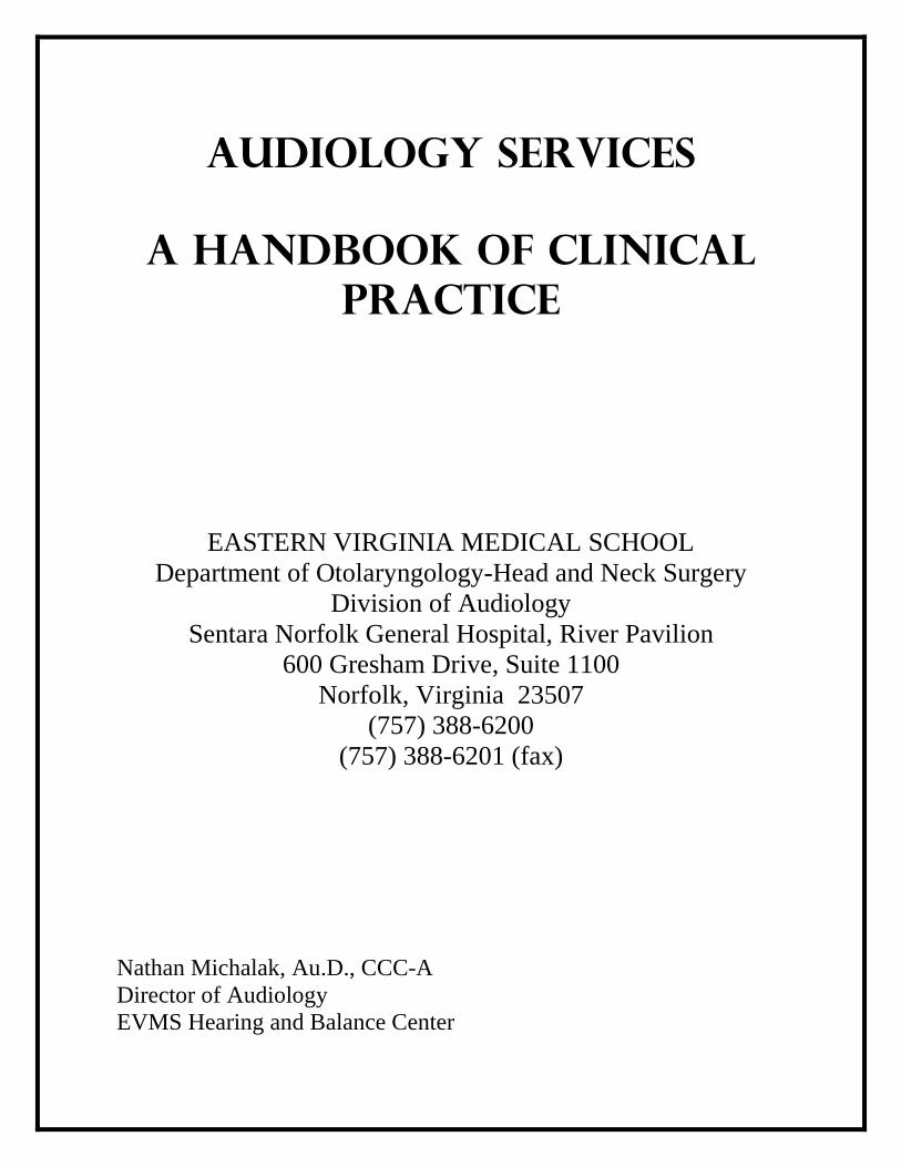
The results of audiograms are most often displayed in graph form. This graph shows the amount of hearing loss expressed in units called decibels at different sound frequencies (also called Hertz). High frequencies correspond to high tones, and low frequencies are low tones. Most audiograms go from around 250 hertz to 4000 hertz. A loss up to 20 decibels on this graph is considered "normal". Hearing losses over 20 decibels are considered abnormal.
The average pure tone thresholds (PTA) of the audiometric frequencies of 500, 1000, and 2000 Hz, is useful in predicting thresholds for speech as well as establishing the degree of handicap imposed by a hearing loss. Scale of Hearing Impairment based on the PTA at 500, 1000, and 2000 Hz is a function of several variables including age: PTA Degree of Handicap ≤ 15 dB HL None 16 – 25 dB HL Mild 26 – 40 dB HL Mild-to-Moderate 41 – 65 dB HL Moderate 66 – 90 dB HL Severe ≥ 91 dB HL Profound
In the audiogram to the left, the red tracings are for the right ear, and the black tracings are for the left. The round circles represent the air conduction and the brackets are the bone conduction. Low frequencies are to the left and high frequencies are on the right. A normal audiogram would show all hearing loss of 20 dB or less. (The measurements would all be above 20 dB. This audiogram shows a normal tracing on the left, and a sensorineural (nerve) hearing loss on the right
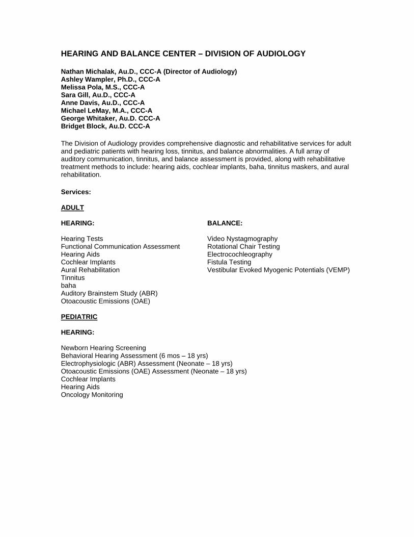
In this audiogram there is a sensorineural hearing loss on both sides in the high frequencies.
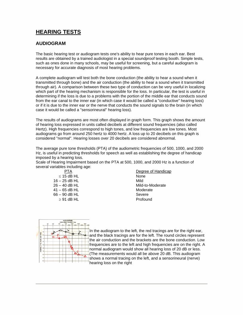
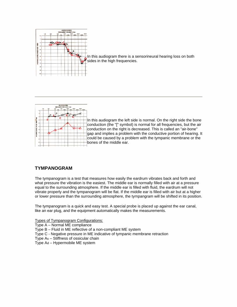
In this audiogram the left side is normal. On the right side the bone conduction (the "[" symbol) is normal for all frequencies, but the air conduction on the right is decreased. This is called an "air-bone" gap and implies a problem with the conductive portion of hearing. It could be caused by a problem with the tympanic membrane or the bones of the middle ear.
TYMPANOGRAM
The tympanogram is a test that measures how easily the eardrum vibrates back and forth and what pressure the vibration is the easiest. The middle ear is normally filled with air at a pressure equal to the surrounding atmosphere. If the middle ear is filled with fluid, the eardrum will not vibrate properly and the tympanogram will be flat. If the middle ear is filled with air but at a higher or lower pressure than the surrounding atmosphere, the tympangram will be shifted in its position.
The tympanogram is a quick and easy test. A special probe is placed up against the ear canal, like an ear plug, and the equipment automatically makes the measurements.
Types of Tympanogram Configurations: Type A – Normal ME compliance Type B – Fluid in ME reflective of a non-compliant ME system Type C - Negative pressure in ME indicative of tympanic membrane retraction Type As – Stiffness of ossicular chain Type Ad – Hypermobile ME system
AUDITORY BRAINSTEM RESPONSE (ABR)
The ABR is a method employed to assess the functions of the ears, cranial nerves, and various brain functions of the lower part of the auditory system. The procedure is to generate a brief click or tone pip from an earphone or headphone and measuring the elicited neuronal action potentials by surface electrodes, typically placed at the vertex of the scalp and ear lobes. The potential of the signal in microvoltage is averaged and charted against the time (millisecond), and is a variation of the electroencephalogram (EEG)
ABR audiometry is a safe and painless test of auditory pathway and brainstem function in response to auditory or (click) stimuli. The procedure was first described by Jewett and Williston in 1971. ABR audiometry is the most common application of auditory evoked responses. There are typically five identifiable waves numbered (I-V); however, only waves I, III, and V are prominent enough to have significant diagnostic value. The physiologic origin of these waveforms is as follows:
• Wave I – Distal Auditory Nerve • Wave II – Proximal Auditory Nerve • Wave III – Cochlear Nucleus • Wave IV – Lateral Lemniscus • Wave V – Inferior Colliculus
USEFULLNESS OF THE DIAGNOSTIC ABR
Auditory brainstem response (ABR) audiometry is considered an effective screening tool in the evaluation of suspected retrocochlear pathology such as an acoustic neuroma or vestibular schwannoma, as well as auditory threshold estimation in infants/children and difficult to test patients.
SYMPTOMS OF VIII NERVE PATHOLOGY
Clinical symptoms may include but are not limited to the following:
• Asymmetrical or unilateral sensorineural hearing loss • Asymmetrical high-frequency hearing loss • Unilateral tinnitus • Unilaterally or bilaterally poor word recognition scores as compared with degree of
sensorineural hearing loss • Perceived distortion of sounds when peripheral hearing is essentially normal
AUDITORY BRAINSTEM EVALUATION
In addition to retrocochlear pathologies, many factors may influence ABR results, including the degree of sensorineural hearing loss, asymmetry of hearing loss, test parameters, and other patient factors. These influences must be factored in when performing and analyzing an ABR result.
Findings suggestive of retrocochlear pathology may include any 1 or more of the following:
• Absolute latency interaural difference wave V (IT5) - Prolonged • I-V interpeak interval interaural difference - Prolonged • Absolute latency of wave V - Prolonged as compared with normative data • Absolute latencies and interpeak intervals latencies I-III, I-V, III-V - Prolonged as
compared with normative data • Absent auditory brainstem response in the involved ear
In general, ABR exhibits a sensitivity of over 90% and a specificity of approximately 70-90%.
Sensitivity for small tumors is not as high. For this reason, a symptomatic patient with a normal ABR result should receive a follow-up audiogram in 6 months to monitor for any changes in hearing sensitivity or tinnitus. The ABR may be repeated if indicated. Alternatively, MRI with gadolinium enhancement, which has become the new criterion standard, can be used to identify very small (3-mm) vestibular schwannomas.
PEDIATRIC HEARING TESTS
Behavioral audiologic evaluation measures for children (6 months corrected age and older) include:
VISUAL REINFORCMENT AUDIOMETRY
The VRA technique typically utilizes a light or animated toy placed at a 45-90 degree angle to one side of the infant, which is flashed on and off to reinforce a head turn response to the sound stimulus. The sound stimuli may consist of frequency-modulated pure tones, speech or narrow-band noise. During training trials, the audiologist will simultaneously present the sound stimuli concurrently with the visual reinforcer. Once the baby has become familiar with the paired presentations, the sound stimulus will be presented in isolation. When the baby’s head turn response occurs, the visual reinforcement is activated. Stimulus intensity can then be decreased in order to determine the softest level where a head turn response can be obtained. The testing can be then be repeated using a variety of frequencies.
CONDITIONED ORIENTING RESPONSE AUDIOMETRY
COR differs from VRA in that two separate visual reinforcers are used, however, at EVMS these terms are largely used synonymously. In using COR, reinforcers are placed 45-90 degrees to either side of the infant’s head with sound stimuli delivered either through matched soundfield speakers or via insert earphones. The task is for the infant to hear the stimulus and be able to localize to the source of the stimulus. Once the child correctly turns their head toward the sound source, the visual stimulus is presented on that side to reinforce their correct response.
CONDITIONED PLAY AUDIOMETRY
Children in the age range between 24 months and 5 years can undergo assessment of their hearing status through the use of conditioned play audiometry. Using this technique, the child is engaged in a game like activity whereby they are asked to perform a specific task (e.g., dropping a block in a bucket or putting a piece in a puzzle, etc.) every time a sound is heard. Sound stimuli can be presented via speakers in soundfield, through earphones, or through the use of a bone conduction oscillator.
Biometric and Electrophysiologic evaluation for children (This incorporates all neonates, infants, and children who cannot perform behavioral testing under headphones for ear specific information):
The ABR can also be used to approximate auditory thresholds. This is accomplished by presenting a click which consists of essentially all frequencies combined into a single stimulus (similar to white light). This encourages neural synchrony by stimulating multiple frequencies simultaneously in temporal succession. An ABR waveform is elicited at supra-threshold levels followed by a reduction in intensity until the ABR waveform dissipates into a flat line. Waveform V is the most robust of the ABR waveforms, therefore, this wave acts as the marker of interest while decreasing intensity and subsequently the amplitude of the waveform. The intensity level preceding complete dissipation of the ABR waveform is considered to be the electrophysiologic threshold. This is essentially a test of auditory nerve function; however, good correlation has been demonstrated with behavioral thresholds in the sound booth under headphones.
As a result of the resonant characteristics of the ear canal, an emphasis between 1,500-4,000 Hz is attributed to the click ABR. For more frequency specific testing, quick tone bursts can be utilized to generate ABR waveforms at more frequency specific locations. This is helpful in identifying particular slopes and patterns of hearing loss. This information is especially critical when programming hearing aids for children.
AUDITORY STEADY STATE RESPONSE
A new variation of the ABR which allows for multiple frequency threshold testing is the Auditory Steady State Response (ASSR). This is an auditory evoked potential elicited through the use of modulated pure tones which can be used to predict hearing sensitivity of individuals of all ages. ASSR technology is particularly sensitive in differentiating between severe to profound hearing loss. Additionally, in some versions, both ears can be presented with up to four frequencies at the same time, making it easier to acquire a larger amount of auditory information simultaneously.
OTOACOUSTIC EMISSIONS
The primary purpose of otoacoustic emission (OAE) tests is to determine cochlear status, specifically hair cell function. This information can be used to (1) screen hearing (particularly in neonates, infants, or individuals with developmental disabilities), (2) partially estimate hearing sensitivity within a limited range, (3) differentiate between the sensory and neural components of sensorineural hearing loss, and (4) test for functional (feigned) hearing loss. The information can be obtained from patients who are sleeping or even comatose because no behavioral response is required.
The normal cochlea does not just receive sound; it also produces low-intensity sounds called OAEs. These sounds are produced specifically by the cochlea and, most probably, by the cochlear outer hair cells as they expand and contract. The presence of cochlear emissions was hypothesized in the 1940s on the basis of mathematical models of cochlear nonlinearity.
However, OAEs could not be measured until the late 1970s, when technology created the extremely sensitive low-noise microphones needed to record these responses.
The 4 types of otoacoustic emissions are as follows:
• Spontaneous otoacoustic emissions (SOAEs) - Sounds emitted without an acoustic stimulus (ie, spontaneously)
• Transient otoacoustic emissions (TOAEs) or transient evoked otoacoustic emissions (TEOAEs) - Sounds emitted in response to an acoustic stimuli of very short duration; usually clicks but can be tone-bursts
• Distortion product otoacoustic emissions (DPOAEs) - Sounds emitted in response to 2 simultaneous tones of different frequencies
• Sustained-frequency otoacoustic emissions (SFOAEs) - Sounds emitted in response to a continuous tone
OAEs measure only the peripheral auditory system, which includes the outer ear, middle ear, and cochlea. The response only emanates from the cochlea, but the outer and middle ear must be able to transmit the emitted sound back to the recording microphone. OAE testing often is used as a screening tool to determine the presence or absence of cochlear function, although analysis can be performed for individual cochlear frequency regions. OAEs cannot be used to fully describe an individual's auditory thresholds, but they can help question or validate other threshold measures (eg, in suspected functional [feigned] hearing loss), or they can provide information about the site of the lesion.
Using current technology, most researchers and clinicians find a correlation between frequency-specific analysis of TOAEs/DPOAEs and cochlear hearing loss. However, at this juncture, the correlation cannot fully describe auditory threshold.
OAE ANATOMY AND PHYSIOLOGY
When sound is used to elicit an emission, it is transmitted through the outer ear, where the auditory stimulus is converted from an acoustic signal to a mechanical signal at the tympanic membrane and is transmitted through the middle ear ossicles; the stapes footplate moves at the oval window, causing a traveling wave in the fluid-filled cochlea. The cochlear fluid's traveling wave moves the basilar membrane; each portion of the basilar membrane is maximally sensitive to only a limited frequency range. The arrangement is a tonotopic gradient. Regions closest to the oval window are more sensitive to high-frequency stimuli. Regions further away are most sensitive to lower-frequency stimuli. Therefore, for OAEs, the first responses returned and recorded by the probe microphone emanate from the highest-frequency cochlear regions because the travel distance is shorter. Responses from the lower-frequency regions, closer to the cochlear apex; arrive later.
When the basilar membrane moves, the hair cells are set into motion and an electromechanical response is elicited, while an afferent signal is transmitted and an efferent signal is emitted. The efferent signal is transmitted back through the auditory pathway, and the signal is measured in the outer ear canal. As described above, the responses from the high-frequency region arrive first, progressively followed by responses from lower-frequency regions. Outer hair cells are
located in the organ of Corti on the basilar membrane. These hair cells are motile; an electrochemical response elicits a motoric response. The 3 rows of outer hair cells have stereocilia arranged in a W formation. The stereocilia are linked to each other and, therefore, move as a unit. These are the outer hair cells believed to underlie OAE generation.
DISTORTION PRODUCT OTOACOUSTIC EMISSIONS
The relative merits of TOAEs and DPOAEs are widely discussed. EVMS uses DPOAE protocols primarily for the reason that greater frequency specificity and can be used to record at higher frequencies than TOAEs. Therefore, DPOAEs may be as particularly useful for early detection of cochlear damage as they are for ototoxicity and noise-induced damage.
Depending on the methodology employed, DPOAEs often can be recorded in individuals with mild-to-moderate hearing losses for whom TOAEs are absent; however, the accuracy of DPOAEs in estimating actual hearing sensitivity is not fully resolved (research continues in this area). DPOAEs frequently correspond to the audiometric configuration of a cochlear hearing loss, which is helpful in some patients.
AUDIOLOGIC TREATMENT
FUNCTIONAL COMMUNICATION ASSESSMENT (FCA)
The EVMS FCA successfully identifies and treats various communication difficulties using a series of innovative speech tests and treatment methods. Approximately 1 in 6 Americans have some degree of auditory communication dysfunction. The intent of this program is to treat these patients to prevent or reduce emotional turmoil, anxiety, depression, and social isolationism. Using the obtained test information, a Customized Communication Enhancement Plan will be created to improve communication and quality of life. A series of clinically proven recommendations will be provided for those who are interested in enhancing their communication performance. Given the communication impairment(s) identified through the FCA evaluation, some or all of the the following recommendations are provided to facilitate maximum communication enhancement:
1. Aural Rehabilitation Class - This is a class that meets once per month, which can be attended multiple times if desired. Friends and family members are also welcome. This class will address how to understand hearing and hearing loss; how hearing loss affects communication; what some of the negative social and psychological consequences can be; and address the best methods and strategies to enhance speech understanding.
2. Hearing Aids - When a hearing loss is present, the easiest and most effective way to correct a communication difficulty is by making all parts of speech audible. This makes the fine points of speech more discernable for increased clarity and understanding. The audiologist will select the most appropriate device based on functional communication testing, degree of hearing loss, and individualized needs.
3. Auditory Training - The goal of auditory training is to help discriminate specific sounds in order to improve speech understanding. This is a process by which the patient is reintroduced to how to hear and understand speech through the use of hearing aids.
Using hearing aids will make speech sound different from what the patient is used to (this is a good thing). Good speech understanding requires very fine discriminations of pitch, loudness, and timing. An example of fine discrimination is the difference between the sound "s" like the first sound in "sun" and "f" like the first sound in "fun." A CD based auditory training program with the use of hearing aids will initiate increased benefit and generate faster progress than with hearing aids alone.
4. Alternative Listening Device(s) - At times additional help is needed for specific situations. Alternative Listening Devices (ALDs) are designed to assist listening and/or communication in select environments. An example of an ALD might include: amplified (telephone, alarm clock, smoke alarm), FM system, cell phone adaptor, iPod/Mp3 adaptor, television headset, etc.
5. Neuropsychological Referral - As the brain ages it is often less effective at processing complex or competing stimuli. This may be compounded by a hearing loss which demands even greater cognitive resources to make sense of the degraded speech sounds. It is also possible through our functional communication assessment to identify auditory cognitive impairments that are separate from hearing loss. Given the findings from todays test results, it is possible that a greater cognitive issue may be a factor regarding poor communication. In this situation, a referral to the Neuropsychology Center is warranted: Michael L. Stutts, Ph.D. Clinical Psychologist and Professor Co-Director, Department of Psychiatry and Behavioral Sciences Eastern Virginia Medical School Hofheimer Hall, Suite 564 825 Fairfax Avenue Norfolk, VA 23507 Phone: 757-446-8400 Fax: 757-446-8401
6. Outtake Measures - Depending on the specific areas of difficulty, a variety of outtake measures will be used to assess progress. This will take place at varying points during treatment and will involve concise methods to sample communication enhancement. The greatest measure, however, is the amount of benefit perceived in real world day to day experiences.
HEARING AIDS AND DEVICES FOR HEARING LOSS
When a hearing loss is present, the easiest and most effective way to correct a communication difficulty is by making all parts of speech audible. This makes the fine points of speech more discernable for increased clarity and understanding. The audiologist will select the most appropriate device based on functional communication testing, degree of hearing loss, and individual needs. EVMS uses a wide range of hearing aid manufacturers to provide patients with the highest quality and most recent digital technology available.
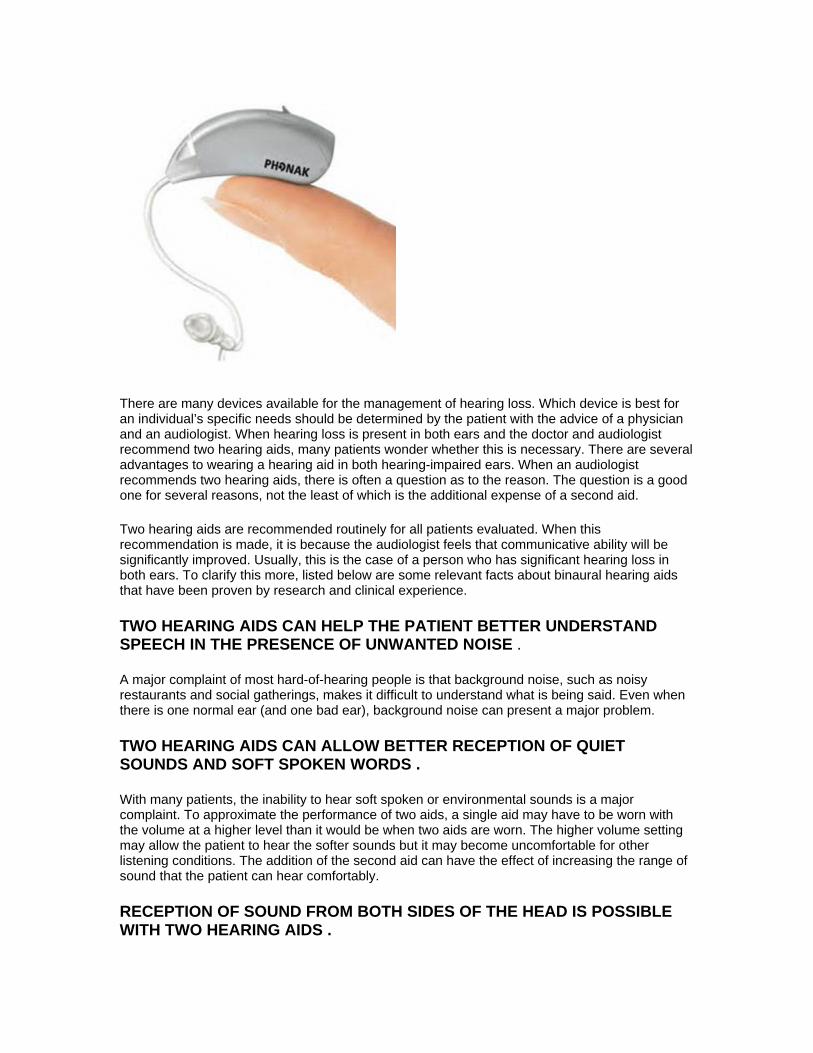
There are many devices available for the management of hearing loss. Which device is best for an individual’s specific needs should be determined by the patient with the advice of a physician and an audiologist. When hearing loss is present in both ears and the doctor and audiologist recommend two hearing aids, many patients wonder whether this is necessary. There are several advantages to wearing a hearing aid in both hearing-impaired ears. When an audiologist recommends two hearing aids, there is often a question as to the reason. The question is a good one for several reasons, not the least of which is the additional expense of a second aid.
Two hearing aids are recommended routinely for all patients evaluated. When this recommendation is made, it is because the audiologist feels that communicative ability will be significantly improved. Usually, this is the case of a person who has significant hearing loss in both ears. To clarify this more, listed below are some relevant facts about binaural hearing aids that have been proven by research and clinical experience.
TWO HEARING AIDS CAN HELP THE PATIENT BETTER UNDERSTAND SPEECH IN THE PRESENCE OF UNWANTED NOISE .
A major complaint of most hard-of-hearing people is that background noise, such as noisy restaurants and social gatherings, makes it difficult to understand what is being said. Even when there is one normal ear (and one bad ear), background noise can present a major problem.
TWO HEARING AIDS CAN ALLOW BETTER RECEPTION OF QUIET SOUNDS AND SOFT SPOKEN WORDS .
With many patients, the inability to hear soft spoken or environmental sounds is a major complaint. To approximate the performance of two aids, a single aid may have to be worn with the volume at a higher level than it would be when two aids are worn. The higher volume setting may allow the patient to hear the softer sounds but it may become uncomfortable for other listening conditions. The addition of the second aid can have the effect of increasing the range of sound that the patient can hear comfortably.
RECEPTION OF SOUND FROM BOTH SIDES OF THE HEAD IS POSSIBLE WITH TWO HEARING AIDS .
The addition of a second hearing aid reduces the need for rotating the head around to face the speaker, making communication easier and more comfortable.
THE ABILITY TO LOCATE THE SOURCE OF A SOUND IS IMPROVED WITH TWO HEARING AIDS.
The ability to identify the direction of a sound source allows a person to react more appropriately to his environment.
Other hearing assistive devices:
• Telephone amplifiers for both land lines and cell phones • Infrared systems for TV and theaters • FM systems • Devices for monitoring of smoke detectors, telephone and door bells, and alarms • Implantable hearing aids • Bone anchored hearing aids
COCHLEAR IMPLANTS
Cochlear implantation is a life-changing event to many patients with hearing losses that are not effectively managed with well-fit hearing aids. With the experience most implant users are able to understand spoken speech in the everyday world. Some patients are able to enjoy music and/or hold conversations on the phone. The implant does not restore normal hearing, and many patients describe its sound as mechanical. However, with time and practice, the brain adapts to the new “sound” and speech signals and environmental sounds begin to sound more natural.The primary benefits of a cochlear implant are: (1) aid in lip reading; (2) perception of environment sounds; (3) aid in monitoring the volume of one’s own voice.
The ideal candidates for cochlear implantation are adults or children with recent hearing loss and young children whose hearing loss is identified very early. People with prior hearing experience adjust very well and often very quickly to hearing with a cochlear implant. They report significant satisfaction and improved quality of life. Deaf children implanted before the age of 5 also do very well. With time and rehabilitation, they develop very good speech understanding as well as speech production skills. The majority of these children are able to attend mainstream schools. Children who are implanted before the age of 2 seem to do even better than those implanted between 2 and 5 years old.
For adults and teens who have been deaf for most of their lives, were educated with manual language, and live in the deaf community, cochlear implantation has been very controversial. Not only are there medical issues, but more importantly social issues. Fortunately, the strong opposition to implantation from the deaf community is lessening. More and more prelingually deafened and long term deafened patients are seeking information on cochlear implants. For those people who are good lip-readers, who have some spoken language skills, and who are committed to learning and adapting to the use of the implant, cochlear implantation may provide significant benefit. Users like this report better communication with hearing family and friends, better use of environmental sounds, and improvement in quality of life.
This technology is continuing to improve thanks to the work of many scientists throughout the world. Current research includes improvement of speech processing technology, improving performance by implanting bilateral devices and preserving residual hearing, expanding the
eligibility requirements by looking at patients with more residual hearing and prelingual deafness, improving the design of the device to one day be 100% implantable, and improving the function of the inner ear by preventing injury to the sensitive inner ear structures with new medications and potentially gene therapy.
WHAT IS A COCHLEAR IMPLANT?
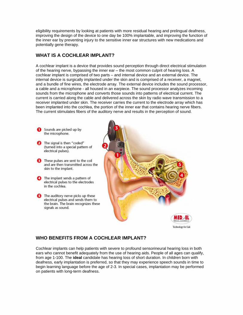
A cochlear implant is a device that provides sound perception through direct electrical stimulation of the hearing nerve, bypassing the inner ear – the most common culprit of hearing loss. A cochlear implant is comprised of two parts – and internal device and an external device. The internal device is surgically implanted under the skin and is comprised of a receiver, a magnet, and a bundle of fine wires, the electrode array. The external device includes the sound processor, a cable and a microphone - all housed in an earpiece. The sound processor analyzes incoming sounds from the microphone and converts those sounds into patterns of electrical current. The current is carried along the cable and delivered across the skin by radio wave transmission to a receiver implanted under skin. The receiver carries the current to the electrode array which has been implanted into the cochlea, the portion of the inner ear that contains hearing nerve fibers. The current stimulates fibers of the auditory nerve and results in the perception of sound.
WHO BENEFITS FROM A COCHLEAR IMPLANT?
Cochlear implants can help patients with severe to profound sensorineural hearing loss in both ears who cannot benefit adequately from the use of hearing aids. People of all ages can qualify, from age 1-100. The ideal candidate has hearing loss of short duration. In children born with deafness, early implantation is preferred, so that they may experience speech sounds in time to begin learning language before the age of 2-3. In special cases, implantation may be performed on patients with long-term deafness.
WHAT KINDS OF HEARING LOSSES ARE TREATED WITH A COCHLEAR IMPLANT?
The cochlear implant is designed for patients with sensorineural hearing loss that have failed conventional treatment including medications and hearing aids. Sensorineural hearing loss is a specific type of hearing loss, defined as any abnormality of the inner ear or auditory nerve that prevents transfer of electrical signals to the auditory nucleus (the brain’s center for hearing).
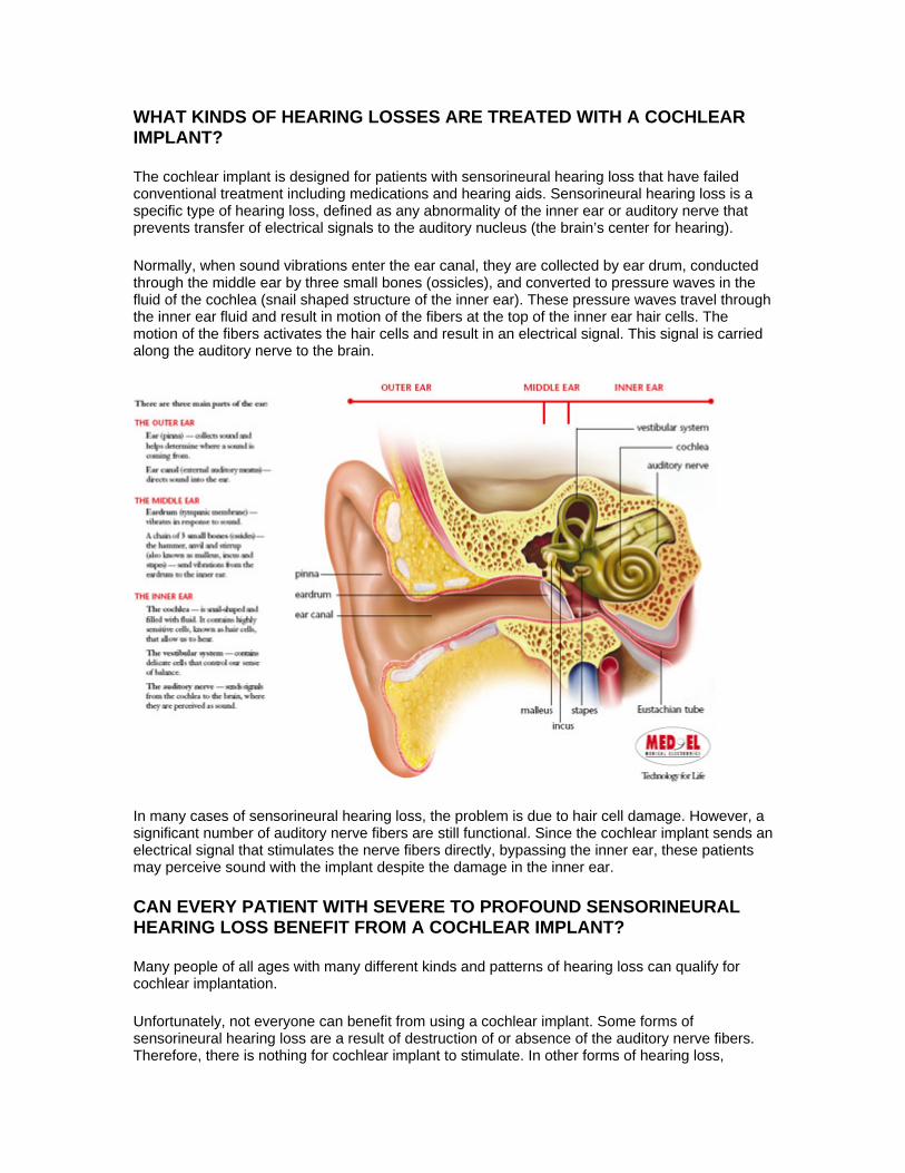
Normally, when sound vibrations enter the ear canal, they are collected by ear drum, conducted through the middle ear by three small bones (ossicles), and converted to pressure waves in the fluid of the cochlea (snail shaped structure of the inner ear). These pressure waves travel through the inner ear fluid and result in motion of the fibers at the top of the inner ear hair cells. The motion of the fibers activates the hair cells and result in an electrical signal. This signal is carried along the auditory nerve to the brain.
In many cases of sensorineural hearing loss, the problem is due to hair cell damage. However, a significant number of auditory nerve fibers are still functional. Since the cochlear implant sends an electrical signal that stimulates the nerve fibers directly, bypassing the inner ear, these patients may perceive sound with the implant despite the damage in the inner ear.
CAN EVERY PATIENT WITH SEVERE TO PROFOUND SENSORINEURAL HEARING LOSS BENEFIT FROM A COCHLEAR IMPLANT?
Many people of all ages with many different kinds and patterns of hearing loss can qualify for cochlear implantation.
Unfortunately, not everyone can benefit from using a cochlear implant. Some forms of sensorineural hearing loss are a result of destruction of or absence of the auditory nerve fibers. Therefore, there is nothing for cochlear implant to stimulate. In other forms of hearing loss,
structural abnormalities of the inner ear prevent surgical implantation. In many forms of sensorineural deafness, the status of the auditory nerve fibers is unknown. In such cases, it can be difficult to predict the potential benefit of receiving a cochlear implant. Currently, researchers are studying the role of auditory nerve fiber survival to better predict performance before implantation. The duration of hearing loss and the age at which it occurred also seem to influence performance with an implant.
HOW DO I KNOW IF I AM A CANDIDATE?
Candidacy for cochlear implantation is determined by a team of specialists which includes an Otologist (ear doctor), an audiologist, a speech therapist, an educational specialist, a social worker, and a psychologist. The Otologist will obtain a complete history, perform a thorough exam, and order appropriate testing. The audiologist will evaluate your hearing by performing an audiogram (hearing test) and will also determine the appropriateness of amplification use. Other team members will also meet with and perform evaluations when necessary. A review board then will evaluate this information to determine whether you are cochlear implant candidate.
WHAT IS INVOLVED IN HAVING A COCHLEAR IMPLANT OPERATION?
Cochlear implantation surgery is performed under general anesthesia and takes approximately one-two hours. An incision is made behind the ear and operating with the aid of a microscope, the surgeon drills away a portion of the mastoid bone (hard bone behind the ear) to gain access to the inner ear. A small opening is created into the cochlear. The electrode array is threaded into the cochlea and its outer end is attached to the bone of the skull. The incision is closed and the head bandaged. Most patients go home the next morning. Patients return the following week for suture removal.
WHEN CAN I BEGIN USING THE IMPLANT?
The patient returns at approximately one month following surgery to begin using the sound processor. The processor “hook-up” and programming is performed by the audiologist. The initial programming takes several hours. During this visit the audiologist and patient design a “MAP”. This MAP is the set of parameters of electrode stimulation that give the patient the best hearing. In the subsequent few months, there are a series of visits to the audiologist to test and adjust the signal levels for the electrodes in the array. These settings can be updated at each visit over a period of weeks or months, as the patient becomes more comfortable with experiencing sound again. At first, sounds may be quite different than remembered prior to the hearing loss. Environmental sounds are usually the first to be identified or relearned. With training and experience, the patient’s perception of speech improves.
ARE THERE ANY ALTERNATIVES TO A COCHLEAR IMPLANT?
Currently, cochlear implantation is limited to patients who cannot benefit significantly from using conventional hearing aids and other hearing assistive devices. Use of some combination of lip reading, cued speech, sign language, and powerful hearing aids are alternatives to implant surgery. An audiologist can direct you to sources for counseling and assistance that emphasize these alternatives.
WHAT ARE THE BENEFITS OF THE COCHLEAR IMPLANT?
The improvement in auditory perception (hearing) following cochlear implantation varies widely from patient to patient. Nearly all patients have some perception of sound. Even if this awareness is only the detection of the presence of absence of a sound in the environment, it can be
substantial aid to lip-reading. In most cases, implantees have some additional ability to discriminate pitch and loudness, which further enhances the benefit of the device. In some patients, the implant may even provide enough hearing to understand speech without lip-reading, to permit use of the telephone and to enjoyment of music.
WHAT ARE THE RISKS OF COCHLEAR IMPLANTATION?
Risks associated with cochlear implantation include those associated with the surgery itself, such as bleeding, infection, problems with anesthesia or healing, dizziness, or injury to the facial nerve. In addition, there are risks associated with the implant, such as mechanical or electrical failure, rejection, infection and problems what would require removal or replacement of the implant. Your surgeon will discuss all of these risks with you. Usually any and all hearing present in the operative ear prior to surgery will be completely lost at time of implant surgery. New techniques and devices are being developed with the goal of preserving residual hearing.
COCHLEAR IMPLANTATION AND MENINGITIS
Recently a possible link between cochlear implantation and meningitis has been suggested. The Center for Disease Control and Prevention (CDC), the Food and Drug Administration (FDA) and others published a study addressing this issue in the July 31, 2003 issue of The New England Journal of Medicine. In 4264 children implanted before age six, they found an incidence of meningitis of just over one half of one percent (0.6% or 26 patients). The authors reported that: 1) Meningitis occurred more often in children with cochlear implants than in children of the same age group in the general population. 2) Children who received a cochlear implant with a positioner were more likely to get bacterial meningitis than children with other types of cochlear implants. 3) Children with a cochlear implant who had congenital inner ear malformations alone or in association with cerebrospinal fluid leaks were at increased risk for bacterial meningitis.
Although this study is vitally important, there is one major weakness. The study infers that children with implants are at a 30 fold increased risk of meningitis compared to a normal child. This is misleading. Hearing-impaired children are at increased risk of meningitis, even without implants. In the New England article, 11.5 percent of the implanted children had an inner ear deformity that put them at risk for meningitis, with or without an implant. 23 percent of the implanted children in the study had meningitis BEFORE receiving their cochlear implant. In another report of a child who developed meningitis after a cochlear implant, the non-implanted ear was found to be the source of the infection. The implant and the implanted ear were in no way involved.
Based on this and other studies, The CDC and FDA recommend that children and adults with cochlear implants should be vaccinated for pneumococcus and children should also be vaccinated for H. Influenza. Otitis media should be treated aggressively with antibiotics and/or ventilation tubes.
See vaccination schedule
More information about cochlear implantation and meningitis can be found at:
• http://www.entnet.org/ent-press/cochlear_commentary.cfm - commentary by the American Academy of Otolaryngology – Head and Neck Surgery on cochlear implants and meningitis
The baha is a surgically implantable system for treatment of hearing loss that works through direct bone conduction. It has been used since 1977, and was cleared by the FDA in 1996 as a treatment for conductive and mixed hearing losses in the United States. In 2002, the FDA approved its use for the treatment of unilateral sensorineural hearing loss.
baha is used to help people with chronic ear infections, congenital external auditory canal atresia and single sided deafness who cannot benefit from conventional hearing aids. The system is surgically implanted and allows sound to be conducted through the bone rather than via the middle ear – a process known as direct bone conduction.
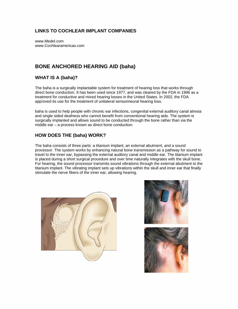
HOW DOES THE (baha) WORK?
The baha consists of three parts: a titanium implant, an external abutment, and a sound processor. The system works by enhancing natural bone transmission as a pathway for sound to travel to the inner ear, bypassing the external auditory canal and middle ear. The titanium implant is placed during a short surgical procedure and over time naturally integrates with the skull bone. For hearing, the sound processor transmits sound vibrations through the external abutment to the titanium implant. The vibrating implant sets up vibrations within the skull and inner ear that finally stimulate the nerve fibers of the inner ear, allowing hearing.
WHO IS A CANDIDATE FOR THE (baha) SYSTEM?
The baha is used to rehabilitate people with conductive and mixed loss hearing impairment. This includes people with chronic infection of the ear canal , people with absence of or a very narrow ear canal as a result of a congenital ear malformation, infection, or surgery, and people with a single sided hearing loss as a result of surgery for a vestibular schwannoma (a tumor of the balance and hearing nerves).
Chronic Ear Infection Treatment for hearing losses with the baha is suitable for people with a conductive or mixed hearing impairment caused by a chronic infection of the middle or outer ear that results in a persistent and unpleasant discharge. The first goal, of course, is to manage the infection. In rare cases, chronic infections fail to respond to treatment, but are determined to be non-threatening. In other cases, infections respond to treatment, but recur with use of a conventional in-the-canal hearing aid. When a hearing aid is placed in a susceptible ear canal, a chronic or recurrent infection may be aggravated by the obstruction of the canal and the resulting excessive humidity and lack of drainage. In these cases, the baha may be a good solution for hearing rehabilitation.
The baha sound processor transmits sound directly to the hearing nerve without involving the ear canal. With baha there is no occlusion of the ear canal to aggravate infection. A baha sound processor offers sound quality at least as good as a conventional air conduction device. For those who need high levels of amplification, problems related to feedback and discomfort are usually resolved.
Congenital Hearing Loss Congenital conductive hearing loss caused by a malformation of the middle or external ear resulting in a missing or incomplete ear canal (external auditory canal atresia) are effectively managed with a baha. Traditionally people with this type of hearing loss have been offered an old-fashioned bone conducting hearing aid. These are either held on the head using a steel spring headband or included in the frame of a pair of glasses. Traditional bone conductors have several disadvantages. The sound quality is poor as the skin acts as a barrier for the sound to travel to the inner ear. They are uncomfortable – patients complain of pain and headaches due to the constant pressure of the headband. They are also cumbersome, obtrusive and insecure.
The baha system can be a real solution for people with this type of impairment. The BAHA sound processor is directly integrated to the skull bone. Because of this direct interface, the BAHA offers significantly better sound quality than that of a traditional bone conductor. The BAHA sound processor works without pressure on the skin avoiding the headaches and soreness associated with the conventional bone conductor. BAHA offers excellent wearing comfort and a better aesthetic result.
BAHA FOR UNILATERAL DEAFNESS
One ear does not provide adequate hearing in many situations. Patients with severe hearing loss on one side, but normal hearing in the other ear have difficulty understanding speech in background noise (such as group conversations and restaurants) and determining which direction sound comes from. Unilateral deafness can result from viral infections, trauma, acoustic neuromas and other ear tumors and ear surgery.
Until recently, the best available approach for providing help in this situation has been the CROS (contralateral routing of offside signal) hearing aid. This technique utilized hearing aid microphones worn in both ears and routed sound from the deaf ear to the hearing ear. Unfortunately, most patients were unsatisfied with this system. Common complaints include the
cosmetic appearance and discomfort of the headband, and the use of a hearing aid mold in the good ear. Most patients felt the benefit from the device is not worth the disadvantages.
The baha, now an FDA cleared solution for unilateral deafness, provides a completely unique benefit. The baha device is placed on the side of the deaf ear, transfers sound through bone conduction, and stimulates the cochlea of the normal hearing ear. The baha effectively transmits sounds from the bad side to the normal ear and ultimately results in a sensation of hearing from a deaf ear. Stereo hearing results in improved understanding of speech, especially in background noise and aids in the localization of sound.
The baha offers significant advantages to the traditional CROS hearing aid. The device is placed behind the ear leaving the canal open. It is worn under the hair and is not perceptible to others. Because it is held in place by a clip and directly integrated with the skull bone, there is no need for a head band and pressure against the skin of the head. In recent clinical trials patients prefer the sound and speech clarity achieved with the baha verses the CROS and verses the unaided condition.
BALANCE
VIDEONYSTAGMOGRAPHY (VNG)
Videonystagmography (VNG) technology for testing inner ear and central motor functions. VNG testing is considered the new standard for testing inner ear functions over Electronystagmography (ENG), because VNG measures the movements of the eyes directly through infrared cameras, instead of measuring the mastoid muscles around the eyes with electrodes like the previous ENG version. VNG testing is more accurate, more consistent, and more comfortable for the patient. By having the patient more comfortable and relaxed, consistent and accurate test results are more easily achieved.
VNG testing is used to determine if a vestibular (inner ear) disease may be causing a balance or dizziness problem, and is one of the only tests available today that can decipher between a unilateral (one ear) and bilateral (both ears) vestibular loss. VNG testing is a series of tests designed to document a person’s ability to follow visual objects with their eyes and how well the eyes respond to information from the vestibular system.
This test also addresses the functionality of each ear and if a vestibular deficit may be the cause of a dizziness or balance problem. To monitor the movements of the eyes, infrared goggles are placed around the eyes to record eye movements during testing. VNG testing is non-invasive, and only minor discomfort is felt by the patients during testing as a result of wearing goggles. There are three main parts to a VNG test:
OCULAR MOTOR TESTING
The patient will be asked to follow objects with their eyes that jump from place to place, stand still, or move smoothly across a light emitting screen. The audiologist will evaluate any latency, velocity, or accuracy errors in the patient’s ability to follow the visual targets. This may indicate a central or neurological problem, or possibly a problem in the pathway connecting the vestibular system to the brain.
POSITIONAL AND POSITIONING TESTS
The audiologist will move the patient’s head and body into various positions to make sure that there are no inappropriate eye movements (nystagmus), when the patient’s head is in different positions. Part of these test are looking at the inner ear system and the condition of endolymph fluid in the semi-circular canals. The audiologist is verifying that small calcium carbonate particles called otoconia are not suspended in the fluid and causing a disturbance to the flow of the fluid know as benign paroxysmal positional vertigo (BPPV).
BPPV is a common cause of dizziness. About 20% of all dizziness is due to BPPV. While BPPV can occur in children, the older you are, the more likely it is that your dizziness is due to BPPV. About 50% of all dizziness in older people is due to BPPV.
The symptoms of BPPV include dizziness or vertigo, lightheadedness, imbalance, and nausea. Activities which bring on symptoms will vary among persons, but symptoms are almost always precipitated by a change of position of the head with respect to gravity. Getting out of bed or rolling over in bed are common "problem" motions.
CALORIC TESTING
The audiologist stimulates both inner ears (one at a time) with warm and then cold air. The patient’s eye movements are monitored using infrared goggles to make sure that both ears can sense this stimulation. This test will confirm that the vestibular system for each ear is working and responding to stimulation; this is the only test available that can decipher between a unilateral and bilateral damage.
Rotational Vestibular Testing (RVT)
Rotational chair testing was first introduced by Bárány in 1907. He initially designed the chair for vestibular ocular reflex (VOR) testing with impulsive rotation in mind. His first test consisted of manual rotating the chair 10 times over 20 seconds followed by a sudden stop of the chair to analyze the postrotary nystagmus of the patient.
Rotational chair testing has undergone numerous changes since that time and now has additional applications, including testing of visual-vestibular interaction, optokinetic after-nystagmus (OKAN), high-velocity sinusoidal testing, and off-vertical axis rotation (OVAR).
The purpose of rotational chair testing is to determine whether or not dizziness may be due to a disorder of inner ear or brain. This test measures the slow phase velocity of the vestibular ocular reflex (VOR) while slowly being turned in a chair that rotates back and forth. Rotational chair testing is usually ordered in addition to ENG/VNG (caloric) testing to confirm a diagnosis and increase accuracy.
These tests both determine if the semi-circular canals of the inner ear are functioning properly, but ENG/VNG tests by themselves may be falsely positive or falsely negative if not administered properly.
SINUSOIDAL HARMONIC ACCELERATATION TEST
The rotational chair test objectively measures the semicircular canals at a higher (and more physiologic) frequency than with caloric testing. The test is an integration of responses from both the right and left vestibular systems, unlike caloric testing which tests them independently. This test measures dizziness by recording the eye movements (nystagmus) while the chair is being rotated. The computer controls the movement of the testing cylinder and compares the nystagmus movements with the movement of the testing cylinder looking for any discrepancies.
As a general rule, patients with inner ear diseases become less dizzy than do normal persons because of their vestibular deficient.
The patient is rotated in a chair at a velocity of from 0.01 to 0.64 Hz. The slow component of the physiologically induced nystagmus is analyzed in terms of phase, gain, and symmetry of eye movement. Symmetry between vestibular systems is measured by comparing the peak slow-wave velocities between left and right rotations of the patient. In acute vestibular lesions, the symmetry measure shows weakness on the affected side, though confounding factors such as compensation, labyrinthine irritation, and cerebellar lesions may render the symmetry test unreliable. Rotatory chair testing is generally more palatable to patients than caloric testing (especially pediatric patients). It is useful in monitoring changes in vestibular function over time, in monitoring compensation after acute injury, and in monitoring residual labyrinthine function in patients with no response during caloric testing.
FIXATION SUPPRESSION TEST
This test measures nystagmus while a person is being rotated in the cylinder while looking at a dot of light that is rotating with them. This would be similar to concentrating on your finger while holding it in front of your eyes, and spinning yourself in circles. This confuses the vestibular system and nystagmus can be measured. The ability to suppress fixation on the dot is impaired by central nervous system conditions and improved by bilateral vestibular loss.
OPTOKINETIC TEST (OPK)
OPK testing measures the dizziness caused by the subject looking at different strips of lights that appear on the wall of the testing cylinder. The eyes will follow a stripe and then make a quick (movement) saccade to catch up with the next stripe, resulting in the pattern of optokinetic nystagmus. Although low in diagnostic power, Optokinetic testing is sometimes useful in the diagnosis of central vestibular conditions and unilateral peripheral vestibular asymmetry.
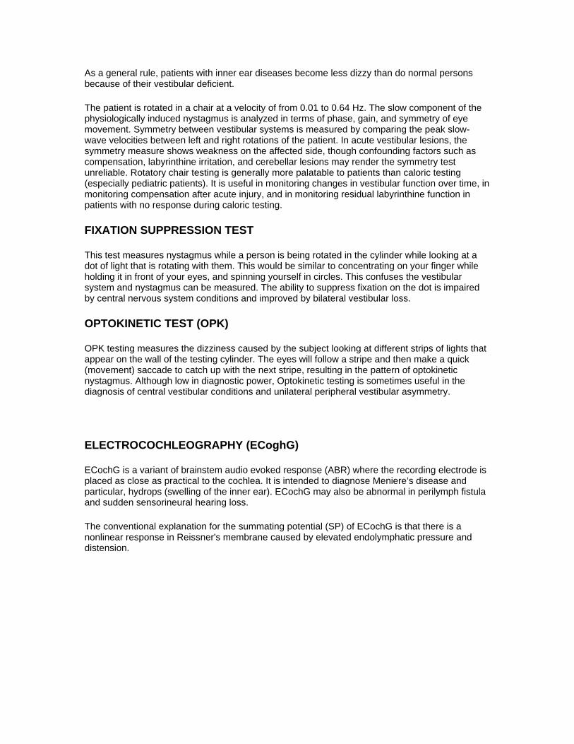
ELECTROCOCHLEOGRAPHY (ECoghG)
ECochG is a variant of brainstem audio evoked response (ABR) where the recording electrode is placed as close as practical to the cochlea. It is intended to diagnose Meniere’s disease and particular, hydrops (swelling of the inner ear). ECochG may also be abnormal in perilymph fistula and sudden sensorineural hearing loss.
The conventional explanation for the summating potential (SP) of ECochG is that there is a nonlinear response in Reissner's membrane caused by elevated endolymphatic pressure and distension.
The technique involves placing an electrode into the outer ear canal, so that it is close to the ear drum. Our preference at EVMS is to use a commercial tymptrode electrode, which is a soft gel electrode that is placed on the eardrum. Other variants include a gold sponge, wire, or spring placed in the ear canal, or a needle that transfixes the ear drum. In general, these are either less effective or more painful.
The sensitivity of the SP/AP ratio to Meniere's disease 60-71%. The sensitivity is said to be higher if it is performed during a symptomatic period, however, it is difficult to get patients scheduled for the test while symptomatic.
TINNITUS
WHAT IS TINNITIS?
Tinnitus is noise heard in the head or ears. It is extremely common - occurring in 20% of the population of the United States. No one person describes his tinnitus like the next person.
ECoghG is interpreted by comparing the amplitude of the summating potential (SP), derived from the inner hair cells of the cochlea to the action potential (AP), derived from the distal auditory nerve. This measurement is known as the SP/AP amplitude ratio.
It may be described as:
• low or high pitched, • containing more that one sound, • extremely loud and distracting or very soft and hardly noticeable • intermittent • constant • subjective (heard only by the person) • objective (heard by others nearby) • pulsatile
Tinnitus is a symptom, not a disease itself. It is frequently, but not always, associated with hearing loss. It can be thought of as a sign of irritation or injury to the hearing system, much like pain or temperature sensations of the skin. Most people experience tinnitus at some point in time. For example, noise exposure such as a concert or a loud party may result in a loud ringing in the ears noticed when returning home to a quiet environment. This kind of tinnitus is temporary, resolving in a few hours. The presence of persistent tinnitus and tinnitus associated with hearing loss should prompt an evaluation by an ENT or otologist/neurotologist. Pulsatile tinnitus, tinnitus with balance problems, and unilateral tinnitus also require evaluation for the underlying etiology.
SUBJECTIVE TINNITUS
WHAT IS IT? Most tinnitus is heard only by the patient. There are many potential causes of this type of tinnitus. The inner ear, hearing nerve, and brain are very delicate and may be injured by one or more of several different mechanisms including age, genetic and hereditary disease, infection, allergy, inflammation, tumors, metabolic problems such as diabetes or low- or high-thyroid, loud noise, prescription and over-the-counter medications, nicotine, and -last but not at all least- stress and fatigue.
WHY DOES IT MAKE ME FEEL SO BAD? Most people with tinnitus are not bothered by it. But for the 20% that are, it can be significantly disruptive to one’s work and family life. Because tinnitus can stimulate the centers of the brain responsible for emotion, some patients experience significant anxiety, depression, irritability and other strong emotional responses.
The hearing system and emotion centers of the brain are intimately linked to the autonomic nervous system. The autonomic nervous system controls all the functions of our body, and performs most functions automatically, beyond conscious control. Because it is closely linked to emotion center, certain emotions result in physical changes in the body; for example, anger can increase the rate of heartbeat. It is responsible for the "fight or flight" reaction, the reaction that prepares the body to react to danger. It is this response that makes the hairs stand up, pupils of the eye dilate, the respiratory rate increase, and blood drain out of the face as it is shunted to muscles of the arms and legs. The autonomic reaction to tinnitus often results in problems with sleep, inability to pay attention to issues other than tinnitus, a high level of reactiveness, and suppression of positive emotions.
HOW IS IT EVALUATED? Although the physician can not “hear” the tinnitus there a good tests which may help quantify the pitch and loudness of the tinnitus. A hearing test is also important during the evaluation. Hyperacusis is an abnormal sensitivity to normal every-day sounds that often accompanies tinnitus. It can also be measured.
Of course, the most important part of an evaluation for tinnitus is a thorough history and physical exam by an ENT or otologist. A hearing test is performed. Special tests such as blood tests, MRI, CT, and auditory brainstem response are sometimes needed.
HOW IS IT TREATED? There are many approaches for the treatment of tinnitus. If a metabolic, drug, infectious, or inflammatory cause is identified specific treatment can be initiated. This may include changing or eliminating medications, managing elevated blood sugars, and treatment with antibiotics, antivirals, and/or anti-inflammatories. If a tumor is found it may be treated with surgery or radiation, or it may be simply watched.
Most tinnitus is idiopathic, meaning a specific cause is not identified. Simple measures may significantly alleviate the tinnitus.
Self-Help Techniques to reduce tinnitus:
• Avoid loud sound. • Eliminate nicotine and caffeine. • Avoid stressful situations. • Rest. Get at least 8 hours of uninterrupted sleep every night. • Avoid too much quiet. Use a fan, noise maker (ocean waves, birds, rain sounds) or quiet
radio music at night to mask the irritating tinnitus as you are trying to sleep. During the day, you may find yourself less bothered by the tinnitus if your mind and body are engaged in activities and there are other sounds in the room for your brain to listen to.
• Avoid focusing on the tinnitus by frequently discussing it with your family.
In cases not responsive to these basic measures, treatment with the physician may include:
• stress reduction • relaxation techniques including massage and acupuncture • treatment for depression and anxiety • sleeping aids • hearing aids to improve underlying hearing loss • family support • professional support groups such as the American Tinnitus Association • Tinnitus Retraining Therapy • Gingko biloba. Some patients find this herbal medication helpful, but the scientific data is
inconclusive. Gingko biloba may increase the risk of bleeding.