Clinical Occlusal References in Prosthetic Procedures INTRODUCTION STATIC RELATIONSHIP The Journal of Gnathology Vol. 7, No. 1, 1988 J. Romerowski, D.C.D., D.S.O. G. Bresson, D.C.D. Paris, France Too often prosthetic procedures refer to theories and technics. However theories and technics are only means to reach the pur- poses and not ends by themselves. The functioning mouth is and must remain the only reference. "Nature to be commanded must be obeyed" ( F. Bacon). Applying a prosthetic philosophy does not lead necessarily to success. Each patient needs his own personal solution. Precise observation of the functioning stomatognathic apparatus teaches us more than theories, although theories can point out what should be observed. What kind of references the practitioner should look for? First it must be determined that the relative position of the man- dible to the cranium does not show any pathologic symptoms. Second, the relationship between the maxillary and the mandibu- lar arches should be considered under the two following aspects: This relationship, commonly called intercuspation which would be better named occlusion*, is used during mastication and degluti- tion. During chewing, the mean duration of interarch contact is about 194 milliseconds.1 Swallowing supposesthe mandibular arch to be fastened to the maxillary arch to serve as a fixed part to allow for contraction of the hyoidian and pharyngian muscles. The mean duration of the interarch contact is about 694 milliseconds.1, that is 3.5 times the duration of the same contacts during chewing. It must be also kept in mind that swallowing occurs from 1500 to 2000 times per 24 hours, twice more time during the awake period • occlusion: from ob + claudere to shut, close (Webster's Third New International Dictionary. 1966). 93
Transcript
Clinical Occlusal References in Prosthetic Procedures
INTRODUCTION
STATIC RELATIONSHIP
The Journal of Gnathology Vol. 7, No. 1, 1988
J. Romerowski, D.C.D., D.S.O. G. Bresson, D.C.D. Paris, France
Too often prosthetic procedures refer to theories and technics. However theories and technics are only means to reach the pur-poses and not ends by themselves. The functioning mouth is and must remain the only reference. "Nature to be commanded must be obeyed" ( F. Bacon). Applying a prosthetic philosophy does not lead necessarily to success. Each patient needs his own personal solution. Precise observation of the functioning stomatognathic apparatus teaches us more than theories, although theories can point out what should be observed.
What kind of references the practitioner should look for?
First it must be determined that the relative position of the man-dible to the cranium does not show any pathologic symptoms.
Second, the relationship between the maxillary and the mandibu-lar arches should be considered under the two following aspects:
This relationship, commonly called intercuspation which would be better named occlusion*, is used during mastication and degluti-tion.
During chewing, the mean duration of interarch contact is about 194 milliseconds.1 Swallowing supposes the mandibular arch to be fastened to the maxillary arch to serve as a fixed part to allow for contraction of the hyoidian and pharyngian muscles. The mean duration of the interarch contact is about 694 milliseconds.1, that is 3.5 times the duration of the same contacts during chewing. It must be also kept in mind that swallowing occurs from 1500 to 2000 times per 24 hours, twice more time during the awake period
• occlusion: from ob + claudere to shut, close (Webster's Third New International Dictionary. 1966).
93
Romerowski, Bresson
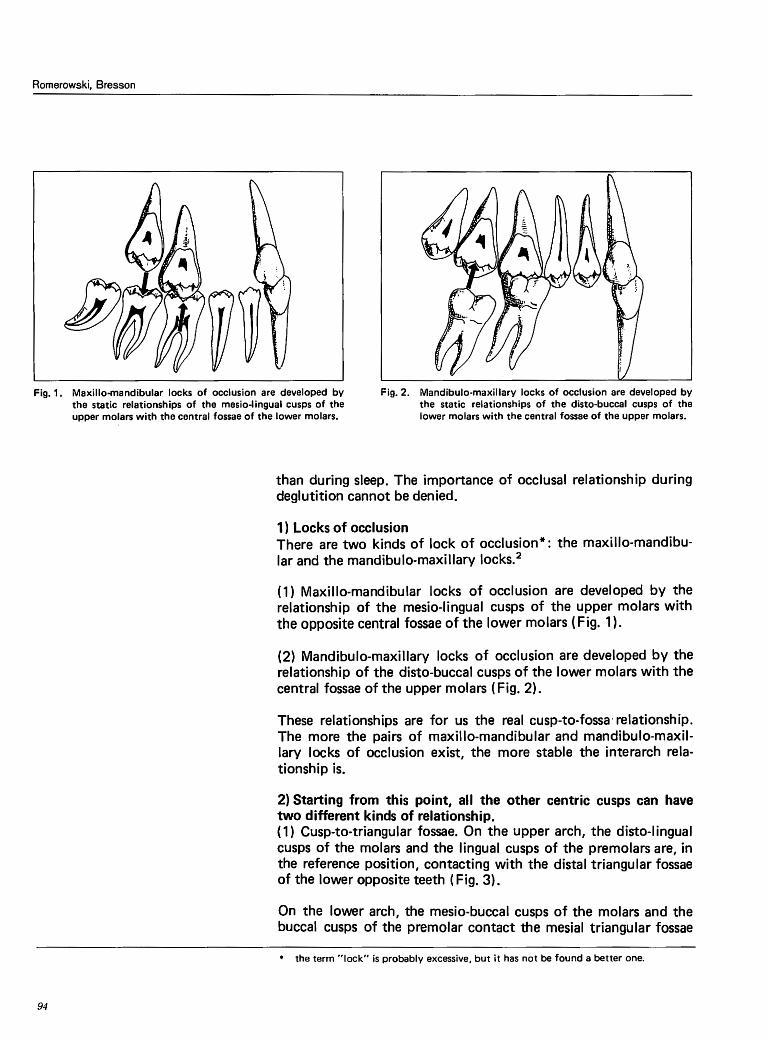
Fig. 1. Maxilla-mandibular locks of occlusion are developed by the static relationships of the mesio-lingual cusps of the upper molars with the central fossae of the lower molars.
Fig. 2. Mandibulo-maxillary locks of occlusion are developed by the static relationships of the disto-buccal cusps of the lower molars with the central fossae of the upper molars.
94
than during sleep. The importance of occlusal relationship during deglutition cannot be denied.
1) Locks of occlusion There are two kinds of lock of occlusion*: the maxillo-mandibu-lar and the mandibulo-maxillary locks.2
( 1) Maxilla-mandibular locks of occlusion are developed by the relationship of the mesio-lingual cusps of the upper molars with the opposite central fossae of the lower molars (Fig. 1 ).
(2) Mandibulo-maxillary locks of occlusion are developed by the relationship of the disto-buccal cusps of the lower molars with the central fossae of the upper molars ( Fig. 2).
These relationships are for us the real cusp-to-fossa· relationship. The more the pairs of maxillo-mandibular and mandibulo-maxil-lary locks of occlusion exist, the more stable the interarch rela-tionship is.
2) Starting from this point, all the other centric cusps can have two different kinds of relationship. ( 1) Cusp-to-triangular fossae. On the upper arch, the disto-lingual cusps of the molars and the lingual cusps of the premolars are, in the reference position, contacting with the distal triangular fossae of the lower opposite teeth ( Fig. 3).
On the lower arch, the mesio-buccal cusps of the molars and the buccal cusps of the premolar contact the mesial triangular fossae
• the term "lock" is probably excessive, but it has not be found a better one.
Clinical Occlusal References in Prosthetic Procedures
Fig. 3. Except the mesio-lingual cusps of the upper molars, all the maxillary lingual cusps are in a cusp to triangular fossae relationship.
Fig. 4. Except the disto-buccal cusps of the lower molars, all the mandibular buccal cusps are in a cusp to triangular fossae relationship.
Fig. 5. Except the mesio-lingual cusps of the upper molars, all the maxillary lingual cusps are in a cusp to embrasure relationship.
Fig. 6. Except the disto-buccal cusps of the lower molars, all the mandibular buccal cusps are in a cusp to embrasure rela-tionship.
The Journal of Gnathology Vol. 7, No. 1, 1988
of the upper opposite teeth ( Fig. 4). This type of relationship is less than 15% of natural dentitions. It is a one to one tooth rela-tionship.3·4
(2) Cusp-to-embrasure relationship. The cusps that are not in-volved in the locks of occlusion are contacting the peripheral ridges of the two opposite marginal ridges (Fig. 5, 6). It is a one to two teeth relationship and represents about 85% of natural cases.5 •6
Very often, this type of relationship has been condemned because of the risk of food impaction into the gingival embrasure (Fig. 7). But why is it that this type of relationship works in 85% of the natural cases without food impactions? It seems that all the
95
Clinical Occlusal References in Prosthetic Procedures
Fig. 10. The disto-lingual cusps of the upper molars are in a cusp to embrasure relationship as the lingual cusps of the premolars are in a cusp to triangular fossa relationship (as described by Charles Stuart).
Fig. 11. The last millimeters of the cycle of mastication are the most important relative to functional dental anatomy.
THE KINEMATIC RELATIONSHIPS
The Journal of Gnathology Vol. 7, No. 1, 1988
pictures explaining the inpropriety of such a relationship are incor-rect. In fact, teeth work together. Each dental arch is a functional unit. When a tooth has to support a stress from food or occlusion, the adjacent teeth help by the acceptance of a part of the stress through the proximal contact zones (M. de Stefanis) (Fig. 8). 7
Therefore, food impactions are the result of a wrong contour of the anatomy of the structures that surround the embrasures.
It appears that the reestablishment of a cusp-to-triangular fossa relationship, without any anatomical distortion (dysmorphia} al lows for a better transmission of the stresses along the corono-radicu lar axis of the teeth and should, each time this is possible, improved ( Fig. 9).
(3) Charles Stuart described the clinical situations where the rela-tionship was a mixture combining cusp to triangular fossae and cusp-to-embrasure-relationships (Fig. 10). 8 This relationship is acceptable if the locks of occlusion keep the stability of the inter-cuspation and if the unity of each dental arch is maintained by the continuity of the interproximal contact zones.
The cycle of mastication begins with a vertical downward motion of the mandible. Then the mandible is directed laterally to the food and then comes back into intercuspation. I nterarch rela-tionship is involved only in the last millimeters of the cycle (Fig. 11}. The clinical mandibular movements on the patient as well as the tests with the articu later mounted models, are normally ob- . served from centric occlusion to excentric positions. That is ex-
97
Clinical Occlusal References in Prosthetic Procedures
Fig. 14. In this situation of the sagittal static relationship be· tween the canines, the registered marks occur on the lingual ridge of the upper canine and on the distal cusp ridge of the lower canine.
The Journal of Gnathology Vol. 7, No. 1, 1988
> its strategic position at the corner of the dental arch > the particularly strong anatomy of its root and supporting al-
veolar bone > its crown length which is the longest of all the teeth > the high level of its proprioceptive capacity
In the lateral mandibular movement, the first contact between opposite teeth occurs normally on the lingual surface ofthe upper canine. The control of the occluding movement depends essen-tially on the quality of the gliding contact on this lingual surface. This assertion can be demonstrated by clinical observation. Patients who develop a unilateral mastication, chew only on the side where the lingual surface of the upper canine can easily be reached and used for the control of the occluding movement. In case of an im-portant overjet or a cross bite at the level of the upper canine, mastication does function on the other side.
If no other tooth is associated with the canine, the functional kinematic relationship is called canine protection and the clinical tested motion is an immediate disclusion (Fig. 13). If an articula-tion recording device is placed between the teeth to register the gliding contacts, on the posterior teeth the centric position marks are the only registrations. On the canines, two types of registration depending on the relationship of the opposite canines in the sagit-tal plane can be found:
(1) The gliding contact occurs between the lingual ridge of the upper canine and the distal cusp ridge of the lower canine (Fig. 14);
(2) The gliding contact appears between the mesial ridge of the
99
The Journal of Gnathology Vol. 7, No. 1, 1988
Clinical Occlusal References in Prosthetic Procedures
lingual surface of the upper canine and the distal cusp ridge of the lower canine ( Fig. 15). In this last case, on the lower tooth a large surface is produced by the engagement of the contact point area during the occluding movement.
4) Sometimes, one or more incisors are associated with the canine protection { Fig. 16). This relationship is called anterior group function.
5) If during the terminal occluding movement, one or more poste-rior teeth are related by gliding contacts during canine function, the relationship is a posterior group function (Fig. 17).
( 1) If all the posterior teeth are involved, it is a total posterior group function ( Fig. 17).
(2) If only some posterior tooth or teeth are concerned, it is a partial posterior group function (Fig. 18). The same term applies to all the mediate disclusions: late or progressive disclusion.
(3) Again the registered contacts on the occlusal surfaces depend on the relative position of the mandibular arch to the maxillary arch in the sagittal plane.
(4) The contacts occur on the central ridges of the upper buccal cusps ( Fig. 19) or
(5) On the distal ridges of the buccal cusps and then on the mesial marginal ridges of the upper teeth (Fig. 20). In this last case, the contact spots on the distal ridges of the lower buccal cusps are induced by the travel of the contact point areas during the closing motion (Fig. 20).
The gliding contact on the upper canine remains during all the period of intercuspation.
(6) Sometimes, if the contralateral mandibular condyle pathway is not very inclined, gliding contacts appear between the central ridges of the lower lingual cusps and the peripheral ridges of the upper lingual cusps. If the gliding contact on the lingual surface of the upper canine is never lost, this kind of kinematic relationship is acceptable, but it should not be created in a prosthetic proce-dure.
For a good function of the stomatognathic complex, the upper canine must be involved in the end of every lateral mandibular movement through a gliding contact on its lingual surface.
The lack of canine function can be accepted as an adaptative situation, but it predisposes for pathology and dysfunction. Every
101
Romerowski, Bresson
REFERENCES
104
1. Lundeen (H.C.), Gibbs (C.H.): Advances in Occlusion. John Wright. London. PSG Inc. Boston. Bristol, 1982.
3. Stuart, C.E.: Full Mouth Waxing Technique. Quintessence Publishing Co., Inc. Chicago, Berlin, Rio de Janeiro, Tokyo, 1983.
4. Thomas, P.K., Tateno, G.: Gnathological Occlusion. Shorin. Tokyo and Denar Corporation Anaheim edit. 1979.
5. Payne, E.V.: Functional Waxing Technique. in Practical Crown and Bridge Prost-hodontics by Wilson and Lang. The Blakiston Division New York, MacGraw Hill Book Co. edit. 1962.
6. Lundeen, H.C.: Introduction a I' Anatomie Occlusale. J. Prelat edit. Paris, 1973. 7. De Stefanis, M.: Personnal Communication. Portland, Oregon, 1977. 8. Stuart, C.E.: Oral Rehabilitation and Occlusion. Post Graduate Education, School
of Dentistry, University of California, San Francisco. 9. MacHorris,W.H.: Lecture. The European Academy of Gnathology, Paris, 1982.
10. Riis, D., Gidden, 0.: lnterdental Discrimination of Small Thickness Differences. J. Prosth. Dent., 24:3, 1970.
11. Kawamura, Y., Nishiyama, T., Funakoshi, M.: A study on Topognosis of the Human Teeth. J. Osaka Univ. Dent. Sch .• 7: 1-5, 1967.
12. Jeanmonod, A.: Occlusodontologie. Applications Cliniques, Paris edit. CdP (1988). 13. D'Amico, A.: The Canine Teeth: Normal Functional Relation of the Natural Teeth
of Man. J. South. Calif. Dent. Ass .• 26: 1-74, 1958. 14. D'Amico, A.: Application of the Concept of the Functional Relation of the Canine
Teeth. J. South. Calif. Dent. Ass., 27: 39-58, 1959.
Prof. Dr. Jean Romerowski 31 rue Marbeuf F. 75006 Paris France Teaching Assistant Gerard Bresson 6 impasse des Hottes F. 51150 Ambonnay Tours s. Marne France