1 Department of Prosthodontics and Dental Materials, University of Siena, 53100 Siena, Italy;[email protected] (D.I.K.P.); [email protected] (E.F.C.)
2 Department of Endodontics and Restorative Dentistry, School of Dental Medicine, University of Siena,53100 Siena, Italy; [email protected]
3 Department of Neurosciences, Reproductive and Odontostomatological Sciences, University of Naples“Federico II”, 80131 Naples, Italy; [email protected]
4 Department of Periodontology, School of Dental Medicine, University of Siena, 53100 Siena, Italy;[email protected]
5 Department of Endodontics and Restorative Dentistry, School of Dental Medicine, University of Genoa,16121 Genoa, Italy; [email protected]
6 Department of Orthodontics, University of Padua, 35122 Padua, Italy; [email protected] Private Practitioner, 25025 Brescia, Italy; [email protected]* Correspondence: [email protected]; Tel.: +39-0577-233131; Fax: +39-0577-233117
Abstract: Background: The aim of this study was to collect long-term restorative and endodonticoutcomes of endodontically treated teeth (ETT). Methods: 298 teeth were included in the studyand were recalled up to 18 years with a media of 10.2 years. At baseline, 198 sample teeth (66.44%)showed symptoms and 164 (55%) had periapical radiolucency. The most frequently used obturationtechniques were warm gutta-percha in 80% of cases, and by carrier in 20%. A total of 192 ETT wererestored by direct resin composite restorations, and 106 posts were luted. Moreover, 75 (25.16%) directrestorations remained as final restorations, 137 single crowns (45.97%), 42 (14.09%) partial adhesivecrowns, and 42 (14.09%) abutments of fixed bridges were the final treatments. Descriptive andinferential statistics were performed (α = 0.05). A Cox regression model was made. Results: resultsshowed success for 92.6% of ETT up to 18 years, 2.68% (8 ETT) showed irreversible failures, and14 (4.69%) reversible complications. Four ETT (1.34%) failed because of root fracture and the other four(1.34%) because of endodontic complications. Eight ETT (2.69%) showed non-irreversible periodontalcomplications and the other six (2.01%) prosthodontic complications. Accordingly, with Kaplan–Meier analysis, the survival rate after 18 years was 97.3% (Interval of Confidence (IC) 95.1–98.3).The presence of a short or long (at least 1 mm related to radiographic apex) quality endodonticfilling displayed a statistically significant higher risk of complication (hazard ratio (HR) = 17.00(IC 5.68–56.84). Furthermore, a clinically detectable not precise coronal margins predicts the presenceof any clinical complication with a hazard ratio almost seven times higher than endodonticallytreated teeth with a proper margin (HR = 6.89 (IC 2.03–23.38)), while the presence of lucency at thebaseline did not affect the risk of complication (HR = 0.575 (IC 0.205–1.61)). The presence of post,tooth position in the arch, and the type of it did not show a high-risk rate (HR = 1.85, 1.98, and 2.24,respectively). Conclusions: a correct filling (at the apex) of root canals combined with proper coronalmargins allow obtaining a long-term high success rate in teeth with a periapical lesion at the baseline.The use of a post or not, when its placement is related to the residual amount of the crown, does notchange the final outcome of the ETT.
Long-term survival of endodontically treated teeth (ETT) depends on correct and well-sealing restoration and the principles of endodontic therapy, by outlining the biology ofthe dental pulp and periradicular tissues, the etiology and pathophysiology of the diseaseprocesses, and the measures to diagnose, prevent, and cure the different disorders thathave been established [1,2].
Different parameters were proposed to define endodontic “success” and the existingdata on endodontic therapy outcome must be carefully interpreted. Differences in theassessment of teeth during follow-up were noted, such as the radiographic assessmentmethod, the radiographic criteria for success (loose and strict), the unit of outcome measure(root and tooth), and the length of follow-up [3].
Criteria setting the threshold for success at the complete resolution of the periapicalradiolucency have been described as “strict” [4] or “stringent” [5], while choosing a merereduction in the size of the periapical radiolucency [6], has been described as setting a“loose” [4] or “lenient” [5] threshold. The frequency of adoption of these two thresholdsin previous studies has been similar; the expected success rates using “strict” criteriawould be lower than those based on “loose” criteria. Regarding periapical periodontitisin radiographs, a scoring system for registration and evaluation was proposed [7]. Thissystem provides an ordinal scale of five scores, ranging from healthy to severe periodontitiswith exacerbating features. It is based on radiographs with verified histological diagnosisand can be suitable in epidemiological studies.
Another outcome measure, “functional retention”, has been introduced [5]. A rootcan be considered “functional” when no clinical signs and symptoms are present, indepen-dently from the presence or absence of periradicular radiolucency [8,9].
“Functional retention” of teeth after root canal treatment is a similar but less lenientoutcome measure than “survival”.
The “strict criteria” are based on remission of clinical signs and symptoms, andlamina dura’s complete restitutio ad integrum [10] were followed in this clinical study.Considering that the shortest recall in this clinical study was performed at 48 months, therewas reasonable time for complete healing of the wide majority of periapical lesions (whenpresent) and, in this way, “strict” criteria were followed.
Endodontic treated teeth must be restored in order to function and for esthetic pur-poses. Restoration of ETT can be performed with or without a post [11], by direct or indirectrestoration. The best treatment is still under discussion, but it depends on the amount ofthe residual crown, the anatomy of roots, etc. [12–16].
The type of restoration made on top of the endodontic treated root is another clinicalkey factor to guarantee longevity to the tooth and, in particular, the precision of the coronalmargin of the restoration, independently, if an indirect or direct restoration is made [17–19].Another important aspect is how the endodontic treated root can be built up, using or notposts [20]. It is still an open question whether a post is needed or can be avoided [11,21,22].
In order to obtain predictable results, clinical trials are needed. Clinical trials areconsidered more reliable than in vitro tests and can be retrospective or prospective [23,24].
Prospective clinical studies are usually performed in specialized centers; specificparameters are evaluated with a limited number of samples. Retrospective studies cancollect a wider number of samples and may reflect the clinical behavior of practitioners.
The aim of this retrospective clinical study was to collect long-term restorative andendodontic outcomes of ETT restored by different clinical procedures.
The tested null hypotheses were: (1) there was no difference in the endodontic outcomeof ETT with and without periapical lesion at the beginning of the treatment; (2) there wasno difference in the endodontic outcome between endodontic treatments with or withoutprecise coronal margins; (3) there was no difference between ETT restored with or withoutpost; (4) there was no difference between ETT restored by direct or indirect restoration.
J. Clin. Med. 2021, 10, 908 3 of 12
2. Materials and Methods2.1. Study Population
Over a 20-year period (March 1999 to March 2019), one expert endodontist (DP)conducted 298 endodontic treatments in 205 patients (143 men, age range: 19 to 61 years;169 women, age range: 19 to 71 years); follow-ups were done with reference to the dentalrecords. Patients were in need of different endodontic therapies. Consecutive patients wereselected from the authors’ offices. Only primary endodontic treated teeth or nonsurgicalretreatments, with a follow-up of at least 18 months or longer, were included in this survey,with patients who returned for oral hygiene recalls in 2019.
All procedures performed in this study involving human participants, were in accor-dance with the ethical standards of the institutional committee, and with the 1964 Helsinkideclaration and its later amendments or comparable ethical standards. Informed writtenconsent was obtained from all individual participants included in the study. Collection andanalysis of the data were approved by the Ethical Committee of the University of Siena.
Inclusion criteria were the following: age: 42 (±7.9) years (range 19 to 71); sex: 169 F,143 M; periodontally healthy or successfully treated patients in need of one or moreendodontic treatments.
Exclusion criteria were the following: individuals who were not yet adults (<18 years),pregnancy, disabilities, previous prosthodontic restorations of abutment teeth, deep defects(close to pulp, <1 mm distance), or pulp capping, heavy occlusal contacts or history ofbruxism, systemic disease or severe medical complications, allergic history concerningmethacrylates, rampant caries, xerostomia, lack of compliance.
2.2. Sample Characteristics
A total of 298 teeth were included in the study, of which, 52 were premolars (26%),170 molars (52%), and 76 anterior teeth (21%, II e V sextant); 103 ETT belonged to themandible (30.68%) and 195 (69.32%) to maxillae.
Several endodontic peculiarities were analyzed. At baseline, 198 sample teeth (66.44%)showed symptoms (tenderness/pain to percussion) and 164 (55%) had periapical radiolu-cency. Regarding ETT with radiolucency, 90 of them (54.87%) were in coincidence withtheir need for retreatment and 74 (45.13%) were necrotic teeth. The most frequently usedobturation techniques were warm gutta-percha in 80% of cases, by carrier in 20%, and hotgutta-percha in other cases, mainly when curve canals were treated.
After being endodontically treated, 192 ETT were restored by direct resin compositerestorations, and 106 posts were luted. A total of 75 (25.16%) direct restorations remainedas final restoration, 137 single crowns (45.97%), 42 (14.09%) partial adhesive crowns, and42 (14.09%) abutments of fixed bridges were the final treatments.
2.3. Original Endodontic Therapy Procedure
For each tooth, the following preoperative data were recorded: demographic data,tooth location, number of root canals, previous endodontic treatment, clinical signs andsymptoms, vitality tests, and radiographic periapical status. Based on these findings, thepreoperative condition was classified as one of the following: vital (healthy or irreversiblyinflamed pulpitis), non-vital, endodontically treated, with or without periapical lesion, andsymptomatic or asymptomatic.
For each tooth, the following intra-operative data were recorded: number of treatmentsessions; inter-appointment dressing (if used); the occurrence of procedural complicationssuch as perforation, breakage of files and flare-up; length of canal filling (at apical level,1 mm short or more and beyond); and temporary restoration placed. A conservativeendodontic cavity (CEC) access was performed using a long shaft round diamond burand endodontic dedicated ultrasonic tips. After straight-line access preparation wasobtained, root canals were negotiated with pre-curved stainless steel K-type files (Maillefer,Bailague, Switzerland), size 0.8 or 10 ISO (International Standard Organization) to themajor apical foramen. Working length was measured using an electronic apex locator
J. Clin. Med. 2021, 10, 908 4 of 12
(Root ZX Morita, Tokyo, Japan), established at electronic 0 and, in most cases, checkedwith an intraoperative X-ray. Due to the long period of time that has been taken intoconsideration in this study, different shaping techniques and instruments have been used.From 1999 to 2003 a crown-down approach was utilized to give correct shaping to thecanals; pre-flaring was performed with a manual pre-curved stainless steel K-file with a#25 tip 0.02 taper, then the shaping was performed with a Ni-Ti rotary file system, whichhad tip size #25 for all instruments and a different taper (QANTEC Kerr, Kerrville, TX,USA From 2003 to 2013, a simultaneous technique was introduced in the clinical procedure,utilizing Ni-Ti rotary files with different tip sizes and different tapers (Mtwo, Sweden eMartina, Italy). From 2013 to 2019, a mixed technique was adopted: pre-flaring and glidepath were performed to length with a nickel-titanium #10 tip size and 0.04 taper rotaryfile, followed by a nickel-titanium #15 tip size and 0.05 taper rotary file (Mtwo, Sweden eMartina, Italy). All canals were shaped with the M-Wire alloy rotary instrument ProTaperNext (Maillefer, Bailague, Switzerland) to a length of up to a #25 tip size and a variabletaper. The apical diameter was measured (apical gauging) using nickel-titanium manualK-type files, NiTi Flex (Maillefer, Bailague, Switzerland), and the shaping of the apicalthird was refined, where needed. Irrigation was copious and frequent using heated 5.25%sodium hypochlorite NiClor (NiClor, Ogna, Bologna, Italy) deposited with side-vented30-G needles. After instrumentation, the root canals were irrigated with 17% EDTA solutionTubuliniclean (Ogna, Bologna, Italy), for 3 min, followed again by several 1-min irrigationswith heated 5.25% sodium hypochlorite solution.
The canals were dried with dedicated sterile paper points, filled with dedicatedgutta-percha cones ProTaper Next (Maillefer, Bailague, Switzerland), and zinc oxide-basedendodontic sealer (Pulp Canal Sealer, Kerr, Germany) using a continuous wave of con-densation technique (80%) or a carrier-based technique (Thermafil, Dentsply, Konstanz,Germany) in roots with curve canals, depending on the root canal anatomy. A post wasplaced when the remaining coronal structure was less than 50% [25]. A temporary restora-tion was performed using zinc oxide based cement placed on the pulp chamber floorcovered by a layer of glass ionomer cement (GCem, GC Co., Tokyo, Japan).
The post space was prepared using the drill provided by the manufacturer. Fiber-reinforced composite post was adapted to the anatomy of the root. Post length was adaptedto the length of the post space. The post surface was cleaned with phosphoric acid andtreated with a silane-coupling agent. For adhesive cementation, the dentinal surface wasetched with phosphoric acid for 10 s and pretreated with a dual-cure adhesive beforethe post was cemented with a dual-cure resin. Aesthetic Plus fiber posts in combinationwith the All Bond 2 bonding system and proprietary C&B resin cement (Bisco) were usedbetween 1999 and 2008. GC fiber posts, in combination with Gradia Core (GC), were usedfrom 2009 to 2018. Porcelain to fused metal crowns were cemented with Fuji Cem (GC)until 2015, whilst more recently, zirconia full crowns were luted with G-Cem adhesivecement (GC). When direct restorations were placed, cuspal coverage was made, and therestorations were made using resin composite materials in combination with proprietarybonding systems. From 1999 to 2010, Gradia (GC) resin composite in combination withG-Bond (GC) was used. After 2010, a combination between G-aenial resin composite (GC)and G-Bond Plus (GC) was used. More recently (from 2016), the same resin was used incombination with GPremio bond (GC).
2.4. Follow-Up
For each tooth, the following postoperative data were recorded: the treatment andrecall period, the presence or absence of signs and symptoms, the presence or absenceof apical lesion, the presence and type of restoration, and the type of build-up with orwithout a post. Only primary endodontic treated teeth or nonsurgical retreatments witha follow-up of at least 18 months or longer were included in this survey with a mediaof 10.2 years. The follow-up sessions were performed with patients who returned to theoffices during oral hygiene recalls during 2019. Among all patients who returned for a
J. Clin. Med. 2021, 10, 908 5 of 12
recall, 298 teeth were selected for this survey. All of the recorded information from thefiles were transferred to a computerized database. The clinical follow-up examinationswere performed by the primary author (D.P.). For teeth examined more than once, only thefindings of the final examination during 2019 were considered. Traumatized teeth, injuredwith luxation, intrusion, extrusion, avulsion, or horizontal fractures, and teeth requiringendodontic surgery, were excluded from this study.
2.5. Criteria of Evaluation
When only the endodontic treatments were evaluated, the following criteria of theEuropean Society of Endodontology 1994 [25] were used to judge the success rate ofroot canal therapy: (1) clinical examination: the absence of pain, swelling, and othersymptoms, no sinus tract, and no loss of function; and (2) radiographic examination:the periodontal ligament space was normal on the original diagnostic radiograph, andit remained unchanged on recall radiographs, or healing of a radiolucent area visible onthe original preoperative radiograph was observed and the periodontal ligament spacereturned to normal. For radiographic examination, PAI (Peri Apical Index) scores wereused [7].
Therefore, cases were considered as failures in the presence of pain, swelling, and sinustract. Radiographically, failures were identified when a lesion appeared after endodontictreatment, when a preexisting lesion increased in size, and when a lesion remained thesame or only diminished in size. Multi-rooted teeth were assessed according to the rootthat appeared the worst.
Debonding of the post was registered when the crown dislodged or/and moved.Loss of retention was registered when mobility was detected between the crown and theabutment, when saliva was expressed at the margin of the crown when pressure wasapplied, or when an explorer could easily be inserted between the tooth and the crown.
Coronal fracture of the direct restoration was registered when visible.Carious lesions were recorded when a dental explorer could penetrate the dentin at
the cervical margin of the crown or the direct restoration as assessed through radiographsand/or clinically.
Ceramic fractures (chipping) were registered clinically and from photographs.Possible “marginal leakage” was clinically evaluated with a sharp explorer along the
margins and radiographically.Periodontal involvement was recorded when a periodontal disease (not existing at the
baseline) was visible around the sample tooth according to periodontal parameters [26].The two examining operators (A.M. and V.M.) were calibrated before the examination.
Calibration was done in 30 cases. The two examiners made their own evaluations. In casethey disagreed, the single cases were reevaluated and an agreement was found.
In this clinical study, the endodontic and restorative treatments made for each toothwere evaluated in a single sample. For that, when the global treatments were considered, itwas decided to avoid misunderstanding between the definitions of success and survival.Success was defined by the percentage of endodontic treatments and restorations thatremained in situ without any modification. Survival was defined by the percentage ofendodontic treatments and restorations that remained in situ, with modifications, but stillunder clinical acceptability. Failure was defined by the percentage of teeth that needed tobe replaced [27].
Data were collected based on predetermined criteria. Percentages of teeth with orwithout apical periodontitis were recorded, as well as adequate root canal treatment (AE),inadequate root canal treatment (IE), adequate seal of the restoration (AR), and inadequateseal of the restoration (IR).
2.6. Radiographic Method and Evaluation
When evaluating treatment results, the first clinical and radiographic examinationwas performed by the primary author (D.P.) when the 298 followed cases were randomly
J. Clin. Med. 2021, 10, 908 6 of 12
selected from patient files, recorded by handwriting, and the recorded information wastransferred to a computerized database. The final evaluation was done together with twoother observers after calibration (A.M. and V.M.). For radiographic examination, PAI scoreswere used [7].
2.7. Statistical Analysis
Descriptive and inferential statistics were performed using the Stata 15 IC statisticalpackage, and the significance level was set at α = 0.05. The minimum, mean, and maximumfollow-ups were calculated for all endodontic treatments. The overall failure incidencerate was according to the total failure events (tooth extraction) and to the presence of anycomplication (free of the event) divided by the total tooth-years during the total follow-upperiod; the 95% confidence intervals (95% CI) were estimated according to the Poissondistribution. Both the cumulative survivals performed were recorded using the Kaplan–Meier analysis. Cox regression analysis was used to examine the risk factors for any clinicalevent during the follow-up period. The results were presented as hazard ratio (HR) with95% confidence intervals (CI).
3. Results
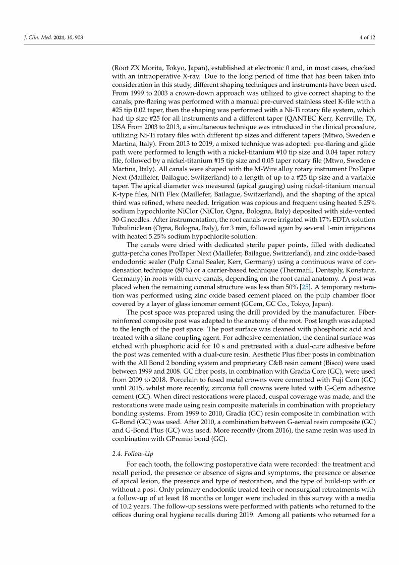
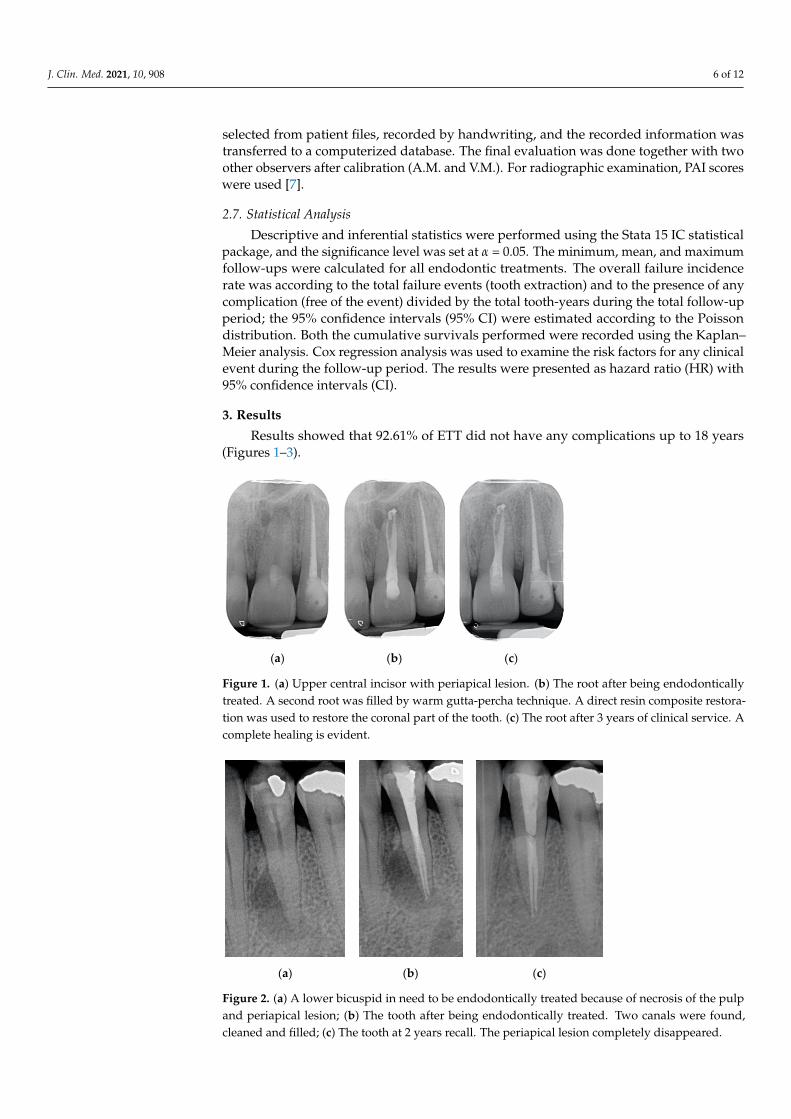
Results showed that 92.61% of ETT did not have any complications up to 18 years(Figures 1–3).
J. Clin. Med. 2021, 10, x FOR PEER REVIEW 6 of 12
2.6. Radiographic Method and Evaluation When evaluating treatment results, the first clinical and radiographic examination
was performed by the primary author (D.P.) when the 298 followed cases were randomly selected from patient files, recorded by handwriting, and the recorded information was transferred to a computerized database. The final evaluation was done together with two other observers after calibration (A.M. and V.M.). For radiographic examination, PAI scores were used [7].
2.7. Statistical Analysis Descriptive and inferential statistics were performed using the Stata 15 IC statistical
package, and the significance level was set at α = 0.05. The minimum, mean, and maxi-mum follow-ups were calculated for all endodontic treatments. The overall failure inci-dence rate was according to the total failure events (tooth extraction) and to the presence of any complication (free of the event) divided by the total tooth-years during the total follow-up period; the 95% confidence intervals (95% CI) were estimated according to the Poisson distribution. Both the cumulative survivals performed were recorded using the Kaplan–Meier analysis. Cox regression analysis was used to examine the risk factors for any clinical event during the follow-up period. The results were presented as hazard ratio (HR) with 95% confidence intervals (CI).
3. Results Results showed that 92.61% of ETT did not have any complications up to 18 years
(Figures 1–3). Regarding the quality of root filling, 264 teeth (88.59%) showed good filling (Figures
1–3) (gutta-percha at the radiologic apex), 22 roots (7.38%) short filling (shorter than 1 mm from the radiographic apex), and 12 (4.03%) long filling (longer than 1 mm of the radio-graphic apex).
(a) (b) (c)
Figure 1. (a) Upper central incisor with periapical lesion. (b) The root after being endodontically treated. A second root was filled by warm gutta-percha technique. A direct resin composite resto-ration was used to restore the coronal part of the tooth. (c) The root after 3 years of clinical service. A complete healing is evident.
Figure 1. (a) Upper central incisor with periapical lesion. (b) The root after being endodonticallytreated. A second root was filled by warm gutta-percha technique. A direct resin composite restora-tion was used to restore the coronal part of the tooth. (c) The root after 3 years of clinical service. Acomplete healing is evident.
J. Clin. Med. 2021, 10, x FOR PEER REVIEW 7 of 12
(a) (b) (c)
Figure 2. (a) A lower bicuspid in need to be endodontically treated because of necrosis of the pulp and periapical lesion; (b) The tooth after being endodontically treated. Two canals were found, cleaned and filled; (c) The tooth at 2 years recall. The periapical lesion completely disappeared.
(a) (b) (c)
Figure 3. (a) A first lower molar with periapical lesion and in need to be retreated. (b) The molar immediately after being endodontically treated. Four canals (one in a radix entomolaris) were de-tected and treated. (c) The molar after 4 years. The good health of the periapical areas can be noted. The crown was restored with an adhesive esthetic onlay.
There were no statistically significant differences among outcomes of vital root and necrotic and retreatment with radiolucency.
At the recall, 284 (94.42%) ETT showed good coronal margin, 11 (4.38%) not-so good margins (margin was filled non-precise with the sharp explorer), and 3 (1.2%) clear open-ing of the margins radiographically.
Success was recorded in 276 ETT (92.6%), 2.68% (8 ETT) showed irreversible failures, and 14 (4.69%) reversible complications.
Four ETT (1.34%) failed because of root fracture and the other four (1.34%) because of endodontic complications.
Eight ETT (2.69%) showed non-irreversible periodontal complications and the other six (2.01%) prosthodontic complications.
The placement or not of a post did not show different outcomes. Sixteen failures were found when a full crown was cemented on the ETT. Failures
were recorded in three cases in combination with direct restorations, in one case with a partial crown, and two cases as the abutment of a bridge.
The survival rate of both “failure” and “free of the event” of endodontically treated teeth, determined by the Kaplan–Meier analysis over an 18-year period, is shown in Figures 4 and 5. The survival rate after 18 years was 97.3% (Interval of Confidence(IC) 95.1-98.3). All of the extractions, except one, occurred during the first 2 years of service. The cumulative survival to any complication displays that they occurred in the vast majority during the
Figure 2. (a) A lower bicuspid in need to be endodontically treated because of necrosis of the pulpand periapical lesion; (b) The tooth after being endodontically treated. Two canals were found,cleaned and filled; (c) The tooth at 2 years recall. The periapical lesion completely disappeared.
J. Clin. Med. 2021, 10, 908 7 of 12
J. Clin. Med. 2021, 10, x FOR PEER REVIEW 7 of 12
(a) (b) (c)
Figure 2. (a) A lower bicuspid in need to be endodontically treated because of necrosis of the pulp and periapical lesion; (b) The tooth after being endodontically treated. Two canals were found, cleaned and filled; (c) The tooth at 2 years recall. The periapical lesion completely disappeared.
(a) (b) (c)
Figure 3. (a) A first lower molar with periapical lesion and in need to be retreated. (b) The molar immediately after being endodontically treated. Four canals (one in a radix entomolaris) were de-tected and treated. (c) The molar after 4 years. The good health of the periapical areas can be noted. The crown was restored with an adhesive esthetic onlay.
There were no statistically significant differences among outcomes of vital root and necrotic and retreatment with radiolucency.
At the recall, 284 (94.42%) ETT showed good coronal margin, 11 (4.38%) not-so good margins (margin was filled non-precise with the sharp explorer), and 3 (1.2%) clear open-ing of the margins radiographically.
Success was recorded in 276 ETT (92.6%), 2.68% (8 ETT) showed irreversible failures, and 14 (4.69%) reversible complications.
Four ETT (1.34%) failed because of root fracture and the other four (1.34%) because of endodontic complications.
Eight ETT (2.69%) showed non-irreversible periodontal complications and the other six (2.01%) prosthodontic complications.
The placement or not of a post did not show different outcomes. Sixteen failures were found when a full crown was cemented on the ETT. Failures
were recorded in three cases in combination with direct restorations, in one case with a partial crown, and two cases as the abutment of a bridge.
The survival rate of both “failure” and “free of the event” of endodontically treated teeth, determined by the Kaplan–Meier analysis over an 18-year period, is shown in Figures 4 and 5. The survival rate after 18 years was 97.3% (Interval of Confidence(IC) 95.1-98.3). All of the extractions, except one, occurred during the first 2 years of service. The cumulative survival to any complication displays that they occurred in the vast majority during the
Figure 3. (a) A first lower molar with periapical lesion and in need to be retreated. (b) The molar immediately after beingendodontically treated. Four canals (one in a radix entomolaris) were detected and treated. (c) The molar after 4 years. Thegood health of the periapical areas can be noted. The crown was restored with an adhesive esthetic onlay.
Regarding the quality of root filling, 264 teeth (88.59%) showed good filling (Figures 1–3)(gutta-percha at the radiologic apex), 22 roots (7.38%) short filling (shorter than 1 mmfrom the radiographic apex), and 12 (4.03%) long filling (longer than 1 mm of the radio-graphic apex).
There were no statistically significant differences among outcomes of vital root andnecrotic and retreatment with radiolucency.
At the recall, 284 (94.42%) ETT showed good coronal margin, 11 (4.38%) not-so goodmargins (margin was filled non-precise with the sharp explorer), and 3 (1.2%) clear openingof the margins radiographically.
Success was recorded in 276 ETT (92.6%), 2.68% (8 ETT) showed irreversible failures,and 14 (4.69%) reversible complications.
Four ETT (1.34%) failed because of root fracture and the other four (1.34%) because ofendodontic complications.
Eight ETT (2.69%) showed non-irreversible periodontal complications and the othersix (2.01%) prosthodontic complications.
The placement or not of a post did not show different outcomes.Sixteen failures were found when a full crown was cemented on the ETT. Failures
were recorded in three cases in combination with direct restorations, in one case with apartial crown, and two cases as the abutment of a bridge.
The survival rate of both “failure” and “free of the event” of endodontically treatedteeth, determined by the Kaplan–Meier analysis over an 18-year period, is shown inFigures 4 and 5. The survival rate after 18 years was 97.3% (Interval of Confidence (IC)95.1–98.3). All of the extractions, except one, occurred during the first 2 years of service. Thecumulative survival to any complication displays that they occurred in the vast majorityduring the first 5 years of service. Notwithstanding after 18 years of observation, thesuccess rate was 92.6% (IC 90.1–94.71).
Cox Regression Analysis
A Cox regression model was built to verify the predictive potential for survival datato any complication of clinical and radiographic variables. The independent variables wereevaluated in terms of the hazard ratio (Table 1). The final model obtained by the command“all sets” (Stata 15 IC displays a risk of any complication five times higher for the presenceof a full crown as a final restoration (HR = 5.03 (IC 1.39–18.20)) in comparison to anyother restorative procedure. The presence of a non-perfect quality of the endodontic filling(short or long) displayed a statistically significant higher risk of complication (HR = 17.00(IC 5.68–56.84). Furthermore, clinically detectable non-precise margins predict the presenceof any clinical complication with a hazard ratio almost 7 times higher than endodonticallytreated teeth with proper coronal margins (HR = 6.89 (IC 2.03–23.38), while the presence oflucency at the baseline did not affect the risk of complication (HR = 0.575 (IC 0.205–1.61).
J. Clin. Med. 2021, 10, 908 8 of 12
The presence of post, tooth position in the arch, and type did not show high-risk rate(HR = 1.85, 1.98, and 2.24, respectively).
J. Clin. Med. 2021, 10, x FOR PEER REVIEW 8 of 12
first 5 years of service. Notwithstanding after 18 years of observation, the success rate was 92.6% (IC 90.1–94.71).
Figure 4. The survival rate after 18 years was 97.3% (Interval of Confidence (IC) 95.1–98.3). All of the extractions, except, one occurred during the first 2 years of service.
Figure 5. The cumulative survival to any complication displays that they occurred in the vast ma-jority during the first 5 years of service. Notwithstanding, after 18 years of observation, the success rate was 92.6% (IC 90.1–94.71).
Cox Regression Analysis A Cox regression model was built to verify the predictive potential for survival data
to any complication of clinical and radiographic variables. The independent variables were evaluated in terms of the hazard ratio (Table 1). The final model obtained by the command “all sets” (Stata 15 IC displays a risk of any complication five times higher for the presence of a full crown as a final restoration (HR = 5.03 (IC 1.39–18.20)) in comparison to any other restorative procedure. The presence of a non-perfect quality of the endodontic filling (short or long) displayed a statistically significant higher risk of complication (HR = 17.00 (IC 5.68–56.84). Furthermore, clinically detectable non-precise margins predict the presence of any clinical complication with a hazard ratio almost 7 times higher than en-dodontically treated teeth with proper coronal margins (HR = 6.89 (IC 2.03–23.38), while the presence of lucency at the baseline did not affect the risk of complication (HR = 0.575
0 0
0 0
0 0
0 0
Figure 4. The survival rate after 18 years was 97.3% (Interval of Confidence (IC) 95.1–98.3). All of theextractions, except, one occurred during the first 2 years of service.
J. Clin. Med. 2021, 10, x FOR PEER REVIEW 8 of 12
first 5 years of service. Notwithstanding after 18 years of observation, the success rate was 92.6% (IC 90.1–94.71).
Figure 4. The survival rate after 18 years was 97.3% (Interval of Confidence (IC) 95.1–98.3). All of the extractions, except, one occurred during the first 2 years of service.
Figure 5. The cumulative survival to any complication displays that they occurred in the vast ma-jority during the first 5 years of service. Notwithstanding, after 18 years of observation, the success rate was 92.6% (IC 90.1–94.71).
Cox Regression Analysis A Cox regression model was built to verify the predictive potential for survival data
to any complication of clinical and radiographic variables. The independent variables were evaluated in terms of the hazard ratio (Table 1). The final model obtained by the command “all sets” (Stata 15 IC displays a risk of any complication five times higher for the presence of a full crown as a final restoration (HR = 5.03 (IC 1.39–18.20)) in comparison to any other restorative procedure. The presence of a non-perfect quality of the endodontic filling (short or long) displayed a statistically significant higher risk of complication (HR = 17.00 (IC 5.68–56.84). Furthermore, clinically detectable non-precise margins predict the presence of any clinical complication with a hazard ratio almost 7 times higher than en-dodontically treated teeth with proper coronal margins (HR = 6.89 (IC 2.03–23.38), while the presence of lucency at the baseline did not affect the risk of complication (HR = 0.575
0 0
0 0
0 0
0 0
Figure 5. The cumulative survival to any complication displays that they occurred in the vast majorityduring the first 5 years of service. Notwithstanding, after 18 years of observation, the success ratewas 92.6% (IC 90.1–94.71).
Table 1. Considered independent variables. (FullC = full crown; FilQuality = quality of endodonticfilling; Symt = presence of symptoms; Seal = quality of restoration margins).
_t Hazard Ratio Std. Err. z P > |z| (95% Conf. Interval)
Post 1.444597 1.288705 0.41 0.680 0.251421 8.300259
Seal 4.425534 4.166967 1.58 0.014 0.6990313 28.01785
J. Clin. Med. 2021, 10, 908 9 of 12
4. Discussion
The long-term survival and success rates of ETT are similar and/or better than thoseof implants available in the literature [28–30]. For that, it is mandatory to safe natural teethas many as possible. High success and survival rates of ETT are mainly related to thequality of the endodontic treatment and the restorative procedure used to save the tooth inclinical services [31].
In this clinical study, many parameters were collected and statistically evaluated ona long-term basis. The clinical samples were followed up to 18 years and failures wereobserved mainly within the first years of clinical service. This finding showed that whenroot fracture was avoided by covering cusps with the crown or the direct restoration,the teeth were protected from occlusal loading [32]. Moreover, when partial or completehealing of the periapical lesion was achieved, the restored root remained in clinical servicewithout any clinical sign or symptom. These findings were in agreement with previousreports [8,33–35].
The presence of signs and symptoms—including the presence of periapical lucency—did not influence the final outcomes. In fact, around half of ETT showed a radiolucencyvisible at the baseline, and of them, approximately 50% were present in teeth in need ofretreatment, whilst the others were necrotic teeth. No differences were found in the finaloutcomes among teeth with radiolucency at the baseline (necrotic and roots in need ofretreatment) and those without (vital teeth). For that, the first null hypothesis—that therewas no difference in the endodontic outcome of ETT, with or without periapical lesions atthe beginning of the treatment—was accepted
The numbers of failures due to tooth fractures, endodontic, periodontal, or prosthodon-tic reasons, were limited to 22 of 298 ETT. Of the recorded failures, 14 were reported asrepairable; eight were catastrophic failures and, consequently, needed root extraction. Thesuccess rate was around 92% (Figures 1–3), the survival rate around 4.69%, and only lessthan 2.69% were irreversible failures. The success and survival rates of this clinical studywere a little higher than several others [36–38]. Another important aspect related to thefailure was the fact that irreversible failures mainly took over in the first two years andwithin the first 5 years, when cumulated as reversible and irreversible failures. It can bespeculated that “biological” complications can come out rather quickly, and periodontaland prosthodontic complications in a longer time, but after 5 years of clinical service, it canbe expected that an ETT can stay in clinical service for many more years.
One-third of the restored ETT were in the mandible and two-thirds in the maxilla, butno statistically significant differences were found in the outcome.
The endodontic standardized procedures used in this study were strictly followed,which could be considered other important factors that determine high-quality outcomes.At the baseline (immediately after endodontic treatment was completed) a precise rootfilling at the radiographic apex of root canals was recorded (approximately 92%), whilein less than 5% the root filling was short, and in less than 4% too long. The quality ofendodontic treatment, and in particular of root filling, could be an important factor used topredict a positive outcome [17–19].
From the result of this clinical study, there was no difference between final outcomes ofvital teeth and second root canal treatments. It was expected that the presence of periapicaltranslucency, teeth already endodontically treated, and/or necrotic teeth can determinelower success and survival rates. These data can be related to the high-quality root canalfillings and bacteria-tight post-endodontic restorations that were made in this clinicalstudy [39,40].
Regarding the survival and success rates of the restoration made on ETT, severalaspects can be pointed out. First, the presence (or not) of the post did not make statisticallysignificant differences. For that, the null hypothesis—that there was no difference betweenETT restored with or without a post—was accepted. This might be because posts wereplaced when clinically indicated. This study is in agreement with other authors [20].
J. Clin. Med. 2021, 10, 908 10 of 12
The type of restoration was evaluated in relation to success, survival, and failure rates.Statistically, there were no differences between the different types of restorations.
However, accordingly, with Cox regression analysis evaluating the high-risk ratio,the placement of a crown on the ETT raises the risk of failure, five times more than anyother type of restoration. This could partially be because nearly 75% of ETT were restoredwith a full or partial crown, or abutment of the bridge, raising the number of possiblefailures combined with crowns. A wider number of samples should be evaluated in orderto confirm these findings.
It should note that, in this study, the clinician performing the work was an expertendodontist. The variable “operator” could be considered one of the most important factorsconcerning the outcomes in dentistry [41]. Experience, knowledge, and skill of the operatorcan justify the high rate of success and survival for up to 18 years.
When risk ratio analysis was performed, restoration with a proper quality of obtu-ration, and a good marginal coronal seal, were significant factors to obtain long-termhigh-quality outcomes. These findings are in agreement with several other studies [17–19].
For that, the null hypothesis—that there was no difference in endodontic outcomebetween endodontic treatments, with or without precise coronal margins—was rejected.
Several limitations of this study can be pointed out. First, the limited number of ETTshould be expanded; moreover, the outcomes are mainly related to the skill and knowledgeof one expert operator, and it would be interesting to enlarge the number of operators.
The study findings should be confirmed by other (similar) multi-center clinical trials,possibly prospectively made.
5. Conclusions
From the findings of this clinical study, the following conclusion can be drawn: acorrect filling (three-dimensional obturation) of root canals, which is the final result of aproper treatment protocol, combined with a good coronal marginal seal, allows obtaining along-term high success rate in teeth with a periapical lesion at the baseline.
The presence of a periapical lesion at the baseline does not decrease the quality of thefinal outcome.
Author Contributions: Conceptualization, S.G., S.B. and M.F.; data curation, S.G., N.D. and A.M.;formal analysis, N.D.; funding acquisition, V.M. and M.F.; investigation, D.I.K.P., S.B., V.M. andE.F.C.; methodology, D.I.K.P., S.G., S.B., E.F.C. and M.F.; resources, A.M.; software, N.D. and E.F.C.;supervision, S.G.; validation, G.S.; visualization, G.S.; writing—original draft, D.I.K.P., S.G., S.B.,E.F.C. and M.F.; writing—review and editing, G.S. and M.F. All authors have read and agreed to thepublished version of the manuscript.
Funding: This research received no external funding.
Institutional Review Board Statement: The study was conducted according to the guidelines of theDeclaration of Helsinki and approved by the Institutional of Ethical Committee of University of Siena(protocol code PR001; date of approval 21 October 2019).
Informed Consent Statement: Informed consent was obtained from all subjects involved in thestudy. Written informed consent has been obtained from the patients to publish this paper.
Conflicts of Interest: The authors declare no conflict of interest.
References1. Torabinejad, M.; Bahjri, K. Essential elements of evidence-based endodontics: Steps involved in concluding clinical research. J.
Endod. 2005, 31, 563–569. [CrossRef]2. Berghenholtz, G.; Kvist, T. Evidence-base endodontics. Endod. Top. 2014, 31, 3–18. [CrossRef]3. Boucher, Y.; Matossian, L.; Rilliard, F.; Machtou, P. Radiographic evaluation of the prevalence and technical quality of root canal
treatment in a French subpopulation. Int. Endod. J. 2002, 35, 229–238. [CrossRef]4. Friedman, S.; Mor, C. The success of endodontic therapy healing and functionality. J. Calif. Dent. Assoc. 2004, 32, 493–503.5. Bender, I.B.; Seltzer, S.; Soltanoff, W. Endodontic success of a reappraisal of criteria. I. Oral Surg. Oral Med. Oral Pathol. 1966, 22,
6. Bender, I.B.; Seltzer, S.; Soltanoff, W. Endodontic success of a reappraisal of criteria. II. Oral Surg. Oral Med. Oral Pathol. 1966, 22,790–802. [CrossRef]
7. Orstavik, D.; Kerekes, K.; Eriksen, H.M. The periapical index: A scoring systems for radiographic assessment of apical periodonti-tis. Endod. Dent. Traumatol. 1986, 2, 20–34. [CrossRef] [PubMed]
8. Farzaneh, M.; Abitbol, S.; Friedman, S. Treatment outcome in endodontics: The Toronto study. Phases I and II: Orthograderetreatment. J. Endod. 2004, 30, 627–633. [CrossRef]
9. Friedman, S.; Abitbol, S.; Lawrence, H.P. Treatment outcome in endodontics: The Toronto Study. Phase 1: Initial treatment. J.Endod. 2003, 29, 787–793. [CrossRef]
10. Ng, Y.L.; Mann, V.; Rahbaran, S.; Lewsey, J.; Gulabivala, K. Outcome of primary root canal treatment: A systematic review of theliterature—Part 2. Influence of clinical factors. Int. Endod. J. 2008, 41, 6–31. [CrossRef] [PubMed]
11. Ferrari, M.; Ferrari Cagidiaco, E.; Goracci, C.; Sorrentino, R.; Zarone, F.; Grandini, S.; Joda, T. Posterior partial crowns out oflithium disilicate (LS2) with or without posts: A randomized controlled prospective clinical trial with a 3-year follow up. J. Dent.2019, 83, 12–17. [CrossRef] [PubMed]
12. Martino, N.; Truong, C.; Clark, A.E.; O’Neill, E.; Hsu, S.M.; Neal, D.; Esquivel-Upshaw, J.F. Retrospective analysis of survivalrates of post-and-cores in a dental school setting. J. Prosthet. Dent. 2020, 123, 434–441. [CrossRef]
13. Schwartz, R.S.; Robbins, J.W. Post placement and restoration of endodontically treated teeth: A literature review. J. Endod. 2004,30, 289–301. [CrossRef] [PubMed]
14. Juloski, J.; Radovic, I.; Goracci, C.; Vulicevic, Z.R.; Ferrari, M. Ferrule effect: A literature review. J. Endod. 2012, 38, 11–19.[CrossRef]
15. Juloski, J.; Apicella, D.; Ferrari, M. The ferrule height on stress distribution within a tooth restored with fiber posts and ceramiccrown: A finite element analysis. Dent. Mater. 2014, 30, 1304–1315. [CrossRef] [PubMed]
16. Vallittu, P.K. Are we misusing fiber posts? Guest editorial. Dent. Mater. 2016, 32, 125–126. [CrossRef] [PubMed]17. Trope, M.; Ray, H.L. Resistance to fracture of endodontically treated teeth. Oral Surg. Oral Med. Oral Pathol. 1992, 73, 99–102.
[CrossRef]18. Tronstad, L.; Asbjornsen, K.; Doving, I.; Pedersen, I.; Ericksen, H.M. Influence of coronal restorations on the periodical health of
of coronal restoration versus the quality of root canal fillings on success of root canal treatment: A systematic review andmeta-analysis. J. Endod. 2011, 37, 895–902. [CrossRef]
20. Zicari, F.; Van Meerbeek, B.; Debels, E.; Lesaffre, E.; Naert, I. An up to 3-Year Controlled Clinical Trial Comparing the Outcome ofGlass Fiber Posts and Composite Cores with Gold Alloy-Based Posts and Cores for the Restoration of Endodontically TreatedTeeth. Int. J. Prosthodont. 2011, 24, 363–372.
21. Ploumaki, A.; Bilkhair, A.; Tuna, T.; Stampf, S.; Strub, J.R. Success rates of prosthetic restorations on endodontically treated teeth;a systematic review after 6 years. Long-term Clinical Outcomes of Endodontically Treated Teeth Restored with or without FiberPost–retained Single-unit Restorations. J. Oral Rehabil. 2013, 40, 618–630. [CrossRef]
22. Guldener, K.A.; Lanzrein, C.L.; Guldener, B.E.S.; Lang, N.P.; Ramseier, C.A.; Salvi, G.E. Long-term Clinical Outcomes ofEndodontically Treated Teeth Restored with or without Fiber Post–retained Single-unit Restorations. J. Endod. 2017, 43, 188–193.[CrossRef] [PubMed]
23. Morimoto, S.; Rebello de Sampaio, F.B.; Braga, M.M.; Sesma, N.; Özcan, M. Survival Rate of Resin and Ceramic Inlays, Onlays,and Overlays: A Systematic Review and Meta-analysis. J. Dent. Res. 2016, 95, 985–994. [CrossRef] [PubMed]
24. Zhang, X.; Pei, X.; Pei, X.; Wan, Q.; Chen, J.; Wang, J. Success and Complication Rates of Root-Filled Teeth Restored with ZirconiaPosts: A Critical Review. Int. J. Prosthodont. 2019, 32, 411–419. [CrossRef] [PubMed]
25. Sorrentino, R.; Goracci, C.; Zarone, F.; Tay, F.R.; Garciía-Godoy, F.; Ferrari, M. Effect of post-retained composite restorations andamount of coronal residual structure on the fracture resistance of endodontically-treated teeth. Am. J. Dent. 2007, 20, 269–274.
26. European Society of Endodontology. Quality guidelines for endodontic treatment: Consensus report of the European Society ofEndodontology. Int. Endod. J. 2006, 39, 921–930. [CrossRef] [PubMed]
27. Loe, H.; Silness, J. Periodontal disease in pregnancy. I. Prevalence and severity. Acta. Odontol. Scand. 1963, 21, 533–551. [CrossRef]28. Anusavice, K.J. Standardizing failure, success, and survival decisions in clinical studies of ceramic and metal-ceramic fixed dental
prostheses. Dent. Mater. 2012, 28, 102–111. [CrossRef]29. Iqbal, M.K.; Kim, S. For teeth requiring endodontic treatment, what are the differences in outcomes of restored endodontically
endodontic treatment and single-tooth implants. J. Endod. 2006, 32, 822–827. [CrossRef] [PubMed]31. Gatten, D.L.; Riedy, C.A.; Hong, S.K.; Johnson, J.D.; Cohenca, N. Quality of life of endodontically treated versus implant treated
patients: A university-based qualitative research study. J. Endod. 2011, 37, 903–909. [CrossRef] [PubMed]32. Buvha, B.; Giovarruscio, M.; Rahim, N.; Bitter, K.; Mannocci, F. The restoration of root filled teeth: A review of the clinical
33. Dias, M.C.R.; Martins, J.N.R.; Chen, A.; Quaresma, S.A.; Luis, H.; Carames, J. Prognosis of indirect Composite Resin CuspalCoverage on Endodontically Treated Premolars and Molars: An In Vivo Prospective Study. J. Prosthodont. 2018, 27, 598–604.[CrossRef]
34. Sundqvist, G.; Fidgor, D.; Persson, S.; Sjögren, U. Microbiologic analysis of teeth with failed endodontic treatment and theoutcome of conservative retreatment. Oral Surg. Oral Med. Oral Pathol. Oral Radiol. Endod. 1998, 85, 86–93. [CrossRef]
35. Ørstavick, D.; Qvist, V.; Stoltze, K. A multivariate analysis of the outcome of endodontic treatment. Eur. J. Oral Sci. 2004, 112,224–230. [CrossRef]
36. Imura, N.; Pinheiro, E.T.; Gomes, B.P.F.A.; Zaia, A.A.; Ferraz, C.C.R.; Souza-Filho, F.J. The outcome of endodontic treatment: Aretrospective study of 2000 cases performed by a specialist. J. Endod. 2007, 33, 1278–1282. [CrossRef]
37. Ng, Y.L.; Mann, V.; Gulabivale, K. Tooth survival following non-surgical root canal treatment: S systematic review of the literature.Int. Endod. J. 2010, 43, 171–189. [CrossRef]
38. Dammaschke, T.; Steven, D.; Kaup, M.; Ott, K.H.R. Long-term survival of root canal treated teeth: A retrospective study over 10years. J. Endod. 2003, 10, 638–643. [CrossRef]
39. Dammaschke, T.; Nykiel, K.; Sagheri, D.; Schafer, E. Influence of coronal restorations on the fracture resistance of root canal-treatedpremolar and molar teeth: A retrospective study. Austr. Endod. J. 2013, 39, 48–56. [CrossRef]
40. Kirkevang, L.L.; Vaeth, M.; Horsted-Bindslev, P.; Wenzel, A. Longitudinal study of periodical and endodontic status in a Danishpopulation. Int. Endod. J. 2006, 39, 100–107. [CrossRef]