41
Clinical Pathology
| Date post: | 02-Jan-2016 |
| Category: |
Documents |
| Upload: | marcia-tyler |
| View: | 218 times |
| Download: | 0 times |
Clinical Pathology
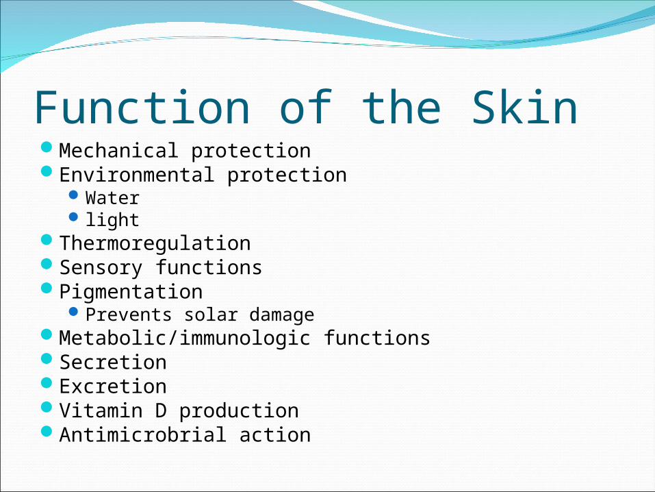
Function of the SkinMechanical protectionEnvironmental protection
Water light
ThermoregulationSensory functionsPigmentation
Prevents solar damageMetabolic/immunologic functionsSecretionExcretionVitamin D productionAntimicrobrial action

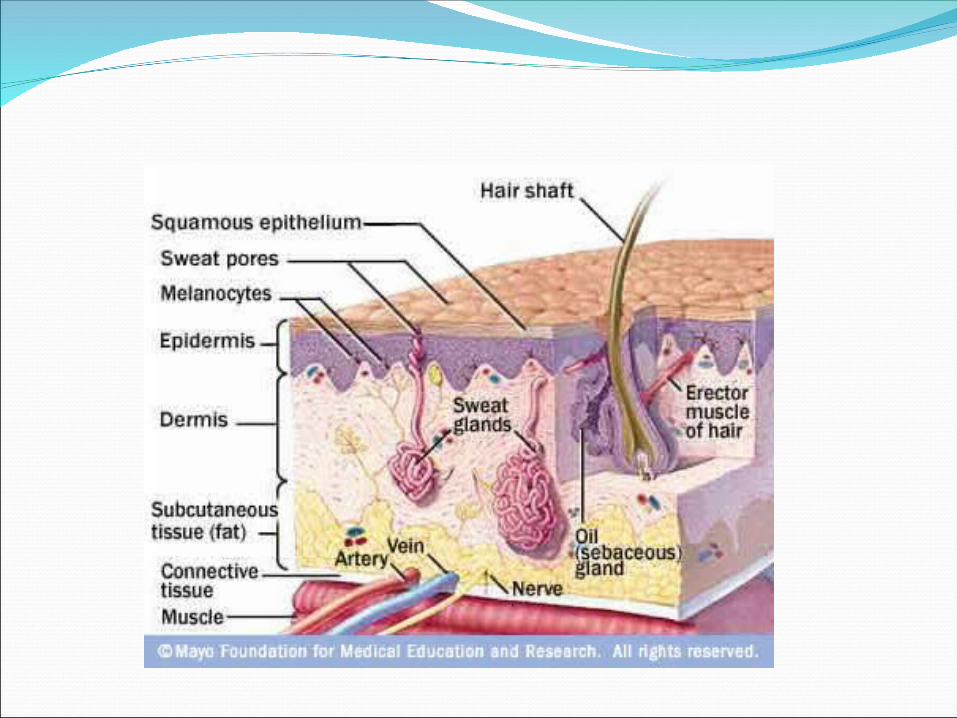
Structure of the SkinEpidermis
Squamous keratinized epithelium (5 layers) sits on basement membrane
DermisCollagen fibers, blood, lymphatic vessels,
nerves, fibroblasts, ground substance.Subcutaneous layerHair follicles
Epidermal invaginations into the dermis.

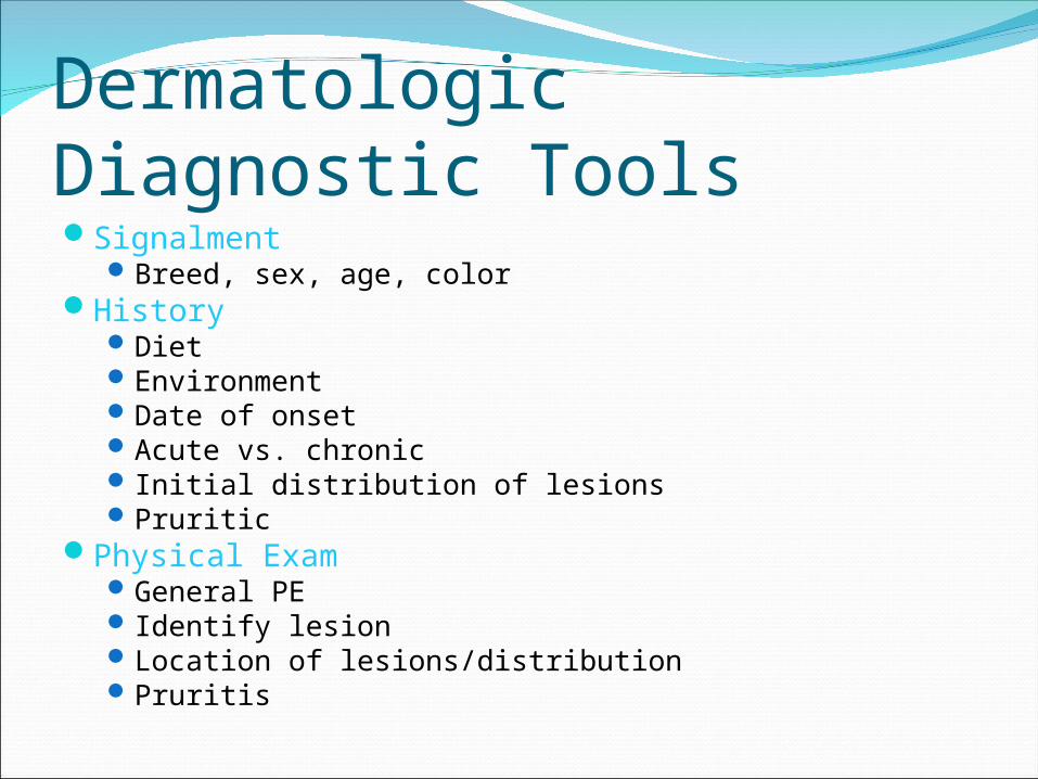
Dermatologic Diagnostic ToolsSignalment
Breed, sex, age, colorHistory
DietEnvironmentDate of onsetAcute vs. chronicInitial distribution of lesionsPruritic
Physical ExamGeneral PEIdentify lesionLocation of lesions/distributionPruritis


Diagnostic TestsWood’s lampSkin scrapingTape strip testDirect smear/impression smearsFungal cultures/ microscopic examsBacterial cultureBiopsyFine needle aspirateSwab


Skin Biopsy Punch


Dermatologic Terms for LesionsPrimary Lesion:
Develop spontaneously as a direct reaction of the underlying disease.
Secondary Lesion:Evolve from primary lesions.
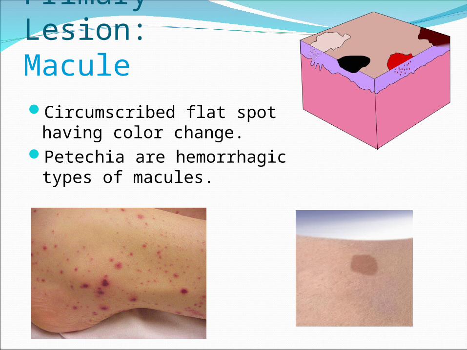
Primary Lesion: MaculeCircumscribed flat spot
having color change.Petechia are hemorrhagic
types of macules.
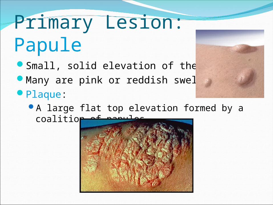
Primary Lesion: PapuleSmall, solid elevation of the skinMany are pink or reddish swellingsPlaque:
A large flat top elevation formed by a coalition of papules.
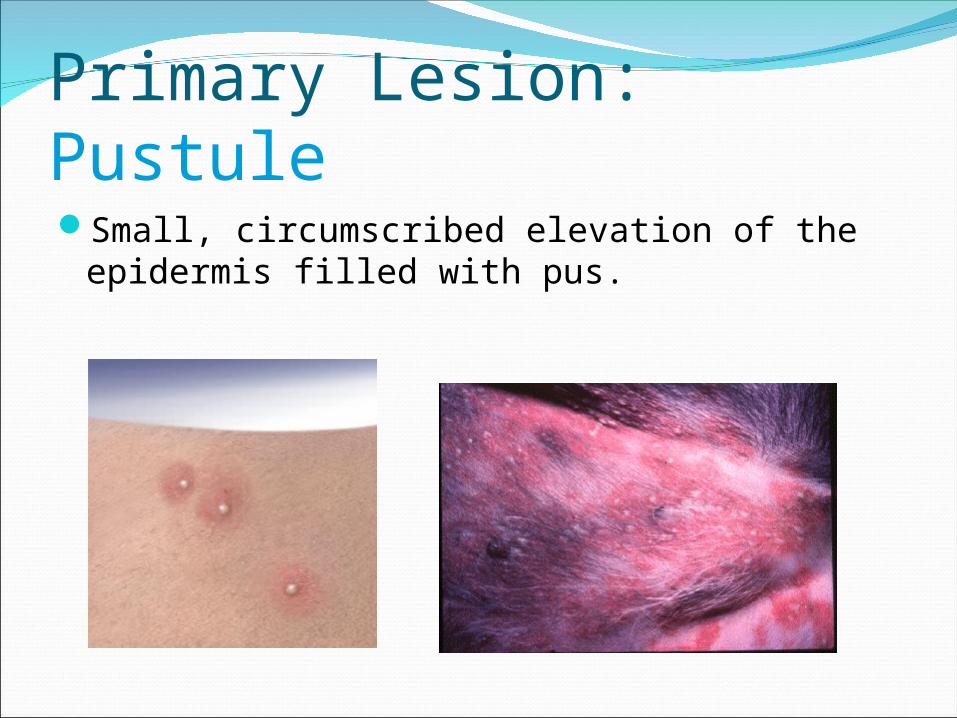
Primary Lesion: PustuleSmall, circumscribed elevation of the
epidermis filled with pus.
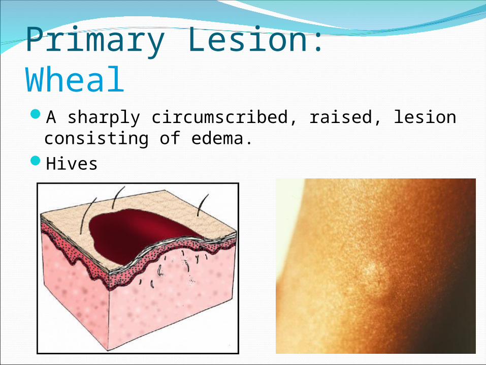
Primary Lesion: WhealA sharply circumscribed, raised, lesion
consisting of edema.Hives
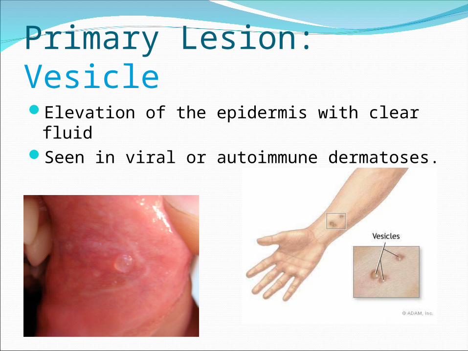
Primary Lesion: VesicleElevation of the epidermis with clear fluidSeen in viral or autoimmune dermatoses.
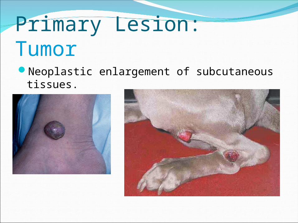
Primary Lesion: TumorNeoplastic enlargement of subcutaneous
tissues.
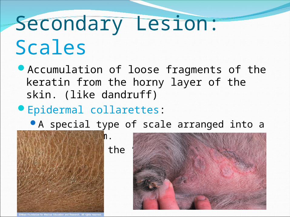
Secondary Lesion: ScalesAccumulation of loose fragments of the
keratin from the horny layer of the skin. (like dandruff)
Epidermal collarettes:A special type of scale arranged into a circular
rim.Remnants of the “roof” of a vesicle or pustule.
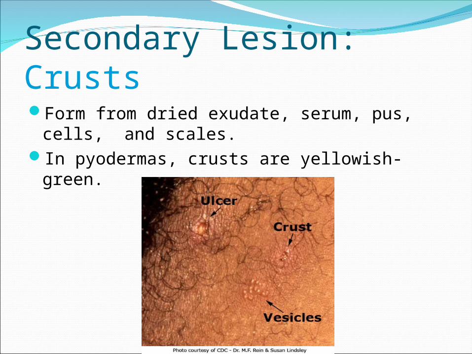
Secondary Lesion: CrustsForm from dried exudate, serum, pus, cells,
and scales.In pyodermas, crusts are yellowish-green.
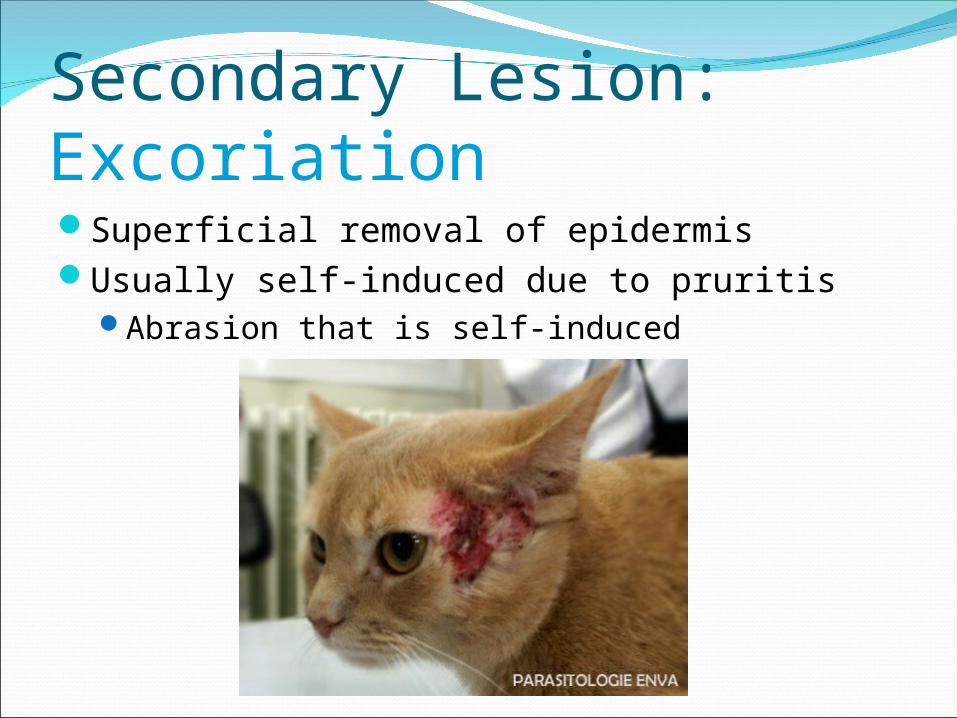
Secondary Lesion: ExcoriationSuperficial removal of epidermisUsually self-induced due to pruritis
Abrasion that is self-induced
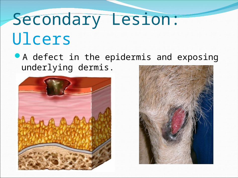
Secondary Lesion: UlcersA defect in the epidermis and exposing
underlying dermis.
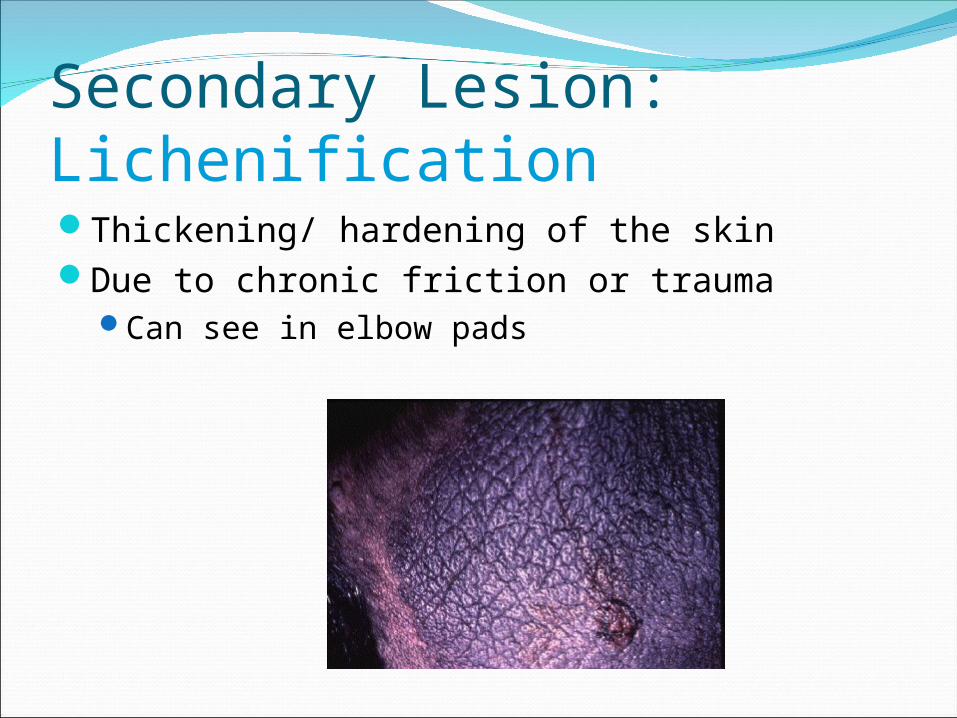
Secondary Lesion: LichenificationThickening/ hardening of the skinDue to chronic friction or trauma
Can see in elbow pads

Secondary Lesion: HyperpigmentationAbnormal pigment of the skin

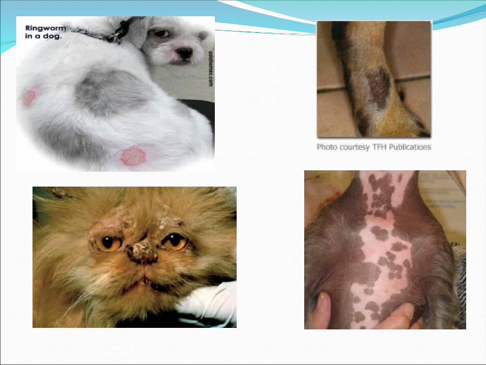
Dermatophytosis/RingwormInfection of the hair shafts and skin.Microsporum canis most common causeIn rare instances- Microsporum gyseum and
Trichophyton spp. Infective spores in soil, by direct contact, and by
environmental fomites, ventilation.Trauma to skin may promote infectionOther risk factors:
AgeImmune competence
Lesions may be circular, irregular, crusts, scales, hair thinning.
Classification of DermatophytesAnthropophilic:
Inhabit people onlyZoophilic:
Inhabit both animals and peopleGeophilic:
Free-living saprophytes in soil. May be contaminant in cultures.
Microsporum gypseum only species that causes lesions in animals.
Diagnosis:
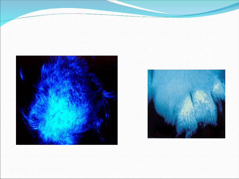
Woods Lamp:50% of Microsporum canis strains will
fluoresce under a woods lamp.Looking for an “apple green” fluorescence
Fungal Culture:Saboraud’s medium or Dermatophyte Test
Medium (DTM) specifically designed for ringworm diagnosis.
Color change before 10 daysMicroscopic Exam of the colony:
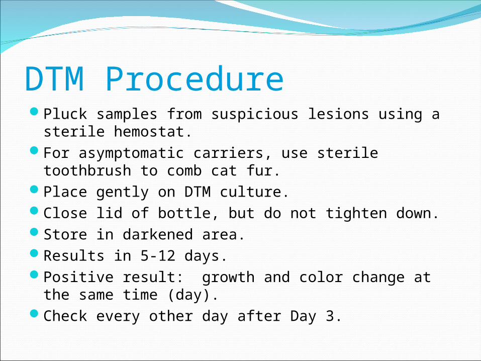
DTM ProcedurePluck samples from suspicious lesions using a sterile
hemostat.For asymptomatic carriers, use sterile toothbrush to
comb cat fur.Place gently on DTM culture.Close lid of bottle, but do not tighten down.Store in darkened area.Results in 5-12 days.Positive result: growth and color change at the same
time (day).Check every other day after Day 3.
Special DTM notes:Pigs
Often have contamination from geophilic/saprphytic fungi.
Swab lesion with alcohol, let dry, then collect sample.


Dermatophyte Identification using Colony Morphology Microsporum canis: Surface is
white and woolly. Reverse side is yellow.
Microsporum gypseum: Surface is coarsely powdery, light tan to cinnamon brown. Reverse is brownish yellow.
Trichophyton mentagrophytes: Surface is cream colored and powdery. Reverse is yellowish to brown.

Microsporum canisConfirm with microscopic examMacroconidia have thick walls, spindle
shaped 8-15 cells and possess a terminal knob.

Microsporum gypseumSpindle shaped but broader with no terminal
knobs. Less than 6 cells on macroconidia.

Trichophyton MentagrophytesFew macroconidia, slender and cigar shaped
with thin walls. Microconidia are numerous and arranged in grape-like clusters.

Direct Microscopic examination of RingwormSelect a few hairs or skin scrape. May be suspended in mineral oil, through direct tape
method or placed in a drop of 20% KOH (if use this method, gently heat and let stand for 10-15 minutes).
Examine under low and high power for fungal spores.
If looking at colonies, tease out a little colony material and place on slide.
Gently touch 2 cm strip of clear tape to surface of colony and then stain with new methylene blue or lactophenol cotton blue stains.
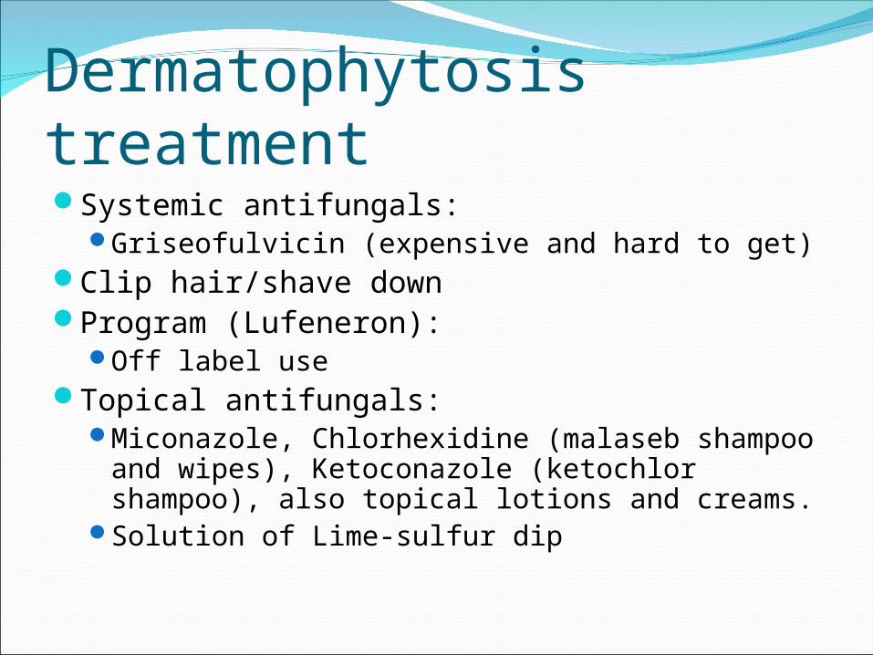
Dermatophytosis treatmentSystemic antifungals:
Griseofulvicin (expensive and hard to get)Clip hair/shave downProgram (Lufeneron):
Off label useTopical antifungals:
Miconazole, Chlorhexidine (malaseb shampoo and wipes), Ketoconazole (ketochlor shampoo), also topical lotions and creams.
Solution of Lime-sulfur dip