29
Clinical Pearls: How I Test Nancy Berliner, M.D. Chief, Division of Hematology Brigham and Women’s Hospital Intensive Review of Internal Medicine July 6, 2020

Clinical Pearls: How I Test
Nancy Berliner, M.D.Chief, Division of Hematology
Brigham and Women’s HospitalIntensive Review of Internal Medicine
July 6, 2020

Nancy Berliner, M.D.
H. Franklin Bunn Professor of Medicine
Chief, Division of Hematology, BWH
Yale Medical School
Residency and Chief Residency at BWH
Hematology Fellowship at BWH
Editor-in-Chief, Blood

Disclosure Information: Nancy Berliner
I have no financial relationships to disclose.
AND
I will NOT include discussion of off-label or investigational use of any products in my
presentation.

Reticulocyte count
Corrected Reticulocyte Count/Reticulocyte Index
Reticulocyte Index = Retic count X Hematocrit
Normal Hct (45)
Reticulocyte Index in a normal healthy adult is between 1 and 2
Evaluation of Anemia

Low retic index
Iron deficiency
Anemia of inflammation
Sideroblastic anemia
Thalassemias
Renal failure
Aplastic anemia
Hypothyroidism
B12/folate deficiency
MDS
Alcohol liver disease
Hemolytic anemias
Blood loss
Low MCV
Normal MCV
High MCV
Reticulocyte Index and MCV
High retic index
Evaluation of Anemia

• Iron Studies: Fe/TIBC/Ferritin
• Erythropoietin level
• B12, folate
• CRP/ESR • Important for interpretation of ferritin, as it is an acute
phase reactant
• In true iron deficiency, one cannot raise ferritin to over about 100
Evaluation of Hypoproliferative Anemia
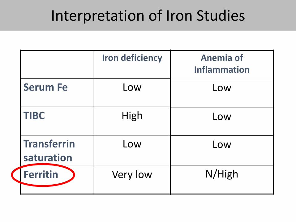
Iron deficiency
Serum Fe Low
TIBC High
Transferrinsaturation
Low
Ferritin Very low
Anemia of Inflammation
Low
Low
Low
N/High
Iron Deficiency AnmiaInterpretation of Iron Studies

Anemia of Inflammation
• Characterized by low serum Fe/TIBC in setting of elevated ferritin
• Associated with a wide variety of clinical disorders• Infections (bacterial endocarditis)
• Rheumatologic Disease (rheumatoid arthritis, SLE)
• Organ dysfunction (CHF, chronic renal failure)
• Malignancy (MDS, NHL)
• Pathophysiology• Impaired EPO responsiveness of hematopoietic stem cell
• shortened red cell survival
• impaired iron mobilization iron-limited erythropoiesis related to overexpression of hepcidin

Erythropoietin and Anemia of the Elderly
• Epo secretion and Epo responsiveness of HSCs may be altered with age
• Epo levels rise with age in healthy, non-anemicindividuals
• Slope of the rise greater in those without diabetes or hypertension
• Anemic individuals had a lower slope of rise
• Hypothesis: Anemia reflects failure of a normal compensatory rise in Epo levels, reflecting age-related co-morbidities
Even in the setting of a normal creatinine, EPO may help interpret anemia, especially in elderly patients
Ferrucci et al; Blood 2010; 115: 3810
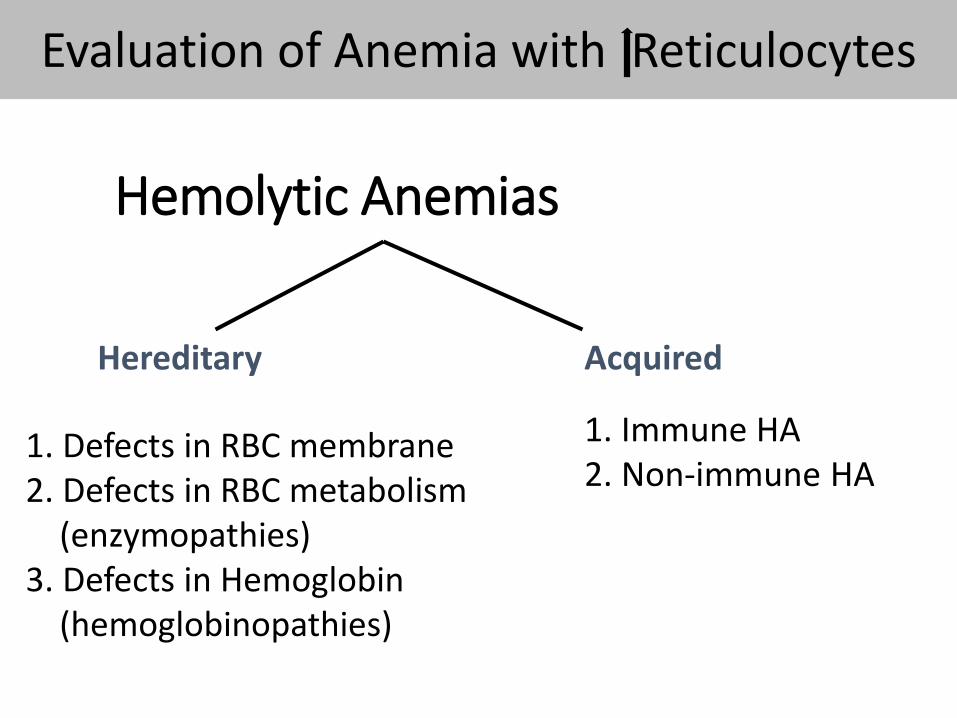
Hemolytic Anemias
Hereditary Acquired
1. Defects in RBC membrane2. Defects in RBC metabolism
(enzymopathies)3. Defects in Hemoglobin
(hemoglobinopathies)
1. Immune HA2. Non-immune HA
Evaluation of Anemia with Reticulocytes

Warm AIHA Cold AIHA
Direct Coombs
IgG or IgG & C3 C3 only
Antibody IgG IgM
Etiology 1. Drugs: Methyldopa, PCN,
Sulfa
2. Malignancy: CLL, NHL
3. Infection
1. Drugs: Quinidine
2. Malignancy: NHL
3. Infection: Mycoplasma
4. Paroxysmal cold hemoglobinuria
Treatment Steroids +/- Danazol
Rituximab
Splenectomy
No role for steroids
Warm pt
Rituximab +/- fludarabine
Autoimmune Hemolytic Anemia


Leukocytosis: Differential Diagnosis
SECONDARY TO OTHER ILLNESSESInfection
Acute: Demargination/release storage poolChronic: Granulomatous dx (leukoerythroblastic)
StressDrug-induced (steroids, b-agonists, lithium)Chronic inflammationPost-splenectomyNon-hematologic malignancyMarrow stimulation (ITP, hemolysis, CMT)
PRIMARY HEMATOLOGIC DISEASECMLOther MPD

Evaluation of Leukocytosis
Neutrophilia is usually reactive, indicative of a normal functioning bone marrow. Bone marrow evaluation is often unnecessary
•Repeat WBC to R/O factitious or artifactual elevation
•Evaluation for acute/chronic infection or inflammation
•FISH for bcr-abl
•Bone marrow exam: r/o granulomatous dx, fungus

Neutropenia: Differential Diagnosis
Congenital Neutropenia• Benign ethnic/familial neutropenia• Severe congenital neutropenia • Cyclic neutropenia• Other rare disorders
Acquired Neutropenia• Autoimmune neutropenia• Drug-induced neutropenia• Chronic idiopathic neutropenia• Primary marrow failure syndromes (MDS, aplasia)

Evaluation of Neutropenia
For Congenital Neutropenia: • Molecular Diagnosis for ELANE, HAX1
• Some advocate for testing for Duffy antigen negative phenotype in suspected ethnic neutropenia
Acquired Neutropenia• Stop possible offending drugs• Flow cytometry for clonality, LGL• Serologic studies for ANA• Anti-neutrophil antibodies are not recommended• R/O MDS: NGS or bone marrow examination

Evaluation of Neutropenia
Stop potential offending drugsBone marrow aspiration/biopsySerologic studies: ANA, viral titers, anti-neutrophil antibodiesR/O Primary malignancy:
Chromosome analysisSucrose-hemolysis test; flow cytometry

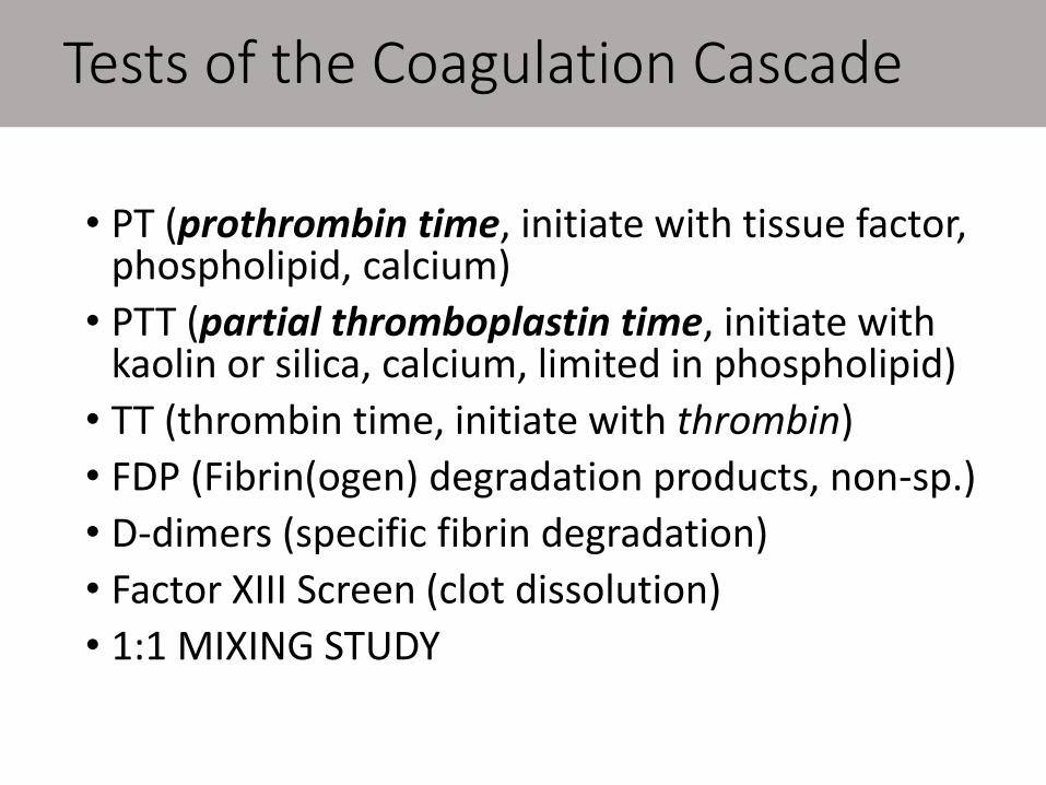
Tests of the Coagulation Cascade
• PT (prothrombin time, initiate with tissue factor, phospholipid, calcium)
• PTT (partial thromboplastin time, initiate with kaolin or silica, calcium, limited in phospholipid)
• TT (thrombin time, initiate with thrombin)
• FDP (Fibrin(ogen) degradation products, non-sp.)
• D-dimers (specific fibrin degradation)
• Factor XIII Screen (clot dissolution)
• 1:1 MIXING STUDY

Causes of PT
Elevated PT:
• Less than 30% of VII (the sole “extrinsic pathway only” protein), X, V, II (common pathway) or fibrinogen
• Inhibitors of fibrin polymerization (FDPs)
• Inhibitors of II or X
• Heparin in vast excess
Most Common Causes
• Vitamin K deficiency
• Warfarin Therapy
• Liver Disease

Causes of PTT
Elevated PTT:
• Any factor level less than 30% except VII,XIII
• Inhibitors of fibrin polymerization (FDP)
• Other inhibitors (lupus anticoagulants)
• Heparin (and warfarin, to lesser degree)
Most Common Causes
• Congenital factor deficiency
• Acquired factor Inhibitors
• DIC
• Dysfibrinogenemia
• Lupus anticoagulant

Interpretation of Mixing Studies for PTTs: deficiency vs inhibitor
PTT PTT PTT
Pt Plasma Nml Plasma 1:1 Mix
Factor 70 sec 30 sec 33 sec
Deficient
Inhibitor 70 sec 30 sec 70 sec


Risks for hypercoagulable states• Inherited
• Acquired: more common• 35% US adults are obese, OR of 2.3 for VTE
• <10% have an inherited thrombophilia
• Mixed: all are additive or synergistic
“Provoked” vs “Unprovoked”• Clear precipitating factor vs idiopathic or unidentified risk
factor• Transient vs persistent provoking factor
• Unprovoked = idiopathic
VTE

The “Hypercoagulable Workup”
Test for Factor V Leiden mutation
PCR for Prothrombin G20210A mutation
Functional assay of Antithrombin
Functional assay of Protein C
Functional assay of Protein S
• Free Protein S Antigen
• Total Protein S Antigen (free + bound to C4bp)
WHAT NOT to test:Homocysteine: FVIIIXIII polymorphisms, IX, XI,XIIPAI-1 4G/5G promoter, PAI-1
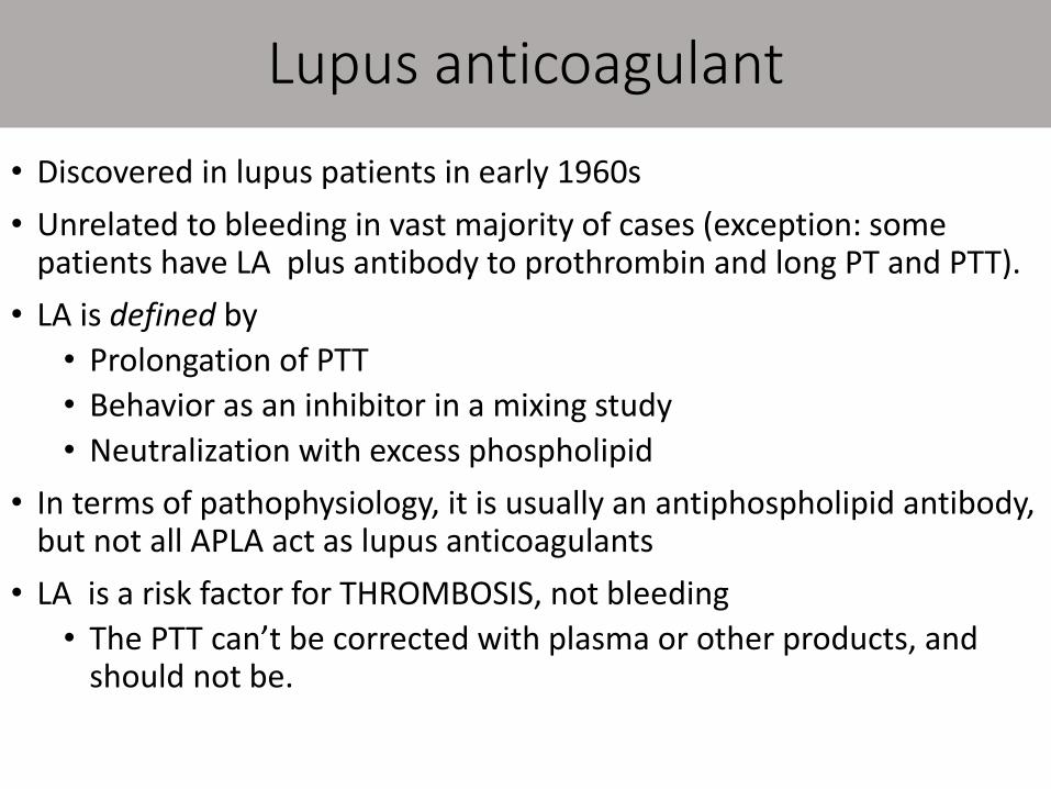
Lupus anticoagulant
• Discovered in lupus patients in early 1960s
• Unrelated to bleeding in vast majority of cases (exception: some patients have LA plus antibody to prothrombin and long PT and PTT).
• LA is defined by
• Prolongation of PTT
• Behavior as an inhibitor in a mixing study
• Neutralization with excess phospholipid
• In terms of pathophysiology, it is usually an antiphospholipid antibody, but not all APLA act as lupus anticoagulants
• LA is a risk factor for THROMBOSIS, not bleeding
• The PTT can’t be corrected with plasma or other products, and should not be.

Tests for Antiphospholipid Antibodies• Lupus anticoagulant:
• Screen: functional clotting assays• Sensitive PTT
• DRVVT
• Kaolin clotting time
• Confirmatory: remove APLA• Platelet neutralization test
• Hexagonal phase phospholipids
• Anticardiolipin and b2-glycoprotein I antibodies• IgG and IgM only
• No diagnostic role for other tests
APLA work-up

Who should be tested?`
Indications of possible inherited hypercoagulable state:
• Age of onset < 50 years
• Recurrent thrombosis
• Positive family history in 1st degree relative
• Unusual location/site
However:
• Avoid indiscriminate testing in the inpatient or ER setting
• There is no need to know immediately—require at least 3 months anticoagulation for VTE regardless of thrombophilia status

Why the controversy?• There are no data that results should affect care• ASH Choosing Wisely Campaign 2013: “do not test in the
setting of provoked VTE due to strong risks”Misinterpretation of the significance of results
• Over treatment in the case of positive results• Duration of therapy determined by provoked vs unprovoked VTE
• False sense of security with negative results• Studies demonstrate increased VTE risk for patients with a family
history of VTE despite negative results
When does it not change care?• Provoked VTE • Antiphospholipid syndrome• Malignancy
Thrombophilia Testing Remains Controversial


