Decisionmaking in Adult Mild Decisionmaking in Adult Mild Traumatic Brain Injury in the Traumatic Brain Injury in the Acute Setting Acute Setting An ACEP / CDC Collaborative An ACEP / CDC Collaborative Project Project Andy Jagoda, MD Andy Jagoda, MD Professor of Emergency Medicine Professor of Emergency Medicine Mount Sinai School of Medicine Mount Sinai School of Medicine New York, New York New York, New York
Transcript
Clinical Policy: Neuroimaging and Clinical Policy: Neuroimaging and Decisionmaking in Adult Mild Traumatic Decisionmaking in Adult Mild Traumatic
Brain Injury in the Acute SettingBrain Injury in the Acute Setting
Professor of Emergency MedicineProfessor of Emergency MedicineMount Sinai School of MedicineMount Sinai School of Medicine
New York, New YorkNew York, New York
Andy Jagoda, MD, FACEP
DisclosuresDisclosures
• Executive Committee, Foundation for Education and Research in Neurologic Emergencies
• No individual financial disclosures
Andy Jagoda, MD, FACEP
IntroductionIntroduction
• Revision of a policy first published in 2002• Project funded by the CDC
Public health issue “Signature casualty of the the war in Iraq
• Approximately 1 million ED visits for TBI Majority of TBI are “mild” Challenge is to identify patients with MTBI
who have neurosurgical lesions or at risk for post concussive symptoms / syndrome
Andy Jagoda, MD, FACEP
IntroductionIntroduction
• Less than 10% of patients with a GCS of 14 / 15 have evidence of an acute intracranial lesion on head CT
• Up to 30% of patients with a GCS of 13 have an acute intracranial lesion on head CT
• Less than 1% of patients with GCS of 14 / 15
Andy Jagoda, MD, FACEP
MTBI: DefinitionMTBI: Definition
• Blunt head injury (or blast?) GCS score of 13 – 15 Any period of observed or self-reported:
Loss of consciousnesstransient confusion, disorientation, or impaired
consciousnessdysfunction of memory (amnesia) around the
injuryneurological or neuropsychological dysfunction
Andy Jagoda, MD, FACEP
MTBI: DefinitionMTBI: Definition
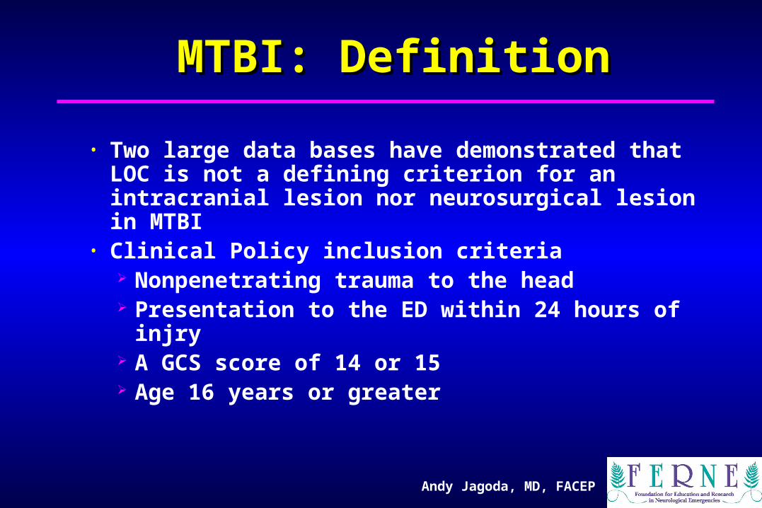
• Two large data bases have demonstrated that LOC is not a defining criterion for an intracranial lesion nor neurosurgical lesion in MTBI
• Clinical Policy inclusion criteria Nonpenetrating trauma to the head Presentation to the ED within 24 hours of injry A GCS score of 14 or 15 Age 16 years or greater
Andy Jagoda, MD, FACEP
GCS and NeuroimagingGCS and Neuroimaging
• Dx of MTBI does not take into account neuroimagingDx of MTBI does not take into account neuroimaging
• Retrospective study, 215 hospitalized patientsRetrospective study, 215 hospitalized patients Mild TBI without complicationsMild TBI without complications
Mild TBI with complications (positive CT)Mild TBI with complications (positive CT)
Moderate TBIModerate TBI
• Mild TBI patients with positive CT performed on Mild TBI patients with positive CT performed on neuropsychiatric testing like moderate TBI neuropsychiatric testing like moderate TBI
• Moderate group had worse function at 6 monthsModerate group had worse function at 6 months
• Length of LOC or amnesia did not differentiate mild from Length of LOC or amnesia did not differentiate mild from moderate groupsmoderate groups
Williams et al. Williams et al. Neurosurgery Neurosurgery 1990;27:422.1990;27:422.
Andy Jagoda, MD, FACEP
2002 Clinical Policy2002 Clinical Policy
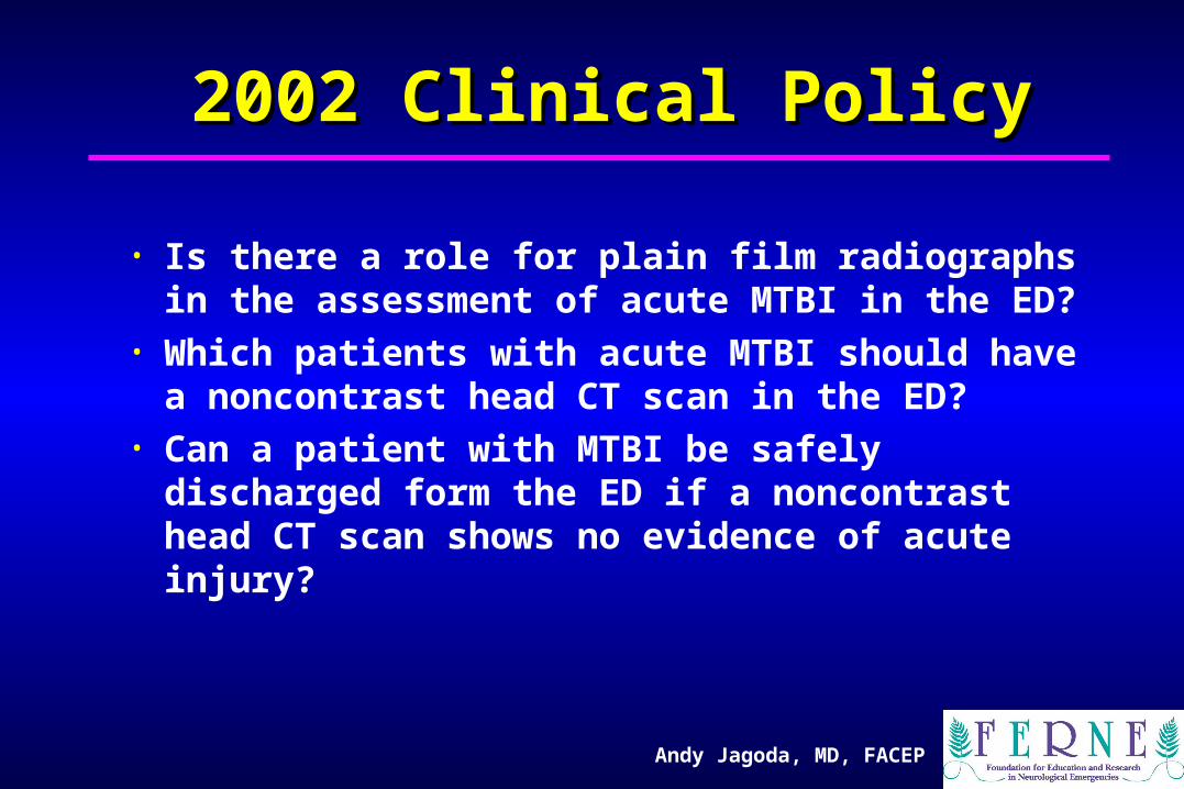
• Is there a role for plain film radiographs in the assessment of acute MTBI in the ED?
• Which patients with acute MTBI should have a noncontrast head CT scan in the ED?
• Can a patient with MTBI be safely discharged form the ED if a noncontrast head CT scan shows no evidence of acute injury?
• Hoffman 2000 Lancet: Meta-analysisHoffman 2000 Lancet: Meta-analysis 20 articles reviewed out of 200 identified20 articles reviewed out of 200 identified Sensitivity .13-.75; PPV of skull fracture in Sensitivity .13-.75; PPV of skull fracture in
prediciting +CT .4prediciting +CT .4 Specificity .9-.99; NPVof skull fracture in Specificity .9-.99; NPVof skull fracture in
predicting +CT .94 predicting +CT .94 • Skull films are not recommended in the evaluation Skull films are not recommended in the evaluation
of MTBI; although the presence of a skull film of MTBI; although the presence of a skull film increases the liklihood of an intracranial lesion, its increases the liklihood of an intracranial lesion, its sensitivity is not high enough to allow it to be a sensitivity is not high enough to allow it to be a useful screenuseful screen
Is there a role for plain skull radiographs in the Is there a role for plain skull radiographs in the evaluation of patients with a MTBI? evaluation of patients with a MTBI?
Andy Jagoda, MD, FACEP
2008 Clinical Policy2008 Clinical Policy
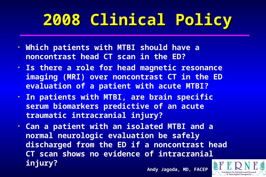
• Which patients with MTBI should have a noncontrast head CT scan in the ED?
• Is there a role for head magnetic resonance imaging (MRI) over noncontrast CT in the ED evaluation of a patient with acute MTBI?
• In patients with MTBI, are brain specific serum biomarkers predictive of an acute traumatic intracranial injury?
• Can a patient with an isolated MTBI and a normal neurologic evaluation be safely discharged from the ED if a noncontrast head CT scan shows no evidence of intracranial injury?
• Presence of any acute intracranial injury on noncontrast head CT scan was chosen as the primary outcome measure for Questions #1,2,3 Lesion requiring a neurosurgical intervention
was the secondary outcome measure• Neurologic deterioration was the primary
outcome measure for Question #4• There is insufficient evidence to use post
concussive symptoms / syndrome as an outcome measure
Andy Jagoda, MD, FACEP
Description of the ProcessDescription of the ProcessStrength of evidence (Class of evidence)
I: Randomized, double blind interventional studies for therapeutic effectiveness; prospective cohort for diagnostic testing or prognosis
II: Retrospective cohorts, case control studies, cross-sectional studies
III: Observational reports; consensus reports
Strength of evidence downgraded based on methodologic flaws
Andy Jagoda, MD, FACEP
Description of the process:Description of the process:
Strength of recommendations:Strength of recommendations:
• A / Standard:A / Standard: Reflects a high degree of Reflects a high degree of
certainty based on Class I studiescertainty based on Class I studies
based on Class II studiesbased on Class II studies
• C / Option:C / Option: Inconclusive certainty based on Inconclusive certainty based on
Class III evidenceClass III evidence
Andy Jagoda, MD, FACEP
Which patients with MTBI should have a Which patients with MTBI should have a noncontrast head CT scan in the ED?noncontrast head CT scan in the ED?
• Level A RecommendationLevel A Recommendation: A noncontrast head Ct : A noncontrast head Ct is indicated in head trauma patients with loss of is indicated in head trauma patients with loss of consciousness or post traumatic amnesia only if consciousness or post traumatic amnesia only if one or more of the following is present: headache, one or more of the following is present: headache, vomiting, age greater than 60 years, drug or vomiting, age greater than 60 years, drug or alcohol intoxication, deficits in short term memory, alcohol intoxication, deficits in short term memory, physical evidence of trauma above the clavicle, physical evidence of trauma above the clavicle, posttraumatic seizure, GCS score less than 15, posttraumatic seizure, GCS score less than 15, coagulopathy, or focal neurologic deficit.coagulopathy, or focal neurologic deficit.
Andy Jagoda, MD, FACEP
Which patients with MTBI should have a Which patients with MTBI should have a noncontrast head CT scan in the ED?noncontrast head CT scan in the ED?
• Haydel 2000 NEJM; Class I study; 2 phasesHaydel 2000 NEJM; Class I study; 2 phases Phase I 520 patients to establish predictive criteriaPhase I 520 patients to establish predictive criteria Phase II 909 patients to validate criteriaPhase II 909 patients to validate criteria 7 predictors identified with 100% sensitivity for 7 predictors identified with 100% sensitivity for
predicting intracranial lesion predicting intracranial lesion in patients with LOCin patients with LOC Use of criteria would decrease head CT by 22%Use of criteria would decrease head CT by 22%
• High sensitivity (95-100%) but low sensitivity (5-30%) High sensitivity (95-100%) but low sensitivity (5-30%) confirmed by large data bases in Italy, Holland, and Spainconfirmed by large data bases in Italy, Holland, and Spain
• Canadian CT Head Rule has higher specificity (50%) BUT Canadian CT Head Rule has higher specificity (50%) BUT the primary outcome measure is neurosurgical lesion and the primary outcome measure is neurosurgical lesion and minor acute intracranial lesions (e.g. small subdurals and minor acute intracranial lesions (e.g. small subdurals and SAH) are not considered important SAH) are not considered important
Andy Jagoda, MD, FACEP
Which patients with MTBI should have a Which patients with MTBI should have a noncontrast head CT scan in the ED?noncontrast head CT scan in the ED?
• Level B RecommendationLevel B Recommendation: A noncontrast head CT : A noncontrast head CT should be considered in head trauma patients should be considered in head trauma patients with with nono loss of consciousness or post traumatic loss of consciousness or post traumatic amnesia if there is a focal neurologic deficit, amnesia if there is a focal neurologic deficit, vomiting, severe, headache, age 65 years or vomiting, severe, headache, age 65 years or greater, physical signs of a basilar skull fracture, greater, physical signs of a basilar skull fracture, GCS score less than 15, coagulapathy, or a GCS score less than 15, coagulapathy, or a dangerous mechanism of injurydangerous mechanism of injury
Smits 13
OR (95%CI)Ibanez8 OR (95%CI)
Fabbri9 OR (95%CI)
Class of evidence II II III
Variable
GCS 14 2 (1-3) 7 (4-14) 19 (14 - 26)
Neurologic deficit 2 (1-3) 7 (2-25) 19 (13-28)
Signs of basilar skull fracture
25 (13-47) 11 (6-23) 10 (6-16)
LOC 2 (1-3) 7 (4-11) 2 (2-3)
Post traumatic amnesia 1.5 (1-2) 3 (2-5) 8 (6-12)-
Headache mild - moderate 1 (0.8-2) 1 (0.8-2) -
Headache – severe 3 (2-6) -
Vomiting 3 (2-4) 4 (2-7) 5 (3-8)
Post traumatic seizure 3 (0.8-10) 2 (0.25-17) 2 (3-5)
Alcohol or drug intoxication 1 (0.6-2) 1 (0.3-3) -
Anticoagulation 2 (1-5) 4 (3-7) 8 (3-9)
Age >65 years - 2 (1-3) 2 (1-3)
Dangerous mechanism 2 (1-4) - 3 (2-4)
Andy Jagoda, MD, FACEP
Is there a role for head MRI over noncontrast CT Is there a role for head MRI over noncontrast CT in the ED evaluation of a paitent with acute MTBIin the ED evaluation of a paitent with acute MTBI
• MRI is up to 30% more sensitive than CT in detecting axonal shear injury
• No studies looked specifically at patients with MTBI within 24 hours of injury
• No studies have shown a clinical correlation with MRI abnormalities with neuropsychologic outcome
• No recommendations
Andy Jagoda, MD, FACEP
In patients with MTBI, are brain specific serum biomarkers In patients with MTBI, are brain specific serum biomarkers predictive of an acute traumatic intracranial injury?predictive of an acute traumatic intracranial injury?
• Traumatic injury results in the release of proteins Neuronal proteins include neuron specific enolase
and tau Astrocyte proteins include S-100B, CK-BB
• S-100B is best studied; found within the serum within 30 minutes with a half life of 97 minutes Elevated in multiple trauma Elevated in marathon runners
• Eight studies reviewed; 2 Class II Sensitivities 90 – 100% Specificities 4 – 65%
Andy Jagoda, MD, FACEP
In patients with MTBI, are brain specific serum biomarkers In patients with MTBI, are brain specific serum biomarkers predictive of an acute traumatic intracranial injury?predictive of an acute traumatic intracranial injury?
• Biberthaler et al: 1309 patients with isolated MTBI and CT scan Cutoff for serum level 0.1 ug / L 7% had an acute intracranial lesion Sensitivity 0.99 (CI 0.96 – 1.0) Specificity 0.30 (CI0.29 – 0.31)
• Level B Recommendation: In patients with MTBI without significant extracranial injuries, a CT should be considered if the S-100B serum level is >0.1 ug / L
Andy Jagoda, MD, FACEP
Can a patient with an isolated MTBI and a normal neurologic Can a patient with an isolated MTBI and a normal neurologic evaluation be safely discharged fromthe ED is a noncontrast head evaluation be safely discharged fromthe ED is a noncontrast head CT scan shows no evidence of intracranial injury?CT scan shows no evidence of intracranial injury?
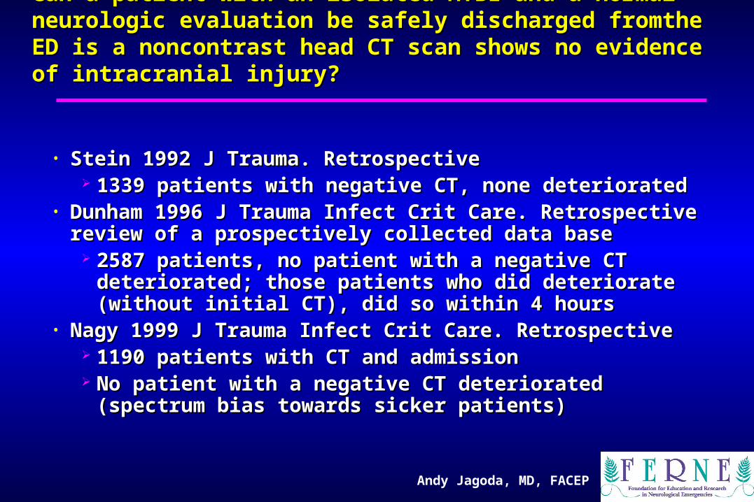
• Dunham 1996 J Trauma Infect Crit Care. Retrospective Dunham 1996 J Trauma Infect Crit Care. Retrospective review of a prospectively collected data basereview of a prospectively collected data base
2587 patients, no patient with a negative CT deteriorated; 2587 patients, no patient with a negative CT deteriorated; those patients who did deteriorate (without initial CT), did those patients who did deteriorate (without initial CT), did so within 4 hoursso within 4 hours
• Nagy 1999 J Trauma Infect Crit Care. Retrospective Nagy 1999 J Trauma Infect Crit Care. Retrospective 1190 patients with CT and admission1190 patients with CT and admission No patient with a negative CT deteriorated (spectrum bias No patient with a negative CT deteriorated (spectrum bias
towards sicker patients)towards sicker patients)
Andy Jagoda, MD, FACEP
Can a patient with an isolated MTBI and a normal neurologic Can a patient with an isolated MTBI and a normal neurologic evaluation be safely discharged from the ED is a noncontrast evaluation be safely discharged from the ED is a noncontrast
head CT scan shows no evidence of intracranial injury?head CT scan shows no evidence of intracranial injury?
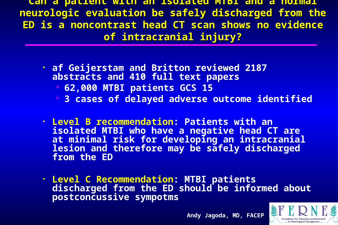
• af Geijerstam and Britton reviewed 2187 abstracts and 410 full text papers 62,000 MTBI patients GCS 15 3 cases of delayed adverse outcome identified
• Level B recommendation: Patients with an isolated MTBI who have a negative head CT are at minimal risk for developing an intracranial lesion and therefore may be safely discharged from the ED
• Level C Recommendation: MTBI patients discharged from the ED should be informed about postconcussive sympotms
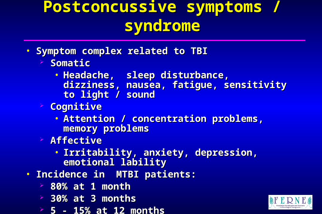
• Symptom complex related to TBISymptom complex related to TBI Somatic Somatic
In patients with a GCS of 15, what is the risk of developing the In patients with a GCS of 15, what is the risk of developing the postconcussive syndrome?postconcussive syndrome?
• Saunders et al. Saunders et al. Ann Emerg MedAnn Emerg Med 1986;15:160. 1986;15:160. 47 consecutive MTBI discharged from the ED47 consecutive MTBI discharged from the ED No patient could remember more than 2 of the 8 items No patient could remember more than 2 of the 8 items
on the home care discharge instructionson the home care discharge instructions 20% denied ever having received instructions20% denied ever having received instructions Third party involvement improved compliance with Third party involvement improved compliance with
instructions to 67%instructions to 67%• Levitt et al. Levitt et al. Amer J Emerg MedAmer J Emerg Med 1994;12:172. 1994;12:172.
23% of MTBI patients discharged from the ED could not 23% of MTBI patients discharged from the ED could not remember any of their discharge instructionsremember any of their discharge instructions
• Studies emphasize importance of involving third parties in Studies emphasize importance of involving third parties in discharge processdischarge process
• Instructions to patients and their families should: Be written at approximately a 6th to 7th grade level Given to the patient and immediate caregiver in both
print and verbal form Layout and type fonts appropriate for low literacy
materials (ie, no type font under 12 points, wide Margins, left justified)
• Patients who have been assessed in the ED using recommendations in this MTBI clinical policy have a very low rate of developing delayed intracranial pathology.
• For patients who have a negative CT or have been deemed too low risk for neuroimaging, home observation including frequent waking or assessment of pupils is not supported by the literature and thus is not recommended.
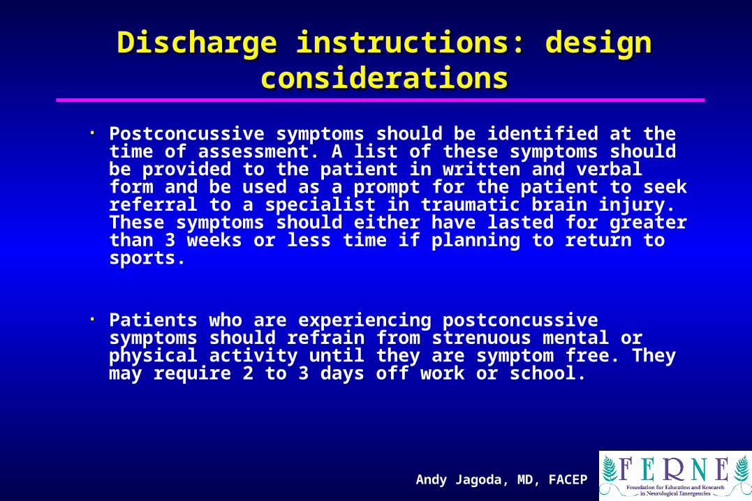
• Postconcussive symptoms should be identified at the time of assessment. A list of these symptoms should be provided to the patient in written and verbal form and be used as a prompt for the patient to seek referral to a specialist in traumatic brain injury. These symptoms should either have lasted for greater than 3 weeks or less time if planning to return to sports.
• Patients who are experiencing postconcussive symptoms should refrain from strenuous mental or physical activity until they are symptom free. They may require 2 to 3 days off work or school.
Andy Jagoda, MD, FACEP
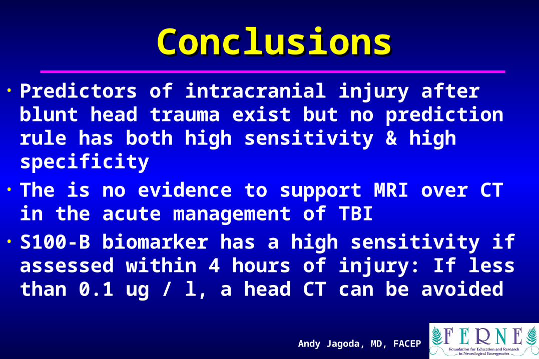
ConclusionsConclusions• Predictors of intracranial injury after blunt
head trauma exist but no prediction rule has both high sensitivity & high specificity
• The is no evidence to support MRI over CT in the acute management of TBI
• S100-B biomarker has a high sensitivity if assessed within 4 hours of injury: If less than 0.1 ug / l, a head CT can be avoided
Andy Jagoda, MD, FACEP
ConclusionsConclusions
• Patients with MTBI who have a normal exam and a negative head CT can be safely discharged
• Patients with MTBI discharged from the ED should receive both verbal and written information on post-concussive symptoms and provided follow up resources