META-ANALYSIS Clinical Presentation of Patients With Tension Pneumothorax A Systematic Review Derek J. Roberts, MD, ∗ †‡§ Simon Leigh-Smith, MBChB,¶ Peter D. Faris, PhD,†Christopher Blackmore, MD, ∗ Chad G. Ball, MD, MSc, ∗ § ∗∗ Helen Lee Robertson, MLIS,†† Elijah Dixon, MD, MSc, ∗∗∗ Matthew T. James, MD, PhD,†‡‡ Andrew W. Kirkpatrick, MD, MHSc, ∗ ‡§ John B. Kortbeek, MD, ∗ ‡§ and Henry T. Stelfox, MD, PhD†‡‡‡ Objective: To determine whether the reported clinical presentation of tension pneumothorax differs between patients who are breathing unassisted versus receiving assisted ventilation. Background: Animal studies suggest that the pathophysiology and physical signs of tension pneumothorax differ by subject ventilatory status. Methods: We searched electronic databases through to October 15, 2013 for observational studies and case reports/series reporting clinical manifestations of tension pneumothorax. Two physicians independently extracted clinical manifestations reported at diagnosis. Results: We identified 5 cohort studies (n = 310 patients) and 156 case series/reports of 183 cases of tension pneumothorax (n = 86 breathing unas- sisted, n = 97 receiving assisted ventilation). Hypoxia was reported among 43 (50.0%) cases of tension pneumothorax who were breathing unassisted versus 89 (91.8%) receiving assisted ventilation (P < 0.001). Pulmonary dysfunc- tion progressed to respiratory arrest in 9.3% of cases breathing unassisted. As compared to cases who were breathing unassisted, the adjusted odds of hypotension and cardiac arrest were 12.6 (95% confidence interval, 5.8–27.5) and 17.7 (95% confidence interval, 4.0–78.4) times higher among cases re- ceiving assisted ventilation. One cohort study reported that none of the patients with tension pneumothorax who were breathing unassisted versus 39.6% of those receiving assisted ventilation presented without an arterial pulse. In contrast to cases breathing unassisted, the majority (70.4%) of those receiving From the ∗ Department of Surgery, University of Calgary, Calgary, Alberta, Canada; †Department of Community Health Sciences, University of Calgary, Calgary, Alberta, Canada; ‡Department of Critical Care Medicine, University of Cal- gary, Calgary, Alberta, Canada; §Regional Trauma Program, University of Calgary and the Foothills Medical Centre, Calgary, Alberta, Canada; ¶United Kingdom Defence Medical Services and Emergency Department, Royal In- firmary of Edinburgh, Scotland, United Kingdom; Alberta Health Sciences Research—Research Analytics, University of Calgary and the Foothills Med- ical Centre, Calgary, Alberta, Canada; ∗∗ Department of Oncology, University of Calgary, Calgary, Alberta, Canada; ††Health Sciences Library, University of Calgary, Calgary, Alberta, Canada; and ‡‡Department of Medicine, University of Calgary, Calgary, Alberta, Canada. Disclosure: Dr Roberts is supported by an Alberta Innovates—Health Solutions Clinician Fellowship Award, a Knowledge Translation Canada Strategic Fund- ing in Health Research Fellowship, and funding from the Canadian Institutes of Health Research. The authors have no conflicts of interest to declare. Supplemental digital content is available for this article. Direct URL citations appear in the printed text and are provided in the HTML and PDF versions of this article on the journal’s Web site (www.annalsofsurgery.com). This is an open-access article distributed under the terms of the Creative Commons Attribution-NonCommercial-NoDerivitives 3.0 License, where it is permissible to download and share the work provided it is properly cited. The work cannot be changed in any way or used commercially. Reprints: Derek J. Roberts, MD, Departments of Surgery, Community Health Sci- ences, and Critical Care Medicine, University of Calgary and the Foothills Medical Centre, Intensive Care Unit Administration, Ground Floor McCaig Tower, Foothills Medical Centre, 3134 Hospital Dr Northwest, Calgary, Al- berta, Canada. E-mail: [email protected]. Copyright C 2014 by Lippincott Williams & Wilkins ISSN: 0003-4932/14/00000-0001 DOI: 10.1097/SLA.0000000000001073 assisted ventilation who experienced hypotension or cardiac arrest developed these signs within minutes of clinical presentation. Discussion: The reported clinical presentation of tension pneumothorax de- pends on the ventilatory status of the patient. This may have implications for improving the diagnosis and treatment of this life-threatening disorder. Keywords: assisted ventilation, breathing unassisted, clinical manifestations, clinical presentation, diagnosis, management, signs and symptoms, tension pneumothorax (Ann Surg 2014;00:1–11) T ension pneumothorax is an uncommon condition with a high mortality rate most frequently reported to occur in prehospital, emergency department, and intensive care unit (ICU) settings. 1–4 This condition is frequently lethal in injured and ventilated ICU patients without early diagnosis and treatment. 5–7 Although the incidence of tension pneumothorax remains poorly estimated, it may occur in up to 1% to 3% of prehospital, major trauma and ICU patients. 1,3,8–12 As many authorities recommend urgent thoracic decompres- sion when the diagnosis is first suspected, health care providers are taught to search for classically described clinical manifestations to recognize patients who may have tension pneumothorax. 5,6,13 Al- though tension pneumothorax is therefore a syndrome diagnosis, available literature sources differ substantially in their descriptions of its clinical presentation. 1 Many of these have been generalized from canine studies of the disorder, 14,15 and do not account for poten- tial differences in pathophysiology and physical signs based on the ventilatory status of the patient (Fig. 1). 1,6,16–21 As misdiagnosis or inappropriate treatment of tension pneu- mothorax can have devastating consequences, 6,22–26 a comprehensive description of its clinical presentation may improve patient care. 1 Thus, we conducted a systematic review to determine whether the reported clinical presentation (and resultant management and out- comes) of tension pneumothorax differs between patients who are breathing unassisted (ie, breathing spontaneously and not receiving positive pressure ventilation) versus receiving assisted (ie, positive pressure) ventilation. Our primary objective was to determine whether available clinical data support animal study observations of poten- tially important differences between subjects of varying ventilatory status in time to, severity, and frequency of presenting hemodynamic complications. 16,17,19 As systematic reviews of case reports and se- ries of other uncommon/emergent conditions have guided clinical practice and future research, 27–31 we synthesized and analyzed data reported by these types of studies alongside a systematic review of observational studies. 1,32–35 METHODS A published protocol details our study methods. 1 This protocol was registered in the PROSPERO Register of Systematic Reviews Annals of Surgery Volume 00, Number 00, 2014 www.annalsofsurgery.com | 1
Transcript
META-ANALYSIS
Clinical Presentation of Patients With Tension PneumothoraxA Systematic Review
Derek J. Roberts, MD,∗†‡§ Simon Leigh-Smith, MBChB,¶ Peter D. Faris, PhD,†‖ Christopher Blackmore, MD,∗
Chad G. Ball, MD, MSc,∗§∗∗ Helen Lee Robertson, MLIS,†† Elijah Dixon, MD, MSc,∗∗∗
Matthew T. James, MD, PhD,†‡‡ Andrew W. Kirkpatrick, MD, MHSc,∗‡§ John B. Kortbeek, MD,∗‡§and Henry T. Stelfox, MD, PhD†‡‡‡
Objective: To determine whether the reported clinical presentation of tensionpneumothorax differs between patients who are breathing unassisted versusreceiving assisted ventilation.Background: Animal studies suggest that the pathophysiology and physicalsigns of tension pneumothorax differ by subject ventilatory status.Methods: We searched electronic databases through to October 15, 2013 forobservational studies and case reports/series reporting clinical manifestationsof tension pneumothorax. Two physicians independently extracted clinicalmanifestations reported at diagnosis.Results: We identified 5 cohort studies (n = 310 patients) and 156 caseseries/reports of 183 cases of tension pneumothorax (n = 86 breathing unas-sisted, n = 97 receiving assisted ventilation). Hypoxia was reported among 43(50.0%) cases of tension pneumothorax who were breathing unassisted versus89 (91.8%) receiving assisted ventilation (P < 0.001). Pulmonary dysfunc-tion progressed to respiratory arrest in 9.3% of cases breathing unassisted.As compared to cases who were breathing unassisted, the adjusted odds ofhypotension and cardiac arrest were 12.6 (95% confidence interval, 5.8–27.5)and 17.7 (95% confidence interval, 4.0–78.4) times higher among cases re-ceiving assisted ventilation. One cohort study reported that none of the patientswith tension pneumothorax who were breathing unassisted versus 39.6% ofthose receiving assisted ventilation presented without an arterial pulse. Incontrast to cases breathing unassisted, the majority (70.4%) of those receiving
From the ∗Department of Surgery, University of Calgary, Calgary, Alberta, Canada;†Department of Community Health Sciences, University of Calgary, Calgary,Alberta, Canada; ‡Department of Critical Care Medicine, University of Cal-gary, Calgary, Alberta, Canada; §Regional Trauma Program, University ofCalgary and the Foothills Medical Centre, Calgary, Alberta, Canada; ¶UnitedKingdom Defence Medical Services and Emergency Department, Royal In-firmary of Edinburgh, Scotland, United Kingdom; ‖Alberta Health SciencesResearch—Research Analytics, University of Calgary and the Foothills Med-ical Centre, Calgary, Alberta, Canada; ∗∗Department of Oncology, Universityof Calgary, Calgary, Alberta, Canada; ††Health Sciences Library, University ofCalgary, Calgary, Alberta, Canada; and ‡‡Department of Medicine, Universityof Calgary, Calgary, Alberta, Canada.
Disclosure: Dr Roberts is supported by an Alberta Innovates—Health SolutionsClinician Fellowship Award, a Knowledge Translation Canada Strategic Fund-ing in Health Research Fellowship, and funding from the Canadian Institutesof Health Research. The authors have no conflicts of interest to declare.
Supplemental digital content is available for this article. Direct URL citationsappear in the printed text and are provided in the HTML and PDF versions ofthis article on the journal’s Web site (www.annalsofsurgery.com).
This is an open-access article distributed under the terms of the Creative CommonsAttribution-NonCommercial-NoDerivitives 3.0 License, where it is permissibleto download and share the work provided it is properly cited. The work cannotbe changed in any way or used commercially.
Reprints: Derek J. Roberts, MD, Departments of Surgery, Community Health Sci-ences, and Critical Care Medicine, University of Calgary and the FoothillsMedical Centre, Intensive Care Unit Administration, Ground Floor McCaigTower, Foothills Medical Centre, 3134 Hospital Dr Northwest, Calgary, Al-berta, Canada. E-mail: [email protected].
assisted ventilation who experienced hypotension or cardiac arrest developedthese signs within minutes of clinical presentation.Discussion: The reported clinical presentation of tension pneumothorax de-pends on the ventilatory status of the patient. This may have implications forimproving the diagnosis and treatment of this life-threatening disorder.
T ension pneumothorax is an uncommon condition with a highmortality rate most frequently reported to occur in prehospital,
emergency department, and intensive care unit (ICU) settings.1–4 Thiscondition is frequently lethal in injured and ventilated ICU patientswithout early diagnosis and treatment.5–7 Although the incidence oftension pneumothorax remains poorly estimated, it may occur in upto 1% to 3% of prehospital, major trauma and ICU patients.1,3,8–12
As many authorities recommend urgent thoracic decompres-sion when the diagnosis is first suspected, health care providers aretaught to search for classically described clinical manifestations torecognize patients who may have tension pneumothorax.5,6,13 Al-though tension pneumothorax is therefore a syndrome diagnosis,available literature sources differ substantially in their descriptionsof its clinical presentation.1 Many of these have been generalizedfrom canine studies of the disorder,14,15 and do not account for poten-tial differences in pathophysiology and physical signs based on theventilatory status of the patient (Fig. 1).1,6,16–21
As misdiagnosis or inappropriate treatment of tension pneu-mothorax can have devastating consequences,6,22–26 a comprehensivedescription of its clinical presentation may improve patient care.1
Thus, we conducted a systematic review to determine whether thereported clinical presentation (and resultant management and out-comes) of tension pneumothorax differs between patients who arebreathing unassisted (ie, breathing spontaneously and not receivingpositive pressure ventilation) versus receiving assisted (ie, positivepressure) ventilation. Our primary objective was to determine whetheravailable clinical data support animal study observations of poten-tially important differences between subjects of varying ventilatorystatus in time to, severity, and frequency of presenting hemodynamiccomplications.16,17,19 As systematic reviews of case reports and se-ries of other uncommon/emergent conditions have guided clinicalpractice and future research,27–31 we synthesized and analyzed datareported by these types of studies alongside a systematic review ofobservational studies.1,32–35
METHODSA published protocol details our study methods.1 This protocol
was registered in the PROSPERO Register of Systematic Reviews
Annals of Surgery � Volume 00, Number 00, 2014 www.annalsofsurgery.com | 1
Roberts et al Annals of Surgery � Volume 00, Number 00, 2014
FIGURE 1. Proposed pathophysiology of tension pneumothorax among subjects who are breathing unassisted (ie, breathingspontaneously and not receiving positive pressure ventilation) versus receiving assisted (ie, positive pressure) ventilation. Tensionpneumothorax results from air moving through a pleural defect into the intrapleural space, leading to progressive atelectasis,pulmonary arterial shunting, and hypoxemia.1,16,17,19 In subjects who are breathing unassisted, theory suggests that the pleuraldefect is a 1-way flap valve that opens during inspiration and closes during expiration, resulting in progressive pneumothoraxvolumes during respiration. Animal studies suggest that subjects receiving assisted ventilation likely present with sudden hemo-dynamic and respiratory compromise whereas those breathing unassisted may more often first present with hypoxemia leadingto progressive respiratory failure over a more delayed period. Among subjects who are breathing unassisted, several compen-satory mechanisms likely prevent hemodynamic compromise during progressive pneumothorax volumes, including increasingrespiratory rates and tidal volumes and increasingly negative contralateral chest excursions.1,6,16,17 Methods by which thesecompensatory mechanisms may maintain arterial blood pressure during tension pneumothorax include incomplete transmissionof pneumothorax-related pressure to the mediastinum and contralateral hemithorax and maintenance of cardiac blood returnthrough a venous siphon effect from increasingly negative contralateral intrathoracic pressures.1,17 In contrast, among thosereceiving assisted ventilation (who are likely incapable of mounting a sufficient compensatory response because of sedation andraised inspiratory pressures), increased intrapleural pressure throughout the respiratory cycle produces an immediate and markeddecrease in cardiac venous return, which likely frequently leads to hypotension and may result in cardiac arrest.1,19–21 PPli indicatesipsilateral intrapleural pressure; PPlc, contralateral intrapleural pressure.
(registration number: CRD42013005826) and developed accordingto recommendations for conducting systematic reviews and meta-analyses.1,35–37
Data SourcesWith assistance from a medical librarian (H.L.R.), we searched
Ovid MEDLINE and EMBASE, PubMed, and the Cochrane Libraryfrom their first available dates to October 15, 2013 without restrictions(see our protocol1 for details regarding database search strategies).To identify additional/ongoing studies, we searched personal files,wrote to colleagues and content experts, and investigated 2 clin-ical trials registries (ClinicalTrials.gov and www.Controlled-Trials
.com). We also used the PubMed “related articles” and GoogleScholar “cited by” features and manually searched reference listsof included articles and relevant review papers identified duringthe search.
Study SelectionTwo physicians (D.J.R. and C.B.) independently reviewed titles
and abstracts of citations identified by the search and selected articlesfor full-text review. Potentially relevant non-English language articleswere translated into English. We included observational (cohort, case-control, and cross-sectional) studies and case reports and series38,39
that reported original data on clinical manifestations of tension
Annals of Surgery � Volume 00, Number 00, 2014 Clinical Presentation of Tension Pneumothorax
Potentially relevant citations identified n = 4160 MEDLINE n = 1175 PubMed n = 1102 EMBASE n = 1854 Cochrane Library n = 29
Irrelevant titles and abstracts excluded n = 2734
Citations retrieved and read in full n = 338
Citations excluded n = 216
• Review article, letter, editorial, technical report, or commentary n = 43 • No data on clinical manifestations of TPTX/clinical manifestations unclear n = 120 • Systematic review of pre-hospital chest decompression for blunt trauma n = 1 • Animal or pre-clinical investigation n = 1 • Age not reported or study participants aged <12 years n = 18 • Respiratory status not specified or unclear n = 8 • Case with previous contralateral pneumonectomy n = 1 • Case of traumatic or ruptured diaphragmatic hernia n = 1• Case of TPTX with tension pneumoperitoneum or pneumopericardium n = 5• Case of loculated or chronic TPTX n = 11• Case of TPTX during thoracic surgery or laparoscopy n = 1• Post-mortem CT, chest X-ray, or autopsy study that failed inclusion criteria n = 1• Case series/report of TPTX during apnea testing for confirmation of brain death n = 2• Duplicate n = 3
Articles included in the systematic review n = 161
Citations identified after duplicates removed n = 3072
Cohort studies n = 5TPTX patients n = 310
Case reports n = 127 Case series n = 29Total case reports n = 81
Cases excluded from within a series n = 25
• Case of simple pneumothorax/other diagnosis n = 16 • No data on clinical manifestations of TPTX n = 6 • Age <12 years n = 1 • Case of TPTX during thoracic surgery n = 1• Case of massive subcutaneous emphysema n = 1
Case reports included in the meta-analysis n = 183Case reports meeting objective TPTX definition n = 103
Articles found through alternate search methods n = 39
FIGURE 2. Flow of articles through the systematic review. CT indicates computed tomography; TPTX, tension pneumothorax.
pneumothorax. We defined clinical manifestations as patient-levelfindings gathered during medical interview, physical examination, orthrough invasive monitoring or treatment equipment or diagnosticstudies.1,40 Studies and reports of fatal cases were included if thecondition causing death was attributed by authors to be tension pneu-mothorax and associated with expulsion of air on thoracic decom-pression or determined by a pathologist to be present on autopsy.1
We excluded studies and reports not describing patient ventilatorystatus and those involving children (defined as age <12 years41), as
their clinical presentation likely differs from older patients becausetheir mediastinum and thoracic wall are more compliant.1,5,16 Wealso excluded studies and reports involving patients with conditionsthat could misrepresent the more common clinical manifestations oftension pneumothorax.1
Disagreements regarding study eligibility were resolved bydiscussion after the entire article had been reread in full. Inter-investigator agreement was quantified using kappa (κ) statistics,42
and the κ-statistic interpretation guidelines suggested by Altman.43
Roberts et al Annals of Surgery � Volume 00, Number 00, 2014
Data ExtractionTwo physicians (D.J.R. and C.B.) independently extracted
data from included studies and case reports using a data extractionspreadsheet.1 Data extracted included study and case characteristicsand reported clinical manifestations, initial management, and out-comes of tension pneumothorax.1 For case reports, the 2 physiciansindependently categorized times from onset of symptoms, a deterio-ration in clinical status, or iatrogenic production of a pneumothoraxto respiratory decompensation/arrest or hypotension/cardiac arrestaccording to whether they were described to occur suddenly (approx-imately 0 to 5 minutes), acutely (approximately >5 to 60 minutes),subacutely (approximately >60 to 180 minutes), or in a more delayedfashion (approximately >180 minutes). Clinical manifestations datawere abstracted as proximal as possible to author’s descriptions ofpretreatment diagnoses of tension pneumothorax. When partial pres-sure of arterial oxygen (PaO2) and fraction of inspired oxygen (FiO2)values were not provided, these were estimated from reported arte-rial oxygen saturation (SpO2) values and oxygen delivery device flowrates using conversion tables.44
Risk of Bias AssessmentFor observational studies, 2 physicians (D.J.R. and C.B.) inde-
pendently evaluated whether tension pneumothorax diagnoses weresupported by radiographic findings/response to thoracic decompres-sion and whether overlap existed between diagnostic criteria andreported clinical manifestations.1 They also evaluated settings fromwhich patients were recruited to determine whether they were likelyrepresentative of the population of tension pneumothorax patients.1
Finally, they assessed whether reported frequencies of clinical mani-festations were precise [(by assessing widths of associated confidenceintervals (CIs)] and whether clinical manifestations were sought thor-oughly and consistently (by determining methods by which theywere gathered and whether this was done similarly across all studypatients).1
For case reports, the 2 physicians independently determinedwhether patient presentations satisfied a published tension pneumoth-orax definition.1 According to this definition, a tension pneumothoraxwas defined as one “that results in significant respiratory or hemody-namic compromise that reverses (or at least significantly improves)on thoracic decompression alone.”1,6
Observational Study Data SynthesisAs included observational studies were limited by clinical het-
erogeneity, planned observational study meta-analyses1 were not con-ducted. Results of these studies were instead described narratively.Exact, 95% CIs surrounding dichotomous clinical manifestationsvariables reported by observational studies were determined usingthe Clopper-Pearson method.45
Case Reports and Series Data Synthesis and AnalysisWe summarized characteristics of reported cases and their de-
scribed clinical manifestations, management, and outcomes as pro-portions, medians (with interquartile ranges), and means (with stan-dard deviations). Dichotomous and continuous variables were com-pared using Fisher exact and Wilcoxon rank sum or matched-pairssigned-ranks tests, respectively.
We estimated unadjusted and adjusted mean differences andodds ratios (ORs) comparing hemodynamic events at tension pneu-mothorax diagnosis between cases who were breathing unassisted andreceiving assisted ventilation. To accommodate for clustering of clin-ical manifestation variables in case series, we conducted these com-parisons using generalized estimating equations with independentcorrelational data structures.46 Model covariates for adjusted analyses
included age; antihypertensive or vasopressor administration beforediagnosis; preexisting shock; history of hypertension, heart failure, orpulmonary disease; and concomitant diagnosis of hemothorax, otherpleural effusion, or new pulmonary disease.1 A separate clustered lo-gistic regression model was used to determine whether subcutaneousemphysema, tracheal deviation, jugular venous distention, ipsilateralpercussion hyperresonance, hypoxia, hypotension, respiratory arrest,or cardiac arrest independently predicted ventilatory status acrossincluded case reports.
To test the robustness of our findings, we conducted sensitivityanalyses in which we recalculated the aforementioned comparisonsusing only those cases that satisfied the published tension pneumoth-orax definition.6 We also explored whether adjusted ORs varied inmagnitude or direction among subgroups of cases.1 We considered2-sided P values of less than 0.05 significant. Stata MP version 13.1(Stata Corp., College Station, TX) was used for statistical analyses.
RESULTS
Study SelectionAmong 4160 citations identified by the search, we included 5
cohort studies (n = 310 total patients),12,47–50 29 case series (median,2 cases per series; range, 1–5), and 127 case reports in the systematicreview (Fig. 2). Inter-investigator agreement on full-text article inclu-sion was good (κ-statistic, 0.75; 95% CI, 0.68–0.82). We requestedsupplementary information on study procedures or reported casesfrom 11 authors, and 10 responded.3,11,12,48,51–56 After excluding 25cases that failed inclusion criteria from within included case series,183 cases were included in the synthesis and analysis of case reportsand series data.
Description of Included Cohort Studies andCase Reports
Characteristics of included cohort studies are presented inTable 1. Studies were published between 2005 and 2014. Three exclu-sively enrolled prehospital trauma patients treated with needle tho-racostomy for suspected tension pneumothoraces,12,48,50 1 includedonly injured patients who received prehospital tube thoracostomy,49
and 1 enrolled ICU patients with both ventilator-associated sim-ple and tension pneumothoraces.47 Mean patient ages ranged from31.5 to 67 years. Three studies included patients receiving assistedventilation,47–49 whereas only 1 reported separately on patients whowere breathing unassisted versus receiving assisted ventilation.50 Thefifth study included 2 groups of patients of which 61% and 87% werereceiving assisted ventilation.12
Among the 183 included cases, 86 (47.0%) were breath-ing unassisted and 97 (53.0%) receiving assisted ventilation (seeTable in Supplemental Digital Content 1, available at http://links.lww.com/SLA/A690, for details regarding case ventilatory statuses).Most (75.4%) cases were reported after the year 1990. The proportionof reported cases who were breathing unassisted increased across thestudy search period from a minority of the total reports before 1994to the majority of them thereafter (see Figure in Supplemental DigitalContent 2, available at http://links.lww.com/SLA/A691).
The Table in Supplemental Digital Content 3, available athttp://links.lww.com/SLA/A692, provides a bibliography of includedcase reports/series and characteristics of individual cases. The demo-graphics and medical history of cases were similar between ventila-tory status groups (Table 2). Mean age of all cases was 45.5 years(standard deviation, 20.2 years). A total of 3.5% of cases who werebreathing unassisted received general anesthesia before diagnosisversus 55.7% receiving assisted ventilation. Bilateral tension pneu-mothoraces were less frequent among cases breathing unassisted(2.3%) versus receiving assisted ventilation (24.4%).
Prehospital 9 (10.5) 0 (0)Emergency department 45 (52.3) 11 (11.3)Intensive care unit 4 (4.7) 25 (25.8)Operating room 3 (3.5) 46 (47.4)Postanesthesia care unit 1 (1.2) 11 (11.3)Hospital ward 11 (12.8) 0 (0)Other in-hospital setting†† 4 (4.7) 2 (2.1)In-hospital setting unclear 9 (11.7) 2 (2.1)
∗Denominator of reported responses is given if different than stated in the column heading. The number of responses in a given category may be greater than the categorytotal if responses are not mutually exclusive.
†Age reported among 92 case reports of patients receiving assisted ventilation and all case reports of those breathing unassisted.‡Other types of chronic lung disease included cystic fibrosis, tuberculosis, sarcoidosis, essential pulmonary hemosiderosis, lung cancer, left upper lobe bronchial atresia,
and an unspecified type of previous lung damage.§Other types of newly diagnosed lung disease included pulmonary hydatid disease and talc-induced pulmonary granulomatosis.¶Two cases in the breathing unassisted group had a massive hemothorax.||These needle lung injuries occurred secondary to liver biopsy, acupuncture, pigtail thoracostomy tube insertion, or instillation of chest wall or intrapleural anesthesia.∗∗Duodenal perforation was secondary to endoscopic retrograde cholangiography in both cases.††Other settings included the endoscopy or angiography suite and the diagnostic imaging department.ALI indicates acute lung injury; ARDS, acute respiratory distress syndrome; COPD, chronic obstructive pulmonary disease; CVC, central venous catheter; NG,
Annals of Surgery � Volume 00, Number 00, 2014 Clinical Presentation of Tension Pneumothorax
Trauma was a relatively commonly reported cause of ten-sion pneumothorax among all cases (Table 2). However, sponta-neous pneumothoraces and gastrointestinal perforation were morefrequently described etiologies among cases breathing unassistedwhereas barotrauma and attempted central venous catheter insertionswere more commonly reported causes among cases receiving assistedventilation.
Risk of Bias AssessmentAn overview of the risk of bias of included cohort studies is
shown in Table 3. In all studies, tension pneumothorax diagnoses werepartly supported by diagnostic imaging findings and/or the cardiores-piratory response of the patient to thoracic decompression. Describedpatient presentations or case definitions partly satisfied the publishedtension pneumothorax definition in 3 cohort studies.48–50 One studycombined some data on clinical manifestations of simple and tensionpneumothoraces together.49
The 2 physicians independently agreed that clinical condi-tions of 103 (57.2%) included cases satisfied the published ten-sion pneumothorax definition (κ-statistic, 0.89; 95% CI, 0.82–0.95).Characteristics of these cases were similar when compared with allcases (see Table in Supplemental Digital Content 4, available athttp://links.lww.com/SLA/A693).
Reported Clinical Presentation of TensionPneumothoraxSigns and Symptoms
Tables 1 and 4 summarize signs and symptoms of tension pneu-mothorax reported by included cohort studies and case reports, re-spectively. Signs and symptoms reported by all included case reportswere similar to those that satisfied the published tension pneumotho-rax definition (see Table in Supplemental Digital Content 5, availableat http://links.lww.com/SLA/A694).
Symptoms and Respiratory Vital SignsSymptoms reported among case reports of patients breath-
ing unassisted included chest pain (52.3%), dyspnea (38.4%), andshortness of breath (31.4%). Respiratory distress was described in41.9% of cases breathing unassisted versus 8.3% receiving assistedventilation.
Many (46.5%) case reports of patients breathing unassisted de-scribed tachypnea. Hypoxia or requirement for supplemental oxygenwas reported among 43 (50.0%) cases who were breathing unassisted
versus 89 (91.8%) receiving assisted ventilation (P < 0.001). Hypoxiawas also reported among 25.0% of patients breathing unassisted ver-sus 11.1% to 50.9% receiving assisted ventilation across 2 includedcohort studies.49,50 The median PaO2/FIO2 ratio among all includedcase reports was 73.0 (interquartile range, 48.4–152.6). One includedcohort study of mechanically ventilated patients with tension pneu-mothoraces reported a mean PaO2/FIO2 ratio of 150.47 Respiratoryarrest occurred in 9.3% of case reports of patients breathing unas-sisted.
Head and Chest ExaminationJugular venous distention (7.1%) and contralateral tracheal
deviation (9.3%) were uncommonly reported by included case reportsand were not described by any of the included cohort studies. Ascompared with cases who were breathing unassisted, subcutaneousemphysema was noted more often (10.5% vs 30.9%; P = 0.001)and contralateral tracheal deviation was noted less often (17.9% vs2.9%; P = 0.004) among cases receiving assisted ventilation. In oneincluded cohort study, subcutaneous emphysema was reported among27.3% of patients receiving assisted ventilation.49
Ipsilateral decreased air entry, percussion hyperresonance, anddecreased thoracic excursions/mobility were the most commonly re-ported chest examination findings among case reports of unilateraltension pneumothoraces. Ipsilateral decreased air entry was also re-ported among 50.0% to 54.5% of patients receiving assisted ventila-tion across 2 included cohort studies.48,49 Hyperresonance to percus-sion was more commonly described among case reports of patientsbreathing unassisted versus receiving assisted ventilation (26.7% vs8.3%; P = 0.001).
Cardiovascular Vital SignsUnadjusted systolic, diastolic, and mean arterial blood pres-
sures were substantially higher among cases who were breathing unas-sisted versus receiving assisted ventilation (Fig. 3). After adjustment,cases who were breathing unassisted had higher reported systolic (126mm Hg vs 94 mm Hg; difference = 32 mm Hg; 95% CI, 19.8–45.0mm Hg; P < 0.001) and mean arterial blood pressures (95.0 mm Hg vs62.8 mm Hg; difference = 32.8 mm Hg; 95% CI, 22.0–43.7 mm Hg;P < 0.001) than those receiving assisted ventilation. Moreover, whencompared with cases who were breathing unassisted, the adjustedodds of hypotension (defined a priori as a mean arterial pressure≤60 mm Hg1) and cardiac arrest were 12.6 (95% CI, 5.8–27.5) and17.7 (95% CI, 4.0–78.4) times higher among those receiving assisted
TABLE 3. Risk of Bias Assessment for the Cohort Studies Included in the Systematic Review
TensionPneumothorax
Diagnosis Supportedby
Source
Response toThoracic
Decompression Radiography
ConditionDescribedSatisfied
PublishedTension PTXDefinition∗
Overlap BetweenDiagnostic Criteria
and ReportedClinical
ManifestationsRepresentative
Patient Population
Diagnosis SoughtThoroughly and
Consistently
Hsu et al47 (2014) Unclear Yes No No Yes UnclearBall et al12 (2010) Yes Partly Unclear Unclear Yes UnclearMistry et al48 (2009) Partly No Partly Unclear Yes UnclearMassarutti et al49 (2006) Partly No Partly Unclear Yes UnclearDavis et al50 (2005) Yes No Partly No Yes Unclear
∗According to this definition, a tension PTX is one that results in significant respiratory or hemodynamic compromise that reverses (or at least significantly improves) on thoracicdecompression alone.
SymptomsChest pain 45 (52.3) 0 (0) NADyspnea 33 (38.4) 0 (0) NAShortness of breath 27 (31.4) 0 (0) NA
Respiratory vital signsRespiratory distress 36 (41.9) 8 (8.3) NATachypnea (RR ≥20/as defined by authors) 40 (46.5) NA NABradypnea (RR ≤12/as defined by authors) 1 (1.2) NA NAHypoxia (SpO2 <92% or PaO2 <60 mm Hg on room
Respiratory arrest 8 (9.3) NA NACardiovascular vital signs
Heart rate, median (IQR)¶ 116 (98–130) 110 (90–136) 0.78Tachycardia (heart rate ≥100/as defined by authors) 37 (43.0) 30 (30.9) 0.09Bradycardia (heart rate ≤60/as defined by authors) 5 (5.8) 8 (8.3) 0.58Hypotension (MAP ≤60 mm Hg/as defined by
Ventricular fibrillation NA 1/12 (8.3) NAPulseless electrical activity NA 9/12 (75.0) NAAsystole NA 2/12 (16.7) NA
∗Contralateral tracheal deviation and contralateral chest signs analyzed only for those patients with unilateral tension pneumothoraces.†Denominator of reported responses is given if different than stated in the column heading.‡The PaO2/FiO2 ratio was able to be computed for 31 and 28 patients who were breathing unassisted versus receiving assisted ventilation, respectively.§Ipsilateral and contralateral refer to the same versus opposite hemithorax affected by tension pneumothorax, respectively.¶Heart rate was reported among 41 case reports of patients who were breathing unassisted versus 33 receiving assisted ventilation, respectively.IQR indicates interquartile range; MAP, mean arterial pressure; NA, not applicable; RR, respiratory rate.
ventilation, with the most commonly reported initial arrest rhythmbeing pulseless electrical activity (75.0%). These increased odds wererobust to a number of sensitivity analyses (see Table in SupplementalDigital Content 6, available at http://links.lww.com/SLA/A695). Oneincluded cohort study also reported that none of the included patientswith a tension pneumothorax who were breathing unassisted versus39.6% of those receiving assisted ventilation presented without anarterial pulse.50
Clustered logistic regression suggested that contralateral tra-cheal deviation was independently associated with an increased oddsof breathing unassisted (OR, 33.3; 95% CI, 3.0–364.5; P = 0.004)whereas hypotension (OR, 8.6; 95% CI, 3.5–31.5; P < 0.001) andsubcutaneous emphysema (OR, 5.9; 95% CI, 1.9–18.4; P = 0.002)were independently associated with an increased odds of having re-ceived assisted ventilation.
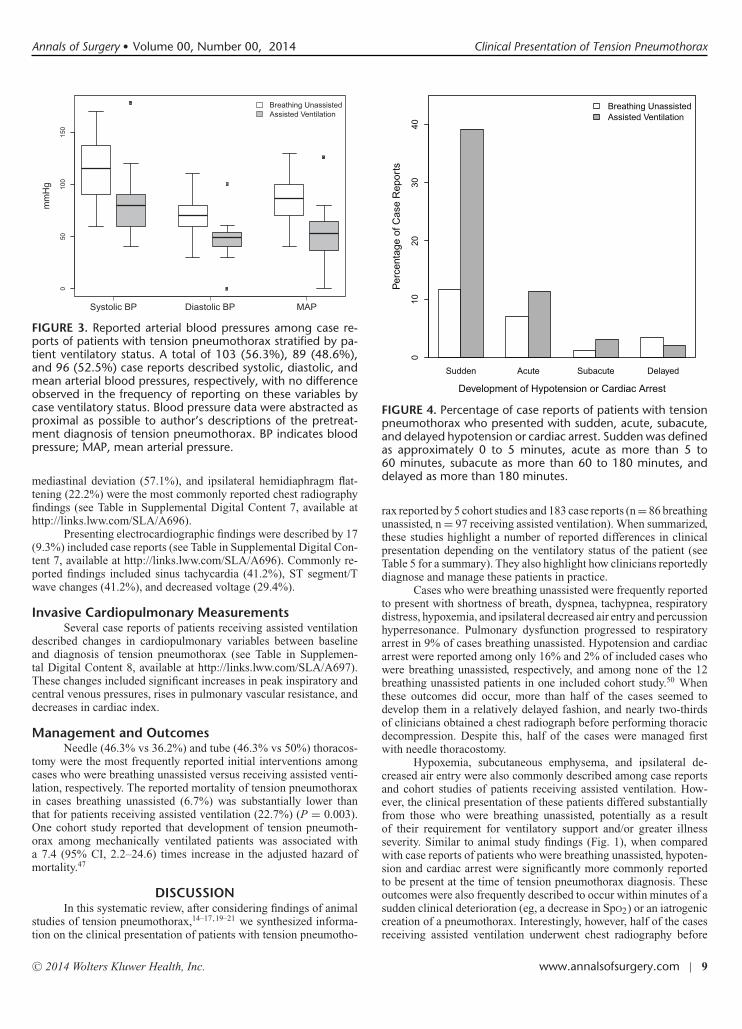
Disease EvolutionApproximate times to development of hypotension/cardiac ar-
rest could be determined for 20 (90.9%) case reports of patientsbreathing unassisted versus 54 (72.0%) receiving assisted ventila-tion. In contrast to cases who were breathing unassisted, the majority(70.4%) of those receiving assisted ventilation who experienced hy-potension or cardiac arrest developed these signs within minutes ofclinical presentation (Fig. 4).
Initial InvestigationsA chest radiograph was obtained before treatment in 55
(64.0%) case reports of patients who were breathing unassisted versus44 (45.4%) receiving assisted ventilation. A pneumothorax occupy-ing greater than 50% of hemithorax volume (55.8%), contralateral
Annals of Surgery � Volume 00, Number 00, 2014 Clinical Presentation of Tension Pneumothorax
FIGURE 3. Reported arterial blood pressures among case re-ports of patients with tension pneumothorax stratified by pa-tient ventilatory status. A total of 103 (56.3%), 89 (48.6%),and 96 (52.5%) case reports described systolic, diastolic, andmean arterial blood pressures, respectively, with no differenceobserved in the frequency of reporting on these variables bycase ventilatory status. Blood pressure data were abstracted asproximal as possible to author’s descriptions of the pretreat-ment diagnosis of tension pneumothorax. BP indicates bloodpressure; MAP, mean arterial pressure.
mediastinal deviation (57.1%), and ipsilateral hemidiaphragm flat-tening (22.2%) were the most commonly reported chest radiographyfindings (see Table in Supplemental Digital Content 7, available athttp://links.lww.com/SLA/A696).
Presenting electrocardiographic findings were described by 17(9.3%) included case reports (see Table in Supplemental Digital Con-tent 7, available at http://links.lww.com/SLA/A696). Commonly re-ported findings included sinus tachycardia (41.2%), ST segment/Twave changes (41.2%), and decreased voltage (29.4%).
Invasive Cardiopulmonary MeasurementsSeveral case reports of patients receiving assisted ventilation
described changes in cardiopulmonary variables between baselineand diagnosis of tension pneumothorax (see Table in Supplemen-tal Digital Content 8, available at http://links.lww.com/SLA/A697).These changes included significant increases in peak inspiratory andcentral venous pressures, rises in pulmonary vascular resistance, anddecreases in cardiac index.
Management and OutcomesNeedle (46.3% vs 36.2%) and tube (46.3% vs 50%) thoracos-
tomy were the most frequently reported initial interventions amongcases who were breathing unassisted versus receiving assisted venti-lation, respectively. The reported mortality of tension pneumothoraxin cases breathing unassisted (6.7%) was substantially lower thanthat for patients receiving assisted ventilation (22.7%) (P = 0.003).One cohort study reported that development of tension pneumoth-orax among mechanically ventilated patients was associated witha 7.4 (95% CI, 2.2–24.6) times increase in the adjusted hazard ofmortality.47
DISCUSSIONIn this systematic review, after considering findings of animal
studies of tension pneumothorax,14–17,19–21 we synthesized informa-tion on the clinical presentation of patients with tension pneumotho-
FIGURE 4. Percentage of case reports of patients with tensionpneumothorax who presented with sudden, acute, subacute,and delayed hypotension or cardiac arrest. Sudden was definedas approximately 0 to 5 minutes, acute as more than 5 to60 minutes, subacute as more than 60 to 180 minutes, anddelayed as more than 180 minutes.
rax reported by 5 cohort studies and 183 case reports (n = 86 breathingunassisted, n = 97 receiving assisted ventilation). When summarized,these studies highlight a number of reported differences in clinicalpresentation depending on the ventilatory status of the patient (seeTable 5 for a summary). They also highlight how clinicians reportedlydiagnose and manage these patients in practice.
Cases who were breathing unassisted were frequently reportedto present with shortness of breath, dyspnea, tachypnea, respiratorydistress, hypoxemia, and ipsilateral decreased air entry and percussionhyperresonance. Pulmonary dysfunction progressed to respiratoryarrest in 9% of cases breathing unassisted. Hypotension and cardiacarrest were reported among only 16% and 2% of included cases whowere breathing unassisted, respectively, and among none of the 12breathing unassisted patients in one included cohort study.50 Whenthese outcomes did occur, more than half of the cases seemed todevelop them in a relatively delayed fashion, and nearly two-thirdsof clinicians obtained a chest radiograph before performing thoracicdecompression. Despite this, half of the cases were managed firstwith needle thoracostomy.
Hypoxemia, subcutaneous emphysema, and ipsilateral de-creased air entry were also commonly described among case reportsand cohort studies of patients receiving assisted ventilation. How-ever, the clinical presentation of these patients differed substantiallyfrom those who were breathing unassisted, potentially as a resultof their requirement for ventilatory support and/or greater illnessseverity. Similar to animal study findings (Fig. 1), when comparedwith case reports of patients who were breathing unassisted, hypoten-sion and cardiac arrest were significantly more commonly reportedto be present at the time of tension pneumothorax diagnosis. Theseoutcomes were also frequently described to occur within minutes of asudden clinical deterioration (eg, a decrease in SpO2) or an iatrogeniccreation of a pneumothorax. Interestingly, however, half of the casesreceiving assisted ventilation underwent chest radiography before
∗Where ∗, ∗∗, ∗∗∗ and ∗∗∗∗ indicate that the sign or symptom was reportedamong approximately 0% to 15%, 15% to 30%, 30% to 45%, or >45% of includedobservational studies or case reports/series.
thoracic decompression and half were initially managed with tubethoracostomy.
Our findings may have implications for improving the diagno-sis and treatment of tension pneumothorax. In contrast to classicalmedical teaching, contralateral tracheal deviation and jugular venousdistention are uncommonly reported clinical manifestations of tensionpneumothorax. Tension pneumothorax may have to be considered inpatients who are breathing unassisted who present with predomi-nantly respiratory signs and symptoms. As those who are breathingunassisted have seldom been reported to present with sudden hemo-dynamic compromise, it may be appropriate to obtain a chest radio-graph in a monitored setting to confirm the diagnosis and lateralizethe disease instead of performing urgent thoracic decompression forpatients who are not in extremis.5,6,13 Thoracic ultrasonography maybe superior to chest radiography for this purpose, as it has a sensi-tivity of approximately 80% to 90% for detection of pneumothoraces(versus approximately 50% for supine chest radiography) and can beperformed rapidly at the bedside.57,58 Conversely, clinicians shouldbe prepared to perform urgent thoracic decompression without chestradiographic confirmation in patients suspected of a tension pneu-mothorax who are receiving assisted ventilation, as these patientshave frequently been reported to present with sudden hemodynamiccompromise and/or cardiac arrest.
Our synthesis and analysis of case reports/series data has sev-eral potential limitations. Our estimates of the frequency of clinicalmanifestations of tension pneumothorax may have been influencedby underreporting of relatively common presentations of tensionpneumothorax or overreporting of presentations that manifestedmore unusual or interesting clinical features.1,59 However, as we canthink of no reason why under- or overreporting would depend on caseventilatory status, it seems unlikely that selection bias would haveinfluenced our between-group comparisons. Furthermore, althoughsome of the included case reports could be argued not to represent
tension pneumothorax, our findings were robust to sensitivityanalyses that included only cases satisfying a published definition.Similarly, as we included case reports of patients with less commonetiologies of tension pneumothorax (eg, gastrointestinal perforation),the validity of combining all cases together may be questioned.1
Despite this, we are unsure why patients with a less common etiologywould present with different clinical manifestations when comparedto those with more common etiologies.1,16,17,19–21 Finally, althoughsome may argue that our findings may be due to unmeasuredconfounding,59 this seems unlikely given that any unmeasuredconfounder that could account for the observed magnitude of theassociation between ventilatory status and hypotension/cardiacarrest would have to be very strongly associated with patientventilatory status and highly predictive of hypotension and cardiacarrest. Thus, as our findings are consistent with results from animalstudies,16,17,19–21 we believe them to be clinically important.
CONCLUSIONSThe reported clinical presentation of tension pneumothorax
depends on the ventilatory status of the patient. This may have impli-cations for improving the diagnosis and treatment of this uncommonyet catastrophic clinical condition.
ACKNOWLEDGMENTSDr Roberts had full access to all of the data in the study and
takes responsibility for the integrity of the data and the accuracy ofthe data analysis. The authors thank the staff of the University ofCalgary Health Sciences Library for obtaining copies of articlesidentified throughout the conduct of the systematic review, SandyCochrane at Multimedia Services at the University of Calgary forassisting with creation of Figure 1, and Kelly Mrklas, MSc, for assist-ing with translation of non-English language articles. The authorsalso thank Eddy S. Lang, MDCM in the Department of EmergencyMedicine at the University of Calgary for reviewing the article andproviding critical input on its findings before submission for peerreview. Dr Roberts is supported by an Alberta Innovates—HealthSolutions Clinician Fellowship Award, a Knowledge TranslationCanada Strategic Training in Health Research Fellowship, andfunding from the Canadian Institutes of Health Research. Thesefunders had no role in the design or conduct of the study; collection,management, analysis, or interpretation of the data; or preparation,review, or approval of the article.
REFERENCES1. Roberts DJ, Leigh-Smith S, Faris PD, et al. Clinical manifestations of tension
pneumothorax: protocol for a systematic review and meta-analysis. Syst Rev.2014;3:3.
2. Cameron PA, Flett K, Kaan E, et al. Helicopter retrieval of primary traumapatients by a paramedic helicopter service. Aust N Z J Surg. 1993;63:790–797.
3. Coats TJ, Wilson AW, Xeropotamous N. Pre-hospital management of patientswith severe thoracic injury. Injury. 1995;26:581–585.
4. Eckstein M, Suyehara D. Needle thoracostomy in the prehospital setting. Pre-hosp Emerg Care. 1998;2:132–135.
5. American College of Surgeons Committee on Trauma. Advanced Trauma LifeSupport (ATLS): Ninth Edition. Chicago, IL: American College of Surgeons;2012.
6. Leigh-Smith S, Harris T. Tension pneumothorax—time for a re-think? EmergMed J. 2005;22:8–16.
7. Chen KY, Jerng JS, Liao WY, et al. Pneumothorax in the ICU: patient outcomesand prognostic factors. Chest. 2002;122:678–683.
8. Fleming WH, Bowen JC. Early complications of long-term respiratory support.J Thorac Cardiovasc Surg. 1972;64:729–738.
9. Kumar A, Pontoppidan H, Falke KJ, et al. Pulmonary barotrauma during me-chanical ventilation. Crit Care Med. 1973;1:181–186.
10. Ludwig J, Kienzle GD. Pneumothorax in a large autopsy population. A studyof 77 cases. Am J Clin Pathol. 1978;70:24–26.
Annals of Surgery � Volume 00, Number 00, 2014 Clinical Presentation of Tension Pneumothorax
11. Warner KJ, Copass MK, Bulger EM. Paramedic use of needle thoracostomyin the prehospital environment. Prehosp Emerg Care. 2008;12:162–168.
12. Ball CG, Wyrzykowski AD, Kirkpatrick AW, et al. Thoracic needle decom-pression for tension pneumothorax: clinical correlation with catheter length.Can J Surg. 2010;53:184–188.
13. Waydhas C, Sauerland S. Pre-hospital pleural decompression and chest tubeplacement after blunt trauma: a systematic review. Resuscitation. 2007;72:11–25.
14. Hilton R. Some effects of artificial pneumothorax on the circulation. J PatholBacteriol. 1925;37:1–8.
15. Simmons DH, Hemingway A, Ricchiuti N. Acute circulatory effects of pneu-mothorax in dogs. J Appl Physiol. 1958;12:255–261.
16. Rutherford RB, Hurt HH, Jr, Brickman RD, et al. The pathophysiology ofprogressive, tension pneumothorax. J Trauma. 1968;8:212–227.
17. Gustman P, Yerger L, Wanner A. Immediate cardiovascular effects of tensionpneumothorax. Am Rev Respir Dis. 1983;127:171–174.
18. Subotich D, Mandarich D. Accidentally created tension pneumothorax in pa-tient with primary spontaneous pneumothorax—confirmation of the experi-mental studies, putting into question the classical explanation. Med Hypothe-ses. 2005;64:170–173.
20. Martin M, Satterly S, Inaba K, et al. Does needle thoracostomy provide ade-quate and effective decompression of tension pneumothorax? J Trauma AcuteCare Surg. 2012;73:1412–1417.
21. Nelson D, Porta C, Satterly S, et al. Physiology and cardiovascular effect ofsevere tension pneumothorax in a porcine model. J Surg Res. 2013;184:450–457.
22. Mines D, Abbuhl S. Needle thoracostomy fails to detect a fatal tension pneu-mothorax. Ann Emerg Med. 1993;22:863–866.
23. Bailey RC, Esberger D. Development of tension pneumothorax after chestdrain insertion. J Accid Emerg Med. 1998;15:128.
25. Rawlins R, Brown KM, Carr CS, et al. Life threatening haemorrhage afteranterior needle aspiration of pneumothoraces. A role for lateral needle aspira-tion in emergency decompression of spontaneous pneumothorax. Emerg MedJ. 2003;20:383–384.
26. Riwoe D, Poncia HD. Subclavian artery laceration: a serious complication ofneedle decompression. Emerg Med Australas. 2011;23:651–653.
27. Bybee KA, Kara T, Prasad A, et al. Systematic review: transient left ventricularapical ballooning: a syndrome that mimics ST-segment elevation myocardialinfarction. Ann Intern Med. 2004;141:858–865.
28. Lyrer P, Engelter S. Antithrombotic drugs for carotid artery dis-section. Cochrane Database Syst Rev. 2010;(10):CD000255. doi:10.1002/14651858.CD000255.pub2.
29. Holty JE, Bravata DM, Liu H, et al. Systematic review: a century of inhalationalanthrax cases from 1900 to 2005. Ann Intern Med. 2006;144:270–280.
30. Andersohn F, Konzen C, Garbe E. Systematic review: agranulocytosis inducedby nonchemotherapy drugs. Ann Intern Med. 2007;146:657–665.
31. Aronson JK, Hauben M. Anecdotes that provide definitive evidence. BMJ.2006;333:1267–1269.
32. Jenicek M. Clinical Case Reporting in Evidence-Based Medicine. New York,NY: Oxford University Press; 2001.
33. Gagnier JJ, Kienle G, Altman DG, et al. The CARE guidelines: consensus-based clinical case report guideline development. J Clin Epidemiol.2014;67:46–51.
34. Selvaraj SA, Chairez E, Wilson LM, et al. Use of case reports and the Ad-verse Event Reporting System in systematic reviews: overcoming barriers toassess the link between Crohn’s disease medications and hepatosplenic T-celllymphoma. Syst Rev. 2013;2:53.
35. Higgins JPT, Green S, eds. Cochrane Handbook for Systematic Reviewsof Interventions Version 5.0.2. The Cochrane Collaboration 2009. www.cochrane-handbook.org. Accessed November 25, 2014.
36. Stroup DF, Berlin JA, Morton SC, et al. Meta-analysis of observationalstudies in epidemiology: a proposal for reporting. Meta-analysis of Obser-
vational Studies in Epidemiology (MOOSE) group. JAMA. 2000;283:2008–2012.
37. Liberati A, Altman DG, Tetzlaff J, et al. The PRISMA statement for re-porting systematic reviews and meta-analyses of studies that evaluate healthcare interventions: explanation and elaboration. Ann Intern Med. 2009;151:W65–W94.
38. Oleckno WA. An overview of epidemiologic study designs. In:Oleckno WA,ed. Epidemiology: Concepts and Methods. Long Grove, IL: Waveland Press,Inc.; 2008:55–84.
39. Dekkers OM, Egger M, Altman DG, et al. Distinguishing case series fromcohort studies. Ann Intern Med. 2012;156:37–40.
40. Richardson WS, Wilson MC, Williams JW Jr, et al. Users’ guides to the medicalliterature: XXIV. How to use an article on the clinical manifestations of disease.Evidence-Based Medicine Working Group. JAMA. 2000;284:869–875.
42. Landis JR, Koch GG. The measurement of observer agreement for categoricaldata. Biometrics. 1977;33:159–174.
43. Altman DG. Practical Statistics for Medical Research. London, United King-dom: Chapman & Hall; 1991.
44. EPIC II Group of Investigators. Conversion tables. http://www.intensive.org/epic2/Documents/Estimation%20of%20PO2%20and%20FiO2.pdf . AccessedJanuary 1, 2014.
45. Clopper CJ, Pearson ES. The use of confidence or fiducial limits illustrated inthe case of the binomial. Biometrika. 1934;26:404–413.
46. Repeated measures and longitudinal data analysis. In: Vittinghoff E, GliddenDV, Shiboski SC, et al. eds. Regression Methods in Biostatistics: Linear, Lo-gistic, Survival, and Repeated Measures Models Second Edition. New York,NY: Springer; 2012:261–308.
47. Hsu CW, Sun SF, Lin HS, et al. Clinical characteristics, hospital outcome andprognostic factors of patients with ventilator-related pneumothorax. MinervaAnestesiol. 2014;80:29–38.
48. Mistry N, Bleetman A, Roberts KJ. Chest decompression during the resus-citation of patients in prehospital traumatic cardiac arrest. Emerg Med J.2009;26:738–740.
49. Massarutti D, Trillo G, Berlot G, et al. Simple thoracostomy in prehospitaltrauma management is safe and effective: a 2-year experience by helicopteremergency medical crews. Eur J Emerg Med. 2006;13:276–280.
50. Davis DP, Pettit K, Rom CD, et al. The safety and efficacy of prehospitalneedle and tube thoracostomy by aeromedical personnel. Prehosp Emerg Care.2005;9:191–197.
51. Tocino IM, Miller MH, Fairfax WR. Distribution of pneumothorax in the supineand semirecumbent critically ill adult. AJR Am J Roentgenol. 1985;144:901–905.
52. Barton ED, Epperson M, Hoyt DB, et al. Prehospital needle aspiration andtube thoracostomy in trauma victims: a six-year experience with aeromedicalcrews. J Emerg Med. 1995;13:155–163.
53. Clark S, Ragg M, Stella J. Is mediastinal shift on chest x-ray of pneumothoraxalways an emergency? Emerg Med (Fremantle). 2003;15:429–433.
54. Lockey D, Crewdson K, Davies G. Traumatic cardiac arrest: who are thesurvivors? Ann Emerg Med. 2006;48:240–244.
55. Allison K, Porter KM, Mason AM. Use of the Asherman chest seal as astabilisation device for needle thoracostomy. Emerg Med J. 2002;19:590–591.
56. Wildgruber M, Rummeny EJ. Bilateral tension pneumothorax. Emerg Med J.2012;29:752.
57. Roberts DJ, Niven DJ, James MT, et al. Thoracic ultrasonography versuschest radiography for detection of pneumothoraces: challenges in derivingand interpreting summary diagnostic accuracy estimates. Crit Care. 2014;18:416.
58. Lichtenstein DA. Lung ultrasound in the critically ill. Ann Intensive Care.2014;4:1.
59. Richason TP, Paulson SM, Lowenstein SR, et al. Case reports describingtreatments in the emergency medicine literature: missing and misleading in-formation. BMC Emerg Med. 2009;9:10.