1 Upper GI Bleeding Tasos Manokas, MD Assistant Professor of Gastroenterology Introduction • GI bleeding results in over 300,000 hospitalizations annually in U.S. • Upper GI bleeding accounts for 75-80% of all acute GI bleeding cases 9 More common in men and elderly 9 Incidence: 50-100 per 100,000 patients/year 9 20,000 deaths annually in United States Clinical presentation • Hematemesis 9 Reflects bleeding proximal to ligament of Treitz • Melena 9 Can be seen with 100 cc blood in UGI tract • Hematochezia 9 Usually lower GI source or very rapid UGI blood loss (1000 cc blood) 9 If associated with bright red NG aspirate, high mortality Prognosis • Wide spectrum of severity 9 Trivial bleeding to variceal bleeding • Emphasis on early identification and intervention of significant bleeds • 2 most important prognostic factors 9 Cause of bleeding (variceal) 9 Underlying comorbid conditions • Mortality from acute UGI bleeding: 5-10% 9 Unchanged over last 50 years despite development, refinement of endoscopic therapy
Transcript
1
Upper GI Bleeding
Tasos Manokas, MDAssistant Professor of Gastroenterology
Introduction• GI bleeding results in over 300,000
hospitalizations annually in U.S.
• Upper GI bleeding accounts for 75-80% of all acute GI bleeding cases
More common in men and elderly
Incidence: 50-100 per 100,000 patients/year
20,000 deaths annually in United States
Clinical presentation• Hematemesis
Reflects bleeding proximal to ligament of Treitz
• MelenaCan be seen with 100 cc blood in UGI tract
• HematocheziaUsually lower GI source or very rapid UGI blood loss (1000 cc blood)If associated with bright red NG aspirate, high mortality
Prognosis• Wide spectrum of severity
Trivial bleeding to variceal bleeding • Emphasis on early identification and intervention
of significant bleeds• 2 most important prognostic factors
Cause of bleeding (variceal)Underlying comorbid conditions
• Mortality from acute UGI bleeding: 5-10%Unchanged over last 50 years despite development, refinement of endoscopic therapy
2
Risk factors• Risk factors associated with increased
Appear as erythema or superficial erosions endoscopicallyCameron’s lesions: linear erosions within a large hiatal hernia
• Rarely associated with significant UGI bleeding• Related to NSAID use, alcohol, or stress gastritis• Bleeding and stress gastritis: < 3% of pts in ICU
High risk: mechanical ventilation > 48 hours, coagulopathy, head injury, extensive burn injuriesProphylaxis: H2-blockers > sucralfate• Limited data on PPI
Aortoenteric Fistula• Rare causes of life-threatening GI bleed• Primary risk factor: Abdominal aortic graft
reconstructionOccur with 0.5% of aortoiliac surgeryMost commonly develop 3-5 years after surgeryMore common with infected grafts
• Most communicate with 3rd portion of duodenum• “Herald” bleed: self-limited bleed hours/days
before severe bleeding
Hemobilia• Bleeding into biliary tree
Vascular communication with bile ducts• Causes: trauma, liver biopsy most common
Also gallstones, vascular aneurysms, liver abscess, neoplasia
• Diagnosis difficultClinical history and endoscopic appearance of blood coming from papillaCan be missed with standard-viewing endoscopeDiagnosis made angiographically
Hemosuccus Pancreaticus• Bleeding into pancreatic duct• Complication of chronic pancreatitis,
pseudocystsAneurysm/pseudoaneurysm of peripancreatic, splenic arteries eroding into pancreatic duct
• Diagnosis difficultClinical history and endoscopic appearance of blood coming from papillaCan be missed with standard-viewing endoscopeDiagnosis made angiographically
5
Vascular Lesions• Vascular ectasia, AVM’s
Associated with connective tissue dz’s(scleroderma, CREST), renal failure, radiation tx, cirrhosis, HHTWatermelon stomach: diffuse, linear AVM’s in gastric antrum; often found in elderly women
• Dieulafoy lesionsLarge, submucosal artery usually located in gastric cardiaModerate to severe bleeding
Neoplasms• Neoplasms
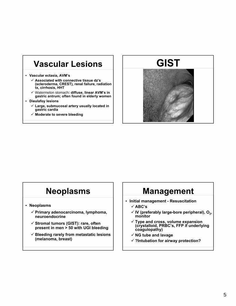
Primary adenocarcinoma, lymphoma, neuroendocrineStromal tumors (GIST): rare, often present in men > 50 with UGI bleedingBleeding rarely from metastatic lesions (melanoma, breast)
GIST
• Initial management - ResuscitationABC’sIV (preferably large-bore peripheral), O2, monitorType and cross, volume expansion (crystalloid, PRBC’s, FFP if underlying coagulopathy)NG tube and lavage?Intubation for airway protection?
Management
6
• HematocritInitial Hct may not reflect degree of blood loss accurately• Hct falls over 24-72 hours as
extravascular fluid enters vascular space to restore volume
Hct may continue to trend down for days after bleeding stops • Not clinically relevant if no signs of
active bleeding (hematemesis, melena, hematochezia)
Laboratory Evaluation
Medical Management• IV proton pump inhibitors (PPI)
Promotes clot stabilization by maintaining intragastric pH > 6Clot lysis by pepsin at pH < 5 • Pepsin irreversibly inactivated at pH > 6
Platelet aggregation improved at pH > 6
IV PPI• Peptic ulcer bleeding
2 large meta-analysis demonstrate significantly lower rebleeding rates and surgery in pt’s treated with IV PPI compared to placebo1,2
• 1 review also found significant benefit in mortality
• All cause UGI bleeding1 large meta-analysis demonstrated no benefit in rebleeding rates, surgery or mortality when compared to placebo3
1Bardou M, et al. Aliment Pharmacol Ther 2005.2Leontiadis G, et al. Cochrane Database Syst Rev 2006.3Dorward S, et al. Cochrane Databse Syst Rev 2006.
• IV octreotideSomatostatin analoguePhysiologic effects:• Decreases gastroduodenal mucosal blood flow• Inhibits gastric acid and pepsin secretion • Stimulates mucus production
Causes splanchnic vasoconstriction and subsequent decrease in splanchnic blood flow Theoretical benefit over PPI in patients with peptic ulcer bleeding• More diverse physiologic effect on upper GI tract
Medical Management
7
IV Octreotide• Peptic ulcer bleeding
1 large meta-analysis showed significant reduction in continued/recurrent bleeding4
• Trend toward significance in all cause UGI bleeding
• All cause UGI bleeding2 RCT’s demonstrated no significant benefit compared to placebo5 or H2 blocker6
1 RCT showed significant benefit in initial hemostasis, blood transfusions, need for surgery, length of hospital stay compared to H2blocker7
4Imperiale T, et al. Ann Intern Med 1997.5Christiansen J, et al. Gastroenterology 1989.6 Lin H, et al. J Clin Gastroenterol 1995.7Lin H, et al. Hepatogastroenterology 1995.
• Within 6 hours of presentation• Indications
Recurrent/continued UGI bleeding• Ongoing hematemesis, active
melena/hematocheziaRisk for variceal hemorrhageHigh risk for recurrent bleeding
Role for urgent endoscopy
Erythromycin before endoscopy
• Shown in multiple studies to improve the quality of endoscopic exam
Some studies also show decreased need for second look EGD
• Given as a single 250 mg IV dose• Must check ECG before giving to assess
QTc
• Clotted blood in stomachEndoscopic Findings
8
Endoscopic Findings• Active bleeding
Endoscopic Findings• Visible vessel
Endoscopic Findings• Adherent clot
Endoscopic Therapy• High risk lesions treated with dual therapy
Repeat Endoscopy• “2nd look” endoscopy often performed 24
hours after initial procedure• In absence of rebleeding, not warranted for
all patientsOnly certain high risk groups shown to benefit
• In patients with rebleeding (rebleeding rate: 20-30%), repeat endoscopy warranted for further treatment
Angiography• Indicated in refractory bleeding
Not amenable to endoscopic therapyPoor surgical candidates
• Requires fast bleeding rate (>0.5 ml/min)• Can embolize left gastric a. or
gastroduodenal a. emperically based on endoscopic localization of bleeding
10
Surgery• Changing role of surgery
No longer used to cure ulcer disease• PPI’s, H.pylori eradication now cures most
cases of PUDNow utilized to stop life-threatening bleeding
• IndicationsBleeding where endoscopy and/or angiography has failedLarge visible vessels (>2-3 mm) along lesser curve of stomach and in duodenal bulb
Prevention of future bleeding• Eliminating NSAID’s• Eradication of H. pylori
Triple therapy for 10-14 days first line therapyBismuth + Metronidazole + Tetracycline• QID dosing can decrease compliance
PPI + Amoxicillin + ClarithromycinIncreasing resistance to metronidazole, clarithromycinMost commonly used regimens cure 80% of cases
H. pylori treatment• Documenting clearance
Failure of therapy associated with ulcer recurrenceSimple, cost-effective, non-invasive tests available (urea breath, stool antigen tests)Must wait 4 weeks after completion of therapy• Must hold PPI 1 week prior to test
Once clearance confirmed, re-infection rare
Variceal BleedingJim Hanje, MD
Assistant Professor of Gastroenterology
Ohio State University Medical Center
11
CirrhosisNormal
Nodules
Irregular surface
Portal systemic collaterals
Distorted sinusoidal architectur
eleads to
increased resistance Portal
vein
Cirrhotic Liver
Splenomegaly
Portal Hypertension
• Progressive complication of cirrhosis
• Marks transition from early compensated cirrhosis to decompensated, end-stage liver disease
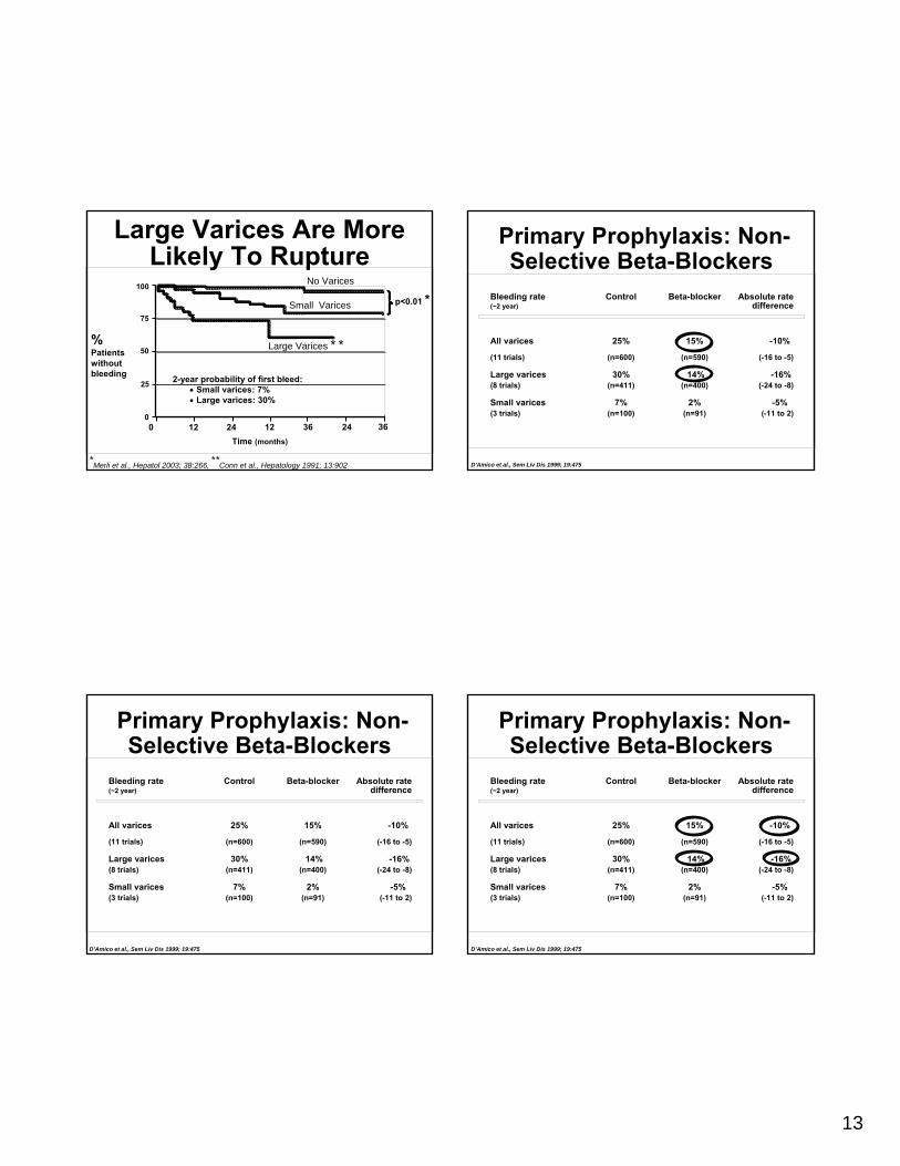
• Nadolol, PropranololTitrated weekly to goal: ↓ HR by 25%
Primary Prophylaxis: Nonselective Beta-Blockers
• Limited utility in clinical practice:Frequent side effect/contraindications (20%)Limited reduction in portal pressures at doses toleratedNeed for long-term/lifelong therapyRebound bleeding with cessation of therapy
Primary Prophylaxis –Endoscopic Band Ligation (EBL)
• Prophylactic EBL every 4 weeks until varicealobliteration
• Esophageal ulcerations form following EBLCan cause dysphagia, chest pain in most patientsPPI BID shown to decrease post-EBL bleeding
• Fewer side effects than B-blockers, but more severe
Bleeding due to esophageal ulcerations, varicealrupture
Primary Prophylaxis: EBL vsBeta-Blockers (BB)
Khuroo, et al., Aliment Pharmacol Ther 2005; 21:347
First hemorrhage SurvivalChen 1998Sarin 1999De 1999Jutabha 2000De la Mora 2000Lui 2002Lo 2004Schepke 2004Total
Relative risk0 1 10 0 1 10 40
Favors EBL Favors EBLFavors BB
Favors BB
15
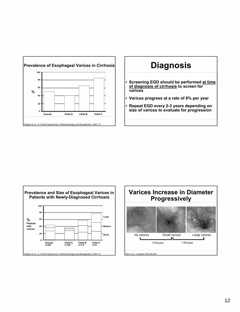
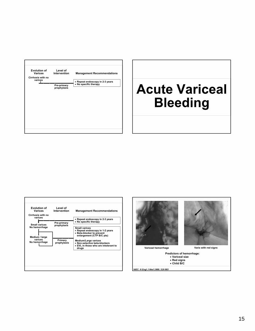
Evolution of Varices
Level of Intervention Management Recommendations
Cirrhosis with no varices
• Repeat endoscopy in 2-3 years• No specific therapyPre-primary
prophylaxis
Evolution of Varices
Level of Intervention Management Recommendations
Cirrhosis with no varices
Small varicesNo hemorrhage
Medium / large varices
No hemorrhage
• Repeat endoscopy in 2-3 years• No specific therapy
Small varices• Repeat endoscopy in 1-2 years• Beta-blocker to prevent
enlargement (CTP B/C pts)
Medium/Large varices• Non-selective beta-blockers• EVL in those who are intolerant to
drugs
Pre-primary prophylaxis
Primary prophylaxis
Acute VaricealBleeding
Predictors of hemorrhage:• Variceal size• Red signs• Child B/C
ABC’sIV (preferably large-bore peripheral), O2, monitorType and cross, volume expansion • Goal Hgb 8 g/dl; over-resuscitation can ↑ portal
pressure and ↑ risk of rebleeding and deathCorrect coagulopathy• FFP, platelets, DDAVP, cryoprecipitate• Recombinant factor VIIa
– Multi-center RCT showed no overall benefit compared to standard therapy BUT…
– CTP B/C patients ↓ bleeding rates
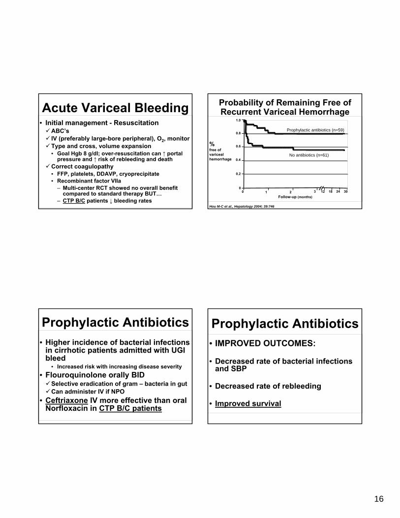
Prophylactic Antibiotics• Higher incidence of bacterial infections
in cirrhotic patients admitted with UGI bleed
• Increased risk with increasing disease severity• Flouroquinolone orally BID
Selective eradication of gram – bacteria in gutCan administer IV if NPO
• Ceftriaxone IV more effective than oral Norfloxacin in CTP B/C patients
Probability of Remaining Free of Recurrent Variceal Hemorrhage
Hou M-C et al., Hepatology 2004; 39:746
Prophylactic antibiotics (n=59)
%free of varicealhemorrhage
1.0
0.6
0.2
0.8
10
No antibiotics (n=61)
02 3 12 30
Follow-up (months)18 24
0.4
Prophylactic Antibiotics• IMPROVED OUTCOMES:
• Decreased rate of bacterial infections and SBP
• Decreased rate of rebleeding
• Improved survival
17
• OctreotideSynthetic analogue of somatostatin• 50ug bolus, followed by 50ug/h continuous
infusion• Safe, minimal side-effects, can be used for 5
daysCauses splanchnic vasoconstriction• Acutely lowers portal pressures by decreasing
splanchnic blood flowDecreases bleeding, no mortality benefit• Minimal benefit when used alone without EBL• Not as potent as other agents, can get
tachyphylaxis
Pharmacologic Therapy
• Endoscopic band ligation (EBL)Treatment of choice for bleeding esophageal varicesSuccessful in 70-90% of casesSuperior to sclerotherapy with decreased rebleeding rates, mortality rates and incidence of complications
• EBL + OctreotideSuperior to either modality aloneShown to significantly reduce rebleeding rates No mortality benefit over banding alone
Endoscopy
Bañares R et al., Hepatology 2002; 35:609
Combination Drug / Endoscopic Therapy More Effective Than Endoscopic Therapy Alone