CLINICAL SCIENCE Screening for hotspot mutations in PI3K, JAK2, FLT3 and NPM1 in patients with myelodysplastic syndromes Joa ˜ o Agostinho Machado-Neto, Fabiola Traina, Mariana Lazarini, Paula de Melo Campos, Katia Borgia Barbosa Pagnano, Irene Lorand-Metze, Fernando Ferreira Costa, Sara T Olalla Saad Hematology and Hemotherapy Center, National Institute of Blood, University of Campinas, Sa ˜ o Paulo, Brazil. INTRODUCTION: Myelodysplastic syndromes encompass a heterogeneous group of clonal hematopoietic stem cell disorders characterized by ineffective hematopoiesis, refractory cytopenia and a tendency to progress toward acute myeloid leukemia. The accumulation of genetic alterations is closely associated with the progression of myelodysplastic syndromes toward acute myeloid leukemia. OBJECTIVE: To investigate the presence of mutations in the points most frequent for mutations (hotspot mutations) in phosphatidylinositol-3-kinase (PI3K), Janus kinase 2 (JAK2), FMS-like tyrosine kinase 3 (FLT3) and nucleophosmin (NPM1), which are involved in leukemia and other cancers, in a population of Brazilian MDS patients. METHODS: Fifty-one myelodysplastic syndromes patients were included in the study. According to French-American- British classification, the patients were distributed as follows: 31 with refractory anemia, 8 with refractory anemia with ringed sideroblasts, 7 with refractory anemia with excess blasts, 3 with refractory anemia with excess blasts in transformation and 2 with chronic myelomonocytic leukemia. Bone marrow samples were obtained and screened for the presence of hotspot mutations using analysis based on amplification with the polymerase chain reaction, sequencing, fragment size polymorphisms or restriction enzyme digestion. All patients were screened for mutations at the time of diagnosis, and 5 patients were also screened at the time of disease progression. RESULTS: In the genes studied, no mutations were detected in the patients at the time of diagnosis. One patient with chronic myelomonocytic leukemia was heterozygous for a Janus kinase 2 mutation after disease progression. CONCLUSIONS: These results show that hotspot mutations in the PI3K, JAK2, FLT3 and NPM1 genes are not common in MDS patients; nevertheless, JAK2 mutations may be present in myelodysplasia during disease progression. KEYWORDS: Hematopoietic Disorder; Acute Leukemia; Myelodysplasia; Mutations; Bone Marrow. Machado-Neto JA, Traina F, Lazarini M, Campos PM, Pagnano KBB, Lorand-Metze I, et al. Screening for hotspot mutations in PI3K, JAK2, FLT3 and NPM1 in patients with myelodysplastic syndromes. Clinics. 2011;66(5):793-799. Received for publication on November 30, 2010; First review completed on January 4, 2011; Accepted for publication on February 11, 2011 E-mail: [email protected]Tel.: 55 19 3289-1089 INTRODUCTION Myelodysplastic syndromes (MDS) encompass a hetero- geneous group of clonal hematopoietic stem cell disorders characterized by ineffective hematopoiesis, refractory cytope- nia and a tendency to progress to acute myeloid leukemia (AML). 1 Low-risk MDS present high levels of intramedullar apoptosis, whereas high-risk MDS show a decrease in apop- tosis, an increase in cell proliferation and a high frequency of evolution to AML. 2,3 The accumulation of genetic alterations is closely associated with the progression of MDS toward AML, and efforts are being made to determine the significance of various genetic aberrations in patients with MDS. 4-6 The same occurs for liver adenomatosis, 7 Rubinstein-taybi syndrome 8 and hemochromatosis. 9 The Phosphatidylinositol-3-kinase (PI3K) and Janus kinase 2 (JAK2) signaling pathways are involved in numerous cellular processes, such as proliferation, apopto- sis and differentiation. 10-12 Mutations in the catalytic subunit of PI3K are frequently observed in several cancers, including AML. 4,5,13 Hotspot mutations occur in exon 9 (E542 and E545) and in exon 20 (H1047), resulting in increased PI3K/Akt activity. 5,14-17 One somatic mutation in the JAK2 gene (V617F) has been identified in myeloproli- ferative disorders such as polycythemia vera (PV) and myelofibrosis. 18 FMS-like tyrosine kinase 3 (FLT3) is a tyrosine kinase re- ceptor that plays an important role in the proliferation and dif- ferentiation of hematopoietic progenitors. 19 Nucleophosmin (NPM1) is a key regulator of hematopoesis that shuttles Copyright ß 2011 CLINICS – This is an Open Access article distributed under the terms of the Creative Commons Attribution Non-Commercial License (http:// creativecommons.org/licenses/by-nc/3.0/) which permits unrestricted non- commercial use, distribution, and reproduction in any medium, provided the original work is properly cited. CLINICS 2011;66(5):793-799 DOI:10.1590/S1807-59322011000500014 793
Transcript
CLINICAL SCIENCE
Screening for hotspot mutations in PI3K, JAK2, FLT3and NPM1 in patients with myelodysplasticsyndromesJoao Agostinho Machado-Neto, Fabiola Traina, Mariana Lazarini, Paula de Melo Campos, Katia Borgia
Barbosa Pagnano, Irene Lorand-Metze, Fernando Ferreira Costa, Sara T Olalla Saad
Hematology and Hemotherapy Center, National Institute of Blood, University of Campinas, Sao Paulo, Brazil.
INTRODUCTION: Myelodysplastic syndromes encompass a heterogeneous group of clonal hematopoietic stem celldisorders characterized by ineffective hematopoiesis, refractory cytopenia and a tendency to progress toward acutemyeloid leukemia. The accumulation of genetic alterations is closely associated with the progression ofmyelodysplastic syndromes toward acute myeloid leukemia.
OBJECTIVE: To investigate the presence of mutations in the points most frequent for mutations (hotspot mutations)in phosphatidylinositol-3-kinase (PI3K), Janus kinase 2 (JAK2), FMS-like tyrosine kinase 3 (FLT3) and nucleophosmin(NPM1), which are involved in leukemia and other cancers, in a population of Brazilian MDS patients.
METHODS: Fifty-one myelodysplastic syndromes patients were included in the study. According to French-American-British classification, the patients were distributed as follows: 31 with refractory anemia, 8 with refractory anemiawith ringed sideroblasts, 7 with refractory anemia with excess blasts, 3 with refractory anemia with excess blasts intransformation and 2 with chronic myelomonocytic leukemia. Bone marrow samples were obtained and screenedfor the presence of hotspot mutations using analysis based on amplification with the polymerase chain reaction,sequencing, fragment size polymorphisms or restriction enzyme digestion. All patients were screened for mutationsat the time of diagnosis, and 5 patients were also screened at the time of disease progression.
RESULTS: In the genes studied, no mutations were detected in the patients at the time of diagnosis. One patientwith chronic myelomonocytic leukemia was heterozygous for a Janus kinase 2 mutation after disease progression.
CONCLUSIONS: These results show that hotspot mutations in the PI3K, JAK2, FLT3 and NPM1 genes are not commonin MDS patients; nevertheless, JAK2 mutations may be present in myelodysplasia during disease progression.
KEYWORDS: Hematopoietic Disorder; Acute Leukemia; Myelodysplasia; Mutations; Bone Marrow.
Machado-Neto JA, Traina F, Lazarini M, Campos PM, Pagnano KBB, Lorand-Metze I, et al. Screening for hotspot mutations in PI3K, JAK2, FLT3 andNPM1 in patients with myelodysplastic syndromes. Clinics. 2011;66(5):793-799.
Received for publication on November 30, 2010; First review completed on January 4, 2011; Accepted for publication on February 11, 2011
Myelodysplastic syndromes (MDS) encompass a hetero-geneous group of clonal hematopoietic stem cell disorderscharacterized by ineffective hematopoiesis, refractory cytope-nia and a tendency to progress to acute myeloid leukemia(AML).1 Low-risk MDS present high levels of intramedullarapoptosis, whereas high-risk MDS show a decrease in apop-tosis, an increase in cell proliferation and a high frequency ofevolution to AML.2,3 The accumulation of genetic alterations isclosely associated with the progression of MDS toward AML,and efforts are being made to determine the significance of
various genetic aberrations in patients with MDS.4-6 The sameoccurs for liver adenomatosis,7 Rubinstein-taybi syndrome8
and hemochromatosis.9
The Phosphatidylinositol-3-kinase (PI3K) and Januskinase 2 (JAK2) signaling pathways are involved innumerous cellular processes, such as proliferation, apopto-sis and differentiation.10-12 Mutations in the catalyticsubunit of PI3K are frequently observed in several cancers,including AML.4,5,13 Hotspot mutations occur in exon 9(E542 and E545) and in exon 20 (H1047), resulting inincreased PI3K/Akt activity.5,14-17 One somatic mutation inthe JAK2 gene (V617F) has been identified in myeloproli-ferative disorders such as polycythemia vera (PV) andmyelofibrosis.18
FMS-like tyrosine kinase 3 (FLT3) is a tyrosine kinase re-ceptor that plays an important role in the proliferation and dif-ferentiation of hematopoietic progenitors.19 Nucleophosmin(NPM1) is a key regulator of hematopoesis that shuttles
Copyright � 2011 CLINICS – This is an Open Access article distributed underthe terms of the Creative Commons Attribution Non-Commercial License (http://creativecommons.org/licenses/by-nc/3.0/) which permits unrestricted non-commercial use, distribution, and reproduction in any medium, provided theoriginal work is properly cited.
This article has received corrections asked by the editor on 2011 in agreement with the ERRATUM published in Volume 66 Number 7. (http://www.scielo.br/pdf/clin/v66n7/v66n7a32.pdf)
between the nucleus and cytoplasm. NPM1 mutations oftenresult in the predominant localization of the protein to thecytoplasm, leading to destabilization of p14ARF and to theinhibition of p53.20 Internal tandem duplications (ITDs) in FLT3and NPM1 mutations are frequent events in the development ofAML, and are associated with prognosis. According to Galeet al,21 it is possible to identify 3 prognostic groups based in thepresence or absence of FTL3 and NPM1 mutations: good (FLT3-ITD2NPM1+), intermediate (FLT3-ITD2NPM12 or FLT3-ITD+NPM1+), and poor prognosis (FLT3-ITD+NPM12).Furthermore, a point mutation in exon 20 of the FLT3 gene(FLT3-D835) has been described in a case of AML.19
Mutations in PI3K, JAK2, FLT3 and NPM1 have beendescribed in cases of MDS; however, additional studies arenecessary to clarify their role in this disease. In this context,the objective of this work was to investigate the occurrence ofthe hotspot mutations E542, E545 and H1047 in PI3K, V617Fin JAK2, ITDs and D835 in FLT3 and exon 12 mutations inNPM1 in MDS patients in a Brazilian population.
MATERIALS AND METHODS
PatientsDNA samples were obtained from bone marrow aspirates
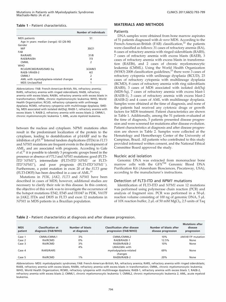
of 51 patients diagnosed with de novo MDS. According to theFrench-American-British (FAB) classification,22 the patientswere classified as follows: 31 cases of refractory anemia (RA),8 cases of refractory anemia with ringed sideroblasts (RARS),7 cases of refractory anemia with excess blasts (RAEB), 3cases of refractory anemia with excess blasts in transforma-tion (RAEBt), and 2 cases of chronic myelomonocyticleukemia (CMML). Using the World Health Organization(WHO) 2008 classification guidelines,23 there were 3 cases ofrefractory cytopenia with unilineage dysplasia (RCUD), 23cases of refractory cytopenia with multilineage dysplasia(RCMD), 8 cases of refractory anemia with ring sideroblasts(RARS), 3 cases of MDS associated with isolated del(5q)(MDS-5q), 7 cases of refractory anemia with excess blast-1(RAEB-1), 3 cases of refractory anemia with excess blast-2(RAEB-2) and 4 cases of AML with multilineage dysplasia.Samples were obtained at the time of diagnosis, and none ofthe patients had received any cytotoxic drugs or growthfactors for MDS treatment. Patient characteristics are shownin Table 1. Additionally, among the 51 patients evaluated atthe time of diagnosis, 5 patients presented disease progres-sion and were screened for mutations after disease evolution.Patient characteristics at diagnosis and after disease progres-sion are shown in Table 2. Samples were collected at theHematology and Hemotherapy Center of the University ofCampinas, Brazil. All patients who contributed to this studyprovided informed written consent, and the National EthicalCommittee Board approved the study.
Nucleic acid isolationGenomic DNA was extracted from mononuclear bone
marrow cells with the GFXTM Genomic Blood DNAPurification Kit (Amersham Biosciences, Piscataway, USA),according to the manufacturer’s instructions.
Detection of FLT3-ITD and NPM1 mutationsIdentification of FLT3-ITD and NPM1 exon 12 mutations
was performed using polymerase chain reaction (PCR) andanalysis of fragment size. PCR was performed in a 50-mLreaction volume consisting of 100 ng of genomic DNA, 5 mLof 10X reaction buffer, 2 mL of 50 mM MgCl2, 2.5 units of Taq
Mutations in Patients with Myelodysplastic SyndromesMachado-Neto JA et al.
CLINICS 2011;66(5):793-799
794
polymerase and 200 nM each of the forward and reverseprimers (Table 3). The reaction conditions were set as follows:5 minutes of denaturing at 94 C followed by 35 cycles of 20seconds at 92 C, 30 seconds at 57 C and 45 seconds at 72 C,with a final step at 72 C for 7 minutes. After dilution (1:20) inwater, 1 mL of each PCR product was mixed with 9 mL of Hi-Di formamide (Applied Biosystems, Foster City, CA) and0.5 mL of GeneScan 500-ROX size marker, and the mixturewas denatured for 5 minutes at 95 C. Samples harboring themutation were identified based on the areas under the curvesrepresenting the wild-type (FLT3:397 bp and NPM1:294 bp)and mutated alleles (FLT3-ITD.397 bp and NPM1.294 bp).
AML patients with FLT3-ITD or NPM1 mutations were usedas positive controls.
Detection of the JAK2 V617F and FLT3-D835mutations
Identification of JAK2 and FLT3 genotypes was performedusing PCR-restriction fragment length polymorphism (PCR-RFLP) analysis. PCR was performed in a 50-mL reactionvolume consisting of 100 ng of genomic DNA, 5 mL of 10Xreaction buffer, 2 mL of 50 mM MgCl2, 2.5 units of Taqpolymerase and 200 nM each of the forward and reverseprimers (Table 3). The reaction conditions were set as follows:
Table 3 - Primer sequences and restriction enzymes
Gene Mutation Primers sequences Restriction enzyme site
FLT3 ITD F: 59-GCAATTTAGGTATGAAAGCCAGC-39
R: 59-CTTTCAGCATTTTGACGGCAACC-39(HEX)
NPM1 exon 12 F: 59-GTGGTAGAATGAAAAATAGAT-39(FAM)
R: 59-CTTGGCAATAGAACCTGGAC-39
JAK2 V617F F: 59-GGGTTTCCTCAGAACGTTGA-39 BsaXI
R: 59-TCATTGCTTTCCTTTTTCACAA-39
FLT3 D835 F: 59-CCGCCAGGAACGTGCTTG-39 Eco321
R:59-GCAGCCTCACATTGCCCC-39
PI3K exon 9 F: 59-TTACAGAGTAACAGACTAGC-39
R: 59-TTTTAGCACTTACCT GTGAC-39
PI3K exon 20 F: 59-AGCTATTCGACAGCAGTGCC-39
R: 59-TTGTGTGGAAGATCCAATCC-39
Figure 1 - PCR and Sequencing of exons 9 and 20 of PI3K. The fragment size of the exon 9 (A) and exon 20 (B) of PI3K are indicated inthe figure. In both figures A and B, lane 1: Ladder 100bp fragments; lane 2: negative control; lanes 3 and 4: amplicons obtained fromgenomic DNA of patient MDS patients (RA). Representative PI3K sequencing from MDS patients, determined by automated sequenceanalysis of exon 9 (C) and 20 (D). The localization of the most frequent hotspot mutations are highlighted in the figure.
CLINICS 2011;66(5):793-799 Mutations in Patients with Myelodysplastic SyndromesMachado-Neto JA et al.
795
5 minutes of denaturing at 94 C followed by 35 cycles of 30seconds at 92 C, 30 seconds at 57 C and 50 seconds at 72 C,with a final step at 72 C for 7 minutes. For RFLP analysis,JAK2 and FLT3 PCR products were digested with BsaXI orEco321 (New England Biolabs, Hitchin, UK), respectively,according to the manufacturer’s protocol, and visualized on a2.5% agarose gel. The normal genotype for JAK2 wasrepresented by a 460-bp fragment, and the heterozygousgenotype was represented by 460-bp, 241-bp and 189-bpfragments, whereas the homozygous mutant genotypeproduced 241-bp and 189-bp fragments. For FLT3-D835, thenormal genotype was represented by 68-bp and 46-bpfragments, and the heterozygous genotype was representedby 114-bp, 68-bp and 46-bp fragments, whereas the homo-zygous mutant genotype produced only a 114-bp fragment.
AML or PV patients with FLT3-D835 and JAK2 V617Fmutations, respectively, were used as positive controls.
Detection of the PI3K E542, E545 and H1047mutations
Screening for PI3K mutations was performed by sequen-cing PCR products. PCR was performed in a 50-mL reactionvolume consisting of 100 ng of genomic DNA, 5 mL of 10Xreaction buffer, 2 mL of 50 mM MgCl2, 2.5 units of Taqpolymerase and 200 nM each of the forward and reverseprimers (Table 3). The reaction conditions were set as follows:5 minutes of denaturing at 94 C followed by 35 cycles of 30seconds at 94 C, 50 seconds at 63 C and 55 seconds at 72 C,with a final step at 72 C for 7 minutes. Sequencing reactionswere performed in both directions with the ABI PRISM
Figure 2 - Fragment analysis of FLT3-ITD and NPM1 mutations. Representative fragment size analysis of a MDS patient with wild-typealleles for FLT3 (A), an AML patient with the FLT3-ITD mutation (B), an MDS patient with wild-type NPM1 (C) and an AML patient with amutation in exon 12 of NPM1 (D). The arrows indicate the presence of the mutant allele.
Mutations in Patients with Myelodysplastic SyndromesMachado-Neto JA et al.
CLINICS 2011;66(5):793-799
796
BigDye terminator version 3.0 cycle sequencing kit, accordingto the manufacturer’s instructions, using either one of theprimers used for amplification (Table 3). After ethanol-sodium acetate precipitation, samples were analyzed on theABI PRISM 3100 Genetic Analyzer.
RESULTS
PI3K mutation analysisSamples from 51 MDS patients were screened for PI3K
mutations; all 51 samples were screened at diagnosis, and 5were screened again after disease progression. We exam-ined exons 9 and 20, as a previous report has shown thatover 75% of the PI3K mutations found in a large number ofcancers are present in these exons.19 The sequencing of PCRproducts showed the absence of mutations in exons 9 and 20of the PI3K gene in all MDS patients. PCR products and thesequences of exons 9 and 20 are presented in figure 1.
FLT3-ITD and NPM1 mutation analysisForty-six MDS patients were screened for FLT3-ITD and
NPM1 exon 12 mutations at diagnosis, and 5 of thesepatients were also screened at the time of disease progres-sion. AML patients with the FLT3-ITD or NPM1 mutationswere used as positive controls. Analysis of DNA samplesfrom the MDS patients showed that all samples includedfragments of normal size, indicating the absence of muta-tions (figure 2).
JAK2 V617F and FLT3-D835 mutation analysisFifty-one MDS patients were screened for JAK2 V617F, and
forty-seven were screened for FLT3-D835 at the time ofdiagnosis. Five patients were screen after disease progression.One PV patient with JAK2 V617F and one AML patient withFLT3-D835 were used as positive controls. RFLP analysisshowed the absence of JAK2 and FLT3-D835 mutations in allMDS patients at the time of diagnosis. Interestingly, we
Figure 3 - JAK2 V617F genotyping. (A) PCR amplification of JAK2: lane 1: 100 bp ladder; lane 2: negative control; lanes 3 to 6 – 460-bpamplicons obtained from the genomic DNA of a patient with PV (3), a CMML patient after disease progression (4) and two MDSpatients (with RA) (5 and 6). (B) BsaXI digestion: lane 1: 100 bp ladder, lane 2: negative control; lanes 3 and 4: digestion patternobserved in a PV patient (3) and in the CMML patient positive for the JAK2 V617F allele after disease progression (4); lanes 5 and 6:digestion pattern observed in two MDS patients (with RA) with wild-type JAK2 alleles.
Figure 4 - FLT3-D835 genotyping. (A) PCR amplification of FLT3: lane 1: 100 bp ladder; lane 2: negative control; lanes 4 to 5: 114-bpamplicons obtained from the genomic DNA of patients with MDS (3-4) and AML (5). (B) Eco321 digestion: lane 1: 100 bp ladder, lane 2:negative control; lanes 3 and 4: digestion pattern observed in two MDS patients negative for the FLT3-D835 allele, lane 5: digestionpattern observed in an AML patient with the FLT3-D835 mutation.
CLINICS 2011;66(5):793-799 Mutations in Patients with Myelodysplastic SyndromesMachado-Neto JA et al.
797
observed the presence of the JAK2 V617F mutation in onepatient with CMML after disease progression (case 1; Table 2).Figures 3 and 4 represent the RFLP analysis of the JAK2 V617and FLT3-D835 mutations, respectively.
DISCUSSION
Acute leukemia results from a combination of muta-tions and changes in protein function that lead to anincrease in proliferation and defects in differentiation andapoptosis.24 Although FLT3 and NPM1 mutations havebeen described with great frequency in cases of AML,25,26
these mutations were not detected in the MDS patientsincluded in this study. As the presence of these mutationswas investigated at the time of diagnosis, screening duringdisease progression could be interesting. Pinheiro andcolleagues27 have reported the acquisition of the FLT3-ITD mutation in 2 of 50 MDS patients included intheir study one year after diagnosis. These patientslater progressed toward AML, suggesting that the ac-quisition of this mutation may be related to leukemictransformation.
JAK2 mutations were not found in the MDS patients in thisstudy at diagnosis. Interestingly, the JAK2 V617F mutationwas identified in one CMML patient after disease progres-sion. Initially, this patient presented with fewer than 5% bonemarrow blasts and lacked the JAK2 V617F mutation. Weobserved the presence of the JAK2 V617F mutation duringdisease progression, with increased white blood cell (WBC)counts and bone marrow blasts (at diagnosis: 9000 WBC/L,3% bone marrow blasts; at disease progression: 60000 WBC/L, 10% bone marrow blasts). JAK2 mutations occur in 10% ofCMML cases and are associated with clinical and morpho-logical features.28 The JAK2 V617F mutation leads toconstitutive activation of the JAK2/STAT3 pathway andaberrant signaling, resulting in growth factor independence,increased proliferation and differentiation failure.29 In lightof the frequency of these events during MDS progression, ourresults suggest that the acquisition of JAK2 mutations may beinvolved in disease progression and should be investigatedin more cases of MDS evolution. This finding is in agreementwith other authors.30,31 A recent publication by Malcovati etal.31 reported 3 patients who evolved from RARS with normalplatelet counts and wild-type JAK2 to RARS-T with JAK2mutation at the time of transformation.
Mutations in exons 9 and 20 of the PI3K gene arefrequently described in cancer.4,5,13 However, we did notobserve the presence of these mutations in the MDS patientsincluded in this study. Constitutive activation of PI3Koccurs in AML and high-risk MDS patients at diagnosis32-34,and mutations in exons 9 and 20 result in constitutiveactivation of this protein.5,14-17 The presence of PI3Kmutations in AML5 justifies the evaluation of these muta-tions in a larger number of MDS patients, as they represent apossible factor involved in disease progression.
The presence or absence of these mutations has prog-nostic value in AML,21 and therefore, the investigation ofsimilar mutations in other myeloid diseases such as MDScould be interesting in the context of developing targetedtherapies. The PI3K/Akt pathway has already been targetedin acute leukemia, and specific PI3K inhibitors, such asLY294002, have been tested in vitro.35 Other members of thePI3K signaling pathway have also been investigated astargets for leukemia treatment. Clinical studies with
rapamycin analogues, which inhibitor mTOR, are currentlyin phase II AML trials, alone or in combination with otherchemotherapeutics.36 Furthermore, FLT3 inhibitors haveshown therapeutic activity in AML patients with FLT3mutations,37 and selective JAK2 inhibitors have been testedin patients with JAK2 mutations.38
In summary, our study has shown that mutations in theJAK2, FLT3, NPM1 and PI3K genes are not common in patientswith MDS at diagnosis and that JAK2 mutations may occur inMDS during disease progression. Further studies may behelpful to understand the involvement of genetic changes andthe impact of these mutations in MDS progression and indifferent subgroups of patients with the disease.
ACKNOWLEDGMENTS
The authors would like to thank Raquel S. Foglio for English review. This
work received financial support from the Conselho Nacional de
Desenvolvimento Cientıfico e Tecnologico (CNPq) and the Fundacao de
Amparo a Pesquisa do Estado de Sao Paulo (FAPESP).
Contributions: Joao Agostinho Machado-Neto contributed to the
selection of patients, carried out all experiments and participated in the
writing of the manuscript; Fabiola Traina contributed to the selection of
patients, clinical follow-up of the patients, analysis of the results and the
writing of the manuscript; Mariana Lazarini provided technical assistance
with the experiments and participated in the writing of the manuscript;
Paula de Melo Campos contributed to the selection of patients and clinical
follow-up of the patients; Katia Borgia Barbosa Pagnano contributed to the
clinical follow-up of the patients and with the techniques to detect the
FLT3-ITD and NPM1 mutation; Irene Lorand-Metze was responsible for
the morphological diagnosis of myelodysplastic syndrome in the patients
included in this study; Fernando Ferreira Costa contributed to the analysis
of the results; Sara T. Olalla Saad was the principal investigator.
REFERENCES
1. List AF, Vardiman J, Issa JP, DeWitte TM. Myelodysplastic syndromes.Hematology Am Soc Hematol Educ Program. 2004:297-317.
2. Parker JE, Mufti GJ. Excessive apoptosis in low risk myelodysplasticsyndromes (MDS). Leuk Lymphoma. 2000;40:1-24, doi: 10.3109/10428190009054877.
3. Parker JE, Mufti GJ, Rasool F, Mijovic A, Devereux S, Pagliuca A. Therole of apoptosis, proliferation, and the Bcl-2-related proteins in themyelodysplastic syndromes and acute myeloid leukemia secondary toMDS. Blood. 2000;96:3932-8.
4. Levine DA, Bogomolniy F, Yee CJ, Lash A, Barakat RR, Borgen PI, et al.Frequent mutation of the PIK3CA gene in ovarian and breast cancers.Clin Cancer Res. 2005;11:2875-8, doi: 10.1158/1078-0432.CCR-04-2142.
5. Karakas B, Bachman KE, Park BH. Mutation of the PIK3CA oncogene inhuman cancers. Br J Cancer. 2006;94:455-459.
6. Baxter EJ, Scott LM, Campbell PJ, East C, Fourouclas N, Swanton S, et al.Acquired mutation of the tyrosine kinase JAK2 in human myeloproli-ferative disorders. Lancet. 2005;365:1054-61.
7. Lerario AM, Brito LP, Mariani BM, Fragoso MC, Machado MA, TeixeiraR. A missense TCF1 mutation in a patient with mody-3 and liveradenomatosis. Clinics. 2009;65:1059-60, doi: 10.1590/S1807-59322010001000024.
8. Torres LC, de Lourdes Lopes Chauffaille M, Delboni TP, Okay TS,Carneiro-Sampaio M, Sugayama S. Rubinstein-taybi syndrome: a femalepatient with a de novo reciprocal translocation t(2; 16)(q36.3; p13.3) anddysgranulopoiesis. Clinics. 2009;65:107-9, doi: 10.1590/S1807-59322010000100016.
9. Bittencourt PL, Marin ML, Couto CA, Cancado EL, Carrilho FJ, GoldbergAC. Analysis of HFE and non-HFE gene mutations in Brazilian patientswith hemochromatosis. Clinics. 2009;64:837-1.
11. Stroud RM, Wells JA. Mechanistic diversity of cytokine receptorsignaling across cell membranes. Sci STKE. 2004;2004(231):re7, doi: 10.1126/stke.2312004re7.
12. Ihle JN, Kerr IM. Jaks and Stats in signaling by the cytokine receptorsuperfamily. Trends Genet. 1995;11:69-74, doi: 10.1016/S0168-9525(00)89000-9.
13. Muller CI, Miller CW, Hofmann WK, Gross ME, Walsh CS, Kawamata N,et al. Rare mutations of the PIK3CA gene in malignancies of thehematopoietic system as well as endometrium, ovary, prostate and
Mutations in Patients with Myelodysplastic SyndromesMachado-Neto JA et al.
osteosarcomas, and discovery of a PIK3CA pseudogene. Leuk Res.2007;31:27-32, doi: 10.1016/j.leukres.2006.04.011.
14. Qiu W, Schonleben F, Li X, Ho DJ, Close LG, Manolidis S, et al. PIK3CAmutations in head and neck squamous cell carcinoma. Clin Cancer Res.2006;12:1441-6, doi: 10.1158/1078-0432.CCR-05-2173.
15. Hafner C, Lopez-Knowles E, Luis NM, Toll A, Baselga E, Fernandez-Casado A, et al. Oncogenic PIK3CA mutations occur in epidermal neviand seborrheic keratoses with a characteristic mutation pattern. ProcNatl Acad Sci U S A. 2007;104:13450-4, doi: 10.1073/pnas.0705218104.
16. Zhao L, Vogt PK. Helical domain and kinase domain mutations inp110alpha of phosphatidylinositol 3-kinase induce gain of function bydifferent mechanisms. Proc Natl Acad Sci U S A. 2008;105:2652-7, doi: 10.1073/pnas.0712169105.
17. Riener MO, Bawohl M, Clavien PA, Jochum W. Rare PIK3CA hotspotmutations in carcinomas of the biliary tract. Genes ChromosomesCancer. 2008;47:363-7, doi: 10.1002/gcc.20540.
18. Tefferi A. Classification, diagnosis and management of myeloprolifera-tive disorders in the JAK2V617F era. Hematology Am Soc Hematol EducProgram. 2006:240-5.
19. Small D, Levenstein M, Kim E, Carow C, Amin S, Rockwell P, et al. STK-1, the human homolog of Flk-2/Flt-3, is selectively expressed in CD34+human bone marrow cells and is involved in the proliferation of earlyprogenitor/stem cells. Proc Natl Acad Sci U S A. 1994;91:459-63, doi: 10.1073/pnas.91.2.459.
20. Cheng K, Grisendi S, Clohessy JG, Majid S, Bernardi R, Sportoletti P, et al.The leukemia-associated cytoplasmic nucleophosmin mutant is anoncogene with paradoxical functions: Arf inactivation and induction ofcellular senescence. Oncogene. 2007;26:7391-400, doi: 10.1038/sj.onc.1210549.
21. Gale RE, Green C, Allen C, Mead AJ, Burnett AK, Hills RK, et al. Theimpact of FLT3 internal tandem duplication mutant level, number, size,and interaction with NPM1 mutations in a large cohort of young adultpatients with acute myeloid leukemia. Blood. 2008;111:2776:84.
22. Bennett JM, Catovsky D, Daniel MT, Flandrin G, Galton DA, GralnickHR, et al. Proposals for the classification of the myelodysplasticsyndromes. Br J Haematol. 1982;51:189-99.
23. Vardiman JW, Thiele J, Arber DA, Brunning RD, Borowitz MJ, Porwit A,et al. The 2008 revision of the World Health Organization (WHO)classification of myeloid neoplasms and acute leukemia: rationale andimportant changes. Blood. 2009;114:937-51, doi: 10.1182/blood-2009-03-209262.
24. Steelman LS, Pohnert SC, Shelton JG, Franklin RA, Bertrand FE,McCubrey JA. JAK/STAT, Raf/MEK/ERK, PI3K/Akt and BCR-ABL incell cycle progression and leukemogenesis. Leukemia. 2004;18:189-218,doi: 10.1038/sj.leu.2403241.
25. Lin P, Jones D, Medeiros LJ, Chen W, Vega-Vazquez F, Luthra R.Activating FLT3 mutations are detectable in chronic and blast phase ofchronic myeloproliferative disorders other than chronic myeloidleukemia. Am J Clin Pathol. 2006;126:530-3.
26. Grundler R, Miething C, Thiede C, Peschel C, Duyster J. FLT3-ITD andtyrosine kinase domain mutants induce 2 distinct phenotypes in a murinebone marrow transplantation model. Blood. 2005;105:4792-9, doi: 10.1182/blood-2004-11-4430.
27. Pinheiro RF, de Sa Moreira E, Silva MR, Alberto FL, Chauffaille Mde L.FLT3 internal tandem duplication during myelodysplastic syndromefollow-up: a marker of transformation to acute myeloid leukemia. CancerGenet Cytogenet. 2008;183:89-93, doi: 10.1016/j.cancergencyto.2008.02.006.
28. Pich A, Riera L, Sismondi F, Godio L, Davico Bonino L, Marmont F, et al.JAK2V617F activating mutation is associated with the myeloproliferativetype of chronic myelomonocytic leukaemia. J Clin Pathol. 2009;62:798-801, doi: 10.1136/jcp.2009.065904.
29. James C, Ugo V, Le Couedic JP, Staerk J, Delhommeau F, Lacout C, et al.A unique clonal JAK2 mutation leading to constitutive signalling causespolycythaemia vera. Nature. 2005;434:1144-8, doi: 10.1038/nature03546.
30. Hellstrom-Lindberg E. Significance of JAK2 and TET2 mutations inmyelodysplastic syndromes. Blood Rev. 2010;24:83-90, doi: 10.1016/j.blre.2010.01.002.
31. Malcovati L, Della Porta MG, Pietra D, Boveri E, Pellagatti A, Galli A,et al. Molecular and clinical features of refractory anemia with ringedsideroblasts associated with marked thrombocytosis. Blood.2009;114:3538-45, doi: 10.1182/blood-2009-05-222331.
32. Chapuis N, Tamburini J, Cornillet-Lefebvre P, Gillot L, Bardet V,Willems L, et al. Autocrine IGF-1/IGF-1R signaling is responsible forconstitutive PI3K/Akt activation in acute myeloid leukemia: therapeuticvalue of neutralizing anti-IGF-1R antibody. Haematologica. 2010;95:415-23, doi: 10.3324/haematol.2009.010785.
33. Tamburini J, Elie C, Bardet V, Chapuis N, Park S, Broet P, et al.Constitutive phosphoinositide 3-kinase/Akt activation represents afavorable prognostic factor in de novo acute myelogenous leukemiapatients. Blood. 2007;110:1025-8, doi: 10.1182/blood-2006-12-061283.
34. Nyakern M, Tazzari PL, Finelli C, Bosi C, Follo MY, Grafone T, et al.Frequent elevation of Akt kinase phosphorylation in blood marrow andperipheral blood mononuclear cells from high-risk myelodysplasticsyndrome patients. Leukemia. 2006;20:230-8, doi: 10.1038/sj.leu.2404057.
35. Billottet C, Grandage VL, Gale RE, Quattropani A, Rommel C,Vanhaesebroeck B, et al. A selective inhibitor of the p110delta isoformof PI 3-kinase inhibits AML cell proliferation and survival and increasesthe cytotoxic effects of VP16. Oncogene. 2006;25:6648-59, doi: 10.1038/sj.onc.1209670.
36. Park S, Chapuis N, Tamburini J, Bardet V, Cornillet-Lefebvre P, WillemsL, et al. Role of the PI3K/AKT and mTOR signaling pathways in acutemyeloid leukemia. Haematologica.95:819-28, doi: 10.3324/haematol.2009.013797.
37. Weisberg E, Barrett R, Liu Q, Stone R, Gray N, Griffin JD. FLT3 inhibitionand mechanisms of drug resistance in mutant FLT3-positive AML. DrugResist Updat. 2009;12:81-9, doi: 10.1016/j.drup.2009.04.001.
38. Verstovsek S. Therapeutic potential of Janus-activated kinase-2 inhibitorsfor the management of myelofibrosis. Clin Cancer Res. 2010;16:1988-996,doi: 10.1158/1078-0432.CCR-09-2836.
CLINICS 2011;66(5):793-799 Mutations in Patients with Myelodysplastic SyndromesMachado-Neto JA et al.