In comparison with the lower extremity, there is relatively paucity literature reporting survival and clinical results of allograftreconstructions after excision of a bone tumor of the upper extremity. We analyze the survival of allograft reconstructions in theupper extremity and analyze the final functional score according to anatomical site and type of reconstruction. A consecutiveseries of 70 allograft reconstruction in the upper limb with a mean followup of 5 years was analyzed, 38 osteoarticular allografts,24 allograft-prosthetic composites, and 8 intercalary allografts. Kaplan-Meier survival analysis of the allografts was performed,with implant revision for any cause and amputation used as the end points. The function evaluation was performed using MSTSfunctional score. Sixteen patients (23%) had revision surgery for 5 factures, 2 infections, 5 allograft resorptions, and 2 localrecurrences. Allograft survival at five years was 79% and 69% at ten years. In the group of patients treated with an osteoarticularallograft the articular surface survival was 90% at five years and 54% at ten years.The limb salvage rate was 98% at five and 10 years.We conclude that articular deterioration and fracture were the most frequent mode of failure in proximal humeral osteoarticularreconstructions and allograft resorption in elbow reconstructions.Thebest functional scorewas observed in the intercalary humeralallograft.
1. Introduction
Excisions of a bone tumor in the upper extremity may resultin a large residual osseous defect and the loss of periarticularsoft-tissue stabilizers of the shoulder [1–10], elbow [11, 12],or wrist [13–15] with potentially deleterious effects on bothfunction and viability of the limb. For these locations, thereare different reconstructions options including prostheticdevices [3, 5–7], biological constructs either with autografts[5, 6] or allografts [1–15], or the combination of allograft withprosthesis [7–11].
Reconstruction with a massive allograft is preferred inour service due to the possibility of obtaining supportingmechanical loads and the ability to attach host ligaments andmuscles to the grafts.
The purpose of this study was to investigate the survivalof allograft reconstructions in the medium to long term, todetermine factors associated with their failure, and to analyze
the final functional score compared to the anatomical site andthe type of the reconstruction.
2. Patients and Methods
From January 1990 to December 2008, we performed aconsecutive series of 72 patients with a musculoskeletaltumor from the upper limb who underwent reconstructionwith a massive allograft. Two patients were excluded due to alack of adequate followup data, leaving 70 cases for analysis.
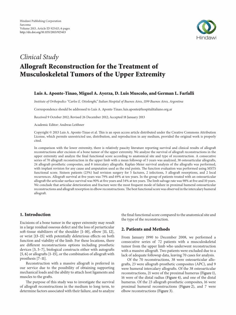
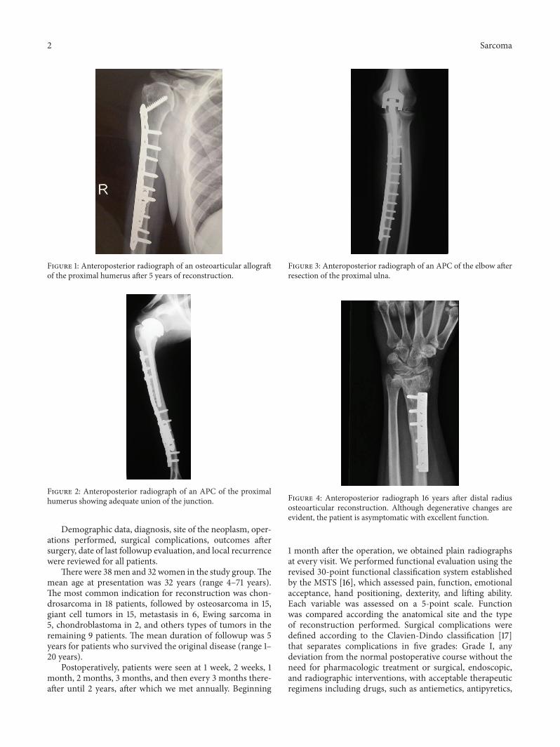
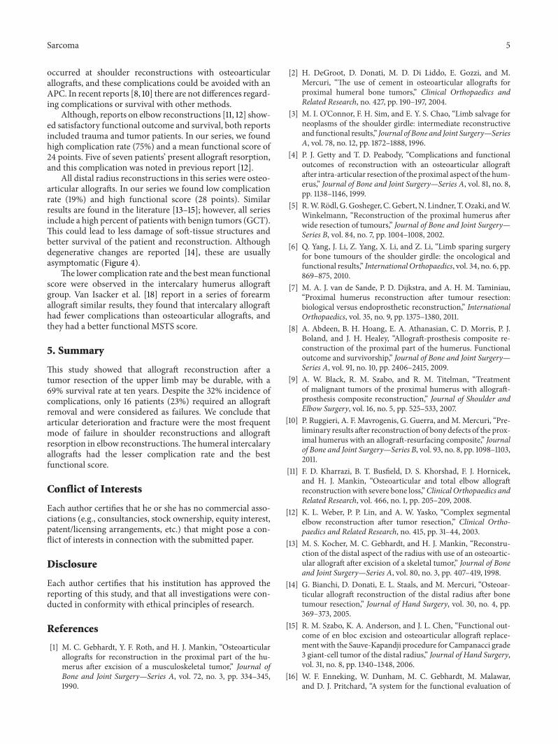
Of the 70 reconstructions, 38 were osteoarticular allo-grafts, 23 were allograft-prosthetic composites (APC), and 9were humeral intercalary allografts. Of the 38 osteoarticularreconstructions, 21 were of the proximal humerus (Figure 1),16 were of the distal radius (Figure 4), and one of the distalhumerus. Of the 23 allograft-prosthetic composites, 16 wereproximal humeral reconstructions (Figure 2), and 7 wereelbow reconstructions (Figure 3).
2 Sarcoma
Figure 1: Anteroposterior radiograph of an osteoarticular allograftof the proximal humerus after 5 years of reconstruction.
Figure 2: Anteroposterior radiograph of an APC of the proximalhumerus showing adequate union of the junction.
Demographic data, diagnosis, site of the neoplasm, oper-ations performed, surgical complications, outcomes aftersurgery, date of last followup evaluation, and local recurrencewere reviewed for all patients.
There were 38men and 32 women in the study group.Themean age at presentation was 32 years (range 4–71 years).The most common indication for reconstruction was chon-drosarcoma in 18 patients, followed by osteosarcoma in 15,giant cell tumors in 15, metastasis in 6, Ewing sarcoma in5, chondroblastoma in 2, and others types of tumors in theremaining 9 patients. The mean duration of followup was 5years for patients who survived the original disease (range 1–20 years).
Postoperatively, patients were seen at 1 week, 2 weeks, 1month, 2 months, 3 months, and then every 3 months there-after until 2 years, after which we met annually. Beginning
Figure 3: Anteroposterior radiograph of an APC of the elbow afterresection of the proximal ulna.
Figure 4: Anteroposterior radiograph 16 years after distal radiusosteoarticular reconstruction. Although degenerative changes areevident, the patient is asymptomatic with excellent function.
1 month after the operation, we obtained plain radiographsat every visit. We performed functional evaluation using therevised 30-point functional classification system establishedby the MSTS [16], which assessed pain, function, emotionalacceptance, hand positioning, dexterity, and lifting ability.Each variable was assessed on a 5-point scale. Functionwas compared according the anatomical site and the typeof reconstruction performed. Surgical complications weredefined according to the Clavien-Dindo classification [17]that separates complications in five grades: Grade I, anydeviation from the normal postoperative course without theneed for pharmacologic treatment or surgical, endoscopic,and radiographic interventions, with acceptable therapeuticregimens including drugs, such as antiemetics, antipyretics,
Sarcoma 3
0 50 100 150 200 250Months of followup
0
0.2
0.4
0.6
0.8
1
Cum
ulat
ive s
urvi
val
Survival functionCensored
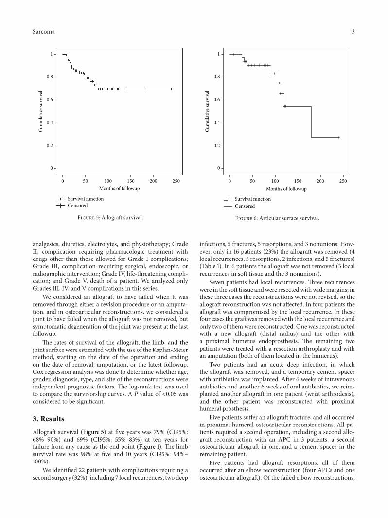
Figure 5: Allograft survival.
analgesics, diuretics, electrolytes, and physiotherapy; GradeII, complication requiring pharmacologic treatment withdrugs other than those allowed for Grade I complications;Grade III, complication requiring surgical, endoscopic, orradiographic intervention; Grade IV, life-threatening compli-cation; and Grade V, death of a patient. We analyzed onlyGrades III, IV, and V complications in this series.
We considered an allograft to have failed when it wasremoved through either a revision procedure or an amputa-tion, and in osteoarticular reconstructions, we considered ajoint to have failed when the allograft was not removed, butsymptomatic degeneration of the joint was present at the lastfollowup.
The rates of survival of the allograft, the limb, and thejoint surface were estimated with the use of the Kaplan-Meiermethod, starting on the date of the operation and endingon the date of removal, amputation, or the latest followup.Cox regression analysis was done to determine whether age,gender, diagnosis, type, and site of the reconstructions wereindependent prognostic factors. The log-rank test was usedto compare the survivorship curves. A 𝑃 value of <0.05 wasconsidered to be significant.
3. Results
Allograft survival (Figure 5) at five years was 79% (CI95%:68%–90%) and 69% (CI95%: 55%–83%) at ten years forfailure from any cause as the end point (Figure 1). The limbsurvival rate was 98% at five and 10 years (CI95%: 94%–100%).
We identified 22 patients with complications requiring asecond surgery (32%), including 7 local recurrences, two deep
0 50 100 150 200 250Months of followup
0
0.2
0.4
0.6
0.8
1
Cum
ulat
ive s
urvi
val
Survival functionCensored
Figure 6: Articular surface survival.
infections, 5 fractures, 5 resorptions, and 3 nonunions. How-ever, only in 16 patients (23%) the allograft was removed (4local recurrences, 5 resorptions, 2 infections, and 5 fractures)(Table 1). In 6 patients the allograft was not removed (3 localrecurrences in soft tissue and the 3 nonunions).
Seven patients had local recurrences. Three recurrenceswere in the soft tissue andwere resectedwithwidemargins; inthese three cases the reconstructions were not revised, so theallograft reconstruction was not affected. In four patients theallograft was compromised by the local recurrence. In thesefour cases the graftwas removedwith the local recurrence andonly two of them were reconstructed. One was reconstructedwith a new allograft (distal radius) and the other witha proximal humerus endoprosthesis. The remaining twopatients were treated with a resection arthroplasty and withan amputation (both of them located in the humerus).
Two patients had an acute deep infection, in whichthe allograft was removed, and a temporary cement spacerwith antibiotics was implanted. After 6 weeks of intravenousantibiotics and another 6 weeks of oral antibiotics, we reim-planted another allograft in one patient (wrist arthrodesis),and the other patient was reconstructed with proximalhumeral prosthesis.
Five patients suffer an allograft fracture, and all occurredin proximal humeral osteoarticular reconstructions. All pa-tients required a second operation, including a second allo-graft reconstruction with an APC in 3 patients, a secondosteoarticular allograft in one, and a cement spacer in theremaining patient.
Five patients had allograft resorptions, all of themoccurred after an elbow reconstruction (four APCs and oneosteoarticular allograft). Of the failed elbow reconstructions,
4 Sarcoma
Table 1: Allograft complications according the different types of reconstructions.
two were converted to an elbow endoprosthesis, two had aresection arthroplasty, and one had a cement spacer.
The three patients who underwent nonunionwere treatedwith autologous bone graft and a new plate, without revisionof the reconstruction.
The articular surface survival (Figure 6) of the groupof patients treated with an osteoarticular allograft was 90%(CI95%: 79%–100%) at five years and 54% (CI95%: 39%–69%)at ten years (Figure 2). All symptomatic articular deteriora-tions occurred in the proximal humeral reconstructions, andnone of them required revision because of this event.
The only independent prognostic factors that were foundto be significant on Cox regression analysis, with revision forany cause as the end point, were the gender of the patient(more frequent in males: 𝑃 = 0.02).
For the patients who retained the reconstruction (54cases), the mean MSTS functional score at last followup was26 of 30 (83%, range 18–30). The best mean functional scorewas observed in the intercalary humeral allograft group.(mean 30: 100%).The worst functional score was observed inproximal humeral osteoarticular allograft group (23 points,range 18–26), and this lower score was mainly relatedwith patients who had a significant articular deterioration(Table 2).
4. Discussion
In comparison with the lower extremity, there is relativelypaucity literature reporting survival and clinical results ofallograft reconstructions after excision of a bone tumor of theupper extremity.We include in this report all reconstructionsdone in the upper extremity done in our unit.
There are some limitations to this study. This is a retro-spective study with a relatively low number of patients andfollowup. In addition, there are many variables related to
the anatomic location of the reconstructions. Despite theselimitations, we believe that this series is one of the largestseries reported in the literature, and our results may providesome trends in the treatment of massive bone defects in theupper limb.
Regarding anatomical site, most publications are relatedto the proximal humerus. Osteoarticular allografts are usedless frequently than in the lower extremity, but there arereports regarding this type of reconstruction in the proxi-mal humerus. Although some authors reported satisfactoryresults with osteoarticular allografts of the proximal humerus[1] and survival rates of 78% at five years [2], recent reportssuggest that better or at least similar results are obtained withallograft prosthesis composite and endoprosthesis recon-structions regarding reconstruction survival and complica-tions [3–8]. Peabody [4] report that due to functional limita-tions as well as an extremely high rate of complications, theydo not use osteoarticular allografts to replace the proximalaspect of the humerus. However, in a recent report [7] thatanalyzed 38 reconstructions of the proximal humerus theendoprosthetic group presented the smallest complicationrate of 21%, compared to 40% in the allograft prosthesis com-posite and 62% in the osteoarticular allograft group.However,in another report that analyzed 45 patients [5] reconstructedafter tumor resection of the proximal humerus they foundthat all limb-salvage procedures for the proximal humeruswere satisfactory for long-term survival, but none of the26 disease-free surviving patients was able to abduct theirshoulder more than 90∘, and only five could achieve activeabduction of more than 30∘. The survival rate was 83% forendoprosthesis, 79% in clavicula prohumero, and 75% inosteoarticular allograft [5].
Reconstructions with APC in the proximal humerusavoid problems of endoprosthesis or osteoarticular allograftsused alone [8–10]. In our series the higher amount of fractures
Sarcoma 5
occurred at shoulder reconstructions with osteoarticularallografts, and these complications could be avoided with anAPC. In recent reports [8, 10] there are not differences regard-ing complications or survival with other methods.
Although, reports on elbow reconstructions [11, 12] show-ed satisfactory functional outcome and survival, both reportsincluded trauma and tumor patients. In our series, we foundhigh complication rate (75%) and a mean functional score of24 points. Five of seven patients’ present allograft resorption,and this complication was noted in previous report [12].
All distal radius reconstructions in this series were osteo-articular allografts. In our series we found low complicationrate (19%) and high functional score (28 points). Similarresults are found in the literature [13–15]; however, all seriesinclude a high percent of patients with benign tumors (GCT).This could lead to less damage of soft-tissue structures andbetter survival of the patient and reconstruction. Althoughdegenerative changes are reported [14], these are usuallyasymptomatic (Figure 4).
The lower complication rate and the best mean functionalscore were observed in the intercalary humerus allograftgroup. Van Isacker et al. [18] report in a series of forearmallograft similar results, they found that intercalary allografthad fewer complications than osteoarticular allografts, andthey had a better functional MSTS score.
5. Summary
This study showed that allograft reconstruction after atumor resection of the upper limb may be durable, with a69% survival rate at ten years. Despite the 32% incidence ofcomplications, only 16 patients (23%) required an allograftremoval and were considered as failures. We conclude thatarticular deterioration and fracture were the most frequentmode of failure in shoulder reconstructions and allograftresorption in elbow reconstructions.The humeral intercalaryallografts had the lesser complication rate and the bestfunctional score.
Conflict of Interests
Each author certifies that he or she has no commercial asso-ciations (e.g., consultancies, stock ownership, equity interest,patent/licensing arrangements, etc.) that might pose a con-flict of interests in connection with the submitted paper.
Disclosure
Each author certifies that his institution has approved thereporting of this study, and that all investigations were con-ducted in conformity with ethical principles of research.
References
[1] M. C. Gebhardt, Y. F. Roth, and H. J. Mankin, “Osteoarticularallografts for reconstruction in the proximal part of the hu-merus after excision of a musculoskeletal tumor,” Journal ofBone and Joint Surgery—Series A, vol. 72, no. 3, pp. 334–345,1990.
[2] H. DeGroot, D. Donati, M. D. Di Liddo, E. Gozzi, and M.Mercuri, “The use of cement in osteoarticular allografts forproximal humeral bone tumors,” Clinical Orthopaedics andRelated Research, no. 427, pp. 190–197, 2004.
[3] M. I. O’Connor, F. H. Sim, and E. Y. S. Chao, “Limb salvage forneoplasms of the shoulder girdle: intermediate reconstructiveand functional results,” Journal of Bone and Joint Surgery—SeriesA, vol. 78, no. 12, pp. 1872–1888, 1996.
[4] P. J. Getty and T. D. Peabody, “Complications and functionaloutcomes of reconstruction with an osteoarticular allograftafter intra-articular resection of the proximal aspect of the hum-erus,” Journal of Bone and Joint Surgery—Series A, vol. 81, no. 8,pp. 1138–1146, 1999.
[5] R.W.Rodl, G.Gosheger, C.Gebert, N. Lindner, T.Ozaki, andW.Winkelmann, “Reconstruction of the proximal humerus afterwide resection of tumours,” Journal of Bone and Joint Surgery—Series B, vol. 84, no. 7, pp. 1004–1008, 2002.
[6] Q. Yang, J. Li, Z. Yang, X. Li, and Z. Li, “Limb sparing surgeryfor bone tumours of the shoulder girdle: the oncological andfunctional results,” International Orthopaedics, vol. 34, no. 6, pp.869–875, 2010.
[7] M. A. J. van de Sande, P. D. Dijkstra, and A. H. M. Taminiau,“Proximal humerus reconstruction after tumour resection:biological versus endoprosthetic reconstruction,” InternationalOrthopaedics, vol. 35, no. 9, pp. 1375–1380, 2011.
[8] A. Abdeen, B. H. Hoang, E. A. Athanasian, C. D. Morris, P. J.Boland, and J. H. Healey, “Allograft-prosthesis composite re-construction of the proximal part of the humerus. Functionaloutcome and survivorship,” Journal of Bone and Joint Surgery—Series A, vol. 91, no. 10, pp. 2406–2415, 2009.
[9] A. W. Black, R. M. Szabo, and R. M. Titelman, “Treatmentof malignant tumors of the proximal humerus with allograft-prosthesis composite reconstruction,” Journal of Shoulder andElbow Surgery, vol. 16, no. 5, pp. 525–533, 2007.
[10] P. Ruggieri, A. F. Mavrogenis, G. Guerra, andM. Mercuri, “Pre-liminary results after reconstruction of bony defects of the prox-imal humerus with an allograft-resurfacing composite,” Journalof Bone and Joint Surgery—Series B, vol. 93, no. 8, pp. 1098–1103,2011.
[11] F. D. Kharrazi, B. T. Busfield, D. S. Khorshad, F. J. Hornicek,and H. J. Mankin, “Osteoarticular and total elbow allograftreconstructionwith severe bone loss,”Clinical Orthopaedics andRelated Research, vol. 466, no. 1, pp. 205–209, 2008.
[12] K. L. Weber, P. P. Lin, and A. W. Yasko, “Complex segmentalelbow reconstruction after tumor resection,” Clinical Ortho-paedics and Related Research, no. 415, pp. 31–44, 2003.
[13] M. S. Kocher, M. C. Gebhardt, and H. J. Mankin, “Reconstru-ction of the distal aspect of the radius with use of an osteoartic-ular allograft after excision of a skeletal tumor,” Journal of Boneand Joint Surgery—Series A, vol. 80, no. 3, pp. 407–419, 1998.
[14] G. Bianchi, D. Donati, E. L. Staals, and M. Mercuri, “Osteoar-ticular allograft reconstruction of the distal radius after bonetumour resection,” Journal of Hand Surgery, vol. 30, no. 4, pp.369–373, 2005.
[15] R. M. Szabo, K. A. Anderson, and J. L. Chen, “Functional out-come of en bloc excision and osteoarticular allograft replace-ment with the Sauve-Kapandji procedure for Campanacci grade3 giant-cell tumor of the distal radius,” Journal of Hand Surgery,vol. 31, no. 8, pp. 1340–1348, 2006.
[16] W. F. Enneking, W. Dunham, M. C. Gebhardt, M. Malawar,and D. J. Pritchard, “A system for the functional evaluation of
6 Sarcoma
reconstructive procedures after surgical treatment of tumors ofthe musculoskeletal system,” Clinical Orthopaedics and RelatedResearch, no. 286, pp. 241–246, 1993.
[17] P. A. Clavien, J. Barkun, M. L. De Oliveira et al., “The clavien-dindo classification of surgical complications: five-year experi-ence,” Annals of Surgery, vol. 250, no. 2, pp. 187–196, 2009.
[18] T. van Isacker,O. Barbier, A. Traore,O. Cornu, F.Mazzeo, andC.Delloye, “Forearm reconstruction with bone allograft followingtumor excision: a series of 10 patients with a mean follow-upof 10 years,” Orthopaedics and Traumatology, vol. 97, no. 8, pp.793–799, 2011.