Purpose. To determine whether subconjunctival lidocaine injection maintains additional anesthetic effect during intravitrealOzurdex injection. Methods. 63 patients who were diagnosed as central or branch retinal vein occlusion and planned to receiveOzurdex injection for macular edema were prospectively included in the study. The patients were randomized into one of the twoanesthetic groups. The first group received topical proparacaine drop and lidocaine applied pledget. The second group receivedsubconjunctival lidocaine injection in addition to the anesthetics in group 1. Results. Mean pain score was 1.90 ± 2.39 in group 1and 1.71 ± 2.09 in group 2 (𝑝 = 0.746). Mean subconjunctival hemorrhage grade was 1.67 ± 0.17 in group 1 and 0.90 ± 0.14 ingroup 2 (𝑝 = 0.001). There was no relationship between the amount of subconjunctival hemorrhage and pain score of the patients.Conclusions. There was no difference in pain scores between the two anesthetic methods.The addition of subconjunctival lidocaineinjection offered no advantage in pain relief compared to lidocaine-applied pledgets.
1. Introduction
Intravitreal injection has become an important treatmentchoice in many vision-threatening diseases. Various pharma-cologic agents such as antibiotics, steroids, and antifungal andantiviral drugs can be delivered into the vitreous cavity by thismethod [1]. Recently, antivascular endothelial growth factor(VEGF) drugs gained popularity especially in treating age-related macular degeneration and diabetic macular edema[2]. Thus intravitreal injections have become one of the mostcommon medical procedures.
Intravitreal injections are usually performed as an officeprocedure, and topical and/or local anesthesia is used dur-ing the injection. The most common anesthesia techniquesare topical anesthetic eye drops, anesthetic-applied pledgetplaced on the conjunctiva, and subconjunctival anestheticinjection [3]. Although all of these techniques are found tobe effective in the previous studies, there is no consensusfor the most effective method [4–7]. On the other handthe studies that investigated the effect of anesthesia methodin intravitreal injections usually evaluated the small-sized
needles. For this reason, the effect of anesthesia in intravitrealOzurdex injection is not known.
Ozurdex (Allergan, Inc., Irvine, CA) is an intravitrealimplant containing 700 𝜇g of dexamethasone in a slow-release preparation. Key features of the drug delivery systemare the sustained-release formulation of the polylactic acid-coglycolic acid (PLGA) matrix material, which dissolvescompletely in vivo, and the single-use applicator for intrav-itreal placement.The United States Food and Drug Adminis-tration (FDA) and European Union (EU) approve it for theintravitreal treatment of macular edema. It is indicated forthe treatment of adult patients with macula edema followingretinal vein occlusion (RVO) (either branch or central) andinflammation of the posterior segment of the eye presentingas noninfectious uveitis. Furthermore, clinical efficacy hasbeen documented in other diseases, such as diabetic macularedema (DME) andpersistentmacular edema (ME) associatedwith uveitis or Irvine-Gass syndrome [8].
The indications for intravitreal Ozurdex implantationhave broadened in the recent years. Consequently, increasing
Hindawi Publishing CorporationJournal of OphthalmologyVolume 2015, Article ID 861535, 5 pageshttp://dx.doi.org/10.1155/2015/861535
2 Journal of Ophthalmology
numbers of Ozurdex implantation are administered. Duringimplantation procedure, patient comfort and eye stabilizationare very important to avoid complications related to appli-cation; calm and painless patient can allow us to completeOzurdex implantation without any problem. Some of the sur-geons are using subconjunctival anesthesia to reduce the painthat occurs while Ozurdex applicator is penetrating throughthe sclera on pars plana. It is arguable if subconjunctivalanesthesia really reduces pain or causes more conjunctivalhemorrhage and edema that could prevent finding the properinjection site and control the leakage after injection.
We conducted a prospective randomized clinical trialcomparing the patients’ comfort and pain with or with-out subconjunctival anesthesia during intravitreal Ozurdeximplantation procedure.
2. Materials and Methods
This prospective randomized study included 63 patients whowere diagnosed as central or branch retinal vein occlusion.The patients underwent intravitreal Ozurdex injection fortheir macular edema related to their diagnosis. All patientswere naıve to any kind of intravitreal injection before enroll-ment. The Ethical Committee of Kocaeli University MedicalFaculty approved this study. After the patients provided awritten consent, each patient was randomized into one ofthe two anesthetic groups. The first group received topicalproparacaine drop and 4% lidocaine-applied pledget. Thesecond group received subconjunctival lidocaine injection inaddition to the anesthetics in group 1.
2.1. The Anesthesia Methods and Injection. First, topi-cal proparacaine hydrochloride ophthalmic solution wasinstilled in eyes of the patients in both groups. Thenproparacaine-applied sterile pledgets were placed into upperand lower conjunctival sac of the patients in both groups for1 minute. Povidone-iodine was used to prepare the ocularsurface for preinjection antisepsis and the periocular areawas covered with an ophthalmic drape. A lid speculumwas placed. All eyes were irrigated with 10% povidone-iodine after draping. A second irrigation with sterile 0.9%NaCl was performed. The patients in group 1 (31 patients)received subconjunctival lidocaine injection before Ozurdeximplantation, and the patients in group 2 (32 patients) didnot receive any anesthesia other than topical proparacainehydrochloride and 4% lidocaine-applied pledget.
This step was followed by the insertion of the Ozurdeximplant into the vitreous cavity through the pars plana using acustomized, single-use 22-gauge applicator in both groups bythe same surgeon. Standard eye calipers were used tomeasurethe distance from the limbus (3.5mm for the pseudophakicpatients, 4mm for the phakic patients). Injections wereperformed in the superotemporal quadrant of the each eye.
2.2. Hemorrhage and Pain Assessment. After the subconjunc-tival lidocaine application the surgeon assessed the gradeof the subconjunctival hemorrhage using a scoring system(none = 0, minor = 1, moderate = 2, and severe = 3).
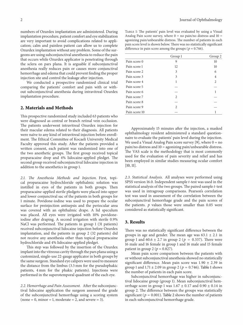
Table 1: The patients’ pain level was evaluated by using a VisualAnalog Pain score survey, where 0 = no pain/no distress and 10 =agonizing pain/unbearable distress. The number of patients in eachpain score level is shown below.There was no statistically significantdifference in pain score among the groups (𝑝 = 0.746).
Approximately 15 minutes after the injection, a maskedophthalmology resident administered a standard question-naire to evaluate the patients’ pain level during the injection.We used a Visual Analog Pain score survey [9], where 0 = nopain/no distress and 10 = agonizing pain/unbearable distress.The VAS scale is the methodology that is most commonlyused for the evaluation of pain severity and relief and hasbeen employed in similar studies measuring ocular comfort[10, 11].
2.3. Statistical Analysis. All analyses were performed usingSPSS version 16.0. Independent sample 𝑡-test was used in thestatistical analysis of the two groups.The paired sample 𝑡-testwas used in intragroup comparisons. Pearson’s correlationtest was used in assessment of the correlation between thesubconjunctival hemorrhage grade and the pain scores ofthe patients. 𝑝 values those were smaller than 0.05 wereconsidered as statistically significant.
3. Results
There was no statistically significant difference between thegroups in age and gender. The mean age was 63.1 ± 2.1 ingroup 1 and 60.6 ± 2.7 in group 2 (𝑝 = 0.337). There were16 male and 16 female in group 1 and 16 male and 15 femalepatient in group 2 (p = 0.827).
Mean pain score comparison between the patients withor without subconjunctival anesthesia showed no statisticallysignificant difference. Mean pain score was 1.90 ± 2.39 ingroup 1 and 1.71 ± 2.09 in group 2 (𝑝 = 0.746). Table 1 showsthe number of patients in each pain score.
Subconjunctival hemorrhage was higher in subconjunc-tival lidocaine group (group 1). Mean subconjunctival hem-orrhage score in group 1 was 1.67 ± 0.17 and 0.90 ± 0.14 ingroup 2. The difference between the groups was statisticallysignificant (𝑝 = 0.001). Table 2 shows the number of patientsin each subconjunctival hemorrhage grade.
Journal of Ophthalmology 3
Table 2: The number of patients in each subconjunctival hem-orrhage grade in both groups is demonstrated. The patients thatreceived additional subconjunctival anesthesia (group 1) presentedwith higher levels of subconjunctival hemorrhage compared to thepatients that received only topical and pledged anesthesia (𝑝 =0.001).
Therewas no relationship between the amount of subcon-junctival hemorrhage and pain score of the patients. Pearson’scorrelation test did not reveal any statistically significantcorrelation (𝑝 = 0.066 and 𝑟 = 0.233).
4. Discussion
The use of corticosteroids for the treatment of ocularinflammatory diseases was first described in the early 1950s.Corticosteroids have anti-inflammatory, antiangiogenic, andantipermeability properties that make them an importanttherapeutic option for a variety of posterior segment diseases[12]. Based on experimental studies, clinical observations,and pathogenic considerations, the intravitreal delivery ofsteroids has been suggested to locally suppress intraocularinflammation, proliferation of cells, and neovascularization[13]. Intravitreal delivery of corticosteroids has allowedmanyposterior segment diseases to be locally treated withoutthe adverse systemic side effects. An increasing number ofophthalmologists use intravitreal steroids for the treatmentof various posterior segment disorders, especially whentraditional therapeutic methods have failed.
A variety of methods have been proposed that achievelonger duration of pharmacologic effect with lower admin-istration frequency and minimal side effects of the intrav-itreal steroids. Novel agents including preservative-free andsustained-release intravitreal implants such as Ozurdex arecurrently approved for ocular use and are being furtherevaluated for the treatment of RVO, DME, uveitis, and AMD.Due to a potential for greater potency, dexamethasone isbeing evaluated alone or in combination with anti-VEGFsas promising options in the emerging armamentarium forthe treatment of several retinal diseases [8]. The safety andtolerability of a sustained-release implant are particularlyimportant due to the long duration of exposure to the drugand the drug vehicle.
The intravitreal injections are usually administered as apart of the office procedure. In order to maintain a successfulinjection the patient should be comfortable and relaxed.This can be achieved by a minimally painful injection.The American Society of Retina Specialists Preferences andTrends Survey from 2010 revealed that 25.44% of the retinaspecialists use topical anesthetic drop, 25.15% use topical
viscous anesthetic, 26.33% use topical anesthetic (drop or vis-cous) + pledget, and 22.19% use subconjunctival injection ofanesthetic for intravitreal injections [3]. Canadian Ophthal-mological Society carried out a survey for retina specialistsin Canada and evaluated the intravitreal injection techniques.They found that 90% of retina specialists routinely used topi-cal proparacaine, lidocaine, or tetracaine drops in preparationfor intravitreal injection. Twenty-five percent routinely usedtopical lidocaine gel, and 16% used it infrequently. Twenty-three percent routinely used a pledget soaked with tetracaineor proparacaine, and 28%used pledgets infrequently. Twenty-three percent routinely used subconjunctival lidocaine injec-tion, and 43% used this technique infrequently [14].
Recently, few studies assessed patient comfort usingdifferent kinds of anesthetic methods during intravitrealinjections. In most of these studies it was found that all ofthese methods were effective [4–7]. Most of these studieshave shown no significant difference in pain score betweenpledget, subconjunctival injection, or topical drops [5, 6, 15–17]. Blaha et al. compared the effectiveness of proparacaine,tetracaine, lidocaine pledget, and subconjunctival injectionof lidocaine. They found no statistical difference in injectionor total procedure pain scores between these methods [5].Davis et al. also evaluated the difference in anesthetic effectbetween topical proparacaine drops, 4% lidocaine-appliedcotton tipped swabs, or 3.5% lidocaine gel. After the injectionthey asked the patients to grade the discomfort associatedwith 3 components of the injection procedure: (1) the lidspeculum; (2) the needle insertion; and (3) the burning sensa-tion from the 5% povidone-iodine solution.They did not findany difference between the groups in any of the factors thatmight cause discomfort during the injection [15]. However,there are also a few studies claiming that there was differencebetween the anesthesia methods. LaHood et al. comparedthe anesthetic effectiveness of topical gel, subconjunctival andcombination of topical gel, and subconjunctival anesthesiafor intravitreal injection in 120 consecutive patients. Theirresults showed that the group receiving topical gel anestheticproduced significantly higher pain scores compared to bothof the other groups [18]. Blaha et al. did not find statisticallysignificant difference between topical proparacaine drops,pledget of 4% lidocaine, and subconjunctival injection of 2%lidocaine. But they reported that proparacaine drops had thelowest average combined with pain score [19].
In most of these studies, researchers evaluated theanesthetic methods in intravitreal injection of ranibizumab(Lucentis, Genentech), bevacizumab (Avastin, Genentech),or triamcinolone (Kenalog, Bristol-Myers Squibb). In orderto perform these injections a smaller sized needle was used(27.5–32 gauges). However, Ozurdex injection is differentthan other intravitreal injections. It has a larger needle, whichis 22 gauges. While inserting the Ozurdex needle it feelsblunter.The long axis of the applicator should be held parallelto the limbus, and the sclera should be engaged at an obliqueangle with the bevel of the needle up (away from the sclera) tocreate a shelved scleral path.The tip of the needle is advancedwithin the sclera for about 1mm (parallel to the limbus) andthen redirected toward the center of the eye and advanceduntil penetration of the sclera is completed and the vitreous
4 Journal of Ophthalmology
cavity is entered (the package insert is available onlineat http://www.allergan.com/assets/pdf/ozurdex pi.pdf). Thisprocedure is more complicated than the quick fine-gauge“pinprick” of other intravitreal therapies. This might result inhigher pain perception than the other intravitreal injections.For this reason, patients might distinguish the differencebetween the anesthetic techniques in this higher pain level. Inour study we compared the anestheticmethods in intravitrealOzurdex injection. We found that subconjunctival lidocaineinjection did not maintain additional decrease in pain.
Kozak et al. compared the gel anesthesia using differentneedle lumens. They observed no difference between 27.5-gauge and 30-gauge needles, but these needles are also smallerthan Ozurdex applicator [7]. For this reason, they cannot becompared with Ozurdex injection. While we were planningour study, we thought that topical proparacaine drops mightnot be enough as a single anesthetic method in Ozurdeximplantation because Ozurdex has a larger applicator. Wecombined proparacaine drops with lidocaine-applied pled-gets in all patients and an additional subconjunctival lido-caine injection in another group of patients. The advantageof using the pledgets is the application of high concentrationof anesthetic to the site of injection. Pledgets might alsohave some disadvantages, such as irritation, corneal edema,and corneal epithelial defects. None of the patients in ourstudy complained about the irritation, andwe did not observeany corneal edema and epithelial defects. In order to avoidthese problems smaller pledgets might be used. Howeversmaller pledgets could be lost in the fornix, and to go fishingaround the fornix to retrieve a pledget might cause additionaldiscomfort for the patient. We believe that comparisonbetween single use of topical proparacaine and 4% lidocaine-applied pledgets in Ozurdex injection must be studied infurther studies.
In our study we found higher incidence of subconjunc-tival hemorrhage in patients that underwent subconjunctivalinjection. It might be aminor complication; however patientsfind the appearance of their eyes bothersome. Additionally, itmight cause difficulty in injection especially when it is com-bined with the chemosis. It makes marking the injection sitelocation more difficult [13]. Other complications that werereported to be related to subconjunctival lidocaine are inad-vertent intravitreal injection and hypersensitivity [20, 21].Subconjunctival hemorrhage and chemosis might also leadto inadvertent intravitreal injection. We did not observe anyof these complications. However we believe that taking therisks that might be caused by subconjunctival chemosis andhemorrhage is unnecessary because subconjunctival lido-caine injection does not maintain additional pain reduction.There is also a controversy about the effect of subconjunctivalanesthesia on endophthalmitis rate. Tustin et al. claim thatsubconjunctival lidocaine is bactericidal and maintains aclinically important antiseptic effect. As a result, they suggestthat application of subconjunctival lidocaine may reducethe incidence of endophthalmitis after intravitreal injection[22]. On the other hand in another prospective randomizedcase control study, subconjunctival anesthesia is found tobe a potential risk factor for postinjection endophthalmitis.The authors hypothesize that compromising the conjunctival
surface before injection allows the introduction of bacteriainto the subconjunctival space. This could act as a source ofinfection for postinjection endophthalmitis [23]. We shouldalso consider this risk while performing subconjunctivallidocaine anesthesia.
In conclusion, there are different methods for painreduction during intravitreal injections. However, there hasbeen no method repeatedly shown to be superior in con-trolling pain. To the best of our knowledge, this study isthe first study that compares the anesthesia techniques inintravitreal Ozurdex implantation. Our study showed thatsubconjunctival lidocaine injection is not mandatory inreducing pain duringOzurdex implantation.On the contrary,it causes conjunctival bleeding and chemosis in the injectionsite, which might cause difficulty in injection. We believethat future investigations to compare the local anesthesiatechniques should be carried out.
Conflict of Interests
The authors declare that there is no conflict of interestsregarding the publication of this paper. The authors have nofinancial or proprietary interest in any product mentioned inthis paper.
References
[1] G. A. Peyman, E. M. Lad, and D. M. Moshfeghi, “Intravitrealinjection of therapeutic agents,” Retina, vol. 29, no. 7, pp. 875–912, 2009.
[2] M. W. Stewart, “The expanding role of vascular endothelialgrowth factor inhibitors in ophthalmology,” Mayo Clinic Pro-ceedings, vol. 87, no. 1, pp. 77–88, 2012.
[3] American Society of Retina Specialists, Preferences and Trends2010 Survey, 2010, http://www.asrs.org/.
[4] L. P. Cintra, L. R. Lucena, J. A. Da Silva, R. A. Costa, I. U. Scott,and R. Jorge, “Comparative study of analgesic effectivenessusing three different anesthetic techniques for intravitreal injec-tion of bevacizumab,” Ophthalmic Surgery Lasers and Imaging,vol. 40, no. 1, pp. 13–18, 2009.
[5] G. R. Blaha, E. P. Tilton, F. C. Barouch, and J. L. Marx, “Ran-domized trial of anesthetic methods for intravitreal injections,”Retina, vol. 31, no. 3, pp. 535–539, 2011.
[6] N. Z. Gregori, M. J. Weiss, R. Goldhardt et al., “Randomizedclinical trial of two anesthetic techniques for intravitreal injec-tions: 4% liquid lidocaine on cotton swabs versus 3.5% lidocainegel,” Expert Opinion on Drug Delivery, vol. 9, no. 7, pp. 735–741,2012.
[7] I. Kozak, L. Cheng, andW. R. Freeman, “Lidocaine gel anesthe-sia for intravitreal drug administration,” Retina, vol. 25, no. 8,pp. 994–998, 2005.
[8] K. G. Kapoor, M. G.Wagner, and A. L.Wagner, “The sustained-release dexamethasone implant: expanding indications in vitre-oretinal disease,” Seminars in Ophthalmology, 2014.
[9] D. P. Price, P. A. McGrath, A. Rafii, and B. Buckingham, “Thevalidation of visual analogue scales as ratio scale measures forchronic and experimental pain,” Pain, vol. 17, no. 1, pp. 45–56,1983.
[10] D. Salo, D. Eget, R. F. Lavery, L. Garner, S. Bernstein, and K.Tandon, “Can patients accurately read visual analog pain scale?”
Journal of Ophthalmology 5
The American Journal of Emergency Medicine, vol. 21, no. 7, pp.515–519, 2003.
[11] P. C. Jacobi, T. S. Dietlein, and F. K. Jacobi, “Comparative studyof topical vs retrobulbar anesthesia in complicated cataractsurgery,” Archives of Ophthalmology, vol. 118, no. 8, pp. 1037–1043, 2000.
[12] N. Floman and U. Zor, “Mechanism of steroid action inocular inflammation: inhibition of prostaglandin production,”Investigative Ophthalmology & Visual Science, vol. 16, no. 1, pp.69–73, 1977.
[13] C. S. Cannon, J. G. Gross, I. Abramson, W. J. Mazzei, andW. R. Freeman, “Evaluation of outpatient experience withvitreoretinal surgery,” British Journal of Ophthalmology, vol. 76,no. 2, pp. 68–71, 1992.
[14] L. Xing, S. J. Dorrepaal, and J. Gale, “Survey of intravitrealinjection techniques and treatment protocols among retinaspecialists in Canada,” Canadian Journal of Ophthalmology, vol.49, no. 3, pp. 261–266, 2014.
[15] M. J. Davis, J. S. Pollack, and S. Shott, “Comparison of topicalanesthetics for intravitreal injections: a randomized clinicaltrial,” Retina, vol. 32, no. 4, pp. 701–705, 2012.
[16] B. Kaderli and R. Avcı, “Comparison of topical and subcon-junctival anesthesia in intravitreal injection administrations,”European Journal of Ophthalmology, vol. 16, no. 5, pp. 718–721,2006.
[17] G. L. Yau, C. S. Jackman, P. L. Hooper, and T. G. Sheidow,“Intravitreal injection anesthesia-comparison of different top-ical agents: a prospective randomized controlled trial,” TheAmerican Journal of Ophthalmology, vol. 151, no. 2, pp. 333.e2–337.e2, 2011.
[18] B. R. LaHood, D. S. Franzco, and A. S. Franzco, “Comparativeassessment of the effectiveness of anesthesia for intravitrealbevacizumab injection,” Clinical & Experimental Ophthalmol-ogy, vol. 39, no. 2, pp. 184–185, 2011.
[19] G. R. Blaha, “Comparison of topical anesthetics for intravitrealinjections: a randomized clinical trial,” Retina, vol. 32, no. 7, pp.1440–1440, 2012.
[20] P. Massaoutis, M. Niscopoulou, and A. Tufail, “Inadvertentintravitreal injection of 0.1mls of unpreserved lidocaine 2%,”The Internet Journal of Ophthalmology and Visual Science, vol.6, no. 1, article 5, 2007.
[21] J. Levy andT. Lifshitz, “Lidocaine hypersensitivity after subcon-junctival injection,”Canadian Journal of Ophthalmology, vol. 41,no. 2, pp. 204–206, 2006.
[22] A. Tustin, S. J. Kim, A. Chomsky, G. B. Hubbard III, and J.Sheng, “Antibacterial properties of 2% lidocaine and reducedrate of endophthalmitis after intravitreal injection,” Retina, vol.34, no. 5, pp. 935–942, 2014.
[23] D.A.M. Lyall, A. Tey, B. Foot et al., “Post-intravitreal anti-VEGFendophthalmitis in the United Kingdom: Incidence, features,risk factors, and outcomes,” Eye, vol. 26, no. 12, pp. 1517–1526,2012.