Clinical StudyEffectiveness of the Virtual Reality System Toyra onUpper Limb Function in People with Tetraplegia: A PilotRandomized Clinical Trial
I. Dimbwadyo-Terrer,1 A. Gil-Agudo,2 A. Segura-Fragoso,3 A. de los Reyes-Guzmán,2
F. Trincado-Alonso,2 S. Piazza,4 and B. Polonio-López5
1Occupational Thinks Research Group, Centro Superior de Estudios Universitarios La Salle (UAM), C/La Salle 10,28023 Madrid, Spain2Biomechanics and Technical Aids Department, National Hospital for Spinal Cord Injury, Finca la Peraleda s/n, 45071 Toledo, Spain3Health Sciences Institute, Avenida de Madrid s/n, Talavera de la Reina, 45600 Toledo, Spain4Neural Rehabilitation Group, Cajal Institute, Spanish National Research Council (CSIC), Avenida Doctor Arce 37,28002 Madrid, Spain5Nursing, Physiotherapy and Occupational Therapy Department, University of Castilla La Mancha,Avenida Real Fabrica de Sedas s/n, Talavera de la Reina, 45600 Toledo, Spain
Correspondence should be addressed to I. Dimbwadyo-Terrer; [email protected]
Received 1 October 2015; Revised 20 December 2015; Accepted 21 December 2015
The aim of this study was to investigate the effects of a virtual reality program combined with conventional therapy in upper limbfunction in people with tetraplegia and to provide data about patients’ satisfaction with the virtual reality system.Thirty-one peoplewith subacute complete cervical tetraplegia participated in the study. Experimental group received 15 sessions with ToyraⓇ virtualreality system for 5 weeks, 30 minutes/day, 3 days/week in addition to conventional therapy, while control group only receivedconventional therapy. All patients were assessed at baseline, after intervention, and at three-month follow-up with a battery ofclinical, functional, and satisfaction scales. Control group showed significant improvements in the manual muscle test (𝑝 = 0,043,partial 𝜂2 = 0,22) in the follow-up evaluation. Both groups demonstrated clinical, but nonsignificant, changes to their arm functionin 4 of the 5 scales used. All patients showed a high level of satisfaction with the virtual reality system.This study showed that virtualreality added to conventional therapy produces similar results in upper limb function compared to only conventional therapy.Moreover, the gaming aspects incorporated in conventional rehabilitation appear to produce high motivation during execution ofthe assigned tasks. This trial is registered with EudraCT number 2015-002157-35.
1. Introduction
The worldwide incidence of spinal cord injury (SCI) liesbetween 10.4 and 83 permillion inhabitants per year [1]. One-third of patients with SCI are reported to have tetraplegia and50% of patients with SCI to have a complete lesion. Relatedto Spain, incidence varies among 12.1 [2] and 13.1 [3], with amean age at the time of injury of 41.8 years, and amale/femaleratio of 2.6. Prevalence of SCI is estimated in 350–380cases per million of inhabitants [2], with more frequencyat the thoracic level (42.7%), following cervical (38.5%) and
lumbosacral (17.8%). Most of the patients with SCI develop atleast one clinical complication in their life, the most commonbeing the loss of muscular control, sensibility, and autonomyfunction below the level of injury [4]. It has been estimatedthat all SCI discharges would need at least 4 hours a day ofspecialized care (occupational therapy, speech therapy, psy-chotherapy, physiotherapy, nursing, etc.), and around 84% ofpeople with tetraplegia will need external help for performingactivities of daily living (ADL) [5].These needs are only a hintof the enormously devastating physical, social, and emotionalburdens that individuals and their families face after a SCI [6].
Hindawi Publishing CorporationBioMed Research InternationalVolume 2016, Article ID 6397828, 12 pageshttp://dx.doi.org/10.1155/2016/6397828
2 BioMed Research International
Impairment of upper limbs (UL) is one of the mostcommon sequels following neurological lesions [7], the lossof arm and hand function being one of the most devastatingconsequences in tetraplegia [1, 8]. In contrast with the lowerlimb, the UL has extensive functionality due to the mobilityof numerous joints that can execute fine movements thanksto complex neuromuscular control [9]. It has been shownthat most people with tetraplegia prefer the recovery of handfunction to that of the bladder, bowel, or even sexual function[10]. Small progresses in arm and hand function may lead toincreased autonomy in daily activities, improving indepen-dence and quality of life [11]. For this reason, improvement inUL function after cervical SCI is a top priority in individualswith tetraplegia [12].
In rehabilitation, considerable amounts of practice arerequired to induce neuroplastic changes and functionalrecovery of neurological motor deficits [13]. However, con-ventional therapies (CT; physical and occupational therapy)do not provide sufficient intensity for optimizing neuro-plasticity because of practical limitations such as its time-consuming and labor-intensive nature, difficulty in trans-portation to special facilities, and need for insurance coverage[14–16]. Furthermore, traditional interventions that requiresimple and repetitive movements may cause monotony andboredom to the patients, reducing motivation for sustainingtreatment [13, 17].
One proposed method to improve rehabilitation is tocomplement conventional therapy with the use of virtualreality (VR) [13]. VR is defined as a simulation of a realenvironment generated by computer in which, through aman-machine interface, it allows the user to interact withcertain elements inside a simulated scenario [18]. VR hasmany advantages for intervention, such as enabling the grad-ing of activities, obtaining precise performance measures,providing a safe and ecologically valid environment, andbeing enjoyable and motivating [13, 19]. In addition, VR canincrease the intensity of the exercise through repetition ofexercises, necessary for inducing neuroplasticity [17].
Furthermore, in spite of the many techniques available tofacilitate therapy, motivation and access are major obstaclesto patients in achieving the necessary dosage of movementsneeded for recovery [16]. Motivation can be defined as a psy-chological property that encourages a person’s action towarda goal by eliciting and/or sustaining goal-directed behavior[20, 21] and has been showed to be a key factor for rehabilita-tion success [22]. The motivation of the subject with the useof VR systems is achieved by making the treatment sessionsmuchmore pleasant and attractive than conventional rehabil-itation [18, 23]. Combining motivational enhancement treat-ments with conventional physical therapy has been shown tobring about increases in compliance with exercises [23].
While a number of VR systems have been used and haveshown promising results in patients with stroke [13, 24, 25], asfar as we know, experiences with people with SCI are scarce.Moreover, there is the need to further understand the relationbetween the detailed characteristics of these systems and theimpact on the recovery of the users [26].
Toyra is a VR rehabilitation system for UL rehabilitationwith people with tetraplegia. It records and reproduces in real
time the movements of the patient through a personalizedavatar displayed on an LCD screen [27]. The VR interfacedisplays several commonobjects, where the patient is asked toreproduce the movements necessary to perform ADL. Toyrasystem allows also assessing UL movements by recordingkinematic variables for the different degrees of freedomduring the execution of analyticalmovements of theUL. Priorpilot studies have shown correlations between kinematic datameasured with Toyra system and functional scales [28], aswell as trends of improvements in kinematic, functional, andclinical variables after theADL-basedVR rehabilitation treat-ment [27], in people with tetraplegia. Moreover, kinematicmetrics have been defined, based on data recorded by the VRsystemToyra, showing promising results in terms of clinicallyrelevant information [29].
In this paper we will discuss the hypothesis that VRcombined with CT will be more effective in improving ULfunction than CT alone.
The aimof this pilot randomized controlled trial was (i) toinvestigate the effects of CT combined with an intensive andrepetitive VR program on UL function in people with sub-acute complete tetraplegia compared with only conventionaltherapy and (ii) to study the satisfaction of patients with theVR system as rehabilitation supplement.
2. Material and Methods
2.1. Participants’ Description. The study included 16 experi-mental subjects (10 males and 6 female) with motor completecervical SCI, 11 ASIA A and 5 ASIA B [30], aged between 24and 62 years, with an injury level between the 5th cervical andthe 8th cervical vertebrae, and a mean of 4,31 ± 2,06monthsafter injury, and 15 control subjects (12 males and 3 female)with motor complete cervical SCI, 10 ASIA A and 5 ASIA B,aged between 19 and 65 years, with an injury level betweenthe 5th cervical and the 8th cervical vertebrae, and a mean of5,60±2,50months after injury (Table 1). Eligible participantsmet the following criteria: (1) at least 18 years of age; (2)less than 12 months from the injury; and (3) motor completespinal cord injury according to the ASIA’s impairment scale atthe level of C5 to C8 (A-B ASIA level). The exclusion criteriaincluded (1) history of traumatic or cognitive pathology thatcan affect the UL movements, (2) technology addiction, (3)epilepsy, and/or (4) pregnancy. All subjects presented normalor corrected-to-normal vision and hearing and tolerated theimmersive VR training successfully.
After the enrolment informed consent was obtained, the31 selected patients were assigned to 2 groups according toa simple randomization technique using sequentially num-bered, opaque sealed envelopes.The envelopes containing thepaper sheet with the type of treatment and a sheet of carbonpaper were obscured with aluminum foil, shuffled. Then acomputer generated random number list was created and aresearch assistant numbered the opaque sealed envelopes andplaced them in a plastic container, in numerical order, to usefor the allocation.
The subjects were unaware of the outcome variablesconsidered in the study, and examiners were unaware of theexperimental group assignment.
BioMed Research International 3
Table 1: Subject’s demographic and clinical characteristics.
The study was carried out in the Biomechanics and Tech-nical Aids Department of the National Spinal Cord InjuryHospital in Toledo (Spain), although the system was installedin the Occupational Therapy Department of the hospital tofacilitate patients’ access. All subjects were recruited from thehospital.
2.2. Ethics Statement. All experimental subjects signed con-sent forms voluntarily. The protocol conformed to the Dec-laration of Helsinki and was approved by the Clinical EthicsCommittee of theHospital Complex of Toledo (Spain) (CEIC48/06-2012). Each participant received oral and writinginformation about experiment’s details. Written consent wasobtained from people able to write. When participant couldnot sign the consent, fingerprint was used and witnessconsent was signed by hospital staff or patients’ relatives.
To carry out a clinical study in the hospital, the LocalEthics Committee has to check and approve the protocol of allstudies. For that reason the present study was not registeredbefore enrolment of participants started.The authors confirmthat all ongoing and related trials for this intervention areregistered.
2.3. Device Description
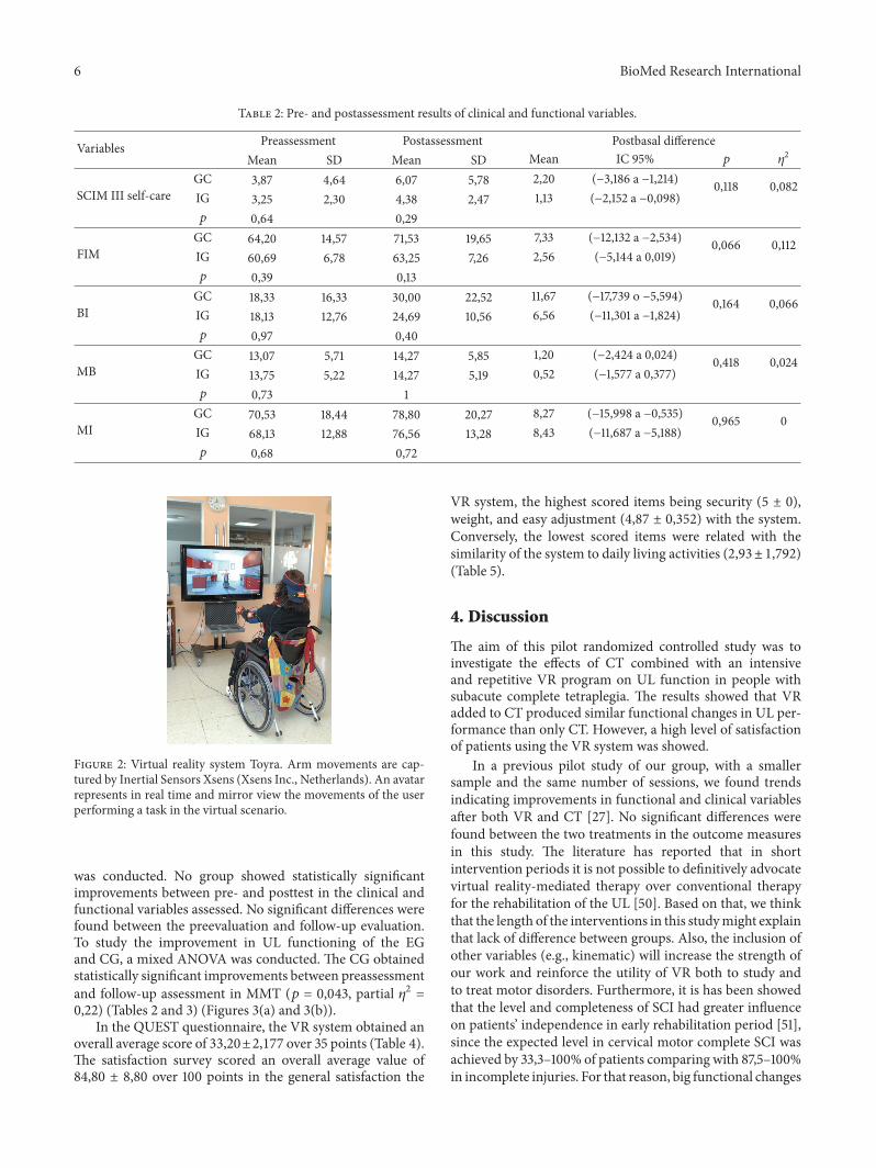
2.3.1. Virtual Reality System. During the experimental proto-col, participants were seated in their own wheelchair.The VRintervention was conducted using the VR system Toyra. ThisVR system consists of a televisionmonitor and a set of inertialsensors Xsens (Xsens Inc., Netherlands) to motion capture,and scenes displayed on an LCD screen [27]. The inertialsensors captured body movements, and the subject thenbecame immersed in the VR scene, interacting with virtualenvironments and objects. The captured inertial sensor dataand UL anthropometric data were used to develop a biome-chanical model that has been previously reported [31].This isa wireless system so subjects moved freely in the real worldwhile manipulating virtual objects in the 3D virtual world.Virtual sessions were designed along therapeutic guidelinesfor SCI interdisciplinary rehabilitation. The system offeredvisual and auditory feedback during the sessions, to increasethe engagement, facilitate the comprehension of the exercises,and deliver a clear sense of progress.
2.3.2. Training Task. In this study, one ADL-based VR gameto induceULmotor skills was used.Themain objective of thisgame was to achieve the maximum degree of autonomy that
is possible in basic ADL. The monitor displayed several dailyobjects (spoon, fork, comb, or sponge), asking the patientto reproduce the movements necessary to perform the cor-responding activities (eating, combing hair, or washing theface). The user was able to choose the preferred avatar, whichrepresented his/her movements in mirror view in real time.Mirror view has shown to add realism and sense of presenceto the practice, as well as feedback about a person’s body pos-ture and quality of movement [32]. The session offered threedifferent difficulty levels, based on changes in objects’ size andheight, and speed of appearance. Subjects performed the taskwith their dominant arm (the arm used to perform the basicdaily living tasks in the real word). If there were doubts, Edin-burgh Inventory was used to established dominance [33].
2.4. OutcomeMeasures. Neurological examinations of all thepatients were performed according to the ASIA standards[30].The right and leftmotor indexes were determined by themanual muscle test (MMT) [34] of C5 and T1 segments fromright and left extremities, respectively.
The functional examination was done by using fourscales.The Functional IndependenceMeasure (FIM) consistsof 18 items organized in six categories, four correspond-ing to motor functions (self-care items, sphincter control,mobility items, and locomotion) and two correspondingto cognitive functions (communication, psychosocial, andcognitive). The lowest and highest scores of the total rangedfrom 18 to 126 [35]. The second scale was The Spinal CordInjury Independence Measure (SCIM III) that has 16 itemsdivided into three functional areas: self-care, respiration andsphincter management, and mobility. Total score can varyfrom 0 (minimal) to 100 (maximal) [36]. Only the self-caresubscore has been considered in this study, because it hasbeen previously shown that the self-care category of the SCIMIII and several of its items correlate well with UL strength andcapacity tests in persons with tetraplegia [37]. The BarthelIndex (BI) consists of 10 items: eating, bathing, grooming,dressing, bowels, bladder, toilet use, transfers (bed to chairand back), mobility (on level surfaces), and stairs. Total scoreis from 0 to 100 [38]. The fourth assessment scale was the ULpart of Motricity Index (MI) which assesses power and rangeof active movement rated for shoulder abduction, elbowflexion, and pinch between the thumb and index finger. Eachmovement is rated on a 0–100 point scale [39].
Clinical and functional results were analyzed based onthe minimal clinically important difference (MCID) defined
4 BioMed Research International
as “the smallest difference in score in the domain of interestwhich patients perceive as beneficial and which would man-date, in the absence of trouble some side effects and excessivecost, a change in the patient’s management” [40], or “thesmallest difference in a score that is considered worthwhile orimportant” [41]. In SCIM self-care subscore, approximately1.06 to 1.22 points are necessary for MCID and 2.65 to3.05 points for substantial meaningful changes [42], whilethere are 22 points in total FIM score [43]. MCID of the BIwas estimated to be 1.85 points in stroke patients [44]. Inthe literature, no estimates were found of MCID for MMTand MI. On the basis of clinical experience and estimatesreported for similar outcome measures in different domains,the MCID was set at 10% of the total range of the scales [45].Based on that, the MCID for theMMTwas 2,5 points and forthe MI 10 points.
In addition tomake a technical assessment of the systems,it is also important to implement a functional evaluationprocedure designed by experts where the users’ opinion anddegree of satisfaction are taken into account. The usabilityconcept is closely linked to the user’s degree of satisfactionwith the product.This concept is used to measure how usefulthe product and the system settings are for the user to achievespecific goals efficiently, effectively, and satisfactorily in aspecific context [46].
To estimate the acceptation and motivation with the VRsystem as rehabilitation supplement we used the QuebecUser Evaluation of Satisfaction with Assistive Technology 2.0(QUEST) and a satisfaction survey. These tests were onlyevaluated in the IG, since CG did not use the VR system.
QUEST is an instrument specifically designed tomeasuresatisfactionwith a broad range of assistive technology devicesin a structured and standardized way [47]. This test wasdesigned to evaluate a person’s satisfaction with his or herassistive device and can be used with adolescents, adults, andelderly people who have acquired an assistive device becauseof physical or sensory impairments [48]. The test includes 12items, related to device characteristics (𝑛 = 8) and assistivetechnology services (𝑛 = 4).The scoringmethod rated from 1(not satisfied at all) to 5 (very satisfied). Only the items relatedto device characteristics were used for this study, due to thelack of external assistive services. Therefore, the maximumpossible score was 5 for each item, and 35 for the total scale.
We also used a satisfaction survey, to identify rehabilita-tion and functional aspects related to the VR system.This wasa Likert scale with 20 items rated from0 (not satisfied at all) to5 (very satisfied), including questions about systems features,VR activities, and motivation. The maximum possible scorewas 5 for each item, and 100 for the total scale.
We have access to a target population in the hospital ofapproximately 100 cervical SCI per year, of which around 30–40% have complete injuries. Based on that, we determinedthat 20% of the total sample could be representative of thepopulation. Furthermore, we have to highlight the scarcenumber of papers published with motor complete cervicalSCI in this field of work, so we considered our sample a goodrepresentation of the target population.
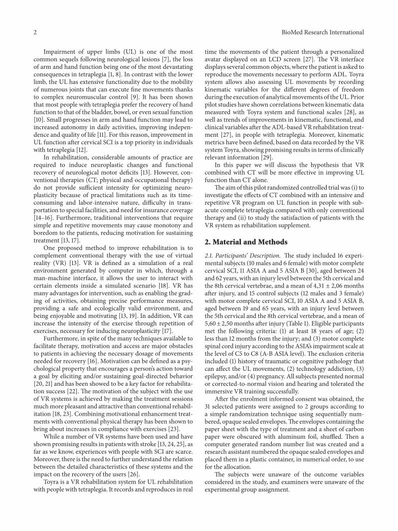
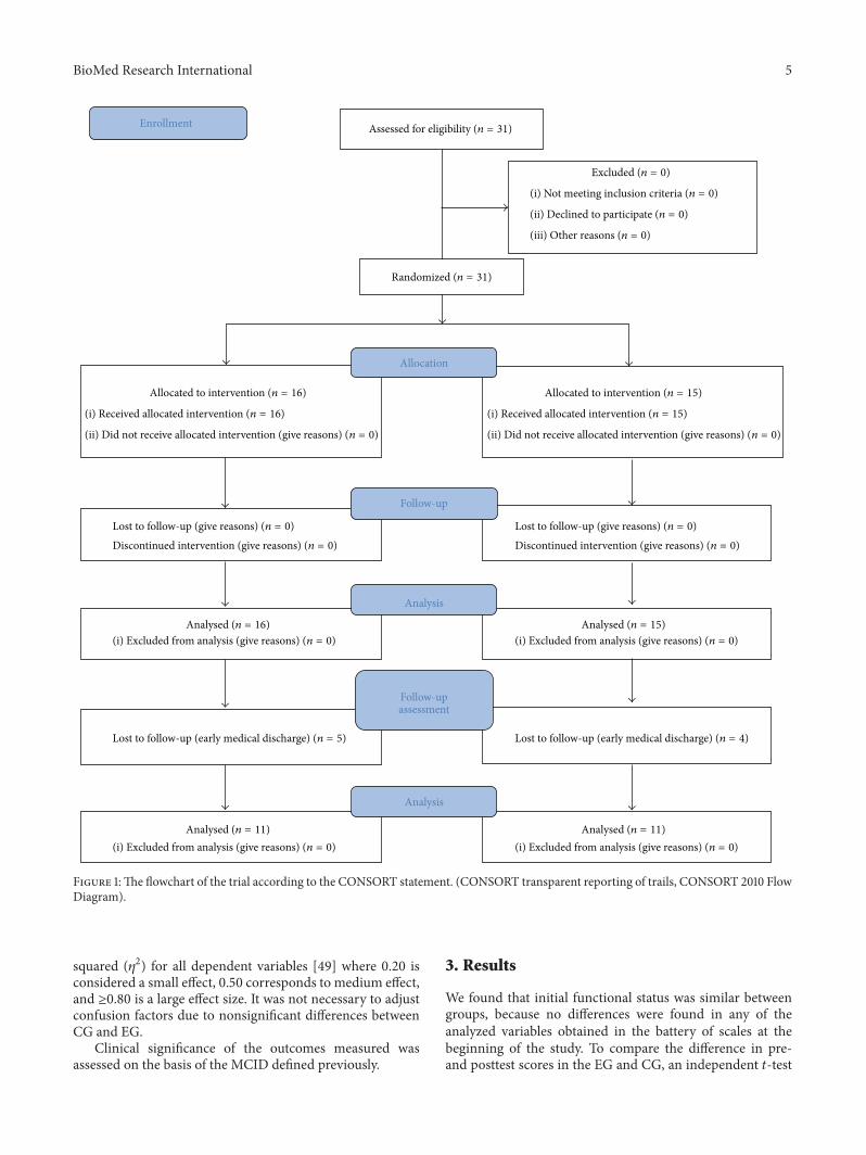
2.5. Study Protocol. The experimental (EG) and control (CG)groups underwent the same CT, which consisted of routineoccupational therapy and physiotherapy such as active andpassive mobilizations, strengthening exercise of UL, andADL training. CT was provided for 1 hour and 30min perday, 5 days per week. EG received 15 sessions with Toyrasystem for 5 weeks, 30 minutes/day, 3 days/week in additionto CT (Figure 1). VR intervention was conducted by oneoccupational therapist who was professionally familiarizedwith VR intervention. CG made only CT (Figure 2).
The date range for participants’ recruitment was fromJanuary 2011 to August 2014. Thirty-one patients (16 for EGand 15 for CG) were assessed before and after interventions.In order to know the stability of the changes, a follow-upassessment was done in 11 subjects of each group after 3months from the VR intervention. The date range for thefollow-up assessment was from February 2013 to November2014. The other patients could not participate due to earlyclinical discharge.
There were no losses or discontinuity during the 5 weeksof study, nor reports of motion sickness or vertigo inducedby the use of the VR system nor muscular pain during thesessions.
2.6. Data Collection. Every subject (CG and EG) was evalu-ated twice: at the beginning of the study and at the end, usinga set of clinical and functional scales.The evaluation was per-formed the day before starting the VR treatment (preassess-ment) and the day after finishing (postassessment). A samplefrom each group (11 subjects in each group) was followed upand assessed 3months after the study (follow-up assessment),to measure the stability of the changes. In this period bothgroups continued with the CT, but VR was not applied.The other persons had to give up the study due to earlymedical discharge, failing to complete the final assessment.The clinical and functional assessment was conducted by anoccupational therapist different to the onewho conducted theVR intervention and familiarized with the scales. The level ofsatisfaction was assessed after VR training to EG subjects.
2.7. Data Analysis and Statistics. The statistical analyses wereconducted using SPSS 17.0 for Windows (SPSS Inc., Chicago,IL).
The independent variables were forms of intervention(CT, CT + VR therapy). The dependent variables includedthe UL functioning (MMT, FIM, SCIM self-care subscore, BI,and MI) and satisfaction with VR system (QUEST and a sat-isfaction survey). Descriptive statistics were used to analyzeclinical and demographic characteristics of the subjects.
The outcome data did not deviate significantly fromnormality according to the Kolmogorov-Smirnov test. Tocompare the mean differences between groups a mixedANOVA was used. Paired 𝑡-tests were performed to comparepre- and postinterventions means changes. The mean ofthe differences between postassessment-preassessment andfollow-up-preassessments and the 95% confidence level (CI)in the dependent variables for each groupwas also calculated.The effect size of interventions was estimated with partial eta
BioMed Research International 5
Allocation
Analysis
Follow-up
Enrollment
assessment
Analysis
Assessed for eligibility (n = 31)
Excluded (n = 0)
(i) Not meeting inclusion criteria (n = 0)
(ii) Declined to participate (n = 0)
(iii) Other reasons (n = 0)
Allocated to intervention (n = 16)
(i) Received allocated intervention (n = 16)
(ii) Did not receive allocated intervention (give reasons) (n = 0)
Lost to follow-up (give reasons) (n = 0)Discontinued intervention (give reasons) (n = 0)
Randomized (n = 31)
Allocated to intervention (n = 15)
(i) Received allocated intervention (n = 15)
(ii) Did not receive allocated intervention (give reasons) (n = 0)
Lost to follow-up (give reasons) (n = 0)Discontinued intervention (give reasons) (n = 0)
Figure 1:The flowchart of the trial according to the CONSORT statement. (CONSORT transparent reporting of trails, CONSORT 2010 FlowDiagram).
squared (𝜂2) for all dependent variables [49] where 0.20 isconsidered a small effect, 0.50 corresponds to medium effect,and ≥0.80 is a large effect size. It was not necessary to adjustconfusion factors due to nonsignificant differences betweenCG and EG.
Clinical significance of the outcomes measured wasassessed on the basis of the MCID defined previously.
3. Results
We found that initial functional status was similar betweengroups, because no differences were found in any of theanalyzed variables obtained in the battery of scales at thebeginning of the study. To compare the difference in pre-and posttest scores in the EG and CG, an independent 𝑡-test
6 BioMed Research International
Table 2: Pre- and postassessment results of clinical and functional variables.
Variables Preassessment Postassessment Postbasal differenceMean SD Mean SD Mean IC 95% 𝑝 𝜂
2
SCIM III self-careGC 3,87 4,64 6,07 5,78 2,20 (−3,186 a −1,214) 0,118 0,082IG 3,25 2,30 4,38 2,47 1,13 (−2,152 a −0,098)𝑝 0,64 0,29
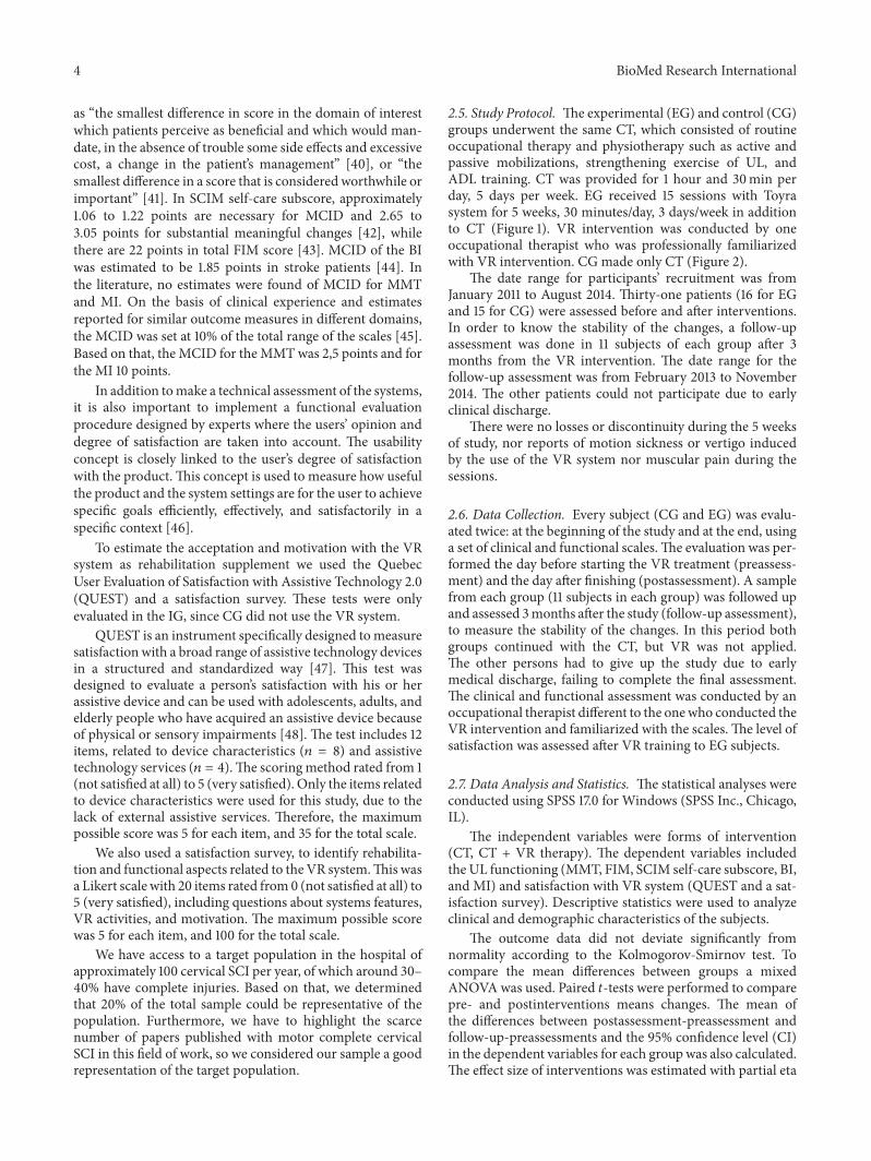
Figure 2: Virtual reality system Toyra. Arm movements are cap-tured by Inertial Sensors Xsens (Xsens Inc., Netherlands). An avatarrepresents in real time and mirror view the movements of the userperforming a task in the virtual scenario.
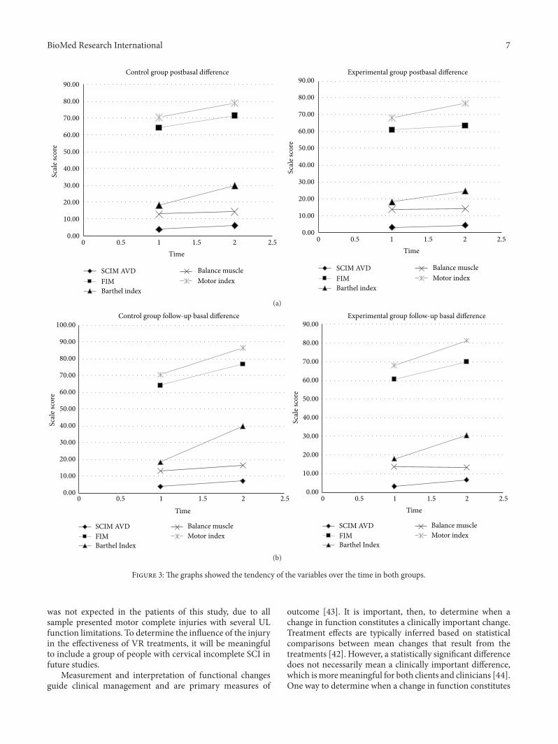
was conducted. No group showed statistically significantimprovements between pre- and posttest in the clinical andfunctional variables assessed. No significant differences werefound between the preevaluation and follow-up evaluation.To study the improvement in UL functioning of the EGand CG, a mixed ANOVA was conducted. The CG obtainedstatistically significant improvements between preassessmentand follow-up assessment in MMT (𝑝 = 0,043, partial 𝜂2 =0,22) (Tables 2 and 3) (Figures 3(a) and 3(b)).
In the QUEST questionnaire, the VR system obtained anoverall average score of 33,20±2,177 over 35 points (Table 4).The satisfaction survey scored an overall average value of84,80 ± 8,80 over 100 points in the general satisfaction the
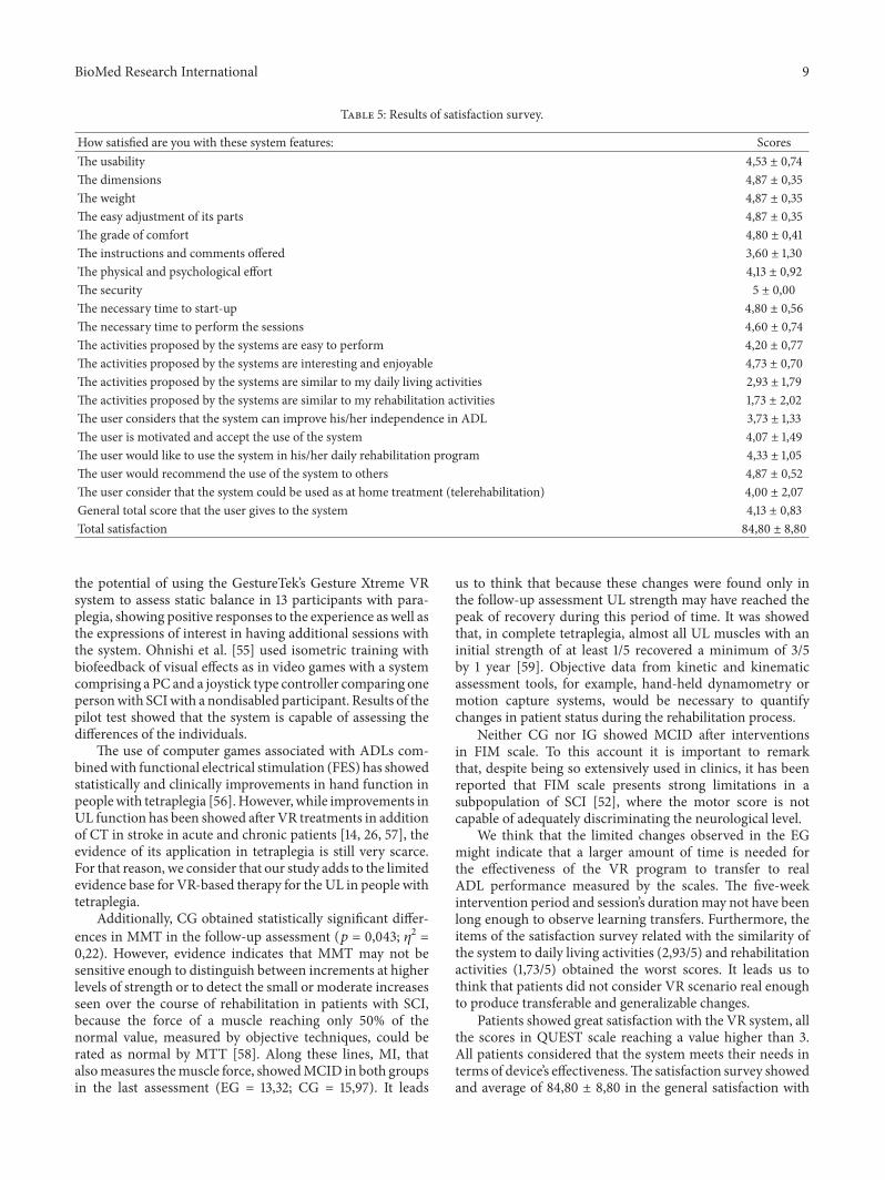
VR system, the highest scored items being security (5 ± 0),weight, and easy adjustment (4,87 ± 0,352) with the system.Conversely, the lowest scored items were related with thesimilarity of the system to daily living activities (2,93±1,792)(Table 5).
4. Discussion
The aim of this pilot randomized controlled study was toinvestigate the effects of CT combined with an intensiveand repetitive VR program on UL function in people withsubacute complete tetraplegia. The results showed that VRadded to CT produced similar functional changes in UL per-formance than only CT. However, a high level of satisfactionof patients using the VR system was showed.
In a previous pilot study of our group, with a smallersample and the same number of sessions, we found trendsindicating improvements in functional and clinical variablesafter both VR and CT [27]. No significant differences werefound between the two treatments in the outcome measuresin this study. The literature has reported that in shortintervention periods it is not possible to definitively advocatevirtual reality-mediated therapy over conventional therapyfor the rehabilitation of the UL [50]. Based on that, we thinkthat the length of the interventions in this studymight explainthat lack of difference between groups. Also, the inclusion ofother variables (e.g., kinematic) will increase the strength ofour work and reinforce the utility of VR both to study andto treat motor disorders. Furthermore, it is has been showedthat the level and completeness of SCI had greater influenceon patients’ independence in early rehabilitation period [51],since the expected level in cervical motor complete SCI wasachieved by 33,3–100% of patients comparing with 87,5–100%in incomplete injuries. For that reason, big functional changes
BioMed Research International 7
0.00
10.00
20.00
30.00
40.00
50.00
60.00
70.00
80.00
90.00
0 0.5 1 1.5 2 2.5
Scal
e sco
re
Control group postbasal difference
SCIM AVDFIMBarthel index
Balance muscleMotor index
SCIM AVDFIMBarthel index
Balance muscleMotor index
0.00
10.00
20.00
30.00
40.00
50.00
60.00
70.00
80.00
90.00
0 0.5 1 1.5 2 2.5
Scal
e sco
re
TimeTime
Experimental group postbasal difference
(a)
0.00
10.00
20.00
30.00
40.00
50.00
60.00
70.00
80.00
90.00
100.00
Scal
e sco
re
Time
Control group follow-up basal difference
0.00
10.00
20.00
30.00
40.00
50.00
60.00
70.00
80.00
90.00
0 0.5 1 1.5 2 2.5 0 0.5 1 1.5 2 2.5
Scal
e sco
re
Time
Experimental group follow-up basal difference
SCIM AVDFIMBarthel Index
Balance muscleMotor index
SCIM AVDFIMBarthel Index
Balance muscleMotor index
(b)
Figure 3: The graphs showed the tendency of the variables over the time in both groups.
was not expected in the patients of this study, due to allsample presented motor complete injuries with several ULfunction limitations. To determine the influence of the injuryin the effectiveness of VR treatments, it will be meaningfulto include a group of people with cervical incomplete SCI infuture studies.
Measurement and interpretation of functional changesguide clinical management and are primary measures of
outcome [43]. It is important, then, to determine when achange in function constitutes a clinically important change.Treatment effects are typically inferred based on statisticalcomparisons between mean changes that result from thetreatments [42]. However, a statistically significant differencedoes not necessarily mean a clinically important difference,which ismoremeaningful for both clients and clinicians [44].One way to determine when a change in function constitutes
8 BioMed Research International
Table 3: Pre- and follow-up assessment results of clinical and functional variables.
Variables Preassessment Follow-up assessment Follow-up basal differenceMean SD Mean SD Mean IC 95% 𝑝 𝜂
2
SCIM III self-careGC 3,87 4,64 7,36 7,08 3,49 (−5,525 a −0,839) 0,944 0IG 3,25 2,30 6,64 7,08 3,39 (−4,893 a −1,652)𝑝 0,64 0,76
Table 4: Results of Quebec User Evaluation of Satisfaction with Assistive Technology (QUEST).
How satisfied are you with these system features: Scores(1) The dimensions (size, height, length, width) of your assistive device? 4,80 ± 0,41(2) The weight of your assistive device? 4,87 ± 0,35(3) The easy in adjusting (fixing, fastening) the parts of your assistive device? 4,87 ± 0,35(4) How safe and secure your assistive device is? 5 ± 0,00(5) How easy it is to use your assistive device? 4,60 ± 0,91(6) How comfortable your assistive device is? 4,80 ± 0,41(7) How effective your assistive device is (the degree to which your device meets your needs)? 4,27 ± 0,88Total satisfaction 33,20 ± 2,17
a clinically important change is to calculate the minimalclinically important difference (MCID) for the measurementinstrument [43]. In interpreting the meaning measures, it isimportant to consider that although small changes may bestatistically significant, they may not be clinically important[41].
Both EG and CG presented MCID in SCIM self-caresubscore after interventions (EG = 1,13; CG = 2,20) andsubstantial meaningful changes in the followed-up monitor-ing (EG = 3,39; CG = 3,49). These results might indicateclinical positive changes after treatments and the preservationof these changes over the time. SCIM is a disability scaledeveloped specifically for SCI persons and has been showedto be the most sensitive to changes in function during therehabilitation of those participants [52]. The MCID foundafter intervention observed in both EG (12,32) and CG(21,67) in the scale BI could indicate that patients in bothgroups might have clinical improvements in independencyafter each treatment, since the MCID of the BI in strokepatients was estimated to be 1.85 points [44]. Indeed, BI level
of dependency was as follows: 0–<20 (total dependency),20–35 (serious dependency), 40–55 (moderate dependency),≥60 (minor dependency), and 100 (total independence). BIappeared to be a reliable test to measure ADL indepen-dence in SCI (Cronbach 𝛼: 0,87), existing strong correlation(Spearman-correlation: 0,69; 𝑝 < 0,0001) between levels ofinjury of people with complete SCI and BI scores [52].
This is in line with results in previous results in stroke[50] where MCID were found in functional tests after bothconventional VR-based treatments. The authors underlinethe necessity to carry out larger trials with bigger samplesto reach strongest conclusions on effectiveness of VR asrehabilitation complement.
To date, research of the applications of VR in the rehabili-tation of SCI is quite limited [53]. Szturm et al. [54] describeda SCI case report using an interactive gaming system, coupledwith the manipulation of common objects, as a form ofrepetitive, task-specific movement therapy. The subject aftertraining was able to fully extend his fingers and to grasp mostobjects. In another study, Kizony et al. [53] demonstrated
BioMed Research International 9
Table 5: Results of satisfaction survey.
How satisfied are you with these system features: ScoresThe usability 4,53 ± 0,74The dimensions 4,87 ± 0,35The weight 4,87 ± 0,35The easy adjustment of its parts 4,87 ± 0,35The grade of comfort 4,80 ± 0,41The instructions and comments offered 3,60 ± 1,30The physical and psychological effort 4,13 ± 0,92The security 5 ± 0,00The necessary time to start-up 4,80 ± 0,56The necessary time to perform the sessions 4,60 ± 0,74The activities proposed by the systems are easy to perform 4,20 ± 0,77The activities proposed by the systems are interesting and enjoyable 4,73 ± 0,70The activities proposed by the systems are similar to my daily living activities 2,93 ± 1,79The activities proposed by the systems are similar to my rehabilitation activities 1,73 ± 2,02The user considers that the system can improve his/her independence in ADL 3,73 ± 1,33The user is motivated and accept the use of the system 4,07 ± 1,49The user would like to use the system in his/her daily rehabilitation program 4,33 ± 1,05The user would recommend the use of the system to others 4,87 ± 0,52The user consider that the system could be used as at home treatment (telerehabilitation) 4,00 ± 2,07General total score that the user gives to the system 4,13 ± 0,83Total satisfaction 84,80 ± 8,80
the potential of using the GestureTek’s Gesture Xtreme VRsystem to assess static balance in 13 participants with para-plegia, showing positive responses to the experience as well asthe expressions of interest in having additional sessions withthe system. Ohnishi et al. [55] used isometric training withbiofeedback of visual effects as in video games with a systemcomprising a PC and a joystick type controller comparing onepersonwith SCIwith a nondisabled participant. Results of thepilot test showed that the system is capable of assessing thedifferences of the individuals.
The use of computer games associated with ADLs com-binedwith functional electrical stimulation (FES) has showedstatistically and clinically improvements in hand function inpeoplewith tetraplegia [56].However, while improvements inUL function has been showed after VR treatments in additionof CT in stroke in acute and chronic patients [14, 26, 57], theevidence of its application in tetraplegia is still very scarce.For that reason, we consider that our study adds to the limitedevidence base for VR-based therapy for the UL in people withtetraplegia.
Additionally, CG obtained statistically significant differ-ences in MMT in the follow-up assessment (𝑝 = 0,043; 𝜂2 =0,22). However, evidence indicates that MMT may not besensitive enough to distinguish between increments at higherlevels of strength or to detect the small or moderate increasesseen over the course of rehabilitation in patients with SCI,because the force of a muscle reaching only 50% of thenormal value, measured by objective techniques, could berated as normal by MTT [58]. Along these lines, MI, thatalsomeasures themuscle force, showedMCID in both groupsin the last assessment (EG = 13,32; CG = 15,97). It leads
us to think that because these changes were found only inthe follow-up assessment UL strength may have reached thepeak of recovery during this period of time. It was showedthat, in complete tetraplegia, almost all UL muscles with aninitial strength of at least 1/5 recovered a minimum of 3/5by 1 year [59]. Objective data from kinetic and kinematicassessment tools, for example, hand-held dynamometry ormotion capture systems, would be necessary to quantifychanges in patient status during the rehabilitation process.
Neither CG nor IG showed MCID after interventionsin FIM scale. To this account it is important to remarkthat, despite being so extensively used in clinics, it has beenreported that FIM scale presents strong limitations in asubpopulation of SCI [52], where the motor score is notcapable of adequately discriminating the neurological level.
We think that the limited changes observed in the EGmight indicate that a larger amount of time is needed forthe effectiveness of the VR program to transfer to realADL performance measured by the scales. The five-weekintervention period and session’s durationmay not have beenlong enough to observe learning transfers. Furthermore, theitems of the satisfaction survey related with the similarity ofthe system to daily living activities (2,93/5) and rehabilitationactivities (1,73/5) obtained the worst scores. It leads us tothink that patients did not consider VR scenario real enoughto produce transferable and generalizable changes.
Patients showed great satisfaction with the VR system, allthe scores in QUEST scale reaching a value higher than 3.All patients considered that the system meets their needs interms of device’s effectiveness.The satisfaction survey showedand average of 84,80 ± 8,80 in the general satisfaction with
10 BioMed Research International
the VR system. In particular, best scored items were security(5 ± 0), weight, and ease of adjustment (4,87 ± 0,352) of thesystem. These results are coherent with the fact that Toyrasystem is based on light inertial sensors that are locatedwith comfortable and ventilated neoprene straps, with theobjective that users feel comfortable during the sessions.Furthermore, patients considered the activities proposed bythe system interesting and enjoyable (4,73 ± 0,70) and feltmotivated with its use (4,07 ± 1,49). Participants expressedthe wish to use Toyra in their daily rehabilitation (4,33±1,05)and at home (4,00 ± 2,07) and would recommend the use ofthe system to others (4,87 ± 0,52). Patients’ motivation wasshown to be an important predictor of long-term changes inquality of life and rehabilitation outcomes [60], as well as toincrease the amount of time that patients are willing to spendin therapy [16].
The treatment and rehabilitation period in completetetraplegia are long, expensive, and exhausting, because of theloss of motor, sensory, and autonomic function. Depressivedisorders, psychosocial problems, and stress are frequentcomplications after this lesion [61]. The incorporation of VRas rehabilitation supplement has showed several personallymotivating factors such as perceived control, curiosity andexploration, and imagination and socially motivating factorsas cooperation, competition, and social interaction [16]. VRsystems may provide more attractive and engagement treat-ments to people with tetraplegia, allowing users to interactwith virtual objects in stimulated and variable environ-ment, providing the user with natural control of movementsusing as many parts of the body as are deemed suitablewithin the context of therapeutic goals, and decreasing thetherapist’s support allowing patients to choose during theirrehabilitation process (e.g., games, characters, and levels).Furthermore, the possibility to play with others can helpmaintain self-esteem and create positive experiences, offeringopportunities for remote and proximal socialization.
For that reason, we deduce that complementing reha-bilitation with a VR system might be useful to increase thedosage of therapy and to augment patient’s engagement andmotivation during the process.
5. Conclusions
This randomized controlled study showed the effects of CTcombined with VR program on UL function in people withcomplete tetraplegia. The results showed that VR added toCT, in comparison with the only application of CT, producesimilar results in UL function. However, both treatmentsseem to produce the minimal clinical changes necessary toconsider clinically important. Moreover, the gaming aspectsincorporated by VR in conventional rehabilitation appearto promote patient motivation and hence adherence to thetreatment. Future research should be implemented with alarger sample size and should increase the dosage of VRtherapy in terms of number of sessions.
Conflict of Interests
The authors declare that there is no conflict of interestsregarding the publication of this paper.
Authors’ Contribution
I. Dimbwadyo-Terrer and A. Segura Fragoso conceivedand designed the experiments. I. Dimbwadyo-Terrer andB. Polonio-Lopez performed the experiments. A. SeguraFragoso, A. de los Reyes-Guzman, and F. Trincado-Alonsoanalyzed the data. I. Dimbwadyo-Terrer and A. Gil-Agudocontributed reagents/materials/analysis tools. I. Dimbwadyo-Terrer, B. Polonio-Lopez, A. Segura Fragoso, A. de los Reyes-Guzman, F. Trincado-Alonso, A. Gil-Agudo, and S. Piazzawrote the paper. I. Dimbwadyo-Terrer, A. Gil-Agudo, B.Polonio-Lopez, and S. Piazza revised the paper criticallyfor important intellectual content. I. Dimbwadyo-Terrer, B.Polonio-Lopez, A. Segura Fragoso, A. de los Reyes-Guzman,F. Trincado-Alonso, A. Gil-Agudo, and S. Piazza providedfinal approval of the version to be published.
Acknowledgments
The authors thank the consortium including FoundationRafael del Pino, Foundation of the Spanish National Hospitalfor Paraplegic Research and Integration (FUHNPAIIN), andINDRA Systems for funding this research.
References
[1] M. Wyndaele and J.-J. Wyndaele, “Incidence, prevalence andepidemiology of spinal cord injury: what learns a worldwideliterature survey?” Spinal Cord, vol. 44, no. 9, pp. 523–529, 2006.
[2] J. Mazaira, F. Labanda, J. Romero et al., “Epidemiologıa de lalesion medular y otros aspectos,” Rehabilitacion, vol. 32, no. 6,pp. 365–372, 1998.
[3] J. Garcıa-Reneses, R. Herruzo-Cabrera, and M. Martinez-Moreno, “Epidemiological study of spinal cord injury in Spain1984-1985,” Paraplegia, vol. 29, no. 3, pp. 180–190, 1991.
[4] J. R. Silver, “A systematic review of the therapeutic interventionsfor heterotopic ossification after spinal cord injury,” Spinal Cord,vol. 49, no. 3, article 482, 2011.
[5] A. Garcıa-Altes, K. Perez, A. Novoa et al., “Spinal cord injuryand traumatic brain injury: a cost-of-illness study,” Neuroepi-demiology, vol. 39, no. 2, pp. 103–108, 2012.
[6] C. T. Liverman, B. M. Altevogt, J. E. Joy, and R. T. John-son, Spinal Cord Injury: Progress, Promise, and Priorities, TheNational Academies Press, Washington, DC, USA, 2005.
[7] M. A. Murphy, K. S. Sunnerhagen, B. Johnels, and C. Willen,“Three-dimensional kinematic motion analysis of a daily activ-ity drinking from a glass: a pilot study,” Journal of NeuroEngi-neering and Rehabilitation, vol. 3, article 18, 2006.
[8] X. Lu, C. R. Battistuzzo, M. Zoghi, and M. P. Galea, “Effectsof training on upper limb function after cervical spinal cordinjury: a systematic review,” Clinical Rehabilitation, vol. 29, no.1, pp. 3–13, 2015.
[9] A. De los Reyes-Guzman, A. Gil-Agudo, B. Penasco-Martın,M. Solıs-Mozos, A. Del Ama-Espinosa, and E. Perez-Rizo,“Kinematic analysis of the daily activity of drinking from a glass
BioMed Research International 11
in a population with cervical spinal cord injury,” Journal ofNeuroEngineering and Rehabilitation, vol. 7, article 41, 2010.
[10] R. W. Hanson and M. R. Franklin, “Sexual loss in relation toother functional losses for spinal cord injured males,” Archivesof Physical Medicine and Rehabilitation, vol. 57, no. 6, pp. 291–293, 1976.
[11] L. R. Hoffman and E. C. Field-Fote, “Functional and corticomo-tor changes in individuals with tetraplegia following unimanualor bimanualmassed practice trainingwith somatosensory stim-ulation: a pilot study,” Journal of Neurologic Physical Therapy,vol. 34, no. 4, pp. 193–201, 2010.
[12] S. Kalsi-Ryan, A. Curt, M. C. Verrier, and M. G. Fehlings,“Development of the Graded Redefined Assessment ofStrength, Sensibility and Prehension (GRASSP): reviewingmeasurement specific to the upper limb in tetraplegia,” Journalof Neurosurgery: Spine, vol. 17, no. 1, supplement, pp. 65–76,2012.
[13] K. R. Lohse, C. G. E. Hilderman, K. L. Cheung, S. Tatla, andH. F. M. Van Der Loos, “Virtual reality therapy for adults post-stroke: a systematic review and meta-analysis exploring virtualenvironments and commercial games in therapy,” PLoS ONE,vol. 9, no. 3, Article ID e93318, 2014.
[14] J.-S. Kwon, M.-J. Park, I.-J. Yoon, and S.-H. Park, “Effects ofvirtual reality on upper extremity function and activities of dailyliving performance in acute stroke: a double-blind randomizedclinical trial,” NeuroRehabilitation, vol. 31, no. 4, pp. 379–385,2012.
[15] C. E. Lang, J. R. MacDonald, D. S. Reisman et al., “Observationof amounts of movement practice provided during strokerehabilitation,”Archives of Physical Medicine and Rehabilitation,vol. 90, no. 10, pp. 1692–1698, 2009.
[16] K. R. Lohse, N. Shirzad, A. Verster, N. Hodges, and H. F. M.Van der Loos, “Video games and rehabilitation: using designprinciples to enhance engagement in physical therapy,” Journalof Neurologic Physical Therapy, vol. 37, no. 4, pp. 166–175, 2013.
[17] H. Sin and G. Lee, “Additional virtual reality training usingXbox kinect in stroke survivors with hemiplegia,” AmericanJournal of Physical Medicine and Rehabilitation, vol. 92, no. 10,pp. 871–880, 2013.
[18] P. L. T. Weiss, R. Kizony, U. Feintuch, and N. Katz, “Virtualreality in neurorehabilitation,” in Textbook of Neural Repair andRehabilitation, M. Selzer, S. Clarke, L. Cohen, P. Duncan, andF. Gage, Eds., vol. 51, no. 8, pp. 182–97, Cambridge UniversityPress, Cambridge, UK, 2006.
[19] O. Bart, T. Agam, P. L. Weiss, and R. Kizony, “Using video-capture virtual reality for children with acquired brain injury,”Disability and Rehabilitation, vol. 33, no. 17-18, pp. 1579–1586,2011.
[20] G. J. Mogenson, D. L. Jones, and C. Y. Yim, “From motivationto action: functional interface between the limbic system andthe motor system,” Progress in Neurobiology, vol. 14, no. 2-3, pp.69–97, 1980.
[21] R. A. Wise, “Dopamine, learning and motivation,” NatureReviews Neuroscience, vol. 5, no. 6, pp. 483–494, 2004.
[22] K. Hafen, J. Bengel, J. Jastrebow, and R. Nubling, “Konzept unddimensionen der reha-motivation,” Pravention und Rehabilita-tion, vol. 12, no. 1, pp. 1–10, 2000.
[23] J. Laut, F. Cappa, O. Nov, M. Porfiri, and D. N. Bonter,“Increasing patient engagement in rehabilitation exercises usingcomputer-based citizen science,” PLoS ONE, vol. 10, no. 3,Article ID e0117013, 2015.
[24] G. Saposnik and M. Levin, “Virtual reality in stroke rehabilita-tion: ameta-analysis and implications for clinicians,” Stroke, vol.42, no. 5, pp. 1380–1386, 2011.
[25] K. E. Laver, S. George, S. Thomas, J. E. Deutsch, and M. Crotty,“Virtual reality for stroke rehabilitation,” Stroke, vol. 43, no. 2,pp. e20–e21, 2012.
[26] M. S. Cameirao, S. B. I. Badia, E. Duarte, A. Frisoli, and P. F.M. J. Verschure, “The combined impact of virtual reality neu-rorehabilitation and its interfaces on upper extremity functionalrecovery in patients with chronic stroke,” Stroke, vol. 43, no. 10,pp. 2720–2728, 2012.
[27] A. Gil-Agudo, I. Dimbwadyo-Terrer, B. Penasco-Martın, A. delos Reyes-Guzman, A. Bernal-Sahun, and A. Berbel-Garcıa,“Experiencia clınica de la aplicacion del sistema de realidadTOyRA en la neuro-rehabilitacion de pacientes con lesionmedular,” Rehabilitacion, vol. 46, no. 1, pp. 41–48, 2012.
[28] I. Dimbwadyo-Terrer, F. Trincado-Alonso, A. I. De la Pena-Gonzalez et al., “Clinical, functional and kinematic correlationsusing the virtual reality system Toyra as upper limb rehabil-itation tool in people with spinal cord injury,” in Proceedingsof the International Congress on Neurotechnology, Electronicsand Informatics (NEUROTECHNIX ’13), A. R. Londral, P.Encarnacao, and J. L. Pons, Eds., pp. 81–88, Vilamoura, Portugal,July 2013.
[29] F. Trincado-Alonso, I. Dimbwadyo-Terrer, A. De Los Reyes-Guzman, P. Lopez-Monteagudo, A. Bernal-Sahun, and A. Gil-Agudo, “Kinematic metrics based on the virtual reality systemtoyra as an assessment of the upper limb rehabilitation in peoplewith spinal cord injury,” BioMed Research International, vol.2014, Article ID 904985, 11 pages, 2014.
[30] R. J. Marino, T. Barros, F. Biering-Sorensen et al., “Internationalstandards for neurological classification of spinal cord injury,”The Journal of Spinal Cord Medicine, vol. 26, supplement 1, pp.S50–S56, 2003.
[31] A. Gil-Agudo, A. Del Ama-Espinosa, A. I. De la Pena-Gonzalez,A. Bernal-Sahun, and E. Rocon, “Applications of upper limbbiomechanical models in spinal cord injury patients,” in Biome-chanics in Applications, V. Klika, Ed., pp. 127–164, InTech, 2011.
[32] P. L. T. Weiss, D. Rand, N. Katz, and R. Kizony, “Video capturevirtual reality as a flexible and effective rehabilitation tool,”Journal of NeuroEngineering and Rehabilitation, vol. 1, no. 1,article 12, 2004.
[33] R. C. Oldfield, “The assessment and analysis of handedness: theEdinburgh inventory,”Neuropsychologia, vol. 9, no. 1, pp. 97–113,1971.
[34] R. L. Lamb, “Manual muscle testing,” in Measurement inPhysical Therapy, J. M. Rothstein, Ed., pp. 47–56, ChurchillLivingstone, New York, NY, USA, 1985.
[35] B. B. Hamilton, J. A. Laughlin, R. C. Fiedler, and C. V.Granger, “Interrater reliability of the 7-level functional indepen-dence measure (FIM),” Scandinavian Journal of RehabilitationMedicine, vol. 26, no. 3, pp. 115–119, 1994.
[36] A. Catz, M. Itzkovich, E. Agranov, H. Ring, and A. Tamir,“SCIM—spinal cord independence measure: a new disabilityscale for patients with spinal cord lesions,” Spinal Cord, vol. 35,no. 12, pp. 850–856, 1997.
[37] C. Rudhe and H. J. A. van Hedel, “Upper extremity functionin persons with tetraplegia: Relationships between strength,capacity, and the spinal cord independence measure,” Neurore-habilitation and Neural Repair, vol. 23, no. 5, pp. 413–421, 2009.
12 BioMed Research International
[38] F. I. Mahoney and D. W. Barthel, “Functional evaluation: theBarthel index,” Maryland State Medical Journal, vol. 14, pp. 61–65, 1965.
[39] G. Demeurisse, O. Demol, and E. Robaye, “Motor evaluationin vascular hemiplegia,” European Neurology, vol. 19, no. 6, pp.382–389, 1980.
[40] R. Jaeschke, J. Singer, and G. H. Guyatt, “Measurement ofhealth status. Ascertaining the minimal clinically importantdifference,” Controlled Clinical Trials, vol. 10, no. 4, pp. 407–415,1989.
[41] R. D. Hays and J. M. Woolley, “The concept of clinicallymeaningful difference in health-related quality-of-life research.How meaningful is it?” PharmacoEconomics, vol. 18, no. 5, pp.419–423, 2000.
[42] G. Scivoletto, F. Tamburella, L. Laurenza, and M. Molinari,“The spinal cord independence measure: how much change isclinically significant for spinal cord injury subjects,” Disabilityand Rehabilitation, vol. 35, no. 21, pp. 1808–1813, 2013.
[43] M. Beninato, K. M. Gill-Body, S. Salles, P. C. Stark, R. M. Black-Schaffer, and J. Stein, “Determination of the minimal clinicallyimportant difference in the FIM instrument in patients withstroke,”Archives of Physical Medicine and Rehabilitation, vol. 87,no. 1, pp. 32–39, 2006.
[44] Y.-W. Hsieh, C.-H. Wang, S.-C. Wu, P.-C. Chen, C.-F. Sheu,and C.-L. Hsieh, “Establishing the minimal clinically importantdifference of the Barthel index in stroke patients,”Neurorehabil-itation and Neural Repair, vol. 21, no. 3, pp. 233–238, 2007.
[45] J. H. Van Der Lee, R. C. Wagenaar, G. J. Lankhorst, T. W.Vogelaar, W. L. Deville, and L. M. Bouter, “Forced use of theupper extremity in chronic stroke patients: results froma single-blind randomized clinical trial,” Stroke, vol. 30, no. 11, pp. 2369–2375, 1999.
[46] A. Jardon, A. M. Gil, A. I. De la Pena, C. A. Monje, and C.Balaguer, “Usability assessment of ASIBOT: a portable robot toaid patients with spinal cord injury,” Disability and Rehabilita-tion: Assistive Technology, vol. 6, no. 4, pp. 320–330, 2011.
[47] L. Demers, M. Monette, Y. Lapierre, D. L. Arnold, and C.Wolfson, “Reliability, validity, and applicability of the Que-bec User Evaluation of Satisfaction with assistive Technology(QUEST 2.0) for adults with multiple sclerosis,” Disability andRehabilitation, vol. 24, no. 1–3, pp. 21–30, 2002.
[48] S.-H. Lee, “Users’ satisfaction with assistive devices in SouthKorea,” Journal of Physical Therapy Science, vol. 26, no. 4, pp.509–512, 2014.
[49] J. Cohen, “Statistical power analysis,” Current Directions inPsychological Science, vol. 1, no. 3, pp. 98–101, 1992.
[50] J. H. Crosbie, S. Lennon, M. C. McGoldrick, M. McNeill, and S.M.McDonough, “Virtual reality in the rehabilitation of the armafter hemiplegic stroke: a randomized controlled pilot study,”Clinical Rehabilitation, vol. 26, no. 9, pp. 798–806, 2012.
[51] S. Mingaila and A. Krisciunas, “Occupational therapy forpatients with spinal cord injury in early rehabilitation,” Medic-ina, vol. 41, no. 10, pp. 852–856, 2005.
[52] J. H. van Tuijl, Y. J. M. Janssen-Potten, and H. A. M. Seelen,“Evaluation of upper extremity motor function tests in tetraple-gics,” Spinal Cord, vol. 40, no. 2, pp. 51–64, 2002.
[53] R. Kizony, L. Raz, N. Katz, H. Weingarden, and P. L. T. Weiss,“Video-capture virtual reality system for patients with paraple-gic spinal cord injury,” Journal of Rehabilitation Research andDevelopment, vol. 42, no. 5, pp. 595–607, 2005.
[54] T. Szturm, J. F. Peters, C. Otto, N. Kapadia, and A. Desai, “Task-specific rehabilitation of finger-hand function using interactivecomputer gaming,” Archives of Physical Medicine and Rehabili-tation, vol. 89, no. 11, pp. 2213–2217, 2008.
[55] K. Ohnishi, E. Goto, F. Sugiki et al., “Home-use upper limbrehabilitation device for cervical spinal cord injured patients,” inComputers Helping People with Special Needs, K. Miesenberger,J. Klaus, W. L. Zagler, and D. Burger, Eds., vol. 3118 of LectureNotes in Computer Science, pp. 880–888, Springer, Berlin,Germany, 2004.
[56] J. Kowalczewski, S. L. Chong, M. Galea, and A. Prochazka, “In-home tele-rehabilitation improves tetraplegic hand function,”Neurorehabilitation and Neural Repair, vol. 25, no. 5, pp. 412–422, 2011.
[57] M. S. Cameirao, S. Bermudez-I Badia, E. Duarte-Oller, and P.F. M. J. Verschure, “Virtual reality based rehabilitation speedsup functional recovery of the upper extremities after stroke: arandomized controlled pilot study in the acute phase of strokeusing the rehabilitation gaming system,” Restorative Neurologyand Neuroscience, vol. 29, no. 5, pp. 287–298, 2011.
[58] L. Noreau and J. Vachon, “Comparison of three methods toassess muscular strength in individuals with spinal cord injury,”Spinal Cord, vol. 36, no. 10, pp. 716–723, 1998.
[59] V. W. Lin and C. M. Bono, Spinal Cord Medicine: Principles andPractice, Demos Medical, New York, NY, USA, 2014.
[60] B. Grahn, C. Ekdahl, and L. Borgquist, “Motivation as apredictor of changes in quality of life and working ability inmultidisciplinary rehabilitation,” Disability and Rehabilitation,vol. 22, no. 15, pp. 639–654, 2000.
[61] K. Nas, L. Yazmalar, V. Sah, A. Aydin, and K. Ones, “Rehabilita-tion of spinal cord injuries,” World Journal of Orthopedics, vol.6, no. 1, pp. 8–16, 2015.


![Review Article Respiratory Management in the Patient with Spinal …downloads.hindawi.com/journals/bmri/2013/168757.pdf · 2019-07-31 · sympathetic nervous system tone [ ].Asaresultofthe](https://static.documents.pub/doc/80x56/5f40527c40d9627dc93c250c/review-article-respiratory-management-in-the-patient-with-spinal-2019-07-31-sympathetic.jpg)


