In our retrospective study we evaluated the efficacy of an improved amniotic membrane (AM) roll-in filling technique (AMR)combined with multilayer amniotic membrane cover to treat corneal perforation and included 46 cornea perforations ≤ 3mm indiameter treated with AMR and 20% C
3F8mixed gas filling of the anterior chamber. Anterior chamber depth, aqueous leakage,
bubble maintenance time, and cornea morphology were monitored after each operation.Themean diameter of corneal perforationwas 1.60 ± 0.55mm (range 0.5–3) and the success rate of the AMR method for corneal perforation reconstruction was 100%after a single operation. Anterior chamber depth was normally reconstructed without AMR break-off, aqueous leak, or othercomplications.Themean time of the C
3F8gas bubble in the anterior chamber was 8.6 ± 2.0 days (range 4–12). At the last follow-up,
all patients’ visual acuity was improved to varying degrees.Themean follow-up time was 11.0±5.6months (range 3–36).The AMRplugging combined with multilayer AM cover is a secure and easy intervention, which led to 100% success in our study. Variousperforations ranging from trauma to infection can be treated with AMR, which is especially practical in those countries wheredonor cornea availability is limited.
1. Introduction
Corneal perforation is one of the most serious complicationsof infections and autoimmune diseases as well as traumata.Penetrating keratoplasty (PK) is an effective treatment forcorneal perforation [1]. However, in clinical practice, thecornea demand is far greater than the cornea supply. More-over, although the success rate of corneal allograft transplan-tation is in general satisfying, infective corneal perforationsare still a high-risk during corneal transplantations, becausethe infective diseases can easily cause corneal graft rejectionand infection, eventually leading to graft failure [2].
Amniotic membrane transplantation (AMT) for thetreatment of corneal ulcers was first introduced by Lee andTseng [3] in 1997. Thereafter, it has been widely used forvarious ocular surface reconstructions. The unique prop-erty of amniotic membranes (AMs) in the reconstructionof ocular surfaces is that the AM contains a remarkablemixture of growth factors and cytokines facilitating prolif-eration and differentiation of epithelial cells, reducing theinflammatory response by inhibiting protease activity, andreducing inflammatory cell activity [4–6]. Therefore, AMT
could promote ocular surface tissue healing of persistentepithelial defects (PEDs), corneal ulcers, and eye burns [7–10]. Clinical results showed that the success rate of treatmentfor corneal ulcers with single ormultilayer AMswas over 80%[10, 11], while for cornea perforation with multilayer AMT itwas about 73% [12]. Furthermore, the treatment for cornealperforations with fibrin glue-assisted augmented AMT wasabout 90% [13, 14]. A newly developed AM roll techniquehas been introduced with a higher success rate for corneaperforation treatments; however, there are few reportedtreatments in the literature [15, 16]. Based on the usageof AM for healing of cornea tissues and the few reportedcases of AMR interventions for cornea perforation treatment,we performed modified AMR interventions combined withmultilayer amniotic membrane cover for a variety of cornealperforations up to 3mm in diameter.
2. Patients and Methods
2.1. Patients. Between July 2007 and April 2011, 46 eyes of46 patients (24 men and 22 women with a mean age of
Hindawi Publishing CorporationBioMed Research InternationalVolume 2016, Article ID 1693815, 8 pageshttp://dx.doi.org/10.1155/2016/1693815
2 BioMed Research International
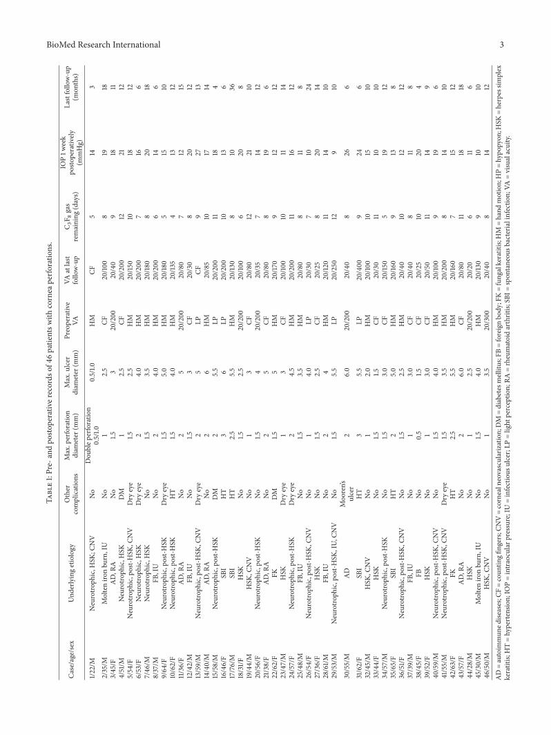
49.4 ± 12.1 years) who suffered from corneal perforationswere consecutively admitted to our hospital after failureof conventional ulcer treatment for 5∼25 days by differentophthalmologists and treated with AMR filling-in combinedwith multilayer AM cover. Five cases of corneal perforation(5 eyes) resulted from corneal bacterial infection causedby foreign bodies and 3 cases from spontaneous bacterialinfections; in 28 eyes corneal perforations were due toherpes simplex keratitis (HSK), in 2 eyes due to molten ironburns, in 6 eyes due to autoimmune diseases, and in 2 eyesdue to fungal keratitis. One of 28 HSK perforations was abiperforation.The time from corneal perforation to receivingsurgery was 2–14 days, and the average was 6.2 ± 2.3 days.All patients were treated with ocular antibiotic drops beforehospitalization and their visual acuities were all found to bebelow 20/200. Clinical data, including patient demography,etiology, surgical procedure, associated therapies, visual acu-ity, and the final outcomes and complications were collectedin a retrospectivemanner (Table 1). Inclusion criterion for ourmodified treatment was size of perforation ≤3mm in diam-eter and exclusion criteria were serious noncorneal diseasessuch as inflammation of the inner structures of the eyeball(entophthalmia) and severe eyelid defect, which would likelyinfluence corneal recovery. The Ethics Committee of the180th Hospital of PLA in Quanzhou, China, approved thisstudy and all patients provided written informed consent.
2.2. Preparation of Preserved Human AM. Preserved humanAMs were obtained from the eye bank of the 180th Hospitalof PLA.The preservationmethodwas as previously described[17]. The AMs derived from patients without infectious dis-eases were peeled from the puerperal placentas and glued toaseptic filter papers, whichwere cut into 3× 3 cmpieces. Afterthe epithelial sides of the membranes were treated with 0.5%sodium hyaluronate to protect them from freezing harm, themembrane sheets were stored in anhydrous glycerol at−45∘C.
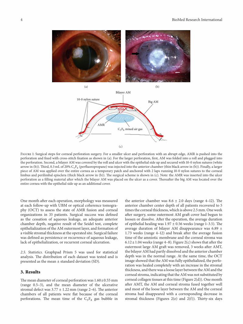
2.3. Surgical Procedure. All operationswere performed by thesame surgeon (Junhua Fan) after patients were anesthetizedwith 2% lidocaine containing 1 : 8 × 10⁴ epinephrine in thesubconjunctival or sub-Tenon’s capsule. After local anesthe-sia, debridement of the ulcer base and necrotic tissue wasperformed with fine forceps and a microsponge, and thepoorly adherent epithelium adjacent to the edge of the ulcerwas removed. Afterwards, a little sodium hyaluronate wasinjected through the perforation into the anterior chamber toseparate the synechia between the iris and the cornea. Afterthe frozen AMs were placed in saline for rehydration andthawing for 10 minutes, they were cut into rectangles withdimensions of about 2 × 4mm and rolled to a length of 2mm.One end of the roll was plugged into the corneal perforationand the other end was spread over the defective stroma zone(Figure 1(a)).
Based on the size of the perforation, two different waysof fixing the AM rolls were used. For the smaller sizedperforation with abrupt edges, we used the suture fixationmethod, in which the suture needle was inserted into theedge from one side of the cornea hole, penetrated throughout
the AM, passed out the edge from the other side of thehole, and then knotted. The depth of the needle insertionwas 2/3 of the cornea thickness. If necessary, crisscrossingsewing was used to anchor the AMR to the inside of the hole(Figure 1(b)). For a larger size of perforation, after the AM rollwas inserted, a single amniotic membrane was folded in halfto become a bilayer amniotic membrane cover. The bilayeramniotic membrane was then trimmed to fit the shape andsize of the corneal ulcer. However, caution must be takento ensure that the outer AM layer was positioned with theepithelial side up because the top of the bilayer AM shouldserve as a basement membrane for reepithelialization. Then,the edge of the bilayer amniotic membrane was sutured tothe healthy corneal tissue around the perforation with a fewinterrupted stitches using 10-0 nylon suture. Afterwards, alarger piece of AM was applied over the entire cornea as atemporary patch and anchored with 2 flaps using running 10-0 nylon sutures to the corneal limbus and perilimbal episclera(Figure 1(b)). After completion of all sewing, 0.3mL of 20%C3F8(perfluoropropane) mixed with air was slowly injected
to separate the iris and the cornea, forming the anteriorchamber. It was appropriate to produce a bubble with adiameter of 5-6mm. After surgery, the conjunctival sac wascleanedwith 1/1000 gentamicin solution and then coatedwithofloxacin eye ointment; both eyes were bandaged for 2 days;the patients were required to reduce eye movements as muchas possible and lie supine on the bed as far as possible tomaintain the C
3F8bubble between the iris and cornea before
the bubble was absorbed. Figure 1(c) shows a scheme of theintervention technique.
2.4. Medical Care after Surgery. Postoperatively, antibiotic,sodium hyaluronate, and mydriatic eye drops togetherwith antibiotic eye ointment were instilled. After the ban-dage was removed, for patients with nonfungal cornealulcer, 0.5% levofloxacin eye drops (Cravit, Santen Phar-maceutical Company, Osaka, Japan), 0.1% fluorometholoneeye drops (Flumetholon, Santen Pharmaceutical Company,Osaka, Japan), 0.1% sodium hyaluronate eye drops (Hyalein0.1, Santen Pharmaceutical Company, Osaka, Japan), and0.5% tropicamide eye drops (Shuangxingming, ZhenshimingPharmaceutical Company, Fuzhou, China) were instilled 4times a day; 0.3% ofloxacin eye ointment (Dikeluo, XinqiPharmaceutical Company, Shenyang, China) was used beforesleeping. For patients with fungal corneal ulcer, 0.1% flu-orometholone eye drops (Flumetholon, Santen Pharma-ceutical Company, Osaka, Japan) were replaced with 5%natamycin eye drops (Natacyn, Alcon Laboratories, Inc.) 6times a day. We followed up each patient daily in the firstweek after surgery, weekly for 3 months postoperatively, andrandomly at appropriate times. During the follow-ups, visualacuity and intraocular pressure (IOP)weremeasured and slit-lamp examinations were performed to assess the degree ofcorneal ulcer healing, graft integrity, signs of inflammation,infection, and changes in the ocular surface and anteriorchamber, as well as for monitoring AMR changes. If thetemporary big AM graft overlay was found to be loosenedor dissolved about 1 week after the operation, we removed it.
BioMed Research International 3Ta
ble1:Pre-
andpo
stoperativ
erecords
of46
patie
ntsw
ithcornea
perfo
ratio
ns.
Case/age/sex
Und
erlyingetiology
Other
complications
Max.perforatio
ndiam
eter
(mm)
Max.ulcer
diam
eter
(mm)
Preoperativ
eVA
VAatlast
follo
w-up
C 3F 8
gas
remaining
(days)
IOP1w
eek
posto
perativ
ely(m
mHg)
Lastfollo
w-up
(mon
ths)
1/22/M
Neurotro
phic,H
SK;C
NV
No
Dou
blep
erforatio
n0.5/1.0
0.5/1.0
HM
CF5
143
2/35/M
Molteniro
nbu
rn,IU
No
12.5
CF20/10
08
1918
3/45/F
AD,R
ANo
1.53
20/200
20/40
918
114/51/M
Neurotro
phic,H
SKDM
12.5
CF20/200
1221
125/54/F
Neurotro
phic,post-H
SK,C
NV
Dry
eye
1.52.5
HM
20/15
010
1812
6/53/F
Neurotro
phic,H
SKDry
eye
24.0
HM
20/200
716
67/46
/MNeurotro
phic,H
SKNo
1.53.5
HM
20/18
08
2018
8/37/M
FB,IU
No
24.0
HM
20/200
614
69/64
/FNeurotro
phic,post-H
SKDry
eye
1.55.0
HM
20/18
05
1510
10/62/F
Neurotro
phic,post-H
SKHT
1.54.0
HM
20/13
54
1312
11/36/F
AD,R
ANo
25
20/200
20/80
712
1512/42/M
FB,IU
No
1.53
CF20/30
820
1213/59/M
Neurotro
phic,post-H
SK,C
NV
Dry
eye
25
LPCF
927
1314/40/M
AD,R
ANo
26
HM
20/85
1017
1415/58/M
Neurotro
phic,post-H
SKDM
25.5
LP20/200
1118
416/46/F
SBI
HT
36
LP20/200
1013
617/76/M
SBI
HT
2.5
5.5
HM
20/13
08
1036
18/31/F
HSK
No
1.52.5
20/200
20/10
06
208
19/44/M
HSK
,CNV
No
13
CF20/80
1221
1020/56/F
Neurotro
phic,post-H
SKNo
1.54
20/200
20/35
714
1221/38/F
AD,R
ANo
25
CF20/80
819
622/62/F
FKDM
1.55
HM
20/17
09
1212
23/47/M
HSK
Dry
eye
13
CF20/10
010
1114
24/57/F
Neurotro
phic,post-H
SKDry
eye
24.5
HM
20/200
1116
1225/48/M
FB,IU
No
1.53.5
HM
20/80
811
826/54/F
Neurotro
phic,post-H
SK,C
NV
No
14.0
LP20/30
710
2427/36/F
HSK
No
1.52.5
CF20/25
820
1428/61/M
FB,IU
No
24
HM
20/12
011
1410
29/53/M
Neurotro
phic,post-H
SK,IU,
CNV
No
1.55.5
LP20/250
129
10
30/55/M
AD
Moo
ren’s
ulcer
26.0
20/200
20/40
826
6
31/62/F
SBI
HT
35.5
LP20/400
924
632/45/M
HSK
,CNV
No
12.0
HM
20/10
010
1510
33/44/F
HSK
No
1.51.5
CF20/30
1110
1034/57/M
Neurotro
phic,post-H
SKNo
1.53.0
CF20/15
05
1912
35/65/F
SBI
HT
25.0
HM
20/16
09
138
36/51/F
Neurotro
phic,post-H
SK,C
NV
No
1.52.5
HM
20/40
1012
1237/39/M
FB,IU
No
13.0
CF20/40
811
838/45/F
FBNo
0.5
1.5CF
20/25
1020
439/52/F
HSK
No
13.0
CF20/50
1114
940
/59/M
Neurotro
phic,post-H
SK,C
NV
No
1.54.0
HM
20/10
09
196
41/55/M
Neurotro
phic,post-H
SK,C
NV
Dry
eye
1.53.5
HM
20/200
814
1042/63/F
FKHT
2.5
5.5
HM
20/16
07
1512
43/57/F
AD,R
ANo
26.0
CF20/80
1118
1844
/28/M
HSK
No
12.5
20/200
20/20
611
645/30/M
Molteniro
nbu
rn,IU
No
1.54.0
HM
20/13
09
1010
46/50/M
HSK
,CNV
No
13.5
20/300
20/40
814
12AD=autoim
mun
edise
ases;C
F=coun
tingfi
ngers;CN
V=cornealneovascularization;DM
=diabetesmellitus;FB=foreignbo
dy;FK=fung
alkeratitis;
HM
=hand
motion;HP=hypo
pyon
;HSK
=herpessim
plex
keratitis;
HT=hypertensio
n;IO
P=intraocularp
ressure;IU
=infectious
ulcer;LP
=light
perceptio
n;RA
=rheumatoidarthritis;
SBI=
spon
taneou
sbacteria
linfectio
n;VA
=visualacuity.
4 BioMed Research International
(a) (b)
10-0 nylon suture
Bilayer AM
AMR
Big AM
C3F8 mixed air gas
(c)
Figure 1: Surgical steps for corneal perforation surgery. For a smaller ulcer and perforation with an abrupt edge, AMR is pushed into theperforation and fixed with cross-stitch fixation as shown in (a). For the larger perforation, first, AM was folded into a roll and plugged intothe perforation. Second, a bilayer AMwas covered by the roll and ulcer with the epithelial side up and secured with 10-0 nylon sutures (whitearrow in (b)).Third, 0.3mL of 20% C
3F8(perfluoropropane) was injected into the anterior chamber (thin black arrow in (b)). Finally, a larger
piece of AM was applied over the entire cornea as a temporary patch and anchored with 2 laps running 10-0 nylon sutures to the corneallimbus and perilimbal episclera (thick black arrow in (b)). The surgical scheme is shown in (c). Note: the AMR was inserted into the ulcerperforation as a filling material after which the bilayer AM was placed on the ulcer as a cover. Thereafter the big AM was located over theentire cornea with the epithelial side up as an additional cover.
One month after each operation, morphology was measuredat each follow-up with UBM or optical coherence tomogra-phy (OCT) to assess the state of AMR fusion and cornealorganizations in 33 patients. Surgical success was definedas the cessation of aqueous leakage, an adequate anteriorchamber depth, negative result of the Seidel test, completeepithelialization of the AM outermost layer, and formation ofa visible stromal thickness at the operated site. Surgical failurewas defined as persistence or recurrence of aqueous leakage,lack of epithelialization, or recurrent corneal ulceration.
2.5. Statistics. Graphpad Prism 5 was used for statisticalanalysis. The distribution of each dataset was tested and ispresented as the mean ± standard deviation (SD).
3. Results
Themean diameter of corneal perforationwas 1.60±0.55mm(range 0.5–3), and the mean diameter of the ulcerativestromal defect was 3.77 ± 1.22mm (range 2–6). The anteriorchambers of all patients were flat because of the cornealperforations. The mean time of the C
3F8gas bubble in
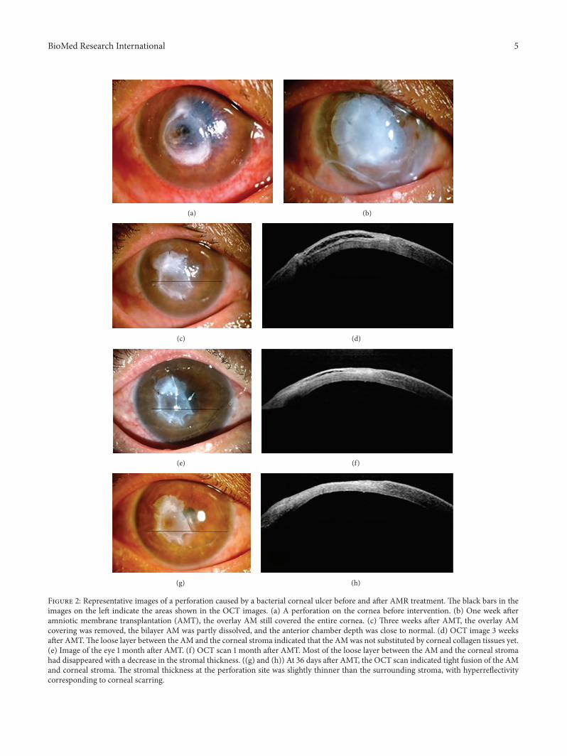
the anterior chamber was 8.6 ± 2.0 days (range 4–12). Theanterior chamber center depth of all patients recovered to 5times the corneal thickness, which is above 2.5mm.Oneweekafter surgery, some outermost AM graft cover had begun toloosen or dissolve. After the operation, the average durationof epithelial healing was 1.97 ± 0.56 weeks (range 1–3.5). Theaverage duration of bilayer AM disappearance was 6.89 ±1.73 weeks (range 4–12) and break after the average fusiontime of the amniotic membrane and the corneal stroma was6.12±1.04weeks (range 4–8). Figure 2(c) shows that after theoutermost large AM graft was removed, 3 weeks after AMT,the bilayer AMhad partly dissolved and the anterior chamberdepth was in the normal range. At the same time, the OCTimage showed that the AMwas fully epithelialized, the perfo-ration was healed completely with an increase in the stromalthickness, and therewas a loose layer between theAMand thecorneal stroma, indicating that theAMwas not substituted bycorneal collagen tissues at this time (Figure 2(d)). Onemonthafter AMT, the AM and corneal stroma fused together welland most of the loose layer between the AM and the cornealstroma had disappeared with a corresponding decrease instromal thickness (Figures 2(e) and 2(f)). Thirty-six days
BioMed Research International 5
(a) (b)
(c) (d)
(e) (f)
(g) (h)
Figure 2: Representative images of a perforation caused by a bacterial corneal ulcer before and after AMR treatment. The black bars in theimages on the left indicate the areas shown in the OCT images. (a) A perforation on the cornea before intervention. (b) One week afteramniotic membrane transplantation (AMT), the overlay AM still covered the entire cornea. (c) Three weeks after AMT, the overlay AMcovering was removed, the bilayer AM was partly dissolved, and the anterior chamber depth was close to normal. (d) OCT image 3 weeksafter AMT.The loose layer between the AM and the corneal stroma indicated that the AMwas not substituted by corneal collagen tissues yet.(e) Image of the eye 1 month after AMT. (f) OCT scan 1 month after AMT. Most of the loose layer between the AM and the corneal stromahad disappeared with a decrease in the stromal thickness. ((g) and (h)) At 36 days after AMT, the OCT scan indicated tight fusion of the AMand corneal stroma. The stromal thickness at the perforation site was slightly thinner than the surrounding stroma, with hyperreflectivitycorresponding to corneal scarring.
6 BioMed Research International
(a) (b)
(c) (d)
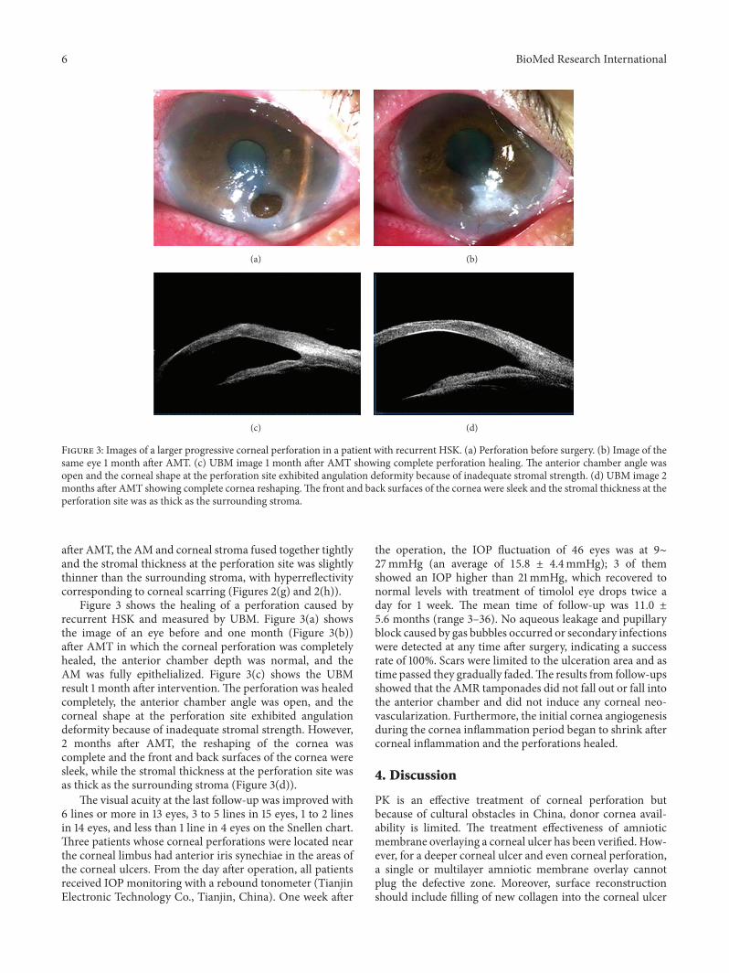
Figure 3: Images of a larger progressive corneal perforation in a patient with recurrent HSK. (a) Perforation before surgery. (b) Image of thesame eye 1 month after AMT. (c) UBM image 1 month after AMT showing complete perforation healing. The anterior chamber angle wasopen and the corneal shape at the perforation site exhibited angulation deformity because of inadequate stromal strength. (d) UBM image 2months after AMT showing complete cornea reshaping.The front and back surfaces of the cornea were sleek and the stromal thickness at theperforation site was as thick as the surrounding stroma.
after AMT, the AM and corneal stroma fused together tightlyand the stromal thickness at the perforation site was slightlythinner than the surrounding stroma, with hyperreflectivitycorresponding to corneal scarring (Figures 2(g) and 2(h)).
Figure 3 shows the healing of a perforation caused byrecurrent HSK and measured by UBM. Figure 3(a) showsthe image of an eye before and one month (Figure 3(b))after AMT in which the corneal perforation was completelyhealed, the anterior chamber depth was normal, and theAM was fully epithelialized. Figure 3(c) shows the UBMresult 1 month after intervention. The perforation was healedcompletely, the anterior chamber angle was open, and thecorneal shape at the perforation site exhibited angulationdeformity because of inadequate stromal strength. However,2 months after AMT, the reshaping of the cornea wascomplete and the front and back surfaces of the cornea weresleek, while the stromal thickness at the perforation site wasas thick as the surrounding stroma (Figure 3(d)).
The visual acuity at the last follow-up was improved with6 lines or more in 13 eyes, 3 to 5 lines in 15 eyes, 1 to 2 linesin 14 eyes, and less than 1 line in 4 eyes on the Snellen chart.Three patients whose corneal perforations were located nearthe corneal limbus had anterior iris synechiae in the areas ofthe corneal ulcers. From the day after operation, all patientsreceived IOP monitoring with a rebound tonometer (TianjinElectronic Technology Co., Tianjin, China). One week after
the operation, the IOP fluctuation of 46 eyes was at 9∼27mmHg (an average of 15.8 ± 4.4mmHg); 3 of themshowed an IOP higher than 21mmHg, which recovered tonormal levels with treatment of timolol eye drops twice aday for 1 week. The mean time of follow-up was 11.0 ±5.6 months (range 3–36). No aqueous leakage and pupillaryblock caused by gas bubbles occurred or secondary infectionswere detected at any time after surgery, indicating a successrate of 100%. Scars were limited to the ulceration area and astime passed they gradually faded.The results from follow-upsshowed that the AMR tamponades did not fall out or fall intothe anterior chamber and did not induce any corneal neo-vascularization. Furthermore, the initial cornea angiogenesisduring the cornea inflammation period began to shrink aftercorneal inflammation and the perforations healed.
4. Discussion
PK is an effective treatment of corneal perforation butbecause of cultural obstacles in China, donor cornea avail-ability is limited. The treatment effectiveness of amnioticmembrane overlaying a corneal ulcer has been verified. How-ever, for a deeper corneal ulcer and even corneal perforation,a single or multilayer amniotic membrane overlay cannotplug the defective zone. Moreover, surface reconstructionshould include filling of new collagen into the corneal ulcer
BioMed Research International 7
bed, thus promoting new basement membrane formationfor rapid epithelialization and suppressing inflammation. Inthe present study, the amniotic membrane was rolled andthen plugged into the defect zone of the cornea; then abilayer amniotic membrane was used to overlay and fix theamniotic roll, and finally a larger amniotic membrane wasused to cover the whole cornea. As a result, enough force wasgenerated to resist the pressure of the anterior chamber andto prevent any aqueous leakage and outward bulging of theAMR during the postoperative period. In addition to fillingthe defective corneal area, it provided an ideal basement forrapid epithelialization and wound healing. Furthermore, thelarge AM covering the whole cornea constitutes amechanicalprotection for the fragile corneal epithelium, allowing suffi-cient oxygenation and hydration of the epithelial cells andsuppressing inflammation [18]. In the present study, ocularsurface inflammation was markedly reduced after AMT, inagreement with previously published reports [12, 13]. It isnoteworthy that AM contains a mixture of growth factors,neurotrophins and cytokines that facilitate proliferation anddifferentiation of epithelial cells, and reduces the inflam-matory response [5, 19, 20]. Additionally, AM can be alsoused to prevent collagenous tissue being uncovered andcollagenolysis.
When amniotic roll plugging was employed, the aqueousfluid could easily ooze along the amniotic membrane roll dueto the anterior chamber pressure resulting in disappearanceof the anterior chamber. To solve this problem, we used agas-solution interfacial tension to prevent aqueous oozingalong the AMR. First, we tried to inject sterile air bubblesinto the anterior chamber but two days later they disappeareddue to absorption, and aqueous fluid again oozed along theAMR.Therefore, we changed to a C
3F8gas mixture injection,
which lasted a relatively long time (more than 1 week) [21].As a result, aqueous fluid oozing was prevented, the depth ofanterior chamber was maintained, and anterior synechia wasprevented.
The technique used in the current studywas not practicedfor corneal perforation sizes ≥3mm in diameter, becausewe were worried that there might be a higher risk of usingthe method for larger cornea perforation sizes. In general,the larger the size of the corneal perforation, the higher thepressure of aqueous humor on the amniotic roll, whichmightlead to complications such as aqueous leakage, hydrops underthe amnioticmembrane, and outward bulging of theAM [12].
Special attention during the operation must be paid tofine suturing of the double amniotic membrane film surface,because if the stitches are too large (>1mm), when injectinggas to the anterior chamber after the operation, the amnioticroll may be extruded from the suture by anterior chamberpressure. Also too large bubble in the anterior chambershould be avoided, because it may cause amniotic roll bulgingand temporary high intraocular pressure after the operation.For larger ulcer surfaces and perforated ulcers with a thinbasement, if the amniotic roll is packed too hard it may causeholes to expand or tear and amniotic roll luxation into theanterior chamber and the interventionmust be redone as PK.
However, for lesions ≤3mm our study showed in thelonger term follow-ups that 2 months after intervention
the results of UBM or anterior segment OCT (AS-OCT)examinations revealed the successful fusion of AM rolls withcorneal tissues which filled the lesion areas. Finally, the AMrolls were replaced with corneal scar tissue and the perfora-tion areas were recovered to the thickness of normal corneaswith both well-shaped corneal sides becoming mellow.
In summary, under conditions when corneal transplan-tations cannot be performed due to a lack of fresh donorcorneas, AMR combined with the multilayer amniotic mem-brane cover method is an effective alternative to conjuncti-val transplantations or conjunctival flap covering surgeries,with a success rate of 100%. The eyesight partly recoveredafter surgery, no neovascularization was induced, and thetechnique was acceptable in terms of cosmesis. Unnecessarytrauma at the donor site can be avoided compared to conjunc-tival flap overlaying. Furthermore, our method might alsoprovide sufficient time to obtain a fresh donor cornea and agood condition for future successful corneal transplantation,thereby converting emergency tectonic PKP into electiveoptical PKP, which is a more favorable procedure with abetter outcome for the patient’s vision. However, long-termstudies with larger samples of patients with various types ofperforations are still needed.
Competing Interests
All authors declare that there are no competing interests.
References
[1] V. Jhanji, A. L. Young, J. S. Mehta, N. Sharma, T. Agarwal, andR. B. Vajpayee, “Management of corneal perforation,” Survey ofOphthalmology, vol. 56, no. 6, pp. 522–538, 2011.
[2] V. Kumar and A. Kumar, “Immunological aspects of cornealtransplant,” Immunological Investigations, vol. 43, no. 8, pp. 888–901, 2014.
[3] S.-H. Lee and S. C. G. Tseng, “Amniotic membrane transplanta-tion for persistent epithelial defects with ulceration,” AmericanJournal of Ophthalmology, vol. 123, no. 3, pp. 303–312, 1997.
[4] G. Colocho, W. P. Graham III, A. E. Greene, D. W. Matheson,and D. Lynch, “Human amniotic membrane as a physiologicwound dressing,” Archives of Surgery, vol. 109, no. 3, pp. 370–373, 1974.
[5] B. K. Na, J. H. Hwang, J. C. Kim et al., “Analysis of humanamniotic membrane components as proteinase inhibitors fordevelopment of therapeutic agent for recalcitrant keratitis,”Placenta, vol. 20, pp. 453–466, 1999.
[6] H. Sato, J. Shimazaki, and K. Shinozaki, “Role of growth factorsfor ocular surface reconstruction after amniotic membranetransplantation,” Investigative Ophthalmology & Visual Science,vol. 39, article S428, 1998.
[7] K. Hanada, J. Shimazaki, S. Shimmura, and K. Tsubota,“Multilayered amniotic membrane transplantation for severeulceration of the cornea and sclera,” American Journal ofOphthalmology, vol. 131, no. 3, pp. 324–331, 2001.
[8] J. S. Kim, J. C. Kim, B. K. Na, J. M. Jeong, and C. Y. Song,“Amniotic membrane patching promotes healing and inhibitsproteinase activity on wound healing following acute cornealalkali burn,” Experimental Eye Research, vol. 70, no. 3, pp. 329–337, 2000.
8 BioMed Research International
[9] F. E. Kruse, K. Rohrschneider, and H. E. Volcker, “Multilayeramniotic membrane transplantation for reconstruction of deepcorneal ulcers,” Ophthalmology, vol. 106, no. 8, pp. 1504–1511,1999.
[10] P. Prabhasawat, N. Tesavibul, andW. Komolsuradej, “Single andmultilayer amniotic membrane transplantation for persistentcorneal epithelial defect with and without stromal thinning andperforation,” British Journal of Ophthalmology, vol. 85, no. 12,pp. 1455–1463, 2001.
[11] A. Solomon, D. Meller, P. Prabhasawat et al., “Amniotic mem-brane grafts for nontraumatic corneal perforations, descemeto-celes, and deep ulcers,” Ophthalmology, vol. 109, no. 4, pp. 694–703, 2002.
[12] M. T. Rodrıguez-Ares, R. Tourino, M. J. Lopez-Valladares, andF. Gude, “Multilayer amniotic membrane transplantation in thetreatment of corneal perforations,” Cornea, vol. 23, no. 6, pp.577–583, 2004.
[13] S. Hick, P. E. Demers, I. Brunette, C. La, M. Mabon, and B.Duchesne, “Amnioticmembrane transplantation andfibrin gluein the management of corneal ulcers and perforations: a reviewof 33 cases,” Cornea, vol. 24, no. 4, pp. 369–377, 2005.
[14] H. K. Kim and H. S. Park, “Fibrin glue-assisted augmentedamniotic membrane transplantation for the treatment of largenoninfectious corneal perforations,” Cornea, vol. 28, no. 2, pp.170–176, 2009.
[15] E. Chan, A. N. Shah, and D. P. S. O’Brart, “‘Swiss roll’amniotic membrane technique for the management of cornealperforations,” Cornea, vol. 30, no. 7, pp. 838–841, 2011.
[16] T. Kawakita, T. Sumi, M. Dogru, K. Tsubota, and J. Shimazaki,“Amniotic membrane transplantation for wound dehiscenceafter deep lamellar keratoplasty: a case report,” Journal ofMedical Case Reports, vol. 1, article 28, 2007.
[17] H. Jiang and J. H. Fan, “The use of sodium hyaluronate incryopreservation of cornea,”Medical Journal of Chinese People’sLiberation Army, vol. 32, pp. 694–696, 2007.
[18] J. Baum, “Thygeson lecture. Amniotic membrane transplanta-tion:why is it effective?”Cornea, vol. 21, no. 4, pp. 339–341, 2002.
[19] Y. Hao, D. H. Ma, D. G. Hwang, W.-S. Kim, and F. Zhang,“Identification of antiangiogenic and antiinflammatory proteinsin human amniotic membrane,” Cornea, vol. 19, no. 3, pp. 348–352, 2000.
[20] N. Koizumi, N. J. Fullwood, G. Bairaktaris, T. Inatomi, S.Kinoshita, and A. J. Quantock, “Cultivation of corneal epithelialcells on intact and denuded human amniotic membrane,”Investigative Ophthalmology and Visual Science, vol. 41, no. 9,pp. 2506–2513, 2000.
[21] W. N. Li, X. X. Li, and J. H. Fan, “Effects of perfluoropane(c3f8) gas combinedwith viscoelastic agents for complicated eyetrauma,”Chinese Journal of Practical Ophthalmology, vol. 27, no.12, pp. 1409–1411, 2009.