Eli Maria Pazzianotto Forti,3 Flávio do Amaral Campos,2 Fabiana Sobral Peixoto-Souza,4
and Dirceu Costa1,4
1 Post Graduation Program of Physiotherapy of Federal University of Sao Carlos (UFSCar), Rod. Washington Luıs,km 235, Sao Carlos, SP, Brazil
2Meridional Hospital, Av. Sao Joao Batista, n 200, Cariacica, ES, Brazil3 Post Graduation Program of Physiotherapy of Methodist University of Piracicaba (UNIMEP), Rod. do Acucar, km 156,Piracicaba, SP, Brazil
4 Post Graduation Program of Physiotherapy of Nove de Julho University (UNINOVE), Av. Dr. Adolpho Pinto, n 109,Barra Funda, Sao Paulo, SP, Brazil
Introduction. Obesity is a condition that causes damage to the respiratory function. However, studies have demonstrated that weightloss due to bariatric surgery has resulted in a huge improvement on some lung volumes, but controversy still persists regarding thebehavior of the respiratory muscle strength and IRV (inspiratory reserve volume). Objective. To evaluate the effect of weight loss,after 1 year of the Roux-en-Y gastric bypass surgery (RYGB), on the lung volumes and the respiratory muscle strength in obesewomen.Methods. 24 obesewomen candidateswere recruited for RYGB. Lung volumes (spirometry) and respiratorymuscle strengthwere evaluated in preoperative period and one year after surgery. Results. There was a significant increase in some lung volumes.However, when examining the components of the VC (vital capacity) separately, an increase in ERV (expiratory reserve volume)and reduction of IRV were observed. Moreover, a statistically significant reduction in the values of respiratory muscle strengthwas recorded: MIP (maximal inspiratory pressure) and MEP (maximal expiratory pressure). Conclusion. Weight loss induced bybariatric surgery provides an increase in some lung volumes of obese women, but reduction in IRV. Additionally, there was also areduction in the respiratory muscle strength.
1. Introduction
Obesity is a condition that causes damage to the variousbody functions, such as cardiovascular, musculoskeletal, andmetabolic functions amongst others [1].The respiratory func-tion is also affected by obesity, as excess fat deposited on thechest wall and the abdominal cavity affects the chest mechan-ics. This results in increased work of breathing, reducedlung volumes, dysfunction of the respiratory muscle, impair-ment in gas exchange, and reduced exercise tolerance [2–9].
A few studies have demonstrated that weight loss due tobariatric surgery has resulted in a huge improvement in somefunctions, such as decrease in hemoglobin and hematocrit[10], decreased heart rate and oxygen consumption [10], andreduction of insulin resistance [11]. In addition, especiallyimproved lung function with increased forced vital capacity(FVC) [3, 12, 13] and forced expiratory volume in one second(FEV1), improved alveolar-capillary diffusion capacity [10]
and improvement in gas exchange [12, 13] have also beenobserved.
2 ISRN Obesity
There is strong evidence supporting the increase in FVCand ERV (expiratory reserve volume) after weight loss [3, 12,13]. However, controversy still persists regarding the behaviorof the respiratory muscle strength and IRV (inspiratoryreserve volume) after weight loss caused by the Roux-en-Ygastric bypass surgery.
The objective of the present study was to evaluate theeffect of weight loss, after 1 year of the Roux-en-Y gastricbypass surgery (RYGB), on the lung volumes and the respi-ratory muscle strength in obese women.
2. Methods and Procedures
2.1. Patients. Were recruited 24 obese women candidatesfor RYGB at the Meridional Hospital. Patients with bodymass index (BMI) 35–50 kg/m2 were included if they metthe minimal criteria for bariatric surgery proposed by theWorld Health Organization (WHO) report of 2000 [1]. Thefollowing were not included in the study: patients sufferingfrompulmonary diseases or those unable to carry out the pul-monary function tests adequately, smokers, patients who didnot attend the reevaluation 1 year after surgery, and patientsrefusing to sign the Informed Consent Term. The presentstudy was approved by the Meridional Hospital Ethics Com-mittee (Protocol number 01/07).
2.2. Pulmonary Function Tests. The evaluation of the pul-monary function was carried out by conventional spirom-etry using a personal computer version of the NDDEasyOne Spirometer Model 2001 (Medizintechnik AG,Zurich, Switzerland). Parameters, such as volume, capacity,and flow of the lungs, were directly evaluated by using theslow vital capacity (SVC), the forced vital capacity (FVC),and the maximum voluntary ventilation (MVV) tests, withvolunteers in a sitting position and a minimum of three rep-etitions, as recommended by the American Thoracic Society(ATS) and the European Respiratory Society (ERS) [14]. Theobtained results were expressed in absolute values and aspercentages of the predicted reference values for the Brazil-ian population [15]. The SVC test produced the followingvariables: vital capacity (VC), tidal volume (VT), inspiratoryreserve volume (IRV), and expiratory reserve volume (ERV).The FVC test allowed the determination of the forced expira-tory volume in 1 s (FEV
1) and the FEV
1/FVC ratio.TheMVV,
a variable that evaluates the respiratory endurance, wasexpressed in liters per minute and as a percentage of the pre-dicted reference value for the Brazilian population [15].
The respiratory muscle strength was determined throughthe maximal static respiratory pressures measured duringforced inspiration and expiration—maximal inspiratory pres-sure (MIP) and maximal expiratory pressure (MEP). Themeasurement was carried out using an aneroid manometer(Wika, Ipero, SP, Brazil), calibrated in centimeter H
2O
(±300 cmH2O), and equipped with a 2mm hole to relieve
the oral pressure.The procedure was carried out as describedby ATS/ERS [16]. MIP and MEP were determined using theresidual volume and the total lung capacity, respectively,with the subjects in a sitting position. The inspiratory and
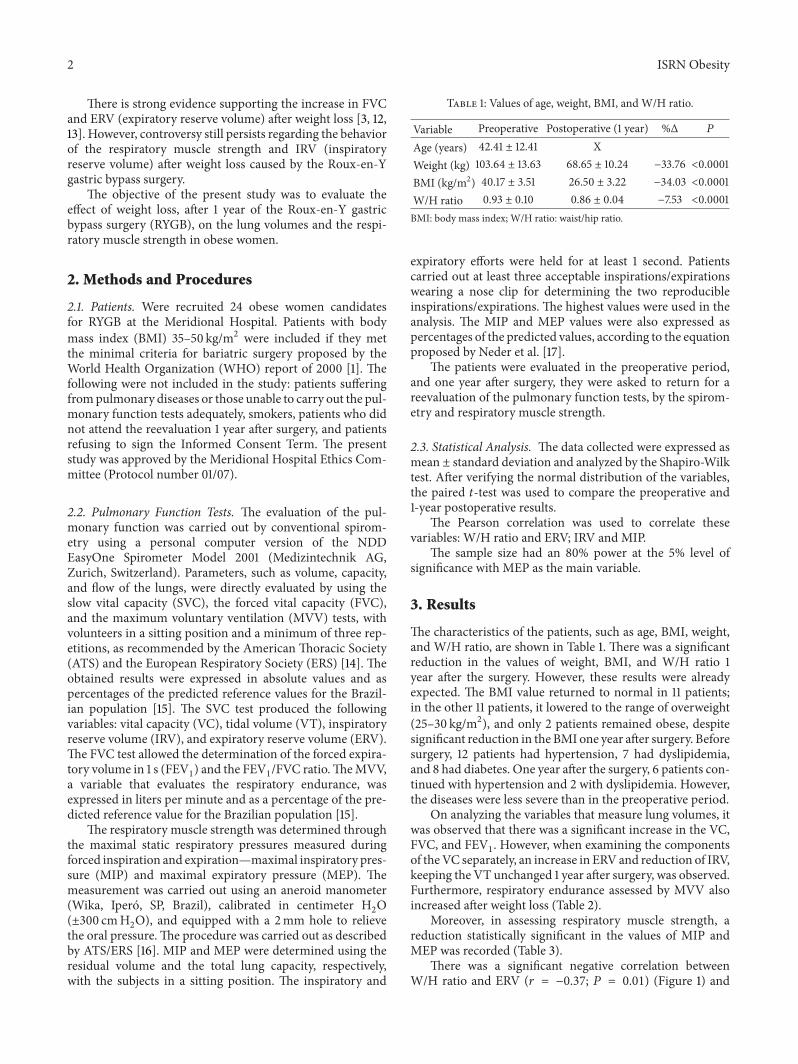
Table 1: Values of age, weight, BMI, and W/H ratio.
Variable Preoperative Postoperative (1 year) %Δ 𝑃
Age (years) 42.41 ± 12.41 XWeight (kg) 103.64 ± 13.63 68.65 ± 10.24 −33.76 <0.0001BMI (kg/m2) 40.17 ± 3.51 26.50 ± 3.22 −34.03 <0.0001W/H ratio 0.93 ± 0.10 0.86 ± 0.04 −7.53 <0.0001BMI: body mass index; W/H ratio: waist/hip ratio.
expiratory efforts were held for at least 1 second. Patientscarried out at least three acceptable inspirations/expirationswearing a nose clip for determining the two reproducibleinspirations/expirations. The highest values were used in theanalysis. The MIP and MEP values were also expressed aspercentages of the predicted values, according to the equationproposed by Neder et al. [17].
The patients were evaluated in the preoperative period,and one year after surgery, they were asked to return for areevaluation of the pulmonary function tests, by the spirom-etry and respiratory muscle strength.
2.3. Statistical Analysis. The data collected were expressed asmean ± standard deviation and analyzed by the Shapiro-Wilktest. After verifying the normal distribution of the variables,the paired 𝑡-test was used to compare the preoperative and1-year postoperative results.
The Pearson correlation was used to correlate thesevariables: W/H ratio and ERV; IRV and MIP.
The sample size had an 80% power at the 5% level ofsignificance with MEP as the main variable.
3. Results
The characteristics of the patients, such as age, BMI, weight,and W/H ratio, are shown in Table 1. There was a significantreduction in the values of weight, BMI, and W/H ratio 1year after the surgery. However, these results were alreadyexpected. The BMI value returned to normal in 11 patients;in the other 11 patients, it lowered to the range of overweight(25–30 kg/m2), and only 2 patients remained obese, despitesignificant reduction in the BMI one year after surgery. Beforesurgery, 12 patients had hypertension, 7 had dyslipidemia,and 8 had diabetes. One year after the surgery, 6 patients con-tinued with hypertension and 2 with dyslipidemia. However,the diseases were less severe than in the preoperative period.
On analyzing the variables that measure lung volumes, itwas observed that there was a significant increase in the VC,FVC, and FEV
1. However, when examining the components
of theVC separately, an increase in ERV and reduction of IRV,keeping the VT unchanged 1 year after surgery, was observed.Furthermore, respiratory endurance assessed by MVV alsoincreased after weight loss (Table 2).
Moreover, in assessing respiratory muscle strength, areduction statistically significant in the values of MIP andMEP was recorded (Table 3).
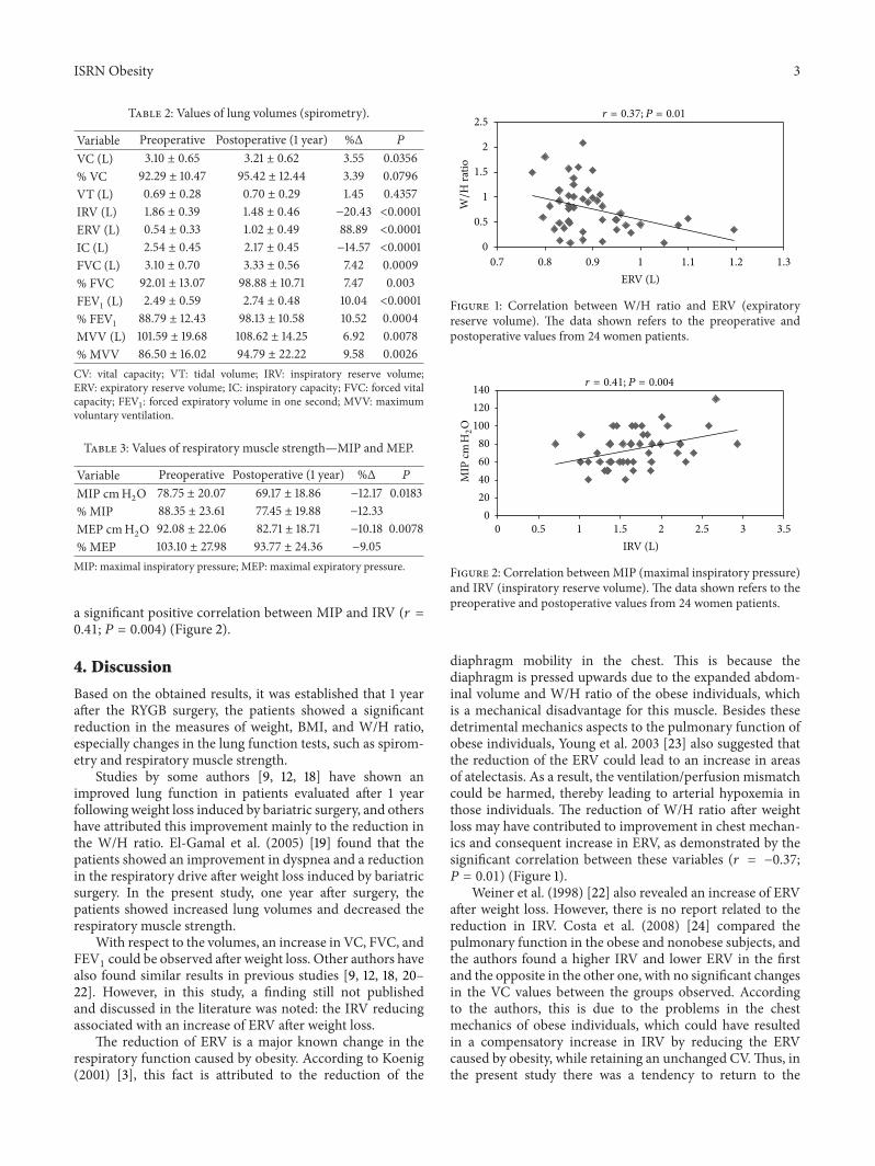
There was a significant negative correlation betweenW/H ratio and ERV (𝑟 = −0.37; 𝑃 = 0.01) (Figure 1) and
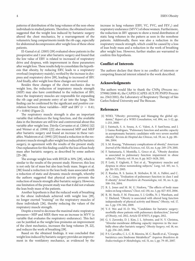
a significant positive correlation between MIP and IRV (𝑟 =0.41; 𝑃 = 0.004) (Figure 2).
4. Discussion
Based on the obtained results, it was established that 1 yearafter the RYGB surgery, the patients showed a significantreduction in the measures of weight, BMI, and W/H ratio,especially changes in the lung function tests, such as spirom-etry and respiratory muscle strength.
Studies by some authors [9, 12, 18] have shown animproved lung function in patients evaluated after 1 yearfollowing weight loss induced by bariatric surgery, and othershave attributed this improvement mainly to the reduction inthe W/H ratio. El-Gamal et al. (2005) [19] found that thepatients showed an improvement in dyspnea and a reductionin the respiratory drive after weight loss induced by bariatricsurgery. In the present study, one year after surgery, thepatients showed increased lung volumes and decreased therespiratory muscle strength.
With respect to the volumes, an increase in VC, FVC, andFEV1could be observed after weight loss. Other authors have
also found similar results in previous studies [9, 12, 18, 20–22]. However, in this study, a finding still not publishedand discussed in the literature was noted: the IRV reducingassociated with an increase of ERV after weight loss.
The reduction of ERV is a major known change in therespiratory function caused by obesity. According to Koenig(2001) [3], this fact is attributed to the reduction of the
0
0.5
1
1.5
2
2.5
0.7 0.8 0.9 1 1.1 1.2 1.3
W/H
ratio
ERV (L)
𝑟 = 0.37; 𝑃 = 0.01
Figure 1: Correlation between W/H ratio and ERV (expiratoryreserve volume). The data shown refers to the preoperative andpostoperative values from 24 women patients.
020406080
100120140
0 0.5 1 1.5 2 2.5 3 3.5IRV (L)
𝑟 = 0.41; 𝑃 = 0.004
MIP
cm H2O
Figure 2: Correlation betweenMIP (maximal inspiratory pressure)and IRV (inspiratory reserve volume). The data shown refers to thepreoperative and postoperative values from 24 women patients.
diaphragm mobility in the chest. This is because thediaphragm is pressed upwards due to the expanded abdom-inal volume and W/H ratio of the obese individuals, whichis a mechanical disadvantage for this muscle. Besides thesedetrimental mechanics aspects to the pulmonary function ofobese individuals, Young et al. 2003 [23] also suggested thatthe reduction of the ERV could lead to an increase in areasof atelectasis. As a result, the ventilation/perfusion mismatchcould be harmed, thereby leading to arterial hypoxemia inthose individuals. The reduction of W/H ratio after weightloss may have contributed to improvement in chest mechan-ics and consequent increase in ERV, as demonstrated by thesignificant correlation between these variables (𝑟 = −0.37;𝑃 = 0.01) (Figure 1).
Weiner et al. (1998) [22] also revealed an increase of ERVafter weight loss. However, there is no report related to thereduction in IRV. Costa et al. (2008) [24] compared thepulmonary function in the obese and nonobese subjects, andthe authors found a higher IRV and lower ERV in the firstand the opposite in the other one, with no significant changesin the VC values between the groups observed. Accordingto the authors, this is due to the problems in the chestmechanics of obese individuals, which could have resultedin a compensatory increase in IRV by reducing the ERVcaused by obesity, while retaining an unchanged CV.Thus, inthe present study there was a tendency to return to the
4 ISRN Obesity
patterns of distribution of the lung volumes of the non-obeseindividuals in studied patients.Therefore, the obtained resultssuggested that the weight loss induced by bariatric surgeryaltered the chest mechanics, by a rearrangement of thevolumetric lung compartments inside the rib cage, especiallyfor abdominal decompression after weight loss of these obesepatients.
El-Gamal et al. (2005) [19] evaluated obese patients in thepreoperative and 1 year after bariatric surgery and found thatthe low value of ERV is related to increased of respiratorydrive and dyspnea, with improvement in these parametersafter weight loss.These results help to consolidate the hypoth-esis that obesity, by reducing ERV, leads to a respiratoryoverload (inspiratory mainly), verified by the increase in dys-pnea and respiratory drive [19], leading to increased of IRV.And finally, after weight loss these changes are reversed.
Besides these changes of the chest mechanics due toweight loss, the reduction of inspiratory muscle strength(MIP) may also have contributed to the reduction of IRV,since the inspiratory muscles are responsible for expandingthe rib cage and promote entry of air into the lungs. Thisfinding can be confirmed by the significant and positive cor-relation between these variables—MIP and IRV (𝑟 = 0.41;𝑃 = 0.004) (Figure 2).
The respiratory muscle strength is also an importantvariable that influences the lung function, and the availabledata in the literature are still few and controversial about thisvariable after weight loss. Davila-Cervantes et al. (2004) [12]and Weiner et al. (1998) [22] also measured MIP and MEPafter bariatric surgery and found an increase in these vari-ables.Wadstrom et al. (1991) [25] found a reduction in the res-piratorymuscle strength afterweight loss induced by bariatricsurgery, in agreement with the results of the present study.One explanation for this finding could be the loss of lean bodymass after bariatric surgery, as described by some authors[25–28].
The average weight loss with RYGB is 30% [29], which issimilar to the results of the present study. However, this lossis not only fat of mass but also lean body mass. Stegen et al.[30] found a reduction in the lean body mass associated witha reduction of static and dynamic muscle strength, wherebythe authors suggested that physical activity prevents thereduction ofmuscle strength after bariatric surgery. However,one limitation of the present study was that it did not evaluatethe lean body mass of the patients.
Another hypothesis is that the reduced work of breathingin the obese individuals achieved with weight loss [19],no longer exerted “training” on the respiratory muscles ofthese individuals [24], thereby reducing the values of therespiratory muscle strength.
Despite a trend to decrease the maximal static respiratorypressures—MIP and MEP, there was an increase in MVV (avariable that evaluates the respiratory endurance). This factcan be justified as the weight loss promotes an improvementof the chest mechanics, increases the lung volumes [9, 22],and reduces the work of breathing [19].
Based on the obtained findings, it was concluded thatweight loss induced by bariatric surgery provides an improve-ment in the ventilatory mechanics, as evidenced by the
increase in lung volumes (ERV, VC, FVC, and FEV1) and
respiratory endurance (MVV)of obesewomen. Furthermore,the reduction in IRV appears to show a trend distribution ofstatic lung volumes in the pattern as seen in the nonobesepatients. Additionally, there was also a reduction in therespiratory muscle strength, which could be caused by a lossof lean body mass and a reduction in the work of breathingafter weight loss. However, further studies are warranted toconfirm this hypothesis.
Conflict of Interests
The authors declare that there is no conflict of interests orcompeting financial interest related to the work described.
Acknowledgments
The authors would like to thank the CNPq (Process no.:579981/2008-8), the CAPES (CAPES AUX PE PNPD Processno. 2562/2011), the Laboratory of Respiratory Therapy of SaoCarlos Federal University andThe Bioscan.
References
[1] WHO, “Obesity: preventing and Managing the global epi-demic,” Report of a WHO Consultation, vol. 894, no. 1–12, pp.1–253, 2000.
[2] J. Faintuch, S. A. Souza, A. C. Valezi, A. F. Sant’Anna, and J.J. Gama-Rodrigues, “Pulmonary function and aerobic capacityin asymptomatic bariatric candidates with very severe morbidobesity,” Revista do Hospital das Clinicas, vol. 59, no. 4, pp. 181–186, 2004.
[3] S. M. Koenig, “Pulmonary complications of obesity,” AmericanJournal of theMedical Sciences, vol. 321, no. 4, pp. 249–279, 2001.
[4] F. Santamaria, S. Montella, L. Greco et al., “Obesity durationis associated to pulmonary function impairment in obesesubjects,” Obesity, vol. 19, no. 8, pp. 1623–1628, 2011.
[5] P. Lotti, F. Gigliotti, F. Tesi et al., “Respiratory muscles anddyspnea in obese nonsmoking subjects,” Lung, vol. 183, no. 5,pp. 311–323, 2005.
[6] Z. Rasslan, R. S. Junior, R. Stirbulov, R. M. A. Fabbri, and C.A. C. Lima, “Evaluation of pulmonary function in class I andII obesity,” Jornal Brasileiro de Pneumologia, vol. 30, no. 6, pp.508–514, 2004.
[7] R. L. Jones and M. M. U. Nzekwu, “The effects of body massindex on lung volumes,”Chest, vol. 130, no. 3, pp. 827–833, 2006.
[8] R. M. Steele, F. M. Finucane, S. J. Griffin, N. J. Wareham, andU. Ekelund, “Obesity is associated with altered lung functionindependently of physical activity and fitness,” Obesity, vol. 17,no. 3, pp. 578–584, 2009.
[9] Y.-F. Wei and H.-D. Wu, “Candidates for bariatric surgery:morbidly obese patients with pulmonary dysfunction,” Journalof Obesity, vol. 2012, Article ID 878371, 6 pages, 2012.
[10] G. S. Zavorsky, D. J. Kim, J. L. Sylvestre, and N. V. Christou,“Alveolar-membrane diffusing capacity improves in the mor-bidly obese after bariatric surgery,” Obesity Surgery, vol. 18, no.3, pp. 256–263, 2008.
[11] P. S. Carvalho, C. L. C. B. Moreira, M. C. Barelli et al., “Cirurgiabariatrica cura Sındrome Metabolica?” Arquivos Brasileiros deEndocrinologia & Metabologia, vol. 51, no. 1, pp. 79–85, 2007.
ISRN Obesity 5
[12] A. Davila-Cervantes, G. Domınguez-Cherit, D. Borunda etal., “Impact of surgically-induced weight loss on respiratoryfunction: a prospective analysis,”Obesity Surgery, vol. 14, no. 10,pp. 1389–1392, 2004.
[13] G. S. Zavorsky, J.M.Murias, D. J. Kim, J. Gow, J. L. Sylvestre, andN.V.Christou, “Waist-to-hip ratio is associatedwith pulmonarygas exhange in the morbidly obese,” Chest, vol. 131, no. 2, pp.362–367, 2007.
[14] M. R. Miller, J. Hankinson, F. Burgos et al., “Standardisationof spirometry,” European Respiratory Journal, vol. 26, no. 2, pp.319–338, 2005.
[15] C. A. De Castro Pereira, T. Sato, and S. C. Rodrigues, “Newreference values for forced spirometry in white adults in Brazil,”Jornal Brasileiro de Pneumologia, vol. 33, no. 4, pp. 397–406,2007.
[16] American Thoracic Society/European Respiratory Society,“ATS/ERS Statement on respiratory muscle testing,” AmericanJournal of Respiratory and Critical Care Medicine, vol. 166, no.4, pp. 518–624, 2002.
[17] J. A. Neder, S. Andreoni, M. C. Lerario, and L. E. Nery,“Reference values for lung function tests. II. Maximal respira-tory pressures and voluntary ventilation,” Brazilian Journal ofMedical and Biological Research, vol. 32, no. 6, pp. 719–727, 1999.
[18] C. Martı-Valeri, A. Sabate, C. Masdevall, and A. Dalmau,“Improvement of associated respiratory problems in morbidlyobese patients after open Roux-en-Y gastric bypass,” ObesitySurgery, vol. 17, no. 8, pp. 1102–1110, 2007.
[19] H. El-Gamal, A. Khayat, S. Shikora, and J. N. Unterborn, “Rela-tionship of dyspnea to respiratory drive pulmonary functiontests in obese patients before and after weight loss,” Chest, vol.128, no. 6, pp. 3870–3874, 2005.
[20] C. Wadstrom, R. Muller-Suur, and L. Backman, “Influence ofexcessive weight loss on respiratory function. A study of obesepateints following gastroplasty,” Acta Chirurgica—EuropeanJournal of Surgery, vol. 157, no. 5, pp. 341–346, 1991.
[21] N. T. Nguyen, M. W. Hinojosa, B. R. Smith, J. Gray, and E.Varela, “Improvement of restrictive and obstructive pulmonarymechanics following laparoscopic bariatric surgery,” SurgicalEndoscopy and Other Interventional Techniques, vol. 23, no. 4,pp. 808–812, 2009.
[22] P. Weiner, J. Waizman, M. Weiner, M. Rabner, R. Magadle, andD. Zamir, “Influence of excessive weight loss after gastroplastyformorbid obesity on respiratorymuscle performance,”Thorax,vol. 53, no. 1, pp. 39–42, 1998.
[23] S. S. Young, S. M. Skeans, T. Austin, and R. W. Chapman,“The effects of body fat on pulmonary function and gasexchange in cynomolgus monkeys,” Pulmonary PharmacologyandTherapeutics, vol. 16, no. 5, pp. 313–319, 2003.
[24] D. Costa, M. C. Barbalho, G. P. S. Miguel, E. M. P. Forti, and J. L.M. C. Azevedo, “The impact of obesity on pulmonary functionin adult women,” Clinics, vol. 63, no. 6, pp. 719–724, 2008.
[25] C. Wadstrom, R. Muller-Suur, and L. Backman, “Influence ofexcessive weight loss on respiratory function. A study of obesepateints following gastroplasty,” Acta Chirurgica—EuropeanJournal of Surgery, vol. 157, no. 5, pp. 341–346, 1991.
[26] D.G.Carey,G. J. Pliego, andR. L. Raymond, “Body compositionandmetabolic changes following bariatric surgery: effects on fatmass, lean mass and basal metabolic rate: six months to one-year follow-up,” Obesity Surgery, vol. 16, no. 12, pp. 1602–1608,2006.
[27] D. G. Carey, G. J. Pliego, R. L. Raymond, and K. B. Skau, “Bodycomposition andmetabolic changes following bariatric surgery:
effects on fat mass, lean mass and basal metabolic rate,”ObesitySurgery, vol. 16, no. 4, pp. 469–477, 2006.
[28] T. Olbers, S. Bjorkman, A. Lindroos et al., “Body composition,dietary intake, and energy expenditure after laparoscopic Roux-en-Y gastric bypass and laparoscopic vertical banded gastro-plasty: a randomized clinical trial,” Annals of Surgery, vol. 244,no. 5, pp. 715–722, 2006.
[29] Z. Pataky, I. Carrard, and A. Golay, “Psychological factors andweight loss in bariatric surgery,” Current Opinion in Gastroen-terology, vol. 27, no. 2, pp. 167–173, 2011.
[30] S. Stegen, W. Derave, P. Calders, C. Van Laethem, and P. Pattyn,“Physical fitness in morbidly obese patients: effect of gastricbypass surgery and exercise training,” Obesity Surgery, vol. 21,no. 1, pp. 61–70, 2011.