Clinical value of Superb Micro-vascular Imaging and ultra-high frequency linear transducer in musculoskeletal ultrasound: - From early detection to diagnosis and treatment evaluation - Akihiro Narita, MD, Ph.D Chief, Department of Radiology Department of Rheumatology, Hokkaido Medical Center for Rheumatic Diseases, Sapporo, Japan Norifumi Sawamukai, MD, Ph.D Director, Rheumatism Center Department of Rheumatology, Hokkaido Medical Center for Rheumatic Diseases, Sapporo, Japan Introduction Rheumatoid arthritis (RA) is a chronic autoimmune disorder which causes structural damage in joints. In a patient with RA, the immune system attacks the joints, membrane thickening and bone erosion. Joint damage and deformation are irreversible, therefore early detection and treatment is critical. In recent years, RA management has improved rapidly, from striving for early detection, to early treatment with biologic agents, with the possibility that biologic-free or drug-free remission in rheumatoid arthritis could be achieved in the near future. Since musculoskeletal ultrasound (MSK US) provides a safe, non-invasive and short time-interval, it is an ideal for the clinical evaluation of RA. The improvements in ultrasound technologies detection, diagnosis and staging, therapeutic monitoring, and post-remission follow-up to provide optimal RA patient management. The clinical value of MSK US in RA evaluation using innovative technologies on the Aplio TM i-series diagnostic ultrasound system will be further discussed hereafter. Clinical value of MSK US on RA In our center, outpatients complaining of joint pain will undergo blood tests, X-ray and MSK US, following consultation with physicians. For patients with no abnormality detected, a MRI or possibly CT may be performed if necessary. modalities. For example X-ray is a fast and inexpensive method for detecting morphology of the bone, cartilage, and tendon. Nevertheless, it has a limitations in detecting synovitis in detail. MRI is superior in the detection of masses such as bone marrow edema, but it is an expensive and a non-real-time method. MSK US has the advantage to evaluate and monitor RA from both an anatomical and vascular perspective. Details of the joint morphology can be detected in grayscale and its vascularity can be quantified in real-time using power Doppler imaging (PDI). In recent years, PDI has been increasingly employed in the clinical setting as it is reported that synovial inflammation is highly associated with joint damage. PDI is a reliable method for detecting, staging and therapeutic monitoring RA. The EULAR (European
Transcript
Clinical value of Superb Micro-vascular Imaging and ultra-high frequency linear transducer in musculoskeletal ultrasound:- From early detection to diagnosis and treatment evaluation -
Akihiro Narita, MD, Ph.DChief, Department of RadiologyDepartment of Rheumatology, Hokkaido Medical Center forRheumatic Diseases, Sapporo, Japan
Norifumi Sawamukai, MD, Ph.DDirector, Rheumatism CenterDepartment of Rheumatology, Hokkaido Medical Center forRheumatic Diseases, Sapporo, Japan
Introduction
Rheumatoid arthritis (RA) is a chronic autoimmune disorder which causes structural damage in joints. In a patient with RA, the immune system attacks the joints,
membrane thickening and bone erosion. Joint damage and deformation are irreversible, therefore early detection and treatment is critical.
In recent years, RA management has improved rapidly, from striving for early detection, to early treatment with biologic agents, with the possibility that biologic-free or drug-free remission in rheumatoid arthritis could be achieved in the near future. Since musculoskeletal ultrasound (MSK US) provides a safe, non-invasive and
short time-interval, it is an ideal for the clinical evaluation of RA. The improvements in ultrasound technologies
detection, diagnosis and staging, therapeutic monitoring, and post-remission follow-up to provide optimal RA patient management.
The clinical value of MSK US in RA evaluation using innovative technologies on the AplioTM i-series diagnostic ultrasound system will be further discussed hereafter.
Clinical value of MSK US on RA
In our center, outpatients complaining of joint pain will undergo blood tests, X-ray and MSK US, following consultation with physicians. For patients with no abnormality detected, a MRI or possibly CT may be performed if necessary.
modalities. For example X-ray is a fast and inexpensive method for detecting morphology of the bone, cartilage, and tendon. Nevertheless, it has a limitations in detecting synovitis in detail. MRI is superior in the detection of masses such as bone marrow edema, but it is an expensive and a non-real-time method.
MSK US has the advantage to evaluate and monitor RA from both an anatomical and vascular perspective. Details of the joint morphology can be detected in grayscale and its vascularity can be quantified in real-time using power Doppler imaging (PDI). In recent years, PDI has been increasingly employed in the clinical setting as it is reported that synovial inflammation is highly associated with joint damage.
PDI is a reliable method for detecting, staging and therapeutic monitoring RA. The EULAR (European
2
League Against Rheumatism)/OMERACT (Outcome Measures in Rheumatology) working group has been developing guidelines for RA assessment. Common indices included DAS28 (Disease Activity Score 28), simplified disease activity index (SDAI), and clinical disease activity index (CDAI).
In MSK US however, artifacts or clutter can degrade the image quality and sensitivity, creating common challenges in making an accurate diagnosis. Furthermore, MSK US is operator-dependent and there are inter-operator differences when acquiring and interpreting images.
The revolutionary Aplio i-series overcomes the above issues by generating grayscale with high resolution and fewer artifacts, with the sensitivity of power Doppler imaging significantly increased. In addition, a new Doppler technology, Superb Micro-vascular Imaging (SMI), was developed to help delineate minute vessels and extreme low-velocity blood flow, which have previously not been detectable using conventional Doppler techniques. These improvements allow clinicians to assess RA with high diagnostic confidence and accuracy for RA assessment.
Conventional MSK US in RA
In our center, MSK US is performed by sonographers for almost all first visit patients complaining of joint pain. As more than 80% of RA symptoms involve the hand and wrist, in our
protocol for RA ultrasound, 26 locations are evaluated including 10 locations on the fingers and 3 locations on the wrist from both hands. Since rheumatoid arthritis is a systemic disease, evaluation of all joints in the body is also conducted.
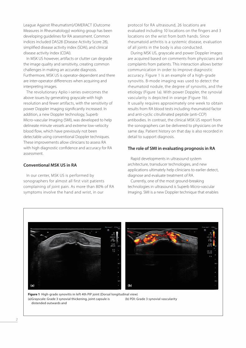
During MSK US, grayscale and power Doppler images are acquired based on comments from physicians and complaints from patients. This interaction allows better communication in order to improve diagnostic accuracy. Figure 1 is an example of a high-grade synovitis. B-mode imaging was used to detect the rheumatoid nodule, the degree of synovitis, and the etiology (Figure 1a). With power Doppler, the synovial vascularity is depicted in orange (Figure 1b). It usually requires approximately one week to obtain results from RA blood tests including rheumatoid factor and anti-cyclic citrullinated peptide (anti-CCP) antibodies. In contrast, the clinical MSK US report from the sonographers can be delivered to physicians on the same day. Patient history on that day is also recorded in detail to support diagnosis.
The role of SMI in evaluating prognosis in RA
Rapid developments in ultrasound system architecture, transducer technologies, and new applications ultimately help clinicians to earlier detect, diagnose and evaluate treatment of RA.
Currently, one of the most ground-breaking technologies in ultrasound is Superb Micro-vascular Imaging. SMI is a new Doppler technique that enables
(a) (b)
Figure 1 High-grade synovitis in left 4th PIP joint (Dorsal longitudinal view)(a) Grayscale: Grade 3 synovial thickening, joint capsule is
visualization of minute vessels with low-velocity flow, which are undetectable with conventional ultrasound techniques, and without the use of a contrast agent. The advantage of SMI is in providing images with high resolution, high sensitivity, high frame rate, and low motion artifacts.
SMI is particularly useful in RA. When abnormal blood flow is detected, bone erosion is likely to progress. The relationship between synovial vascularity and bone erosion is one of the most important indicator for predicting the prognosis for RA, and this is critical for patient management.
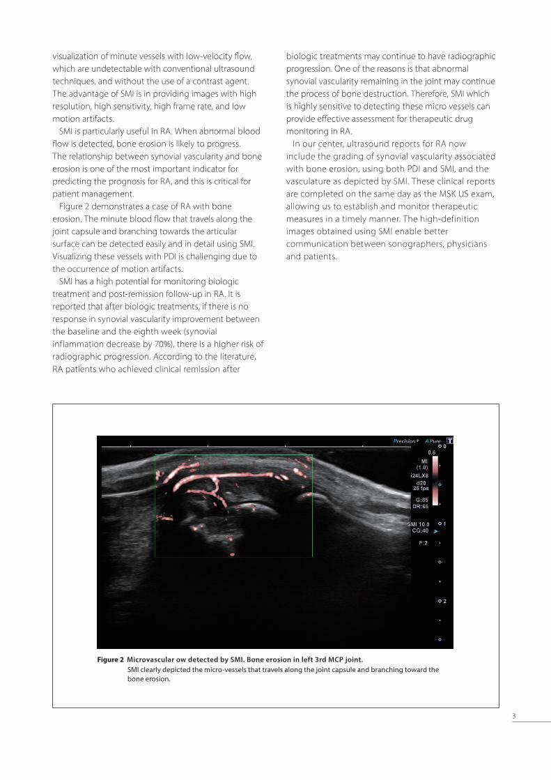
Figure 2 demonstrates a case of RA with bone erosion. The minute blood flow that travels along the joint capsule and branching towards the articular surface can be detected easily and in detail using SMI. Visualizing these vessels with PDI is challenging due to the occurrence of motion artifacts.
SMI has a high potential for monitoring biologic treatment and post-remission follow-up in RA. It is reported that after biologic treatments, if there is no response in synovial vascularity improvement between the baseline and the eighth week (synovial inflammation decrease by 70%), there is a higher risk of radiographic progression. According to the literature, RA patients who achieved clinical remission after
biologic treatments may continue to have radiographic progression. One of the reasons is that abnormal synovial vascularity remaining in the joint may continue the process of bone destruction. Therefore, SMI which is highly sensitive to detecting these micro vessels can provide effective assessment for therapeutic drug monitoring in RA.
In our center, ultrasound reports for RA now include the grading of synovial vascularity associated with bone erosion, using both PDI and SMI, and the vasculature as depicted by SMI. These clinical reports are completed on the same day as the MSK US exam, allowing us to establish and monitor therapeutic measures in a timely manner. The high-definition images obtained using SMI enable better communication between sonographers, physicians and patients.
Figure 2 Microvascular ow detected by SMI. Bone erosion in left 3rd MCP joint.SMI clearly depicted the micro-vessels that travels along the joint capsule and branching toward the bone erosion.
4
Artifact reduction with 24 MHz ultra-high frequency linear transducer
Additional benefits of SMI when combined with the 24 MHz ultra-high frequency linear transducer, PLI-2004BX, are expected, particularly for RA assessment (Figure 3). The newly designed probe has a higher frequency bandwidth compared with conventional transducers, generating crystal-clear images of anatomical structures including muscles, tendons and joints (Figure 4).
The grayscale images coming from the 24 MHz linear probe are exceptional. As SMI offers the benefit of distinct reduction of artifacts and noise, clinicians can perform a diagnosis more quick and easily. When examining RA, the synovial thickening and vascular abnormalities can be detected promptly.
The advanced 24 MHz linear probe is a 1.5D array transducer equipped with Intelligent Dynamic Micro-slice technology (iDMS). iDMS provides
high-flexibility electronic focusing in the lens direction and generates a thin slice beam with continuous focus at all depths. This sharp and homogeneous beam delivers images with high sensitivity, more contrast and elevation resolution.
With its higher axial, lateral and elevational resolution, the ultra-high frequency probe is capable of delivering unprecedented image quality when examining cortical bone, cartilage, and adipose tissues. Compared with conventional probes, the 24 MHz transducer is able to delineate the uneven, hypoechoic synovial thickening in great detail (Figure 3b). The application of 24 MHz transducer on superficial soft-tissue lesions such as lipomas, fibromas, rheumatoid nodules, and cellulitis are also anticipated.
3D imaging with precise positional information using Smart Sensor 3D
3D imaging of synovial vasculature can be useful for grading RA. On the Aplio platform, an application called Smart 3D can be used to obtain 3D images by free-hand scanning using 2D transducers. However, it can be difficult to ensure inter-operator reliability when scanning joints of RA patients with irregular fingers. To acquire accurate rendering, it is necessary to place the probe firmly on the fingers, but this can cause discomfort for the patient.
A newly developed volume rendering technology called Smart Sensor 3D, works in a similar way, but provides more clinical value to 3D MSK US. High resolution 3D images with precise positional
Figure 3 24 MHz ultra-high frequency linear probe PLI-2004BX
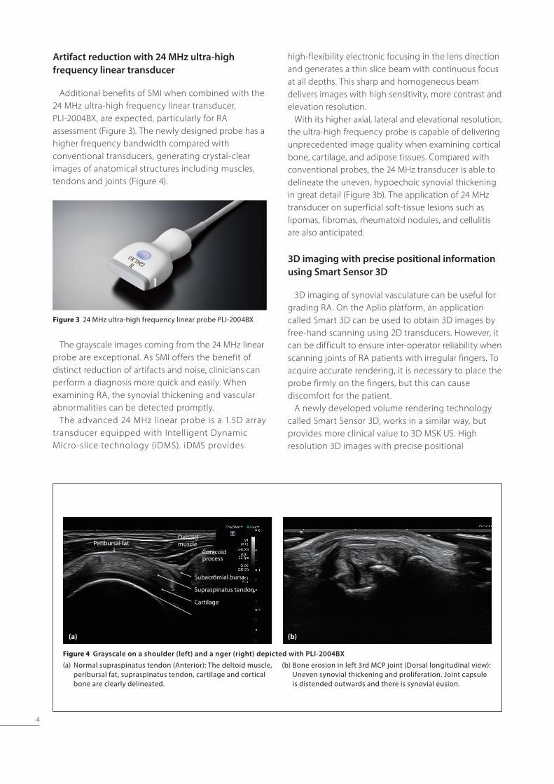
Figure 4 Grayscale on a shoulder (left) and a nger (right) depicted with PLI-2004BX
(a) Normal supraspinatus tendon (Anterior): The deltoid muscle, peribursal fat, supraspinatus tendon, cartilage and cortical bone are clearly delineated.
(b) Bone erosion in left 3rd MCP joint (Dorsal longitudinal view): Uneven synovial thickening and proliferation. Joint capsule is distended outwards and there is synovial eusion.
(a) (b)
Peribursal fatDeltoid muscle
Coracoid process
Subacromial bursa
Supraspinatus tendon
Cartilage
5
information can be acquired utilizing a magnetic field transmitter and a sensor. When incorporating SMI with Smart Sensor 3D, the synovial vasculature can be reconstructed with high sensitivity and accuracy (Figure 5, 6). Thus enabling a more accurate diagnosis and effective treatment evaluation. It also optimizes communication between physicians and sonographers for surgical planning.
Clinical Benets from innovational technologies in MSK US
Optimal patient care can be delivered when there is a more effective communication across health care team. One of the most important benefit in MSK US offered by the improvement of ultrasound technologies is the reinforcement of communication between physicians and sonographers. The improvements in system
architecture, imaging technologies, and innovative applications enable sonographers to perform RA assessment with higher diagnostic accuracy and confidence. The extraordinary image quality obtained by the 24 MHz transducer and SMI allows more detailed observation of minute vasculature, previously unseen with ultrasound. Scoring of RA utilizing both power Doppler and SMI are now standard included in MSK US reports, providing additional information for RA prognosis. Incorporating MSK US with disease activity score (DAS28 and CDAI), blood test results and visual analogue scale (VAS), optimal RA management can be established.
Currently, our protocol suggests scanning at 26 locations. With rapid development of ultrasound technologies, we are looking forward to assessing RA at only one joint.
Figure 5 Synovitis in right 2nd MCP joint (Dorsal longitudinal view)
(a) 2D mSMI (b) Smart Sensor 3D with cSMIVascular abnormalities of synovitis were detected with high sensitivity and continuity
Figure 6 Synovitis in right 3rd MCP joint (Dorsal longitudinal view)
(a) 2D SMI (b) Smart Sensor 3D with SMIAlthough erosion of cortical bone is absent, vessels are concentrated at the position of irregular bone lining.3D SMI has a higher detectability of vasculature.
(a)
(a)
(b)
(b)
6
References
1. Fukae, Jun, et al. "Change of synovial vascularity in a single
finger joint assessed by power Doppler sonography correlated
with radiographic change in rheumatoid arthritis: comparative
study of a novel quantitative score with a semiquantitative
score." Arthritis care & research 62.5 (2010): 657-663.
2. Fukae, Jun, et al. "Radiographic prognosis of finger joint
damage predicted by early alteration in synovial vascularity in
patients with rheumatoid arthritis: potential utility of power
Doppler sonography in clinical practice." Arthritis care & research