POSITION PAPER Clinician’ s Guide to Prevention and Treatment of Osteoporosis F. Cosman & S. J. de Beur & M. S. LeBoff & E. M. Lewiecki & B. Tanner & S. Randall & R. Lindsay Received: 12 June 2014 /Accepted: 24 June 2014 /Published online: 15 August 2014 # The Author(s) 2014. This article is published with open access at Springerlink.com Abstract The Clinician’ s Guide to Prevention and Treatment of Osteoporosis was developed by an expert committee of the National Osteoporosis Foundation (NOF) in collaboration with a multispecialty council of medical experts in the field of bone health convened by NOF. Readers are urged to consult current prescribing information on any drug, device, or pro- cedure discussed in this publication. Keywords Diagnosis . Guide . Osteoporosis . Prevention . Treatment Executive summary Osteoporosis is a silent disease until it is complicated by fractures—fractures that occur following minimal trauma or, in some cases, with no trauma. Fractures are common and place an enormous medical and personal burden on the aging individuals who suffer them and take a major economic toll on the nation. Osteoporosis can be prevented, diagnosed, and treated before fractures occur. Importantly, even after the first fracture has occurred, there are effective treatments to de- crease the risk of further fractures. Prevention, detection, and treatment of osteoporosis should be a mandate of primary care providers. Since the National Osteoporosis Foundation (NOF) first published the Guide in 1999, it has become increasingly clear that many patients are not being given appropriate information about prevention and many patients are not receiving appro- priate testing to diagnose osteoporosis or establish osteoporo- sis risk. Most importantly, many patients who have osteoporosis-related fractures are not being diagnosed with osteoporosis and are not receiving any of the Food and Drug Administration (FDA)-approved, effective therapies. This Guide offers concise recommendations regarding pre- vention, risk assessment, diagnosis, and treatment of osteopo- rosis in postmenopausal women and men age 50 and older. It includes indications for bone densitometry and fracture risk thresholds for intervention with pharmacologic agents. The absolute risk thresholds at which consideration of osteoporo- sis treatment is recommended were guided by a cost- effectiveness analysis. Synopsis of major recommendations to the clinician Recommendations apply to postmenopausal women and men age 50 and older. Universal recommendations & Counsel on the risk of osteoporosis and related fractures. & Advise on a diet that includes adequate amounts of total calcium intake (1000 mg/day for men 50–70; 1200 mg/day F. Cosman (*) : R. Lindsay Helen Hayes Hospital, West Haverstraw, NY, USA e-mail: [email protected]S. J. de Beur Johns Hopkins Bayview Medical Center, Baltimore, MD, USA M. S. LeBoff Brigham and Women’ s Hospital, Boston, MA, USA E. M. Lewiecki New Mexico Clinical Research and Osteoporosis Center, Albuquerque, NM, USA B. Tanner Vanderbilt University Medical Center, Nashville, TN, USA S. Randall National Osteoporosis Foundation, Washington, DC, USA F. Cosman : R. Lindsay Department of Medicine, Columbia University, New York, NY, USA Osteoporos Int (2014) 25:2359–2381 DOI 10.1007/s00198-014-2794-2
Transcript
POSITION PAPER
Clinician’s Guide to Prevention and Treatment of Osteoporosis
F. Cosman & S. J. de Beur &M. S. LeBoff &E.M. Lewiecki &B. Tanner & S. Randall & R. Lindsay
Received: 12 June 2014 /Accepted: 24 June 2014 /Published online: 15 August 2014# The Author(s) 2014. This article is published with open access at Springerlink.com
Abstract The Clinician’s Guide to Prevention and Treatmentof Osteoporosis was developed by an expert committee of theNational Osteoporosis Foundation (NOF) in collaborationwith a multispecialty council of medical experts in the fieldof bone health convened byNOF. Readers are urged to consultcurrent prescribing information on any drug, device, or pro-cedure discussed in this publication.
Osteoporosis is a silent disease until it is complicated byfractures—fractures that occur following minimal trauma or,in some cases, with no trauma. Fractures are common and
place an enormous medical and personal burden on the agingindividuals who suffer them and take a major economic toll onthe nation. Osteoporosis can be prevented, diagnosed, andtreated before fractures occur. Importantly, even after the firstfracture has occurred, there are effective treatments to de-crease the risk of further fractures. Prevention, detection, andtreatment of osteoporosis should be a mandate of primary careproviders.
Since the National Osteoporosis Foundation (NOF) firstpublished the Guide in 1999, it has become increasingly clearthat many patients are not being given appropriate informationabout prevention and many patients are not receiving appro-priate testing to diagnose osteoporosis or establish osteoporo-sis risk. Most importantly, many patients who haveosteoporosis-related fractures are not being diagnosed withosteoporosis and are not receiving any of the Food and DrugAdministration (FDA)-approved, effective therapies.
This Guide offers concise recommendations regarding pre-vention, risk assessment, diagnosis, and treatment of osteopo-rosis in postmenopausal women and men age 50 and older. Itincludes indications for bone densitometry and fracture riskthresholds for intervention with pharmacologic agents. Theabsolute risk thresholds at which consideration of osteoporo-sis treatment is recommended were guided by a cost-effectiveness analysis.
Synopsis of major recommendations to the clinician
Recommendations apply to postmenopausal women and menage 50 and older.
Universal recommendations
& Counsel on the risk of osteoporosis and related fractures.& Advise on a diet that includes adequate amounts of total
calcium intake (1000mg/day for men 50–70; 1200mg/day
F. Cosman (*) : R. LindsayHelen Hayes Hospital, West Haverstraw, NY, USAe-mail: [email protected]
S. J. de BeurJohns Hopkins Bayview Medical Center, Baltimore, MD, USA
M. S. LeBoffBrigham and Women’s Hospital, Boston, MA, USA
E. M. LewieckiNew Mexico Clinical Research and Osteoporosis Center,Albuquerque, NM, USA
B. TannerVanderbilt University Medical Center, Nashville, TN, USA
S. RandallNational Osteoporosis Foundation, Washington, DC, USA
F. Cosman : R. LindsayDepartment ofMedicine, Columbia University, New York, NY, USA
Osteoporos Int (2014) 25:2359–2381DOI 10.1007/s00198-014-2794-2
for women 51 and older and men 71 and older), incorpo-rating dietary supplements if diet is insufficient.
& Advise on vitamin D intake (800–1000 IU/day), includingsupplements if necessary for individuals age 50 and older.
& Recommend regular weight-bearing and muscle-strengthening exercise to improve agility, strength, pos-ture, and balance; maintain or improve bone strength; andreduce the risk of falls and fractures.
& Assess risk factors for falls and offer appropriate modifi-cations (e.g., home safety assessment, balance trainingexercises, correction of vitamin D insufficiency, avoid-ance of central nervous system depressant medications,careful monitoring of antihypertensive medication, andvisual correction when needed).
& Advise on cessation of tobacco smoking and avoidance ofexcessive alcohol intake.
Diagnostic assessment
& Measure height annually, preferably with a wall-mountedstadiometer.
& Bone mineral density (BMD) testing should be performed:
– In women age 65 and older and men age 70 and older– In postmenopausal women and men above age 50–69,
based on risk factor profile– In postmenopausal women and men age 50 and older
who have had an adult age fracture, to diagnose anddetermine degree of osteoporosis
– In all women age 70 and older and all men age 80 andolder if BMD T-score is ≤−1.0 at the spine, total hip, orfemoral neck
– In women age 65 to 69 and men age 70 to 79 if BMD T-score is ≤−1.5 at the spine, total hip, or femoral neck
– In postmenopausal women and men age 50 and olderwith specific risk factors:
& Low-trauma fracture during adulthood (age 50 and older)& Historical height loss (difference between the current
height and peak height at age 20) of 1.5 in. or more (4 cm)& Prospective height loss (difference between the current
height and a previously documented height measurement)of 0.8 in. or more (2 cm)
& Recent or ongoing long-term glucocorticoid treatment– If bone density testing is not available, vertebral imaging
may be considered based on age alone.
& Check for secondary causes of osteoporosis.
& Biochemical markers of bone turnover can aid in riskassessment and serve as an additional monitoring toolwhen treatment is initiated.
Monitoring patients
& Perform BMD testing 1 to 2 years after initiating medicaltherapy for osteoporosis and every 2 years thereafter.
– More frequent BMD testing may be warranted in certainclinical situations.
– The interval between repeat BMD screenings may be lon-ger for patients without major risk factors and who have aninitial T-score in the normal or upper low bone mass range.
& Biochemical markers can be repeated to determine iftreatment is producing expected effect.
Pharmacologic treatment recommendations
& Initiate pharmacologic treatment:
– In those with hip or vertebral (clinical or asymptomatic)fractures
– In those with T-scores ≤−2.5 at the femoral neck, totalhip, or lumbar spine by DXA
– In postmenopausal women and men age 50 and olderwith low bone mass (T-score between −1.0 and −2.5,osteopenia) at the femoral neck, total hip, or lumbar spineby DXA and a 10-year hip fracture probability ≥3 % or a10-year major osteoporosis-related fracture probability≥20% based on the USA-adaptedWHO absolute fracturerisk model (Fracture Risk Algorithm (FRAX®); www.NOF.org and www.shef.ac.uk/FRAX)
& Current FDA-approved pharmacologic options for osteo-porosis are bisphosphonates (alendronate, ibandronate,risedronate, and zoledronic acid), calcitonin, estrogenagonist/antagonist (raloxifene), estrogens and/or hormonetherapy, tissue-selective estrogen complex (conjugatedestrogens/bazedoxifene), parathyroid hormone 1–34(teriparatide), and receptor activator of nuclear factorkappa-B (RANK) ligand inhibitor (denosumab).
& No pharmacologic therapy should be considered indefi-nite in duration. After the initial treatment period, whichdepends on the pharmacologic agent, a comprehensiverisk assessment should be performed. There is no uniformrecommendation that applies to all patients and durationdecisions need to be individualized.
& In adults age 50 and older, after a fracture, institute appropri-ate risk assessment and treatment measures for osteoporosisas indicated. Fracture liaison service (FLS) programs, where
patients with recent fractures may be referred for care coor-dination and transition management, have demonstrated im-provement in the quality of care delivered.
Osteoporosis: impact and overview
Scope of the problem
Osteoporosis is the most common bone disease in humans,representing a major public health problem as outlined inBone Health and Osteoporosis: A Report of the SurgeonGeneral (2004) [1]. It is characterized by low bone mass,deterioration of bone tissue and disruption of bone architec-ture, compromised bone strength, and an increase in the risk offracture. According to the WHO diagnostic classification,osteoporosis is defined by BMD at the hip or lumbar spinethat is less than or equal to 2.5 standard deviations below themean BMD of a young-adult reference population. Osteopo-rosis is a risk factor for fracture just as hypertension is forstroke. The risk of fractures is highest in those with the lowestBMD; however, the majority of fractures occur in patientswith low bone mass rather than osteoporosis, because of thelarge number of individuals with bone mass in this range.
Osteoporosis affects an enormous number of people, of bothsexes and all races, and its prevalence will increase as thepopulation ages. Based on data from the National Health andNutrition Examination Survey III (NHANES III), NOF hasestimated that more than 9.9 million Americans have osteopo-rosis and an additional 43.1 million have low bone density [2].About one out of every two Caucasian women will experiencean osteoporosis-related fracture at some point in her lifetime, aswill approximately one in five men [1]. Although osteoporosisis less frequent in African Americans, those with osteoporosishave the same elevated fracture risk as Caucasians.
Medical impact
Fractures and their complications are the relevant clinical se-quelae of osteoporosis. Themost common fractures are those ofthe vertebrae (spine), proximal femur (hip), and distal forearm(wrist). However, most fractures in older adults are due at leastin part to low bone mass, even when they result from consid-erable trauma. A recent fracture at any major skeletal site in anadult older than 50 years of age should be considered a signif-icant event for the diagnosis of osteoporosis and provides asense of urgency for further assessment and treatment. Themost notable exceptions are those of the fingers, toes, face,and skull, which are primarily related to trauma rather thanunderlying bone strength. Fractures may be followed by fullrecovery or by chronic pain, disability, and death [3].
Hip fractures are associated with an 8 to 36 % excessmortality within 1 year, with a higher mortality in men than
in women [4]; additionally, hip fractures are followed by a 2.5-fold increased risk of future fractures [5]. Approximately 20%of hip fracture patients require long-term nursing home care,and only 40 % fully regain their pre-fracture level of indepen-dence [1]. Although the majority of vertebral fractures areinitially clinically silent, these fractures are often associatedwith symptoms of pain, disability, deformity, and mortality[3]. Postural changes associated with kyphosis may limitactivity, including bending and reaching.
Multiple thoracic fractures may result in restrictive lungdisease, and lumbar fractures may alter abdominal anatomy,leading to constipation, abdominal pain, distention, reducedappetite, and premature satiety. Vertebral fractures, whetherclinically apparent or silent, are major predictors of futurefracture risk, up to 5-fold for subsequent vertebral fractureand 2- to 3-fold for fractures at other sites. Wrist fractures areless disabling but can interfere with some activities of dailyliving as much as hip or vertebral fractures.
Pelvic fractures and humerus fractures are also commonand contribute to increased morbidity and mortality. Fracturescan also cause psychosocial symptoms, most notably depres-sion and loss of self-esteem, as patients grapple with pain,physical limitations, and lifestyle and cosmetic changes.
Economic toll
Annually, two million fractures are attributed to osteoporosis,causing more than 432,000 hospital admissions, almost 2.5million medical office visits, and about 180,000 nursing homeadmissions in the USA [1]. Medicare currently pays for approx-imately 80% of these fractures, with hip fractures accounting for72 % of fracture costs. Due in part to an aging population, thecost of care is expected to rise to $25.3 billion by 2025 [6].
Despite the availability of cost-effective and well-toleratedtreatments to reduce fracture risk, only 23% of women age 67or older who have an osteoporosis-related fracture receiveeither a BMD test or a prescription for a drug to treat osteo-porosis in the 6 months after the fracture [7].
Basic pathophysiology
Bonemass in older adults equals the peak bonemass achievedby age 18–25 minus the amount of bone subsequently lost.Peak bone mass is determined largely by genetic factors, withcontributions from nutrition, endocrine status, physical activ-ity, and health during growth [8].
The process of bone remodeling that maintains a healthyskeleton may be considered a preventive maintenance pro-gram, continually removing older bone and replacing it withnew bone. Bone loss occurs when this balance is altered,resulting in greater bone removal than replacement. The
Osteoporos Int (2014) 25:2359–2381 2361
imbalance occurs with menopause and advancing age. Withthe onset ofmenopause, the rate of bone remodeling increases,magnifying the impact of the remodeling imbalance. The lossof bone tissue leads to disordered skeletal architecture and anincrease in fracture risk.
Figure 1 shows the changes within cancellous bone as aconsequence of bone loss. Individual trabecular plates of boneare lost, leaving an architecturally weakened structure withsignificantly reduced mass. Increasing evidence suggests thatrapid bone remodeling (as measured by biochemical markersof bone resorption or formation) increases bone fragility andfracture risk.
Bone loss leads to an increased risk of fracture that ismagnified by other aging-associated declines in functioning.Figure 2 shows the factors associated with an increased risk ofosteoporosis-related fractures. These include general factorsthat relate to aging and sex steroid deficiency, as well asspecific risk factors, such as use of glucocorticoids, whichcause decreased bone formation and bone loss, reduced bonequality, and disruption of microarchitectural integrity. Frac-tures result when weakened bone is overloaded, often by fallsor certain activities of daily living.
Approach to the diagnosis and managementof osteoporosis
NOF recommends a comprehensive approach to the diagnosisand management of osteoporosis. A detailed history andphysical examination together with BMD assessment, verte-bral imaging to diagnose vertebral fractures, and, when ap-propriate, the WHO 10-year estimated fracture probability areutilized to establish the individual patient’s fracture risk [11].Therapeutic intervention thresholds are based on NOF’s eco-nomic analysis that takes into consideration the cost-effectiveness of treatments and competition for resources inthe USA [12, 13]. The clinician’s clinical skills and pastexperience, incorporating the best patient-based researchavailable, are used to determine the appropriate therapeuticintervention. The potential risks and benefits of all osteoporo-sis interventions should be reviewed with patients and the
unique concerns and expectations of individual patients con-sidered in any final therapeutic decision.
Risk assessment
All postmenopausal women and men age 50 and older shouldbe evaluated for osteoporosis risk in order to determine theneed for BMD testing and/or vertebral imaging. In general, themore risk factors that are present, the greater is the risk offracture. Osteoporosis is preventable and treatable, but be-cause there are no warning signs prior to a fracture, manypeople are not being diagnosed in time to receive effectivetherapy during the early phase of the disease. Many factorshave been associated with an increased risk of osteoporosis-related fracture (Table 1).
Since the majority of osteoporosis-related fractures resultfrom falls, it is also important to evaluate risk factors forfalling (Table 2). The most important of these are personalhistory of falling, muscle weakness and gait, selected medi-cations, balance, and visual deficits [15]. Dehydration is also arisk factor for falls.
Several of these risk factors have been included in theWHO10-year fracture risk model (Table 3). As suggested by theWHO [11], this set of risk factors increases fracture risk inde-pendently of BMD and can be combined with BMD measure-ments to assess an individual patient’s risk of future fracture.
Diagnostic assessment
Consider the possibility of osteoporosis and fracture risk basedon the presence of the risk factors and conditions outlined inTables 1 and 3.Metabolic bone diseases other than osteoporosis,such as hyperparathyroidism or osteomalacia, may be associat-ed with low BMD. Many of these diseases have very specifictherapies, and it is appropriate to complete a history and phys-ical examination before making a diagnosis of osteoporosis onthe basis of a low BMD alone. In patients in whom a specificsecondary, treatable cause of osteoporosis is being considered(Table 1), relevant blood and urine studies (see below) should beobtained prior to initiating therapy. Any adulthood fracture maybe an indication of osteoporosis and should be evaluated ac-cordingly. Consider hip and vertebral fractures as indications of
Normal bone Osteoporo�c bone
Fig. 1 Micrographs of normal vs.osteoporotic bone [9], fromDempster et al., with permissionof The American Society forBone and Mineral Research [9]
2362 Osteoporos Int (2014) 25:2359–2381
osteoporosis unless excluded by the clinical evaluation andimaging. Fractures present a sense of urgency as they signifyincreased fracture risk over the subsequent 5 years [16]. Patientswith recent fractures, multiple fractures, or very low BMDshould be evaluated for secondary etiologies.
Osteoporosis affects a significant number of men, yet thecondition often goes undetected and untreated. The evaluationof osteoporosis inmen requires special consideration as some ofthe laboratory testing to assess underlying causes in men differsfrom those in women. Screening BMD and vertebral imagingrecommendations for men are outlined in Table 8. The 2012Endocrine Society’s Osteoporosis in Men: An Endocrine So-ciety Clinical Practice Guideline provides a detailed approachto the evaluation and treatment of osteoporosis in men [17].
Diagnosis
The diagnosis of osteoporosis is established by measurementof BMD or by the occurrence of adulthood hip or vertebralfracture in the absence of major trauma (such as a motorvehicle accident or multiple story fall). Laboratory testing isindicated to exclude secondary causes of osteoporosis [1, 14,17] (Table 4).
BMD measurement and classification
DXAmeasurement of the hip and spine is the technology usedto establish or confirm a diagnosis of osteoporosis, predictfuture fracture risk, and monitor patients. Areal BMD isexpressed in absolute terms of grams of mineral per squarecentimeter scanned (g/cm2) and as a relationship to twonorms: compared to the BMD of an age-, sex-, andethnicity-matched reference population (Z-score) or comparedto a young-adult reference population of the same sex (T-score). The difference between the patient’s BMD and themean BMD of the reference population, divided by the stan-dard deviation (SD) of the reference population, is used tocalculate T-scores and Z-scores. Peak bone mass is achievedin early adulthood, followed by a decline in BMD. The rate ofbone loss accelerates in women at menopause and continuesto progress at a slower pace in older postmenopausal women(see Fig. 3) and in older men. An individual’s BMD is
presented as the standard deviation above or below the meanBMD of the reference population, as outlined in Table 5. TheBMD diagnosis of normal, low bone mass (osteopenia), oste-oporosis, and severe or established osteoporosis is based onthe WHO diagnostic classification (Table 5) [18].
BMD testing is a vital component in the diagnosis andmanagement of osteoporosis. BMD has been shown to corre-late with bone strength and is an excellent predictor of futurefracture risk. Instead of a specific threshold, fracture riskincreases exponentially as BMD decreases. Although avail-able technologies measuring central (lumbar spine and hip)and peripheral skeletal sites (forearm, heel, fingers) providesite-specific and global (overall risk at any skeletal site) as-sessment of future fracture risk, DXA measurement at the hipis the best predictor of future hip fracture risk. DXA measure-ments of the lumbar spine and hip must be performed byappropriately trained technologists on properly maintainedinstruments. DXA scans are associated with exposure to triv-ial amounts of radiation.
In postmenopausal women and men age 50 and older, theWHO diagnostic T-score criteria (normal, low bone mass, andosteoporosis) are applied to BMD measurement by centralDXA at the lumbar spine and femoral neck [18]. BMD mea-sured by DXA at the one-third (33 %) radius site can be usedfor diagnosing osteoporosis when the hip and lumbar spinecannot be measured or are unusable or uninterpretable [19]. Inpremenopausal women, men less than 50 years of age, andchildren, the WHO BMD diagnostic classification should notbe applied. In these groups, the diagnosis of osteoporosisshould not be made on the basis of densitometric criteriaalone. The International Society for Clinical Densitometry(ISCD) recommends that instead of T-scores, ethnic or race-adjusted Z-scores should be used, with Z-scores of −2.0 orlower defined as either “low bone mineral density for chrono-logical age” or “below the expected range for age” and thoseabove −2.0 being “within the expected range for age” [19].
Who should be tested?
The decision to perform bone density assessment should bebased on an individual’s fracture risk profile and skeletalhealth assessment. Utilizing any procedure to measure bone
Fig. 2 Pathogenesis ofosteoporosis-related fractures,from Cooper and Melton, withmodification [10]
Osteoporos Int (2014) 25:2359–2381 2363
density is not indicated unless the results will influence thepatient’s treatment decision. The US Preventive Services TaskForce recommends testing of all women age 65 and older and
younger women whose fracture risk is equal to or greater thanthat of a 65-year-old white woman who has no additional riskfactors [20].
Table 1 Conditions, diseases, and medications that cause or contribute to osteoporosis and fractures
Lifestyle factors
Alcohol abuse Excessive thinness Excess vitamin A
Frequent falling High salt intake Immobilization
Inadequate physical activity Low calcium intake Smoking (active or passive)
Tamoxifen® (premenopausal use) Thiazolidinediones (such as Actos® and Avandia®) Thyroid hormones (in excess)
From: The Surgeon General’s Report [1], with modification
2364 Osteoporos Int (2014) 25:2359–2381
Table 6 outlines the indications for BMD testing. BMDmeasurement is not recommended in children or adolescentsand is not routinely indicated in healthy young men or pre-menopausal women unless there is a significant fracture his-tory or there are specific risk factors for bone loss.
Vertebral imaging
A vertebral fracture is consistent with a diagnosis of osteopo-rosis, even in the absence of a bone density diagnosis, and isan indication for pharmacologic treatment with osteoporosismedication to reduce subsequent fracture risk [18, 21]. Mostvertebral fractures are asymptomatic when they first occur andoften are undiagnosed for many years. Proactive vertebralimaging is the only way to diagnose these fractures. The
finding of a previously unrecognized vertebral fracture maychange the diagnostic classification, alter future fracture riskcalculations, and affect treatment decisions [22].
Independent of BMD, age, and other clinical risk factors,radiographically confirmed vertebral fractures (even ifcompletely asymptomatic) are a sign of impaired bone qualityand strength and a strong predictor of new vertebral and otherfractures. The presence of a single vertebral fracture increasesthe risk of subsequent fractures 5-fold and the risk of hip andother fractures 2- to 3- fold [23]. Vertebral imaging can beperformed using a lateral thoracic and lumbar spine X-ray orlateral vertebral fracture assessment (VFA), available on mostmodern DXA machines. VFA can be conveniently performedat the time of BMD assessment, while conventional X-raymay require referral to a standard X-ray facility.
Table 2 Risk factors for falls
From: Health Professional’sGuide to the Rehabilitation of thePatient with Osteoporosis [14]
Environmental risk factors
Lack of assistive devices in bathrooms Obstacles in the walking path
Loose throw rugs Slippery conditions
Low level lighting
Medical risk factors
Age Medications causing sedation (narcoticanalgesics, anticonvulsants, psychotropics)
Anxiety and agitation Orthostatic hypotension
Arrhythmias Poor vision
Dehydration Previous falls or fear of falling
Depression Reduced problem solving or mental acuityand diminished cognitive skills
Vitamin D insufficiency [serum 25-hydroxyvitamin D(25(OH)D)<30 ng/ml (75 nmol/L)]
Urgent urinary incontinence
Malnutrition
Neurological and musculoskeletal risk factors
Kyphosis Reduced proprioception
Poor balance Weak muscles/sarcopenia
Impaired transfer and mobility Deconditioning
Diseases listed in Table 1
Table 3 Risk factors included inthe WHO Fracture Risk Assess-ment Model
From: WHO Technical Report[11]
Clinical risk factors included in the FRAX Tool
Current age Rheumatoid arthritis
Gender Secondary causes of osteoporosis: type 1(insulin dependent) diabetes, osteogenesisimperfecta in adults, untreated long-standinghyperthyroidism, hypogonadism or prematuremenopause (<40 years), chronic malnutritionor malabsorption, and chronic liver disease
A prior osteoporotic fracture (including clinicaland asymptomatic vertebral fractures)
Parental history of hip fracture
Femoral neck BMD Current smoking
Low body mass index (BMI, kg/m2) Alcohol intake (3 or more drinks/day)
Oral glucocorticoids ≥5 mg/d of prednisonefor >3 months (ever)
Osteoporos Int (2014) 25:2359–2381 2365
Indications for vertebral imaging
Because vertebral fractures are so prevalent in older individ-uals and most produce no acute symptoms, vertebral imagingtests are recommended for the individuals defined in Table 7.
Once a first vertebral imaging test is done, it only needs to berepeated if prospective height loss is documented or new backpain or postural change occurs [3, 24]. A follow-up vertebralimaging test is also recommended in patients who are beingconsidered for a medication holiday, since stopping medica-tion would not be recommended in patients who have recentvertebral fractures.
Biochemical markers of bone turnover
Bone remodeling (or turnover) occurs throughout life to repairfatigue damage and microfractures in bone and to maintainmineral homeostasis. Biochemical markers of bone remodel-ing [e.g., resorption markers—serum C-telopeptide (CTX)and urinary N-telopeptide (NTX)—and formation markers—serum bone-specific alkaline phosphatase (BSAP),osteocalcin (OC), and aminoterminal propeptide of type Iprocollagen (PINP)] are best collected in the morning whilepatients are fasting.
Biochemical markers of bone turnover may [25]:
& Predict risk of fracture independently of bone density inuntreated patients
& Predict rapidity of bone loss in untreated patients& Predict extent of fracture risk reduction when repeated after
3–6 months of treatment with FDA-approved therapies& Predict magnitude of BMD increases with FDA-approved
therapies& Help determine adequacy of patient compliance and per-
sistence with osteoporosis therapy& Help determine duration of “drug holiday” and when and
if medication should be restarted. (Data are quite limited tosupport this use, but studies are underway.)
Use of WHO FRAX® in the USA
FRAX®was developed to calculate the 10-year probability ofa hip fracture and the 10-year probability of a major osteopo-rotic fracture (defined as clinical vertebral, hip, forearm, orproximal humerus fracture), taking into account femoral neckBMD and the clinical risk factors shown in Table 3 [11]. TheFRAX® algorithm is available at www.nof.org as well as atwww.shef.ac.uk/FRAX. It is also available on newer DXAmachines or with software upgrades that provide the FRAX®scores on the bone density report.
The WHO algorithm used in this Guide was calibrated toUS fracture and mortality rates; therefore, the fracture riskfigures herein are specific for the US population. Economicmodeling was performed to identify the 10-year hip fracturerisk above which it is cost-effective, from the societal perspec-tive, to treat with pharmacologic agents. The US-based eco-nomic modeling is described in one report [12], and the US-
Table 4 Exclusion of secondary causes of osteoporosis
Consider the following diagnostic studies for secondary causes ofosteoporosis
Blood or serum
Complete blood count (CBC)
Chemistry levels (calcium, renal function,phosphorus, and magnesium)
Total testosterone and gonadotropin in younger men
Bone turnover markers
Consider in selected patients
Serum protein electrophoresis (SPEP), serum immunofixation,serum-free light chains
Tissue transglutaminase antibodies (IgA and IgG)
Iron and ferritin levels
Homocysteine
Prolactin
Tryptase
Urine
24-h urinary calcium
Consider in selected patients
Protein electrophoresis (UPEP)
Urinary free cortisol level
Urinary histamine
Fig. 3 Z- and T-scores in women, from ISCD Bone DensitometryClinician Course, Lecture 5 (2008), with permission of the InternationalSociety for Clinical Densitometry
adapted WHO algorithm and its clinical application are illus-trated in a companion report [13].
The latter analyses generally confirm the previous NOFconclusion that it is cost-effective to treat individuals with aprior hip or vertebral fracture and those with a DXA femoralneck T-score ≤−2.5. Previous analyses have established that alumbar spine T-score ≤−2.5 also warrants treatment [26].
FRAX underestimates fracture risk in patients with recentfractures, multiple osteoporosis-related fractures, and those atincreased risk for falling. FRAX® is most useful in patientswith low femoral neck BMD. Utilizing FRAX® in patientswith low BMD at the lumbar spine but a relatively normalBMD at the femoral neck underestimates fracture risk in theseindividuals. Specifically, the WHO algorithm has not beenvalidated for the use of lumbar spine BMD. NOF recommendstreatment of individuals with osteoporosis of the lumbar spineas well as the hip.
Application of US-adapted FRAX® in the USA
& FRAX® is intended for postmenopausal women and menage 50 and older; it is not intended for use in youngeradults or children.
& The FRAX® tool has not been validated in patients cur-rently or previously treated with pharmacotherapy forosteoporosis. In such patients, clinical judgment must beexercised in interpreting FRAX® scores. Patients who
have been off osteoporosis medications for 1 to 2 yearsor more might be considered untreated [27].
& FRAX® can be calculated with either femoral neck BMDor total hip BMD, but, when available, femoral neck BMDis preferred. The use of BMD from nonhip sites is notrecommended.
& The WHO determined that for many secondary causes ofosteoporosis, fracture risk was mediated primarily throughimpact on BMD [28]. For this reason, when femoral neckBMD is inserted into FRAX®, the secondary causes ofosteoporosis button are automatically inactivated.
The therapeutic thresholds proposed in this Guide are forclinical guidance only and are not rules. All treatment decisionsrequire clinical judgment and consideration of individual patientfactors, including patient preferences, comorbidities, risk factorsnot captured in the FRAX® model (e.g., frailty, falls), recentdecline in bone density, and other sources of possible under- oroverestimation of fracture risk by FRAX®.
The therapeutic thresholds do not preclude clinicians orpatients from considering intervention strategies for those who
Table 5 Defining osteoporosis by BMD
WHO definition of osteoporosis based on BMD
Classification BMD T-score
Normal Within 1 SD of the mean level for a young-adultreference population
T-score at −1.0 and above
Low bone mass (osteopenia) Between 1.0 and 2.5 SD below that of the mean level for a young-adult reference population
T-score between −1.0 and −2.5
Osteoporosis 2.5 SD or more below that of the mean level fora young-adult reference population
T-score at or below −2.5
Severe or established osteoporosis 2.5 SD or more below that of the mean level fora young-adult reference population with fractures
T-score at or below −2.5 with one or more fractures
Although these definitions are necessary to establish the presence of osteoporosis, they should not be used as the sole determinant of treatment decisions
Table 6 Indications for BMD testing
Consider BMD testing in the following individuals:
• Women age 65 and older and men age 70 and older, regardless ofclinical risk factors
• Younger postmenopausal women, women in the menopausal transition,and men age 50 to 69 with clinical risk factors for fracture
• Adults who have a fracture at or after age 50
• Adults with a condition (e.g., rheumatoid arthritis) or taking amedication (e.g., glucocorticoids in a daily dose ≥5 mg prednisone orequivalent for ≥3 months) associated with low bone mass or bone loss
Table 7 Indications for vertebral imaging
Consider vertebral imaging tests for the following individualsa:
• All women age 70 and older and all men age 80 and older if BMD T-score at the spine, total hip, or femoral neck is ≤−1.0
•Women age 65 to 69 and men age 70 to 79 if BMD T-score at the spine,total hip, or femoral neck is ≤−1.5
• Postmenopausal women and men age 50 and older with specific riskfactors:
▪ Low-trauma fracture during adulthood (age 50 and older)
▪ Historical height loss of 1.5 in. or more (4 cm)b
▪ Prospective height loss of 0.8 in. or more (2 cm)c
▪ Recent or ongoing long-term glucocorticoid treatment
a If bone density testing is not available, vertebral imaging may beconsidered based on age aloneb Current height compared to peak height during young adulthoodc Cumulative height loss measured during interval medical assessment
Osteoporos Int (2014) 25:2359–2381 2367
do not have osteoporosis by BMD (WHO diagnostic criterionof T-score ≤−2.5), do not meet the cut points after FRAX®, orare not at high enough risk of fracture despite low BMD.Conversely, these recommendations should not mandate treat-ment, particularly in patients with low bone mass above theosteoporosis range. Decisions to treat must still be made on acase-by-case basis.
Additional bone densitometry technologies
The following bone mass measurement technologies includedin Table 8 are capable of predicting both site-specific andoverall fracture risk. When performed according to acceptedstandards, these densitometric techniques are accurate andhighly reproducible [19]. However, T-scores from these tech-nologies cannot be used according to the WHO diagnosticclassification because they are not equivalent to T-scoresderived from DXA.
Universal recommendations for all patients
Several interventions to preserve bone strength can be recom-mended to the general population. These include an adequateintake of calcium and vitamin D, lifelong participation inregular weight-bearing and muscle-strengthening exercise,cessation of tobacco use, identification and treatment of alco-holism, and treatment of risk factors for falling.
Adequate intake of calcium and vitamin D
Advise all individuals to obtain an adequate intake of dietarycalcium. Providing adequate daily calcium and vitamin D is asafe and inexpensive way to help reduce fracture risk. Con-trolled clinical trials have demonstrated that the combinationof supplemental calcium and vitamin D can reduce the risk offracture [29]. A balanced diet rich in low-fat dairy products,fruits, and vegetables provides calcium as well as numerousnutrients needed for good health. If adequate dietary calciumcannot be obtained, dietary supplementation is indicated up tothe recommended daily intake.
Lifelong adequate calcium intake is necessary for the ac-quisition of peak bone mass and subsequent maintenance ofbone health. The skeleton contains 99% of the body’s calciumstores; when the exogenous supply is inadequate, bone tissueis resorbed from the skeleton to maintain serum calcium at aconstant level.
NOF supports Institute of Medicine (IOM) recommenda-tions that men age 50–70 consume 1000 mg/day of calciumand that women age 51 and older and men age 71 and olderconsume 1200 mg/day of calcium [30]. There is no evidencethat calcium intake in excess of these amounts confers
additional bone strength. Intakes in excess of 1200 to1500 mg/day may increase the risk of developing kidneystones, cardiovascular disease, and stroke. The scientific liter-ature is highly controversial in this area [31–34].
Table 9 illustrates a simple method for estimating thecalcium content of a patient’s diet. The average daily dietarycalcium intake in adults age 50 and older is 600 to 700 mg/day. Increasing dietary calcium is the first-line approach, butcalcium supplements should be used when an adequate die-tary intake cannot be achieved.
Vitamin D plays a major role in calcium absorption, bonehealth, muscle performance, balance, and risk of falling. NOFrecommends an intake of 800 to 1000 international units (IU)of vitamin D per day for adults age 50 and older. Institute ofMedicine Dietary Reference Intakes for vitamin D are 600 IU/day until age 70 and 800 IU/day for adults age 71 years andolder [30].
Chief dietary sources of vitamin D include vitamin D-fortified milk (400 IU/quart, although certain products such
Table 8 Additional bone densitometry technologies
CT-based absorptiometry: Quantitative computed tomography (QCT)measures volumetric integral, trabecular, and cortical bone density atthe spine and hip and can be used to determine bone strength, whereaspQCT measures the same at the forearm or tibia. High-resolutionpQCT (HR-pQCT) at the radius and tibia provides measures of volu-metric density, bone structure, and microarchitecture. In postmeno-pausal women, QCT measurement of spine trabecular BMD canpredict vertebral fractures, whereas pQCT of the forearm at theultradistal radius predicts hip but not vertebral fractures. There isinsufficient evidence for fracture prediction in men. QCT and pQCTare associated with greater amounts of radiation exposure than centralDXA or pDXA.
Trabecular Bone Score (TBS) is an FDA-approved technique which isavailable on some densitometers. It may measure themicroarchitectural structure of bone tissue and may improve the abilityto predict the risk of fracture.
The following technologies are often used for community-based screen-ing programs because of the portability of the equipment. Results arenot equivalent to DXA and abnormal results should be confirmed byphysical examination, risk assessment, and central DXA.
Peripheral dual-energy x-ray absorptiometry (pDXA) measures arealbone density of the forearm, finger, or heel. Measurement by validatedpDXA devices can be used to assess vertebral and overall fracture riskin postmenopausal women. There is insufficient evidence for fractureprediction in men. pDXA is associated with exposure to trivialamounts of radiation. pDXA is not appropriate for monitoring BMDafter treatment.
Quantitative ultrasound densitometry (QUS) does not measure BMDdirectly but rather speed of sound (SOS) and/or broadband ultrasoundattenuation (BUA) at the heel, tibia, patella, and other peripheralskeletal sites. A composite parameter using SOS and BUA may beused clinically. Validated heel QUS devices predict fractures in post-menopausal women (vertebral, hip, and overall fracture risk) and inmen 65 and older (hip and nonvertebral fractures). QUS is not asso-ciated with any radiation exposure.
2368 Osteoporos Int (2014) 25:2359–2381
as soymilk are not always supplemented with vitamin D), somefortified juices and cereals (40 to 50 IU/serving or more), saltwater fish, and liver. Some calcium supplements and mostmultivitamin tablets also contain vitamin D. Supplementationwith vitamin D2 (ergocalciferol) or vitamin D3 (cholecalciferol)may be used. Vitamin D2 is derived from plant sources andmaybe used by individuals on a strict vegetarian diet.
Many older patients are at high risk for vitamin D deficien-cy, including patients with malabsorption (e.g., celiac disease)or other intestinal diseases (e.g., inflammatory bowel disease,gastric bypass surgery), chronic renal insufficiency, patientson medications that increase the breakdown of vitamin D(e.g., some antiseizure drugs), housebound patients, chroni-cally ill patients and others with limited sun exposure, indi-viduals with very dark skin, and obese individuals. There isalso a high prevalence of vitamin D deficiency in patients withosteoporosis, especially those with hip fractures, even in pa-tients taking osteoporosis medications [35, 36].
Since vitamin D intakes required to correct vitamin Ddeficiency are so variable among individuals, serum25(OH)D levels should be measured in patients at risk ofdeficiency. Vitamin D supplements should be recommendedin amounts sufficient to bring the serum 25(OH)D level toapproximately 30 ng/ml (75 nmol/L) and a maintenance doserecommended to maintain this level, particularly for individ-uals with osteoporosis. Many patients with osteoporosis willneed more than the general recommendation of 800–1000 IU/day. The safe upper limit for vitamin D intake for the generaladult population was increased to 4000 IU/day in 2010 [30].
Treatment of vitamin D deficiency
Adults who are vitamin D deficient may be treated with50,000 IU of vitamin D2 or vitamin D3 once a week or theequivalent daily dose (7000 IU vitamin D2 or vitamin D3) for8–12 weeks to achieve a 25(OH)D blood level ofapproximately 30 ng/ml. This regimen should be followedby maintenance therapy of 1500–2000 IU/day or whateverdose is needed to maintain the target blood level [37, 38].
Regular weight-bearing and muscle-strengthening exercise
Recommend regular weight-bearing and muscle-strengthening exercise to reduce the risk of falls and fractures[39–42]. Among the many health benefits, weight-bearing andmuscle-strengthening exercise can improve agility, strength,posture, and balance, which may reduce the risk of falls. Inaddition, exercise may modestly increase bone density. NOFstrongly endorses lifelong physical activity at all ages, both forosteoporosis prevention and overall health, as the benefits ofexercise are lost when people stop exercising.
Weight-bearing exercise (in which bones andmuscles workagainst gravity as the feet and legs bear the body’s weight)includes walking, jogging, Tai Chi, stair climbing, dancing,and tennis. Muscle-strengthening exercise includes weighttraining and other resistive exercises, such as yoga, Pilates,and boot camp programs. Before an individual with osteopo-rosis initiates a new vigorous exercise program, such as run-ning or heavy weight-lifting, a clinician’s evaluation isappropriate.
Fall prevention
Major risk factors for falling are shown in Table 2. Inaddition to maintaining adequate vitamin D levels andphysical activity, as described above, several strategieshave been demonstrated to reduce falls. These include,but are not limited to, multifactorial interventions suchas individual risk assessment, Tai Chi and other exerciseprograms, home safety assessment, and modificationespecially when done by an occupational therapist, andgradual withdrawal of psychotropic medication if possi-ble. Appropriate correction of visual impairment mayimprove mobility and reduce risk of falls.
There is a lack of evidence that the use of hip protectors bycommunity-dwelling adults provides statistically significantreduction in the risk of hip or pelvis fractures. Also, there is noevidence that the use of hip protectors reduces the rate of falls.In long-term care or residential care settings, some studies
Table 9 Estimating daily dietary calcium intake
Step 1: Estimate calcium intake from calcium-rich foodsa
Product # of servings/day Estimated calcium/serving, in mg Calcium in mg
Milk (8 oz.) __________ ×300 = __________
Yogurt (6 oz.) __________ ×300 = __________
Cheese (1 oz. or 1 cubic in.) __________ ×200 = __________
Fortified foods or juices __________ ×80 to 1,000b = __________
Subtotal = __________
Step 2: Add 250 mg for nondairy sources to subtotal above +250
Total calcium, in mg = __________
aAbout 75 to 80 % of the calcium consumed in American diets is from dairy productsb Calcium content of fortified foods varies
Osteoporos Int (2014) 25:2359–2381 2369
have shown a marginally significant reduction in hip fracturerisk. There are no serious adverse effects of hip protectors;however, adherence to long-term use is poor [43]. There isadditional uncertainty as to which hip protector to use, as mostof the marketed products have not been tested in randomizedclinical trials.
Cessation of tobacco use and avoidance of excessive alcoholintake
Advise patients to stop tobacco smoking. The use of tobaccoproducts is detrimental to the skeleton as well as to overallhealth [44–47]. NOF strongly encourages a smoking cessationprogram as an osteoporosis intervention.
Recognize and treat patients with excessive alcohol intake.Moderate alcohol intake has no known negative effect onbone and may even be associated with slightly higher bonedensity and lower risk of fracture in postmenopausal women.However, alcohol intake of more than two drinks per day forwomen or three drinks a day for men may be detrimental tobone health, increases the risk of falling, and requires furtherevaluation for possible alcoholism [48].
Pharmacologic therapy
All patients being considered for treatment of osteoporosisshould also be counseled on risk factor reduction including theimportance of calcium, vitamin D, and exercise as part of anytreatment program for osteoporosis. Prior to initiating treat-ment, patients should be evaluated for secondary causes ofosteoporosis and have BMD measurements by central DXA,when available, and vertebral imaging studies when appropri-ate. Biochemical marker levels should be obtained if monitor-ing of treatment effects is planned. An approach to the clinicalassessment of individuals with osteoporosis is outlined inTable 10.
The percentage of risk reductions for vertebral andnonvertebral fractures cited below are those cited in theFDA-approved prescribing information. In the absence ofhead-to-head trials, direct comparisons of risk reductionamong drugs should be avoided.
Who should be considered for treatment?
Postmenopausal women and men age 50 and older presentingwith the following should be considered for treatment:
& A hip or vertebral fracture (clinically apparent or found onvertebral imaging). There are abundant data that patientswith spine and hip fractures will have reduced fracture riskif treated with pharmacologic therapy. This is true for
fracture patients with BMD in both the low bone massand osteoporosis range [49–58]. In patients with a hip orspine fracture, the T-score is not as important as thefracture itself in predicting future risk of fracture andantifracture efficacy from treatment.
& T-score ≤−2.5 at the femoral neck, total hip, or lumbarspine. There is abundant evidence that the elevated risk offracture in patients with osteoporosis by BMD is reducedwith pharmacotherapy [52, 57, 59–70].
& Low bone mass (T-score between −1.0 and −2.5 at thefemoral neck or lumbar spine) and a 10-year probability ofa hip fracture ≥3 % or a 10-year probability of a major
Table 10 Clinical approach to managing osteoporosis in postmenopaus-al women and men age 50 and older
General principles:
• Obtain a detailed patient history pertaining to clinical risk factors forosteoporosis-related fractures and falls
• Perform physical examination and obtain diagnostic studies toevaluate for signs of osteoporosis and its secondary causes
•Modify diet/supplements, lifestyle, and other modifiable clinical riskfactors for fracture
• Estimate patient’s 10-year probability of hip and any majorosteoporosis-related fracture using the US-adapted FRAX and per-form vertebral imagingwhen appropriate to complete risk assessment
• Decisions on whom to treat and how to treat should be based onclinical judgment using this Guide and all available clinicalinformation
Consider FDA-approved medical therapies based on the following:
• Vertebral fracture (clinical or asymptomatic) or hip fracture
• Hip DXA (femoral neck or total hip) or lumbar spine T-score ≤−2.5• Low bone mass (osteopenia) and a US-adapted WHO 10-yearprobability of a hip fracture ≥3 % or 10-year probability of any majorosteoporosis-related fracture ≥20 %
• Patient preferences may indicate treatment for people with 10-yearfracture probabilities above or below these levels
Consider nonmedical therapeutic interventions:
• Modify risk factors related to falling
• Referrals for physical and/or occupational therapy evaluation (e.g.,walking aids and other assistive devices)
•Weight-bearing, muscle-strengthening exercise, and balance training
Follow-up:
• Patients not requiring medical therapies at the time of initialevaluation should be clinically re-evaluated when medically appro-priate
• Patients taking FDA-approved medications should have laboratoryand bone density re-evaluation after 2 years or more frequently whenmedically appropriate
• Vertebral imaging should be repeated if there is documented heightloss, new back pain, postural change, or suspicious finding on chestX-ray, following the last (or first) vertebral imaging test or in patientsbeing considered for a temporary cessation of drug therapy to makesure no new vertebral fractures have occurred in the interval
• Regularly, and at least annually, assess compliance and persistencewith the therapeutic regimen
2370 Osteoporos Int (2014) 25:2359–2381
osteoporosis-related fracture ≥20 % based on the US-adapted WHO algorithm [13, 15, 71, 72].
Although FRAX calculated fracture risk prediction hasbeen confirmed in multiple studies, there are relatively fewdata confirming fracture risk reductions with pharmacothera-py in this group of patients.
US FDA-approved drugs for osteoporosis
Current FDA-approved pharmacologic options for the preven-tion and/or treatment of postmenopausal osteoporosis include,in alphabetical order: bisphosphonates (alendronate,alendronate plus D, ibandronate, risedronate and zoledronicacid), calcitonin, estrogens (estrogen and/or hormone thera-py), estrogen agonist/antagonist (raloxifene), tissue-selectiveestrogen complex (conjugated estrogens/bazedoxifene), para-thyroid hormone (PTH [1–34], teriparatide), and the receptoractivator of nuclear factor kappa-B (RANK) ligand (RANKL)inhibitor denosumab. Please see prescribing information forspecific details of their use.
The antifracture benefits of FDA-approved drugs havemostly been studied in women with postmenopausal osteopo-rosis. There are limited fracture data in glucocorticoid-inducedosteoporosis and in men. FDA-approved osteoporosis treat-ments have been shown to decrease fracture risk in patientswho have had fragility fractures and/or osteoporosis by DXA.Pharmacotherapy may also reduce vertebral fractures in pa-tients with low bone mass (osteopenia) without fractures, butthe evidence supporting overall antifracture benefit is not asstrong. Thus, the clinician should assess the potential benefitsand risks of therapy in each patient and the effectiveness of agiven osteoporosis treatment on reduction of vertebral andnonvertebral fractures.
Note that the intervention thresholds do not take into ac-count the nonskeletal benefits or risks associated with specificdrug use. NOF does not advocate the use of drugs not ap-proved by the FDA for prevention and treatment of osteopo-rosis. Examples of these drugs are listed in Table 11 forinformation only.
Bisphosphonates
Drug efficacy
Alendronate, brand name: Fosamax®, Fosamax Plus D,Binosto™, and generic alendronate Alendronate sodium isapproved by the FDA for the prevention (5 mg daily and35 mg weekly tablets) and treatment (10 mg daily tablet,70 mg weekly tablet, 70 mg weekly tablet with 2,800 or5,600 IU of vitamin D3, and 70 mg effervescent tablet) ofpostmenopausal osteoporosis. Alendronate is also approvedfor treatment to increase bone mass in men with osteoporosis
and for the treatment of osteoporosis in men and womentaking glucocorticoids [73].
Alendronate reduces the incidence of spine and hip frac-tures by about 50 % over 3 years in patients with a priorvertebral fracture or in patients who have osteoporosis at thehip site [49, 59]. It reduces the incidence of vertebral fracturesby 48 % over 3 years in patients without a prior vertebralfracture [74].
Ibandronate, brand name: Boniva® and genericibandronate Ibandronate sodium is approved by the FDAfor the treatment (150 mg monthly tablet and 3 mg every3 months by intravenous injection) of postmenopausal osteo-porosis. Ibandronate is available as a generic preparation inthe USA. The oral preparations are also approved for theprevention of postmenopausal osteoporosis.
Table 11 Non-FDA-approved drugs for osteoporosis
These drugs are listed for information only. Nonapproved agents include:
Calcitriol: This synthetic vitamin D analogue, which promotes calciumabsorption, has been approved by the FDA for managinghypocalcemia and metabolic bone disease in renal dialysis patients. Itis also approved for use in hypoparathyroidism, both surgical andidiopathic, and pseudohypoparathyroidism. No reliable datademonstrate a reduction of risk for osteoporotic fracture.
Genistein: An isoflavone phytoestrogen which is the main ingredient inthe prescription “medical food” product Fosteum® and generallyregarded as safe by the FDA. Genistein may benefit bone health inpostmenopausal women but more data are needed to fully understandits effects on bone health and fracture risk.
Other bisphosphonates (etidronate, pamidronate, tiludronate): Thesemedications vary chemically from alendronate, ibandronate,risedronate, and zoledronic acid but are in the same drug class. At thistime, none is approved for prevention or treatment of osteoporosis.Most of these medications are currently approved for other conditions(e.g., Paget’s disease, hypercalcemia of malignancy, myositisossificans).
PTH (1-84): This medication is approved in some countries in Europe fortreatment of osteoporosis in women. In one clinical study, PTH(1-84)effectively reduced the risk of vertebral fractures at a dose of 100 mcg/day.
Sodium fluoride: Through a process that is still unclear, sodium fluoridestimulates the formation of new bone. The quality of bone mass thusdeveloped is uncertain, and the evidence that fluoride reduces fracturerisk is conflicting and controversial.
Strontium ranelate: This medication is approved for the treatment ofosteoporosis in some countries in Europe. Strontium ranelate reducesthe risk of both spine and nonvertebral fractures, but the mechanism isunclear. Incorporation of strontium into the crystal structure replacingcalciummay be part of its mechanism of effect. These effects have onlybeen documented with the pharmaceutical grade agent produced byServier. This effect has not been studied in nutritional supplementscontaining strontium salts.
Tibolone: Tibolone is a tissue-specific, estrogen-like agent that mayprevent bone loss and reduce menopausal symptoms. It is indicated inEurope for the treatment of vasomotor symptoms of menopause andfor prevention of osteoporosis, but it is not approved for use in theUSA.
Osteoporos Int (2014) 25:2359–2381 2371
Ibandronate reduces the incidence of vertebral fractures byabout 50 % over 3 years, but reduction in risk of nonvertebralfracture with ibandronate has not been documented [50].
Risedronate, brand name: Actonel®, Atelvia™, and genericrisedronate Risedronate sodium is approved by the FDA forthe prevention and treatment (5 mg daily tablet; 35 mg weeklytablet; 35 mg weekly delayed release tablet; 35 mg weeklytablet packaged with six tablets of 500 mg calcium carbonate;75 mg tablets on two consecutive days every month; and150 mg monthly tablet) of postmenopausal osteoporosis.Risedronate is also approved for treatment to increase bonemass in men with osteoporosis and for the prevention andtreatment of osteoporosis in men and women who are eitherinitiating or taking glucocorticoids [75].
Risedronate reduces the incidence of vertebral fractures by41 to 49 % and nonvertebral fractures by 36 % over 3 years,with significant risk reduction occurring within 1 year oftreatment in patients with a prior vertebral fracture [51, 52].
Zoledronic acid, brand name: Reclast® Zoledronic acid isapproved by the FDA for the prevention and treatment (5 mgby intravenous infusion over at least 15 min once yearly fortreatment and once every 2 years for prevention) of osteoporosisin postmenopausal women. It is also approved to improve bonemass in men with osteoporosis and for the prevention andtreatment of osteoporosis in men and women expected to be onglucocorticoid therapy for at least 12 months. Zoledronic acid isalso indicated for the prevention of new clinical fractures inpatients (both women and men) who have recently had a low-trauma (osteoporosis-related) hip fracture [58].
Zoledronic acid reduces the incidence of vertebral fracturesby 70% (with significant reduction at 1 year), hip fractures by41 %, and nonvertebral fractures by 25 % over 3 years inpatients with osteoporosis defined by prevalent vertebral frac-tures and osteoporosis by BMD of the hip [66].
Drug administration
Alendronate (generic and Fosamax) and risedronate (Actonel)tablets must be taken on an empty stomach, first thing in themorning, with 8 oz of plain water (no other liquid). Binosto mustbe dissolved in 4 oz of room temperature water taken on anempty stomach, first thing in the morning. Delayed releaserisedronate (Atelvia) tablets must be taken immediately afterbreakfast with at least 4 oz of plain water (no other liquid). Aftertaking these medications, patients must wait at least 30 minbefore eating, drinking, or taking any other medication. Patientsshould remain upright (sitting or standing) during this interval.
Ibandronate must be taken on an empty stomach, first thingin the morning, with 8 oz of plain water (no other liquid). Aftertaking this medication, patients must remain upright and waitat least 60 min before eating, drinking, or taking any other
medication. Ibandronate, 3 mg/3 ml prefilled syringe, is givenby intravenous injection over 15 to 30 s, once every 3 months.Serum creatinine should be checked before each injection.
Zoledronic acid, 5 mg in 100 ml, is given once yearly oronce every 2 years by intravenous infusion over at least15 min. Patients should be well hydrated and may be pre-treated with acetaminophen to reduce the risk of an acutephase reaction (arthralgia, headache, myalgia, fever). Thesesymptoms occurred in 32% of patients after the first dose, 7 %after the second dose, and 3 % after the third dose.
Drug safety
Side effects are similar for all oral bisphosphonate medica-tions and include gastrointestinal problems such as difficultyswallowing and inflammation of the esophagus and stomach.
All bisphosphonates can affect renal function and arecontraindicated in patients with estimated GFR below30–35 ml/min. Zoledronic acid is contraindicated inpatients with creatinine clearance less than 35 mL/minor in patients with evidence of acute renal impairment.Healthcare professionals should screen patients prior toadministering zoledronic acid in order to identify at-riskpatients and should assess renal function by monitoringcreatinine clearance prior to each dose of zoledronicacid [76]. Eye inflammation can also occur. Any suchcomplication should be reported to the healthcare pro-vider as soon as possible.
There have been rare reports of osteonecrosis of thejaw (ONJ) with long-term use of bisphosphonates forosteoporosis, though ONJ is much more common fol-lowing high-dose intravenous bisphosphonate treatmentfor patients with cancer. The risk of ONJ appears toincrease with duration of treatment beyond 5 years[77].
Although rare, low-trauma atypical femur fracturesmay be associated with the long-term use ofbisphosphonates (e.g., >5 years of use). Pain in thethigh or groin area, which can be bilateral, often pre-cedes these unusual fractures. Patients should be evalu-ated closely for these unusual fractures, including pro-active questioning regarding thigh and groin pain. Forpatients with thigh and groin pain, a stress fracture inthe subtrochanteric region or femoral shaft of the femurmay be present. Bilateral X-ray of the femurs should beordered when an atypical femur fracture is suspected,followed by an MRI or a radionuclide bone scan whenclinical suspicion is high enough [78]. Surgical fixationis required in some cases, whereas medical conservativetreatment is appropriate in other cases. Bisphosphonatesshould be stopped if atypical femur fractures haveoccurred.
2372 Osteoporos Int (2014) 25:2359–2381
Calcitonin
Drug efficacy
Brand name: Miacalcin® or Fortical® and genericcalcitonin Salmon calcitonin is FDA-approved for the treat-ment of osteoporosis in women who are at least 5 yearspostmenopausal when alternative treatments are not suitable.
Miacalcin nasal spray has not been shown to increase bonemineral density in early postmenopausal women.
Calcitonin reduces vertebral fracture occurrence by about30 % in those with prior vertebral fractures but has not beenshown to reduce the risk of nonvertebral fractures [54, 79].Due to the possible association between malignancy andcalcitonin-salmon use, the need for continued therapy shouldbe re-evaluated on a periodic basis.
Drug administration
Two hundred international units delivered as a single dailyintranasal spray. Subcutaneous administration by injectionalso is available.
Drug safety
Intranasal calcitonin can cause rhinitis, epistaxis, and allergicreactions, particularly in those with a history of allergy tosalmon. The FDA has reviewed long-term post-marketingdata concerning calcitonin and the very small increase in therisk of certain cancers. A meta-analysis of 21 randomized,controlled clinical trials with calcitonin-salmon (nasal sprayand investigational oral forms) suggests an increased risk ofmalignancies in calcitonin-salmon-treated patients comparedto placebo-treated patients. The overall incidence of malig-nancies reported in the 21 trials was higher among calcitonin-salmon-treated patients (4.1 %) compared with placebo-treated patients (2.9 %). The data were not sufficient forfurther analyses by specific type of malignancy. Although adefinitive causal relationship between the calcitonin-salmonuse and malignancies cannot be established from this meta-analysis, the benefits for the individual patient should becarefully evaluated against all possible risks [80, 81].
Estrogen/hormone therapy (ET/HT)
Drug efficacy
ET brand names:e.g., Climara®, Estrace®, Estraderm®,Estratab®, Ogen®, Premarin®, Vivelle®; HT brand names:e . g . , A c t i v e l l a ® , F e m h r t ® , P r e m p h a s e ® ,Prempro® Estrogen/hormone therapy is approved by theFDA for the prevention of osteoporosis, relief of vasomotorsymptoms, and vulvovaginal atrophy associated with
menopause.Women who have not had a hysterectomy requireHT, which also contains progestin to protect the uterine lining.
TheWoman’s Health Initiative (WHI) found that 5 years ofHT (Prempro®) reduced the risk of clinical vertebral fracturesand hip fractures by 34 % and other osteoporotic fractures by23 % [69].
Drug administration
ET/HT is available in a wide variety of oral as well as trans-dermal preparations including estrogen only, progestin only,and combination estrogen–progestin. ET/HT dosages includecyclic, sequential, and continuous regimens. If and whentreatment is stopped, bone loss can be rapid and alternativeagents should be considered to maintain BMD.
Drug safety
The WHI reported increased risks of myocardial infarction,stroke, invasive breast cancer, pulmonary emboli, and deepvein thrombosis during 5 years of treatment with conjugatedequine estrogen and medroxyprogesterone acetate(Prempro®) [69]. Subsequent analyses of these data showedno increase in cardiovascular disease in women starting treat-ment within 10 years of menopause [82]. In the estrogen onlyarm of WHI, no increase in breast cancer incidence was notedover 7.1 years of treatment. Other doses and combinations ofestrogen and progestins were not studied and, in the absenceof comparable data, their risks should be assumed to becomparable. Because of the risks, ET/HT should be used inthe lowest effective doses for the shortest duration to treatmoderately severe menopausal symptoms and should be con-sidered primarily for women within the first few years ofmenopause. When ET/HT use is considered solely for pre-vention of osteoporosis, the FDA recommends that approvednonestrogen treatments should first be carefully considered.When ET/HT treatments are stopped, bone loss can be rapidand alternative agents should be considered to maintain BMD.
Estrogen agonist/antagonist (formerly known as SERMs):Raloxifene
Drug efficacy
Ralox i f ene , brand name: Ev i s ta® and gener icraloxifene Raloxifene is approved by the FDA for both pre-vention and treatment of osteoporosis in postmenopausalwomen.
Raloxifene reduces the risk of vertebral fractures by about30 % in patients with a prior vertebral fracture and by about55 % in patients without a prior vertebral fracture over3 years [55]. Reduction in risk of nonvertebral fracture withraloxifene has not been documented. Raloxifene is also
Osteoporos Int (2014) 25:2359–2381 2373
indicated for the reduction in risk of invasive breast cancer inpostmenopausal women with osteoporosis [83–86]. Raloxi-fene does not reduce the risk of coronary heart disease.
Drug administration
Available in a 60-mg tablet form to be taken with or withoutfood.
Drug safety
Raloxifene increases the risk of deep vein thrombosis to adegree similar to that observed with estrogen. It can alsoincrease hot flashes and cause leg cramps.
Conjugated estrogens/bazedoxifene, brand name:Duavee® Conjugated estrogens/bazedoxifene is approvedby the FDA for women who suffer from moderate-to-severehot flashes (vasomotor symptoms) associatedwithmenopauseand to prevent osteoporosis after menopause.
The medication combines conjugated estrogen with anestrogen agonist/antagonist (bazedoxifene). The bazedoxifenecomponent reduces the risk of endometrial hyperplasia (ex-cessive growth of the lining of the uterus) that can occur withthe estrogen component of the drug. Therefore, progestins donot need to be taken with conjugated estrogens/bazedoxifene.
Use of this combination drug significantly increased meanlumbar spine BMD (treatment difference, 1.51 %), at12 months compared to placebo in women who had beenpostmenopausal between 1 and 5 years. Treatment with con-jugated estrogens/bazedoxifene also increased total hip BMD.The treatment difference in total hip BMD at 12 months was1.21 % [87–90].
Drug administration
Available as a tablet containing conjugated estrogens andbazedoxifene 0.45 mg/ 20 mg, to be taken once daily withoutregard to meals.
Drug safety
Conjugated estrogens/bazedoxifene is intended only for post-menopausal women who still have a uterus. Like other productscontaining estrogen, it should be used for the shortest durationconsistent with treatment goals and risks for the individualwoman. When using this drug only for the prevention of
osteoporosis, such use should be limited to women who are atsignificant risk of osteoporosis and only after carefully consider-ing alternatives that do not contain estrogen.
Side effects of conjugated estrogens/bazedoxifene includemuscle spasms, nausea, diarrhea, dyspepsia, upper abdominalpain, oropharyngeal pain, dizziness, and neck pain. Becausethis product contains estrogen, it is approved with the sameBoxedWarning and other Warnings and Precautions that havebeen approved with estrogen products.
Parathyroid hormone: teriparatide
Drug efficacy
PTH(1-34), teriparatide, brand name: Forteo® Teriparatideis approved by the FDA for the treatment of osteoporosis inpostmenopausal women and men at high risk for fracture. It isalso approved for treatment in men and women at high risk offracture with osteoporosis associated with sustained systemicglucocorticoid therapy [91]. Teriparatide reduces the risk ofvertebral fractures by about 65 % and nonvertebral fragilityfractures by about 53 % in patients with osteoporosis, after anaverage of 18 months of therapy [57].
Drug administration
Teriparatide is an anabolic (bone-building) agent administeredby 20 μg daily subcutaneous injection. If and when treatmentis stopped, bone loss can be rapid and alternative agentsshould be considered to maintain BMD. Treatment durationis recommended not to exceed 18 to 24 months.
Drug safety
Side effects of teriparatide include leg cramps, nausea, anddizziness. Because it caused an increase in the incidence ofosteosarcoma in rats (high doses, long duration treatment in therodent), patients with an increased risk of osteosarcoma (e.g.,patients with Paget’s disease of bone and those having priorradiation therapy of the skeleton), bone metastases, hypercal-cemia, or a history of skeletal malignancy should not receiveteriparatide therapy. It is common practice to follow teriparatidetreatment with an antiresorptive agent, usually a bisphospho-nate, to maintain or further increase BMD.
RANKL/RANKL inhibitor: denosumab
Drug efficacy
Denosumab, brand name Prolia® Denosumab is approvedby the FDA for the treatment of osteoporosis in postmeno-pausal women at high risk of fracture. Denosumab reduces theincidence of vertebral fractures by about 68 %, hip fractures
2374 Osteoporos Int (2014) 25:2359–2381
by about 40 %, and nonvertebral fractures by about 20 % over3 years [56]. Denosumab is also indicated to increase bonemass in men at high risk of fracture, treat bone loss in womenwith breast cancer on aromatase inhibitor therapies, and totreat bone loss in men receiving gonadotropin-reducing hor-mone treatment for prostate cancer who are at high risk forfracture.
Drug administration
Administered by a health professional, 60 mg every 6 monthsas a subcutaneous injection.
Drug safety
Denosumab may cause hypocalcemia. Hypocalcemia must becorrected before starting denosumab. Denosumab increasedthe risk of serious skin infections (cellulitis) and skin rash.Denosumab has been rarely associated with the developmentof ONJ, both when used to treat osteoporosis and to treatpatients with cancer (at much higher doses), although it ismuch more common in the latter setting. Denosumab has alsobeen associated rarely with the development of atypical femurfractures. If and when denosumab treatment is stopped, boneloss can be rapid and alternative agents should be consideredto maintain BMD.
Sequential and combination therapy
When osteoporosis is diagnosed in young individuals, choicesof osteoporosis medication may change over time to takeadvantage of the best benefit to risk ratio at each stage of life(sequential monotherapy). For more severe osteoporosis, se-quential treatment with anabolic therapy followed by anantiresorptive agent is generally preferred to concomitantcombination therapy. However, combination therapy withteriparatide and an antiresorptive can be considered in a fewclinical settings in patients with very severe osteoporosis suchas spine and hip fractures. There are few indications forcombining two antiresorptive treatments, but such optionscould be considered in the short term in women who areexperiencing active bone loss while on low dose HT formenopausal symptoms or raloxifene for breast cancerprevention.
Duration of treatment
No pharmacologic therapy should be considered indefinite induration. All nonbisphosphonate medications produce tempo-rary effects that wane upon discontinuation. If these treat-ments are stopped, benefits rapidly disappear. In contrast,bisphosphonates may allow residual effects even after treat-ment discontinuation. Therefore, it may be possible to
discontinue bisphosphonates and retain residual benefitsagainst fracture at least for several years.
Evidence of efficacy beyond 5 years is limited, whereas raresafety concerns such as ONJ and atypical femur fractures be-come more common beyond 5 years [67, 92]. Since there is noextensive evidence base to guide treatment duration decisions,duration decisions need to be individualized [93]. After the initial3- to 5-year treatment period, a comprehensive risk assessmentshould be performed. This should include interval clinical histo-ry, particularly with respect to intercurrent fracture history andnew chronic diseases or medications, as well as height measure-ment, BMD testing, and vertebral imaging if there has been anydocumented height loss during the treatment period. It is reason-able to discontinue bisphosphonates after 3 to 5 years in peoplewho appear to be at modest risk of fracture after the initialtreatment period. In contrast, for those who appear to be at highrisk for fracture, continued treatment with a bisphosphonate or analternative therapy should be considered [94].
Monitoring patients
It is important to ask patients whether they are taking theirmedications and to encourage continued and appropriate com-pliance with their osteoporosis therapies to reduce fracture risk.It is also important to review their risk factors and encourageappropriate calcium and vitamin D intakes, exercise, fall pre-vention, and other lifestyle measures. Furthermore, the need forcontinued medication to treat osteoporosis should be reviewedannually. Duration of treatment must be individualized. Somepatients may be able to discontinue treatment temporarily afterseveral years of therapy, particularly after bisphosphonate ad-ministration [95, 96]. Other patients will need to continuetreatment. If treatment is discontinued, serial monitoring shouldinclude clinical assessment for fractures, falling, any intervalchronic disease occurrence and consideration of serial BMDtesting, use of biochemical markers, and vertebral imaging insome patients.
Accurate yearly height measurement is a critical determi-nation of osteoporosis treatment efficacy. Patients who lose2 cm (or 0.8 in.) or more in height either acutely or cumula-tively should have a repeat vertebral imaging test to determineif new or additional vertebral fractures have occurred since theprior vertebral imaging test.
Serial central DXA testing is an important component ofosteoporosis management. Measurements for monitoring pa-tients should be performed in accordance with medical neces-sity, expected response, and in consideration of local regula-tory requirements. NOF recommends that repeat BMD assess-ments generally agree with Medicare guidelines of every2 years but recognizes that testing more frequently may bewarranted in certain clinical situations.
The following techniques may be used to monitor theeffectiveness of treatment:
Osteoporos Int (2014) 25:2359–2381 2375
Central DXA Central DXA assessment of the hip or lumbarspine is the “gold standard” for serial assessment of BMD.Biological changes in bone density are small compared to theinherent error in the test itself, and interpretation of serial bonedensity studies depends on appreciation of the smallest changein BMD that is beyond the range of error of the test. This leastsignificant change (LSC) varies with the specific instrumentused, patient population being assessed, measurement site,technologist’s skill with patient positioning and test analysis,and the confidence intervals used [97]. Changes in the BMDof less than 3–6 % at the hip and 2–4 % at the spine from testto test may be due to the precision error of the testing itself.Information on how to assess precision and calculate the LSCis available at www.ISCD.org.
QCT Volumetric BMD of the lumbar spine can be used tomonitor age-, disease, and treatment-related BMD changes inmen and women. Precision of acquisition should beestablished by phantom data and analysis precision by re-analysis of patient data.
pDXA, pQCT, and QUS Peripheral skeletal sites do not re-spond with the same magnitude as the spine and hip tomedications and thus are not appropriate for monitoring re-sponse to therapy at this time.
Biochemical markers of bone turnover Suppression of bio-chemical markers of bone turnover after 3–6 months of treat-ment and biochemical marker increases after 1–3 months ofanabolic therapy have been predictive of greater BMD re-sponses and in some cases fracture risk reduction in largeclinical trials. Biochemical marker changes in individuals mustexceed the LSC in order to be clinically meaningful. The LSCis specific to the biomarker being utilized, which is calculatedby multiplying the “precision error” of the specific biochemicalmarker (laboratory provided) by 2.77 (95 % confidence level).Biological variability can be reduced by obtaining samples inthe early morning after an overnight fast. Serial measurementsshould be made at the same time of day at the same laboratory.
Vertebral imaging Once the first vertebral imaging test hasbeen performed to determine prevalent vertebral fractures(indications above), repeat testing should be performed toidentify incident vertebral fractures if there is a change in thepatient’s status suggestive of new vertebral fracture, includingdocumented prospective height loss, undiagnosed back pain,postural change, or a possible finding of new vertebral defor-mity on chest X-ray. If patients are being considered for atemporary cessation of drug therapy, vertebral imaging shouldbe repeated to determine that no vertebral fractures haveoccurred in the interval off treatment. A new vertebral fractureon therapy indicates a need for more intensive or continuedtreatment rather than treatment cessation [95].
Implementation of FLS secondary fracture preventionprograms
FLS programs have been implemented successfully in a num-ber of closed and open settings over the last 15 years, both in theUSA (including the American Orthopedic Association Own theBone program) as well as abroad. These programs have accom-plished a reduction in secondary fracture rates as well as healthcare cost savings [98, 99]. In the USA, Kaiser Permanente’sHealthy Bones program has reduced the expected hip fracturerate by 38 % since 1998 [100]; Geisinger Health Systemachieved $7.8 million in cost savings over 5 years [101].
A Fracture Liaison Service is a coordinated care systemheaded by an FLS coordinator (a nurse practitioner, physician’sassistant, nurse, or other health professional) who ensures thatindividuals who suffer a fracture receive appropriate diagnosis,treatment, and support [102]. The FLS uses established proto-cols to find and assess fracture patients. The program creates apopulation database of fracture patients and establishes a pro-cess and timeline for patient assessment and follow-up care. AnFLS coordinator is frequently based in a hospital and requiressupport from a qualified physician or physician team.
Physical medicine and rehabilitation
Physical medicine and rehabilitation can reduce disability,improve physical function, and lower the risk of subsequentfalls in patients with osteoporosis. Rehabilitation and exerciseare recognized means to improve function, such as activitiesof daily living. Psychosocial factors also strongly affect func-tional ability of the patient with osteoporosis who has alreadysuffered fractures.
Recommendations from theHealth Professional’s Guide toRehabilitation of the Patient with Osteoporosis [14]:
& Evaluate and consider the patient’s physical and functionalsafety as well as psychological and social status, medicalstatus, nutritional status, and medication use before pre-scribing a rehabilitation program.
& Evaluate the patient and her/his current medication useand consider possible interactions and risk for alteredmental status. Intervene as appropriate.
& Provide training for the performance of safe movementand safe activities of daily living, including posture, trans-fers, lifting, and ambulation in populations with or at highrisk for osteoporosis. Intervene as appropriate, e.g., withprescription for assistive device for improved balance withmobility.
& Implement steps to correct underlying deficits wheneverpossible, i.e., improve posture and balance and strengthenquadriceps muscles to allow a person to rise unassisted
2376 Osteoporos Int (2014) 25:2359–2381
from a chair; promote use of assistive devices to help withambulation, balance, lifting, and reaching.
& Evaluate home environment for risk factors for falls andintervene as appropriate.
& Based on the initial condition of the patient, provide acomplete exercise recommendation that includes weight-bearing aerobic activities for the skeleton, postural train-ing, progressive resistance training for muscle and bonestrengthening, stretching for tight soft tissues and joints,and balance training.
& Advise patients to avoid forward bending and exercisingwith trunk in flexion, especially in combination withtwisting.
& As long as principles of safe movement are followed,walking and daily activities, such as housework and gar-dening, are practical ways to contribute to maintenance offitness and bone mass. Additionally, progressive resis-tance training and increased loading exercises, within theparameter of the person’s current health status, are bene-ficial for muscle and bone strength. Proper exercise mayimprove physical performance/function, bone mass, mus-cle strength, and balance, as well as reduce the risk offalling.
& Avoid long-term immobilization and recommend partialbed rest (with periodic sitting and ambulating) only whenrequired and for the shortest periods possible.
& In patients with acute vertebral fractures or chronic painafter multiple vertebral fractures, the use of trunk orthoses(e.g., back brace, corset, posture training support devices)may provide pain relief by reducing the loads on the fracturesites and aligning the vertebra. However, long-term bracingmay lead to muscle weakness and further de-conditioning.
& Effective pain management is a cornerstone in rehabilitationfrom vertebral fractures. Pain relief may be obtained by theuse of a variety of physical, pharmacological, and behav-ioral techniques with the caveat that the benefit of pain reliefshould not be outweighed by the risk of side effects such asdisorientation or sedation which may result in falls.
& Individuals with recent, painful vertebral fractures that failconservative management may be candidates for interven-tions, such as kyphoplasty or vertebroplasty, when per-formed by experienced practitioners.
Conclusions and remaining questions
The Guide has focused on the prevention, diagnosis, andtreatment of osteoporosis in postmenopausal women andmen age 50 and older using the most common existing diag-nostic and treatment methods available. Many additional is-sues urgently need epidemiologic, clinical, and economicresearch. For example:
& How can we better assess bone strength using noninvasivetechnologies and thus further refine or identify patients athigh risk for fracture?
& Can we expand the WHO FRAX™ algorithm to incorpo-rate information on lumbar spine BMD and to considermultiple fractures and recency of fractures in quantitativerisk assessment.
& Can we develop a fracture risk calculator for patients whohave already initiated pharmacologic therapy.
& How can children, adolescents, and young adults maxi-mize peak bone mass?
& What are the precise components (type, intensity, dura-tion, frequency) of an effective exercise program for oste-oporosis prevention and treatment?
& What should be done to identify and modify risk factorsfor falling, and what would be the magnitude of effect onfracture risk in a population?
& How effective are different FDA-approved treatments inpreventing fractures in patients with moderately low bonemass? Do benefits exceed risks?
& What approaches are most effective in treating osteoporo-sis in disabled populations?
& How can we make the diagnosis of vertebral fracturesmore accurate and consistent, particularly mild fractures?
& How long should antiresorptive therapies be continued,and are there long-term side effects as yet unknown?
& Are combination therapies useful and, if so, which drugcombinations are best and when should they be used?
& Can we identify agents or medications that will returnbone mass and bone structure to normal even in thosestarting with severe osteoporosis?
& Should we treat patients to a certain goal and then recon-sider type and/or dose of therapy? If so, what should thatgoal be?
& How should therapeutic agents be sequentially prescribedin order to maximize benefits and minimize risks over thelifespan of the patient?
NOF is committed to continuing the effort to answer theseand other questions related to this debilitating disease, with thegoal of eliminating osteoporosis as a threat to the health ofpresent and future generations. For additional resources onosteoporosis and bone health, visit www.nof.org.
Acknowledgments The NOF acknowledges the members of the 2008Clinican’s Guide Develoment Committee, Bess Dawson-Hughes,Sundeep Khosla, L. Joseph Melton III, Anna N.A. Tosteson, MurrayFavus, and Sanford Baim, and the consultants to the 2014 UpdateCommittee, Karl Insogna, Douglas Kiel, Harold Rosen, and JohnSchousboe.
Disclosure policy It is the policy of NOF to ensure balance, indepen-dence, objectivity, and scientific rigor in all sponsored publications andprograms. NOF requires disclosure of any financial interest or any otherrelationship that the committeemembers have with themanufacturer(s) of
any commercial product(s). All contributors to this publication havedisclosed any real or apparent interest that may have direct bearing onthe subject matter of this program. All potential conflicts have beenresolved to the satisfaction of the NOF. Medication information includedin this guidance follows the US FDA-approved label.
Conflicts of interest None.
Open Access This article is distributed under the terms of the CreativeCommons Attribution Noncommercial License which permits any non-commercial use, distribution, and reproduction in any medium, providedthe original author(s) and the source are credited.
References
1. Office of the Surgeon General (US) (2004) Bone health and osteo-porosis: a report of the Surgeon General. Office of the SurgeonGeneral (US), Rockville (MD). Available from: http://www.ncbi.nlm.nih.gov/books/NBK45513/. Accessed March 2014
2. Wright NC, Looker A, Saag K, Curtis JR, Dalzell ES, Randall S,Dawson-Hughes B (2014) The recent prevalence of osteoporosisand low bone mass based on bone mineral density at the femoralneck or lumbar spine in the United States. J Bone Miner Res. doi:10.1002/jbmr.2269
3. Lewiecki EM, Laster AJ (2006) Clinical review: clinical applica-tions of vertebral fracture assessment by dual-energy x-ray absorp-tiometry. J Clin Endocrinol Metab 91(11):4215–4222
4. Abrahamsen B, van Staa T, Ariely R, Olson M, Cooper C (2009)Excess mortality following hip fracture: a systematic epidemiolog-ical review. Osteoporos Int 20(10):1633–1650
5. Colón-Emeric C, Kuchibhatla M, Pieper C et al (2003) The contri-bution of hip fracture to risk of subsequent fractures: data from twolongitudinal studies. Osteoporos Int 11:879–883
6. Burge R, Dawson-Hughes B, Solomon DH, Wong JB, King AB,Tosteson A (2007) Incidence and economic burden of osteoporosis-related fractures in the United States, 2005–2025. J BoneMiner Res22(3):465–475
7. National Committee for Quality Assurance. HEDIS & performancemeasurement. Proposed changes to existing measure for HEDIS®12015: osteoporosis management in women who had a fracture(OMW). Washington, DC. Available at www.ncqa.org/HEDISQualityMeasurement.aspx. Accessed 2 March 2014
8. Khosla S, Riggs BL (2005) Pathophysiology of age-related boneloss and osteoporosis. Endocrinol Metab Clin N Am 34:1015–1030
9. Dempster DW, Shane E, Horbert W, Lindsay R (1986) A simplemethod for correlative light and scanning electron microscopy ofhuman iliac crest bone biopsies: qualitative observations in normaland osteoporotic subjects. J Bone Miner Res 1(1):15–21
10. Cooper C, Melton LJ 3rd (1992) Epidemiology of osteoporosis.Trends Endocrinol Metab 3(6):224–229
11. Kanis JA, on behalf of the World Health Organization ScientificGroup (2007) Assessment of osteoporosis at the primary health carelevel. Technical Report. World Health Organization CollaboratingCenter for Metabolic Bone Diseases. University of Sheffield, UK
12. Tosteson ANA, Melton LJ 3rd, Dawson-Hughes B, NationalOsteoporosis Foundation Guide Committee et al (2008) Cost-effective osteoporosis treatment thresholds: the United States per-spective. Osteoporos Int 19(4):437–447
13. Dawson-Hughes B, Tosteson ANA, Melton LJ 3rd, NationalOsteoporosis Foundation Guide Committee et al (2008)Implications of absolute fracture risk assessment for osteoporosispractice guidelines in the U.S.A. Osteoporos Int 19(4):449–458
14. National Osteoporosis Foundation (2003) Health professional’sguide to rehabilitation of the patient with osteoporosis. NationalOsteoporosis Foundation, Washington, DC
15. American Geriatrics Society, British Geriatrics Society andAmerican Academy of Orthopaedic Surgeons Panel on FallsPrevention (2001) Guideline for the prevention of falls in olderpersons. J Am Geriatr Soc 49:664–672
16. van den Bergh JP, van Geel TA, Geusens PP (2012) Osteoporosis,frailty and fracture: implications for case finding and therapy. NatRev Rheumatol 8(3):163–172
17. Watts NB, Adler RA, Bilezikian JP, Endocrine Society et al (2012)Osteoporosis in men: an Endocrine Society clinical practice guide-line. J Clin Endocrinol Metab 97(6):1802–1822. (Also available onthe Endocrine Society’s website http://www.endo-society.org/guidelines/Current-Clinical-Practice-Guidelines.cfm)
18. Kanis JA, Melton LJ 3rd, Christiansen C, Johnston CC, Khaltaev N(1994) The diagnosis of osteoporosis. J BoneMiner Res 9(8):1137–1141
19. International Society for Clinical Densitometry. 2013 OfficialPositions—Adult. http://www.iscd.org/official-positions/2013-iscd-official-positions-adult/. Accessed Feb 2014.
20. NelsonHD, Haney EM,ChouR, Dana T, Fu R, Bougatsos C (2010)Screening for osteoporosis: systematic review to update the 2002US Preventive Services Task Force Recommendation. EvidenceSyntheses No. 77. AHRQ Publication No. 10-05145-EF-1.Agency for Healthcare Research and Quality, (US), Rockville
21. Melton LJ, Chrischilles EA, Cooper C, Lane AW, Riggs BL (2005)How many women have osteoporosis? J Bone Miner Res 20(5):886–892
22. Lenchik L, Rogers LF, Delmas PD, Genant HK (2004) Diagnosis ofosteoporotic vertebral fractures: importance of recognition and de-scription by radiologists. Am J Roentgenol 183(4):949–958
23. Ross PD, Davis JW, Epstein RS, Wasnich RD (1991) Pre-existingfractures and bone mass predict vertebral fracture incidence inwomen. Ann Intern Med 114(11):919–923
24. Schousboe JT, Ensrud KE, Nyman JA, Kane RL, Melton LJ 3rd(2005) Potential cost-effective use of spine radiographs to detectvertebral deformity and select osteopenic post-menopausal womenfor amino-bisphosphonate therapy. Osteoporos Int 16(12):1883–1893
25. Burch J, Rice S, Yang H, Neilson A, Stirk L, Francis R, Holloway P,Selby P, Craig D (2014) Systematic review of the use of boneturnover markers for monitoring the response to osteoporosis treat-ment: the secondary prevention of fractures, and primary preventionof fractures in high-risk groups. Health Technol Assess (Winchester,England) 18(11):1–206
26. National Osteoporosis Foundation (2005) Physician’s guide to pre-vention and treatment of osteoporosis. National OsteoporosisFoundation, Washington, DC
27. National Osteoporosis Foundation (NOF) and International Societyfor Clinical Densitometry (ISCD). Recommendations to DXAManufacturers for FRAX® Implementation. Available at http://www.nof.org/files/nof/public/content/resource/862/files/392.pdf.Accessed 28 Jan 2013
28. Kanis JA, Johnell O, Oden A, Johansson H, McCloskey E (2008)FRAX and the assessment of fracture probability in men and wom-en from the UK. Osteoporos Int 19(4):385–397
29. Larsen ER, Mosekilde L, Foldspang A (2004) Vitamin D andcalcium supplementation prevents osteoporotic fractures in elderlycommunity dwelling residents: a pragmatic population-based 3-year intervention study. J Bone Miner Res 19(3):370–378
30. Institute of Medicine (US) Committee to review dietary referenceintakes for vitamin D and calcium (2011). In: Ross AC, Taylor CL,Yaktine AL et al (eds) Dietary reference intakes for calcium andvitamin D. National Academies Press (US), Washington (DC).
Available from: http://www.ncbi.nlm.nih.gov/books/NBK56070/.Accessed March 2014
31. Prentice RL, Pettinger MB, Jackson RD et al (2013) Health risksand benefits from calcium and vitamin D supplementation:Women’s Health Initiative clinical trial and cohort study.Osteoporos Int 24(2):567–580
32. Reid IR, Bolland MJ (2012) Calcium supplements: bad for theheart? Heart 98(12):895–896
33. Bolland MJ, Grey A, Avenell A, Gamble GD, Reid IR (2011)Calcium supplements with or without vitamin D and risk of cardio-vascular events: reanalysis of the Women’s Health Initiative limitedaccess dataset and meta-analysis. BMJ 19:342
34. Moyer VA, U.S. Preventive Services Task Force (2013) Vitamin Dand calcium supplementation to prevent fractures in adults: U.S.Preventive Services Task Force recommendation statement. AnnIntern Med 158(9):691–696
35. LeBoff MS, Hawkes WG, Glowacki J, Yu-Yahiro J, Hurwitz S,Magaziner J (2008) Vitamin D-deficiency and post-fracture changesin lower extremity function and falls in women with hip fractures.Osteoporos Int 19(9):1283–1290
36. HolickMF, Siris ES, Binkley N et al (2005) Prevalence of vitaminDinadequacy among postmenopausal north American women receiv-ing osteoporosis therapy. J Clin EndocrinolMetab 90(6):3215–3224
37. Looker AC, Pfeiffer CM, Lacher DA, Schleicher RL, Picciano MF,Yetley EA (2008) Serum 25-hydroxyvitamin D status of the USpopulation: 1988–1994 compared to 2000–2004. Am J Clin Nutr88(6):1519–1527
38. Wortsman J, Matsuoka LY, Chen TC, Lu Z, Holick MF (2000)Decreased bioavailability of vitamin D in obesity. Am J Clin Nutr72(3):690–693
39. Granacher U, Gollhofer A, Hortobágyi T, Kressig RW, MuehlbauerT (2013) The importance of trunk muscle strength for balance,functional performance and fall prevention in seniors: a systematicreview. Sports Med 43(7):627–641
40. Sherrington C, Whitney JC, Lord SR, Herbert RD, Cumming RG,Close JC (2008) Effective exercise for the prevention of falls: asystematic review and meta-analysis. J Am Geriatr Soc 56(12):2234–2243
41. Choi M, Hector M (2012) Effectiveness of intervention programs inpreventing falls: a systematic review of recent 10 years and meta-analysis. J Am Med Dir Assoc 13(2):188.13–188.e21
42. Gillespie LD, RobertsonMC, GillespieWJ, Sherrington C, Gates S,Clemson LM, Lamb SE (2012) Interventions for preventing falls inolder people living in the community. Cochrane Database Syst Rev12(9), CD007146. doi:10.1002/14651858.CD007146.pub3
43. Gillespie WJ, Gillespie LD, Parker MJ (2010) Hip protectors forpreventing hip fractures in older people. Cochrane Database SystRev 10, CD001255
44. Ayo-Yusuf OA, Olutola BG (2014) Epidemiological associationbetween osteoporosis and combined smoking and use of snuffamong South African women. Niger J Clin Pract 17:174–177
45. Waugh EJ, Lam MA, Hawker GA, McGowan J, Papaioannou A,Cheung AM et al (2009) Risk factors for low bone mass in healthy40–60 year old women: a systematic review of the literature.Osteoporos Int 20:1–21
46. Benson BW, Shulman JD (2005) Inclusion of tobacco exposure as apredictive factor for decreased bone mineral content. Nicotine TobRes 7:719–724
47. Krall EA, Dawson-Hughes B (1999) Smoking increases bone lossand decreases intestinal calcium absorption. J Bone Miner Res 14:215–220
48. MaurelDB,BoisseauN,BenhamouCL, et al (2012)Alcohol and bone:review of dose effects and mechanisms. Osteoporos Int 23(1):1–16
49. Black DM, Cummings SR, Karpf DB et al (1996) Randomised trialof effect of alendronate on risk of fracture in women with existing
vertebral fractures. Fracture Intervention Trial Research Group.Lancet 348(9041):1535–1541
50. Chesnut CH 3rd, Skag A, Christiansen C et al (2004) Effectsof oral ibandronate administered daily or intermittently onfracture risk in postmenopausal osteoporosis. J Bone MinerRes 19(18):1241–1249
51. Harris ST, Watts NB, Genant HK et al (1999) Effects of risedronatetreatment on vertebral and nonvertebral fractures in women withpostmenopausal osteoporosis: a randomized controlled trial.Vertebral Efficacy with Risedronate Therapy (VERT) StudyGroup. JAMA 282(14):1344–1352
52. Reginster J,MinneHW, SorensenOH et al (2000) Randomized trial ofthe effects of risedronate on vertebral fractures in women withestablished postmenopausal osteoporosis. Vertebral Efficacy withRisedronate Therapy (VERT) Study Group. Osteoporos Int 11(1):83–91
53. Kanis JA, Barton IP, Johnell O (2005) Risedronate decreases frac-ture risk in patients selected solely on the basis of prior vertebralfracture. Osteoporos Int 16(5):475–482
54. Chesnut CH 3rd, Silverman S, Andriano K et al (2000) A random-ized trial of nasal spray salmon calcitonin in postmenopausal wom-en with established osteoporosis: the prevent recurrence of osteo-porotic fractures study. PROOF Study Group. Am J Med 109(4):267–276
55. Ettinger B, Black DM, Mitlak BH et al (1999) Reduction of verte-bral fracture risk in postmenopausal women with osteoporosis treat-ed with raloxifene: results from a 3-year randomized clinical trial.Multiple Outcomes of Raloxifene Evaluation (MORE)Investigators. JAMA 282(7):637–645. (Erratum in: JAMA1999;282(22):2124)
56. Cummings SR, San Martin J, McClung MR et al (2009)Denosumab for prevention of fractures in postmenopausal womenwith osteoporosis. N Engl J Med 361(8):756–765, Erratum in: NEngl J Med 2009;361(19):1914
57. Neer RM, Arnaud CD, Zanchetta JR et al (2001) Effect of parathy-roid hormone (1–34) on fractures and bone mineral density inpostmenopausal women with osteoporosis. N Engl J Med344(19):1434–1441
58. Lyles KW, Colón-Emeric CS, Magaziner JS et al (2007) Zoledronicacid and clinical fractures and mortality after hip fracture. N Engl JMed 357(18):1799–1809
59. Black DM, Thompson DE, Bauer DC et al (2000) Fracture riskreduction with alendronate in women with osteoporosis: theFracture Intervention Trial. FIT Research Group. J ClinEndocrinol Metab 85(11):4118–4124
60. Black DM, Schwartz AV, Ensrud KE et al (2006) Effects of con-tinuing or stopping alendronate after 5 years of treatment: theFracture Intervention Trial Long-term Extension (FLEX): a ran-domized trial. JAMA 296(24):2927–2938
61. Bone HG, Hosking D, Devogelaer JP, Alendronate Phase IIIOsteoporosis Treatment Study Group et al (2004) Ten years’ expe-rience with alendronate for osteoporosis in postmenopausal women.N Engl J Med 350(12):1189–1199
62. Miller PD, McClung MR, Macovei L et al (2005) Monthly oralibandronate therapy in postmenopausal osteoporosis: 1-year resultsfrom the MOBILE study. J Bone Miner Res 20(8):1315–1322
63. Reginster JY, Adami S, Lakatos P et al (2006) Efficacy and tolerabilityof once-monthly oral ibandronate in postmenopausal osteoporosis: 2year results from the MOBILE study. Ann Rheum Dis 65(5):654–661
64. Eisman JA, Civitelli R, Adami S et al (2008) Efficacy and tolera-bility of intravenous ibandronate injections in postmenopausal os-teoporosis: 2-year results from the DIVA study. J Rheumatol 35(3):488–497
65. McClung MR, Geusens P, Miller PD, Hip Intervention ProgramStudy Group et al (2001) Effect of risedronate on the risk of hip
fracture in elderly women. Hip Intervention Program Study Group.N Engl J Med 344(5):333–340
66. Black DM, Delmas PD, Eastell R, HORIZON Pivotal Fracture Trialet al (2007) Once-yearly zoledronic acid for treatment of postmen-opausal osteoporosis. N Engl J Med 356(18):1809–1822
67. Sorensen OH, Crawford GM, Mulder H et al (2003) Long-termefficacy of risedronate: a 5-year placebo-controlled clinical experi-ence. Bone 32(2):120–126
68. Cummings, SR, San Martin J, McClung MR et al (2009)FREEDOM Trial. Denosumab for prevention of fractures in post-menopausal women with osteoporosis. N Engl J Med 361(8):756–765. (Erratum in: N Engl J Med. 2009;36(10):191)
69. Rossouw JE, Anderson GL, Prentice RL, Writing Group for theWomen’s Health Initiative Investigators et al (2002) Risks andbenefits of estrogen plus progestin in healthy postmenopausal wom-en: principal results from theWomen’s Health Initiative randomizedcontrolled trial. JAMA 288(3):321–333
70. Anderson GL, Limacher M, Assaf AR, Women’s Health InitiativeSteering Committee et al (2004) Effects of conjugated equine estro-gen in postmenopausal women with hysterectomy: the Women’sHealth Initiative randomized controlled trial. JAMA 291(14):1701–1712
71. Kanis JA, Johansson H, Oden A, Dawson-Hughes B, Melton LJ3rd, McCloskey EV (2010) The effects of a FRAX revision for theUSA. Osteoporos Int 21(1):35–40
72. Ettinger B, Black DM, Dawson-Hughes B, Pressman AR, MeltonLJ 3rd (2010) Updated fracture incidence rates for the US version ofFRAX. Osteoporos Int 21(1):25–33
73. Saag KG, Emkey R, Schnitzer TJ et al (1998) Alendronate for theprevention and treatment of glucocorticoid-induced osteoporosis.Glucocorticoid-Induced Osteoporosis Intervention Study Group.NEJM 339(5):292–299
74. Cummings SR, Black DM, Thompson DE et al (1998) Effect ofalendronate on risk of fracture in women with low bone density butwithout vertebral fractures: results from the Fracture InterventionTrial. JAMA 280(24):2077–2082
75. Eastell R, Devogelaer JP, Peel NF et al (2000) Prevention of boneloss with risedronate in glucocorticoid-treated rheumatoid arthritispatients. Osteoporos Int 11(4):331–337
76. U.S. Food and Drug Administration. Reclast (zoledronic acid): drugsafety communication—new contraindication and updated warningon kidney impairment. Posted 09/01/2011. Available at: http://www. fda .gov /Sa fe ty /MedWatch /Sa fe ty In fo rma t ion /SafetyAlertsforHumanMedicalProducts/ucm270464.htm. Accessed28 Jan 2013
77. Khosla S, Burr D, Cauley J, American Society for Bone andMineral Research et al (2007) Bisphosphonate-associatedosteonecrosis of the jaw: report of a task force of the AmericanSociety for Bone and Mineral Research. J Bone Miner Res 22(10):1470–1491
78. Shane E, Burr D, Abrahmsen B, American Society for Bone andMineral Research et al (2014) Atypical subtrochanteric and diaph-yseal femoral fractures: second report of a task force of theAmerican Society for Bone and Mineral Research. J Bone MinerRes 29(1):1–23
79. Binkley N, Bolognese M, Sidorowicz-Bialynicka A, Vally T, TroutR, Miller C, Buben CE, Gilligan JP, Krause DS, for the OralCalcitonin in Postmenopausal Osteoporosis (ORACAL)Investigators (2012) A phase 3 trial of the efficacy and safety oforal recombinant calcitonin: the oral calcitonin in postmenopausalosteoporosis (ORACAL) trial. J Bone Miner Res 27(8):1821–1829
80. Overman RA, Borse M, Gourlay ML (2013) Salmon calcitonin useand associated cancer risk. Ann Pharmacother 47(12):1675–1684
81. Food and Drug Administration. Questions and answers: changes tothe indicated population for miacalcin (calcitonin-salmon). March11, 2013. Available online: http://www.fda.gov/Drugs/DrugSafety/PostmarketDrugSafetyInformationforPatientsandProviders/ucm388641.htm
82. Manson JE, Allison MA, Rossouw JE et al (2007) Estrogentherapy and coronary-artery calcification. N Engl J Med 356:2591–2602
83. Cauley JA, Norton L, Lippman ME et al (2001) Continued breastcancer risk reduction in postmenopausal women treated with ralox-ifene: 4-year results from the MORE trial. Multiple outcomes ofraloxifene evaluation. Breast Cancer Res Treat 65(2):125–134
84. Martino S, Cauley JA, Barrett-Connor E, CORE Investigators et al(2004) Continuing outcomes relevant to Evista: breast cancer inci-dence in postmenopausal osteoporotic women in a randomized trialof raloxifene. J Natl Cancer Inst 96(23):1751–1761
85. Vogel VG, Costantino JP, Wickerham DL, National SurgicalAdjuvent Breast and Bowel Project (NSABP) et al (2006) Effectsof tamoxifen vs. raloxifene on the risk of developing invasive breastcancer and other disease outcomes: the NSABPStudy of Tamoxifenand Raloxifene (STAR) P-2 trial. JAMA 295(23):2727–2741
86. Barrett-Connor E,Mosca L, Collins P, RaloxifeneUse for The Heart(RUTH) Trial Investigators et al (2006) Effects of raloxifene oncardiovascular events and breast cancer in postmenopausal women.N Engl J Med 355(2):125–137
87. Gennari L, Merlotti D, De Paola V, Martini G, Nuti R (2008)Bazedoxifene for the prevention of postmenopausal osteoporosis.Ther Clin Risk Manag 4(6):1229–1242
89. Mirkin S, Komm BS, Pan K, Chines AA (2013) Effects ofbazedoxifene/conjugated estrogens on endometrial safety and bonein postmenopausal women. Climacteric 16(3):338–346
90. Pinkerton JV, Pickar JH, Racketa J, Mirkin S (2012) Bazedoxifene/conjugated estrogens for menopausal symptom treatment and oste-oporosis prevention. Climacteric 15(5):411–418
91. Saag K, Shane E, Boonen S et al (2007) Teriparatide or alendronatein glucocorticoid-induced osteoporosis. N Engl J Med 357(20):2028–2039
92. Black DM, Reid IR, Boonen S et al (2012) The effect of 3 versus 6years of zoledronic acid treatment of osteoporosis: a randomizedextension to the HORIZON-Pivotal Fracture Trial (PFT). J BoneMiner Res 27:243–254, Erratum in: J Bone Miner Res.2012;27(12):2612
93. Boonen S, Ferrari S, Miller PD (2012) Postmenopausal osteoporo-sis treatment with antiresorptives: effects of discontinuation or long-term continuation on bone turnover and fracture risk—a perspec-tive. J Bone Miner Res 27(5):963–974
94. Black DM, Bauer DC, Schwartz AV, Cummings SR, Rosen CJ(2012) Continuing bisphosphonate treatment for osteoporosis—forwhom and for how long? N Engl J Med 366(22):2051–2053
95. Cosman F, Cauley J, Eastell R, Boonen S, Palermo L, Reid I,Cummings SR, Black DM (2014) Reassessment of fracture risk inwomen after 3 years of treatment with zoledronic acid: when is itreasonable to discontinue treatment? JCEM (in press)
96. Bauer DC, Schwartz A, Palermo L, Cauley J, Hochberg M, SantoraA, Cummings SR, Black DM (2014) Fracture prediction afterdiscontinuation of 4 to 5 years of alendronate therapy: the FLEXstudy. JAMA Intern Med 174(7):1126–1134. doi:10.1001/jamainternmed.2014.1232
97. Bonnick SL (2013) Dual-energy x-ray absorptiometry: interpretingreports and serial measurements. Clin Obstet Gynecol 56(4):677–685
98. Dell R, Greene D (2010) Is osteoporosis disease management costeffective? Curr Osteoporos Rep 8(1):49–55
99. McLellan AR, Wolowacz SE, Zimovetz EA et al (2011) Fractureliaison services for the evaluation and management of patients withosteoporotic fracture: a cost-effectiveness evaluation based on datacollected over 8 years of service provision. Osteoporos Int 22(7):2083–2098
100. Dell R, Greene D, Schelkun SR, Williams K (2008) Osteoporosisdisease management: the role of the orthopaedic surgeon. J BoneJoint Surg Am 90(Suppl 4):188–194
101. Newman ED (2003) A schema for effective osteoporosis manage-ment: outcomes of the Geisinger Health System OsteoporosisProgram. Dis Manag Health Outcome 11(10):611–616
102. Eisman JA, Bogoch ER, Dell R, Harrington JT, McKinney RE,McLellan A, Mitchell PJ, Silverman S, Singleton R, Siris E, for the
ASBMR Task Force on Secondary Fracture Prevention (2012)Making the first fracture the last fracture: ASBMR task force reporton secondary fracture prevention. J Bone Miner Res 27:2039–2046
The Clinician’s Guide to Prevention and Treatment of Osteoporosis 2014is endorsed by the following organizations: American Academy of PainMedicine (AAPM), American Association of Clinical Endocrinologists(AACE), American Orthopaedic Association (AOA), American Osteo-pathic Association (AOA), American Society for Bone and MineralResearch (ASBMR), and International Society for Clinical Densitometry(ISCD).
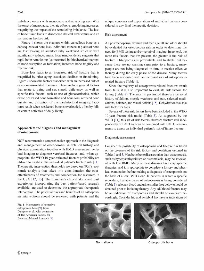

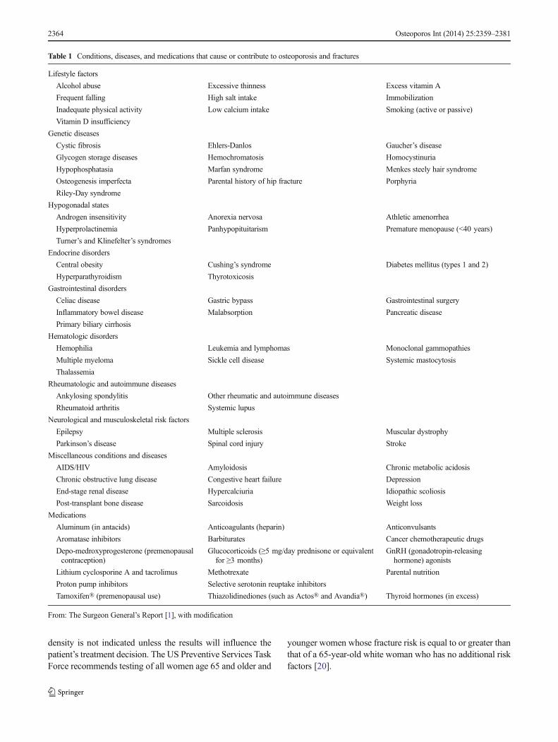
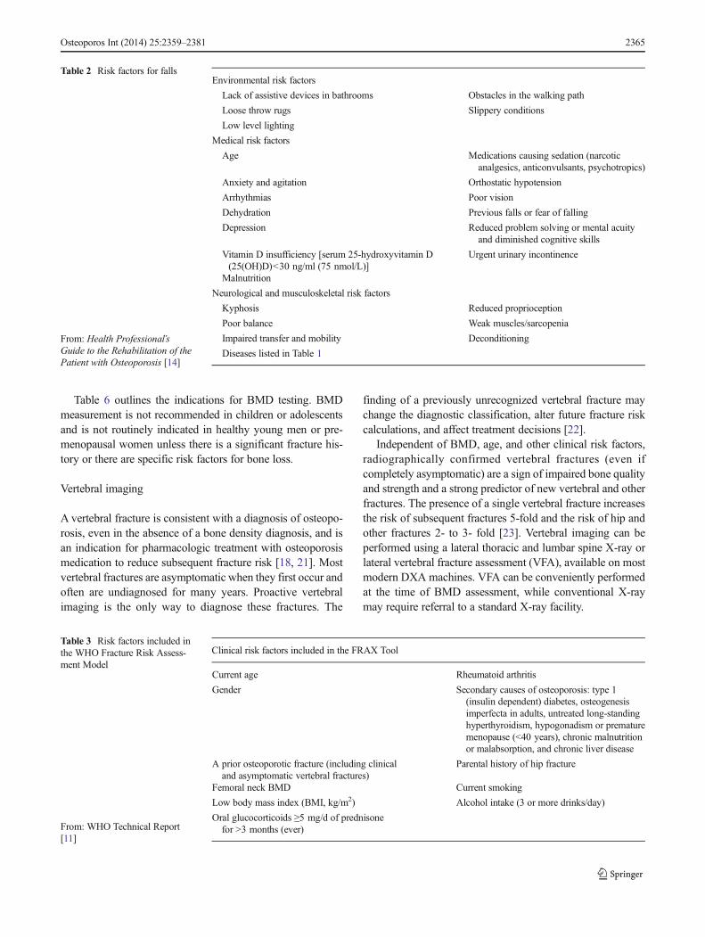
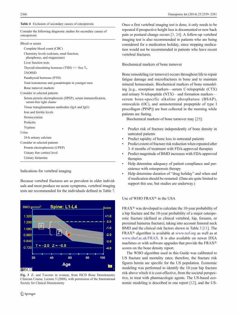
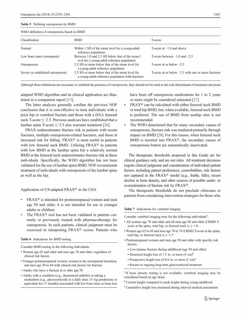

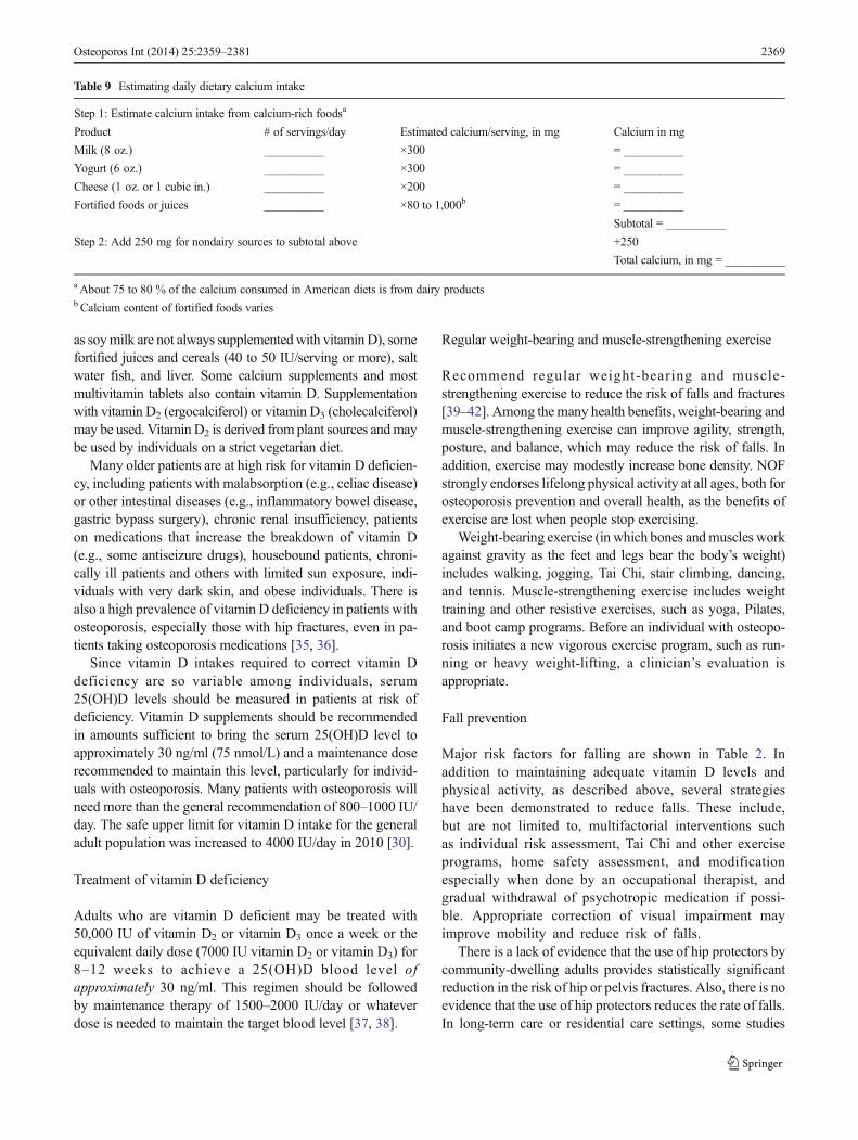


![Review Article Diabetic Osteoporosis: A Review of Its ...mineral density (BMD) in patients with DOP. Shang et al. [, ] found that Bushen Tongluo decoction had the ability ... Yushanjiang](https://static.documents.pub/doc/80x56/60ffebcf977497703253bb3a/review-article-diabetic-osteoporosis-a-review-of-its-mineral-density-bmd.jpg)


