genetic engineering, cloning vectors, characteristics of good vectors. recombinant gene technology. very important concept in the genetic engineering. used as a study handout undergraduate level students.
22
Cloning vectors A cloning vector is a small piece of DNA into which a foreign DNA fragment can be inserted. The insertion of the fragment into the cloning vector is carried out by treating the vehicle and the foreign DNA with a restriction enzyme that creates the same overhang, then ligating the fragments together. There are many types of cloning vectors. Genetically engineered plasmids and bacteriophages (such as phage λ) are perhaps most commonly used for this purpose. Other types of cloning vectors include bacterial artificial chromosomes (BACs) and yeast artificial chromosomes (YACs). Cloning vectors based on E.coli plasmids Simplest cloning vectors based on small bacterial plasmids are the most widespread in gene cloning experiment. A large number of different plasmid vectors are available for use with E.coli and these vectors combine ease of purification with desirable properties such as high transformation efficiency, convenient selectable markers for transformants and recombinants, and the ability to clone reasonably large pieces of DNA. One of the most popular vector used in gene cloning is pBR322, and it's nomenclature is shown below: pBR322 plasmid p- A plasmid BR- The laboratory in which the vector was originally constructed(Bolivar and Rodriguez, the two researchers who developed pBR322). 322- This number distinguishes the plasmid from other plasmids developed in the same laboratory. Important properties of pBR322 plasmid pBR322 is 4363bp in size which helps its purification with ease and recombinant molecules can also be constructed with it. pBR322 carries two sets of antibiotic resistance genes (Figure-1). Either ampicillin or tetracycline resistance can be used as a selectable marker for cells containing the plasmid, and each marker gene includes unique restriction sites that can be used in cloning experiments. The great variety of restriction sites(PstI,PvuI, ScaI,BamHI,HindIII) can be used for insertional inactivation means
Transcript
Cloning vectorsA cloning vector is a small piece of DNA into which a foreign DNA fragment can be inserted. The insertion of the fragment into the cloning vector is carried out by treating the vehicle and the foreign DNA with a restriction enzyme that creates the same overhang, then ligating the fragments together. There are many types of cloning vectors. Genetically engineered plasmids and bacteriophages (such as phage λ) are perhaps most commonly used for this purpose. Other types of cloning vectors include bacterial artificial chromosomes (BACs) and yeast artificial chromosomes (YACs).
Cloning vectors based on E.coli plasmidsSimplest cloning vectors based on small bacterial plasmids are the most widespread in gene cloning experiment. A large number of different plasmid vectors are available for use with E.coli and these vectors combine ease of purification with desirable properties such as high transformation efficiency, convenient selectable markers for transformants and recombinants, and the ability to clone reasonably large pieces of DNA. One of the most popular vector used in gene cloning is pBR322, and it's nomenclature is shown below:
pBR322 plasmid
p- A plasmidBR- The laboratory in which the vector was originally constructed(Bolivar and Rodriguez, the two researchers who developed pBR322).322- This number distinguishes the plasmid from other plasmids developed in the same laboratory.
Important properties of pBR322 plasmid
pBR322 is 4363bp in size which helps its purification with ease and recombinant molecules can also be constructed with it.
pBR322 carries two sets of antibiotic resistance genes (Figure-1). Either ampicillin or tetracycline resistance can be used as a selectable marker for cells containing the plasmid, and each marker gene includes unique restriction sites that can be used in cloning experiments. The great variety of restriction sites(PstI,PvuI, ScaI,BamHI,HindIII) can be used for insertional inactivation means that pBR322 can be used to clone DNA fragments with any of several kinds of sticky ends.
pBR322 has a reasonably high copy number. Normally there are about 15 molecules present in a transformed E.coli cell, but the number can be increased up to 1000-3000, by plasmid amplification in the presence of a protein synthesis inhibitor(chloramphenicol).
Figure 1: Structure of pBR322 plasmid
Other E.coli plasmid cloning vectors
Many other plasmid cloning vectors have been constructed, the majority of which derived from pBR322 plasmid.
pBR327 plasmid
It is a higher copy number plasmid and was constructed by removing a 1089 bp segment from pBR322. This deletion left the ampR tetR genes intact, but changed the replicative and conjugative abilities of the resulting plasmid.
Comparison between pBR322 and pBR327
pBR327 has a higher copy number(30-45 molecules) than pBR322. The higher copy number of pBR327 in normal cells makes this vector more suitable.
The deletion also destroys the conjugative ability of pBR322, making pBR327 a non-conjugative plasmid that can't direct its own transfer to other E.coli cells.
pUC8 Plasmid
pUC8 plasmid is derived from pBR322, remaining only the replication origin and the ampR gene. All the cloning sites are clustered into a short segment of the lacZ' gene carried by pUC8 (Figure-2).
Figure 2: The restriction site cluster in the lacz' gene of pUC8 plasmid
Advantages with pUC8 plasmid
The manipulations involved in construction of pUC8 were accompanied by a chance mutation, within the origin of replication, that results in the plasmid having a copy number 500-700 even before amplification. This has a significant effect on the yield of cloned DNA obtainable from E.coli cells transformed with recombinant pUC8 plasmids.
pUC vectors carry different combinations of restriction sites and show greater flexibility in the types of DNA fragment that can be cloned. Clustering of the restriction sites allows a DNA fragment with two different sticky ends to be cloned without involving linker attachment. DNA cloned into a member of the pUC series can be transferred directly to its M13mp counterpart (Figure-3), because of the same restriction site clusters and it can be analysed by DNA sequencing or in vitro mutagenesis.
Identification of recombinant cells with pUC8 plasmid, can be achieved in one step by plating on to agar medium containing ampicillin with X-gal. A cloning experiment with pUC8 can be carried out in half the time needed with pBR322 and pBR327 because with both pBR322 and pBR327 plasmids, selection of recombinants is a two step procedure, requiring replica-plating from one antibiotic medium to another.
Figure 3: Transfer of a DNA fragment from pUC8 to M13mp8
PGEM3Z plasmid
pGEM3Z is having similarity with a pUC vector because of the presence of the ampR genes and lacZ' genes containing a cluster of restriction sites (Figure-4). The difference is that pGEM3Z has two additional, short pieces of DNA, each of which acts as the recognition site for attachment of an RNA polymerase enzyme. These two promoter sequences lie on either side of the cluster of restriction site used for introduction of new DNA into the pGEM3Z molecule.
Figure 4: Structure of pGEM3Z plasmid
Cloning vectors of M13 bacteriophagePhage DNA molecules carry several genes that are essential for replication, including genes coding for components of the phage protein coat and phage-specific DNA replicative enzymes. Alteration or deletion of any of these genes will impair or destroy the replicative ability of the resulting molecule. So there is only limited scope for modifying the M13 genome and generally phage cloning vectors are only slightly different from parent molecules.
M13mp1 and M13mp2
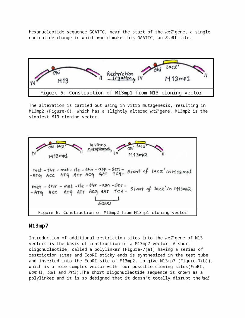
The construction of a M13 cloning vector involves introduction of the lacZ' genes into the intergenic sequence. This results in production of M13mp1, which forms blue plaques on X-gal agar. M13mp1 (Figure-5) contains the hexanucleotide sequence GGATTC, near the start of the lacZ' gene, a single nucleotide change in which would make this GAATTC, an EcoRI site.
Figure 5: Construction of M13mp1 from M13 cloning vector
The alteration is carried out using in vitro mutagenesis, resulting in M13mp2 (Figure-6), which has a slightly altered lacZ' gene. M13mp2 is the simplest M13 cloning vector.
Figure 6: Construction of M13mp2 from M13mp1 cloning vector
M13mp7
Introduction of additional restriction sites into the lacZ' gene of M13 vectors is the basis of construction of a M13mp7 vector. A short oligonucleotide, called a polylinker (Figure-7(a)) having a series of restriction sites and EcoRI sticky ends is synthesized in the test tube and inserted into the EcoRI site of M13mp2, to give M13mp7 (Figure-7(b)), which is a more complex vector with four possible cloning sites(EcoRI, BamHI, SalI and PstI).The short oligonucleotide sequence is known as a polylinker and it is so designed that it doesn't totally disrupt the lacZ' gene; a reading frame is maintained throughout the polylinker, and a functional, though altered, β-galactosidase enzyme is still produced.
Figure 7: Construction of M13mp7
Complex M13 vectors
The complex M13 vectors have more complex polylinkers inserted into the lacz' gene. M13mp8 is a complex vector and it has the ability to take DNA fragments with two different sticky ends. M13mp9 is another vector having the same polylinker but in the reverse orientation. A DNA fragment cloned into M13mp8, if excised by double restriction, and then inserted into M13mp9, will now itself be in the reverse orientation, which is important in DNA sequencing, in which the nucleotide sequence is read from one end of the polylinker into the inserted DNA fragment.
Cloning vectors of λ bacteriophageThe first two types of vectors to be produced were λ-insertion and λ-replacement vectors.
Insertion vectors
An insertion vector possesses at least one unique restriction site into which new DNA can be inserted. λgt10 and λZAPII are two popular insertion vectors. λgt10 can carry up to 8kb of new DNA, inserted into a unique EcoRI site located in the cI gene. Insertional activation of this gene distinguishes recombinants as clear rather than turbid plaques.
Replacement vectors
A λ replacement vector has two recognition sites for the restriction endonuclease used for cloning. These sites flank a segment of DNA that is replaced by the DNA to be cloned. The replaceable fragment often carries additional restriction sites that can be used to cut it up into small pieces. Replacement vectors are generally designed to carry larger pieces of DNA than insertion vectors can handle. λWES.λB' and λEMBL4 are two popular replacement vectors.
CosmidA cosmid is a plasmid that carries a cos site the sequence yielding cohesive ends (Figure-8). Cosmids are hybrids between a phage DNA molecule and a bacterial plasmid, and are designed in such a way that the enzymes that package the λ DNA molecule into the phage protein coat need only the cos sites in order to fuction. A typical cosmid has replication origin, unique restriction sites and selectable markers from the plasmid; therefore selection strategy for obtaining the recombinant vectors is based on that for the contributing plasmid. Cosmid vectors are constructed using recombinant DNA techniques.
Figure 8: A cosmid vector
Cloning experiment with a cosmid
The cosmid vectors are opened by the appropriate restriction enzyme at a unique site, are then mixed with DNA inserts prepared by using the same enzyme and annealed. These DNA fragments are produced usually by partial digestion with a restriction endonuclease, as total digestion produce too small fragments to be cloned with a cosmid. Ligation is carried out so that concatamers are formed (Figure-9). In vitro packaging will cleave the cos sites and place the recombinant cosmids in mature phage particles. These λ phages are then used to infect E.coli culture, infected cells are plated on to a selective medium and antibiotic-resistant colonies are grown.
Figure 9: A cosmid vector is used to clone large fragments of DNA
Yeast vectorsThe yeast Saccharomyces cerevisiae is one of the most important organisms in biotechnology. Development of cloning vectors for yeast has been stimulated greatly by the discovery of a plasmid that is present in most strains of S. cerevisiae. The 2µm circle, as it is called is one of only a very limited number of plasmids found in eukaryotic cells.
Yeast plasmids
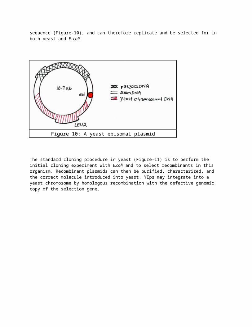
Vectors derived from the 2µm circle are called yeast episomal plasmids or YEps. YEps may contain the entire 2µm plasmid, or include just the 2µm origin of replication. YEp13 illustrates several general features of yeast cloning vectors. It is a shuttle vector. It contains the 2µm origin of replication and the selectable LEU2 gene, along with the entire pBR322 sequence (Figure-10), and can therefore replicate and be selected for in both yeast and E. coli.
Figure 10: A yeast episomal plasmid
The standard cloning procedure in yeast (Figure-11) is to perform the initial cloning experiment with E.coli and to select recombinants in this organism. Recombinant plasmids can then be purified, characterized, and the correct molecule introduced into yeast. YEps may integrate into a yeast chromosome by homologous recombination with the defective genomic copy of the selection gene.
Figure 11: Cloning with a yeast episomal plasmid
Yeast Integrative plasmids or YIps
Yeast vectors that rely on integration into the host chromosome for survival and replication, and are usually used when studying the functionality of a solo gene or when the gene is toxic. Also connected with the gene URA3, that codes an enzyme related to the biosynthesis of pyrimidine nucleotides (T, C).
Yeast Replicative plasmids or YRps
Yeast replicative plasmids which transport a sequence of chromosomal DNA that includes an origin of replication. These plasmids are less stable, as they can "get lost" during the budding.
Yeast Artificial Chromosome(YAC)Artificial chromosomes can be used to clone huge pieces of DNA in yeast. First described in 1983 by Murray and Szostak, a YAC is an artificially constructed chromosome and contains the telomeric, centromeric, and replication origin sequences named autonomous replicating sequence needed for replication and preservation in yeast cells (Figure-12). A YAC is built using an initial circular plasmid, which is typically broken into two linear molecules using restriction enzymes; DNA ligase is then used to ligate a sequence or gene of interest between the two linear molecules, forming a single large linear piece of DNA. The construction of the YAC clone is similar to that for cosmids, in that two end fragments are ligated with target DNA to yield the
complete chromosome, which is then introduced into yeast cells. YAC vectors can accommodate genomic DNA fragments of more than 1 Mb, and hence can be used to clone entire human genes, such as the cystic fibrosis gene, which is 250 kb in length.
The yeast sequences that have been included on pYAC3 are as follows: TEL represents a segment of the telomeric DNA sequence, which is extended by the telomerase enzyme inside the yeast cell. CEN4 is the centromere sequence for chromosome 4 of S.cerevisiae. The ARS (autonomously replicating sequence) functions as a yeast origin of replication.TRP1 and URA3 are yeast selectable markers, one for each end, to ensure that only properly reconstituted YACs survive in the yeast cells. SUP4, which is insertionally inactivated in recombinants (Figure-12), is a gene which is the basis of a red-white color test, which is analogous to blue-white screening in E.coli.
Figure 12: pYAC3
Cloning with pYAC3
The vector is first digested with a combination of BamHI and SnaBI, cutting the molecule into three fragments. The BamHI fragment is removed, leaving two arms, each bounded by one TEL sequence and one SnaBI site. The DNA to be inserted is ligated between the two arms producing the artificial chromosome (Figure-13). The DNA must have blunt ends as SnaBI is a blunt end cutter. Then the artificial chromosome is introduced into S. cerevisiae by protoplast transformation. The yeast strain that is used is a double auxotrophic mutant, trp1- and ura3- which will be converted to trp1+ and ura3+ by the two markers on the artificial chromosome.
Figure 13: Cloning with a YAC vector
Selection in S.cerevisiae
Saccharomyces cerevisiae selectable markers do not normally confer resistance to toxic substances, as in E.coli plasmids, but instead enable the growth of yeast on selective media lacking specific nutrients. Transformants are selected by plating on to minimal medium, on which only cells containing a correctly constructed artificial chromosome will able to grow. If any cells transformed with an incorrect artificial chromosome, will not be able to grow on minimal medium as one of the markers will be absent.
Cloning vectors for higher plantsThere are important potential benefits of gene cloning using higher plants as the host organisms. Three types of vector system have been used with varying degrees of success with higher plants:
Vectors based on naturally occuring plasmids of Agrobacterium. Direct gene transfer using DNA fragments not attached to a plant cloning vector. Vectors based on plant viruses.
Agrobacterium tumefaciens-Ti plasmid
A. tumefaciens is a soil microorganism that causes crown gall disease in many species of dicotyledonous plants. Crown gall occurs when a wound on the stem allows A. tumefaciens to invade the plant. After infection the bacteria cause a cancerous proliferation of the stem tissue in the region of the crown. The ability to cause crown gall disease is associated with the presence of the Ti(Tumour inducing) plasmid (Figure-14) within the bacterial cell. Ti plasmid is a large circular plasmid (greater than 200kb) that carries numerous genes involved in the infection process.
Figure 14: The Ti plasmid
The Ti plasmid is lost when Agrobacterium is grown above 28°C. Such cured bacteria do not induce crown galls, i.e. they become avirulent. The infection part of the plasmid is integrated into the plant chromosomal DNA is called the T-DNA, is between 15 and 30kb in size, depending on the strain. It is maintained in a stable form in the plant cell and is passed on to daughter cells as an integral part of the chromosomes (Figure-15). The T-DNA contains eight or so genes that are expressed in the plant cell and are responsible for the cancerous properties of the transformed cells. These genes also direct synthesis of unusual compounds, called opines, that the bacteria use as nutrients. A.tumefaciens genetically engineers the plant cell for its own purposes.
Figure 15: Integration and expression of T-DNA in plant genome
Production of transformed plants with the Ti plasmid
Generally plant cells and protoplasts are infected with Ti plasmid. Plant cells and protoplasts can be plated on to a selective medium in order to isolate transformants (Figure-16). A mature plant regenerated from transformed cells will contain the cloned gene in every cell and will pass the cloned gene to its offspring. Regeneration of a transformed plant will occur only if the Ti vector has been disarmed so that transformed cells do not display cancerous properties. Disarming is possible because the cancer genes, all of which lie in the T-DNA are not needed for infection process, infectivity being controlled mainly by the virulence region of the Ti plasmid. The only parts of the T-DNA that are involved in infection are two 25 bp repeat sequences found at the left and right borders of the region integrated into the plant DNA. Any DNA placed between these two repeat sequences will be treated as T-DNA and transferred to the plant. It is therefore possible to remove all the cancer genes from the normal T-DNA, and replace them with an entirely new set of genes, without disturbing the infection process.
Figure 16: Transformation of cultured plant cells by recombinant Agrobacterium tumefaciens
The binary vector pBIN19 is a disarmed cloning vector in which the left and right T-DNA borders flank a copy of the lacZ' gene, containing a number of cloning sites, and a kanamycin resistance gene that functions after integration of the vector sequences into the plant chromosome. Ti vectors such as pBIN19 have recently been supplemented by related vectors based on the Ri plasmid of Agrobacterium rhizogenes. Ri plasmid and Ti plasmids are very similar, the main difference being that transfer of the T-DNA from an Ri plasmid to a plant results not in a crown gall but in a hairy root disease, typified by a massive proliferation of a highly branched root system. The possibility of growing transformed roots at a high density in a liquid culture is being explored by biotechnologists as a potential means of obtaining large amounts of protein from genes cloned in plants.
Direct gene transferGenes may be transiently or permanently introduced into cultured eukaryotic cells without the use of a vector in the strict sense. A eukaryotic gene on a bacterial plasmid, may transiently express its product when transfected into a cell line, even if the plasmid doesn't replicate in that type of cell. Alternatively DNA introduced by transfection or microinjection may become stably integrated into the cell's chromosomal DNA. This process normally require significant sequence similarity between the incoming DNA and the genome in animal cells but, in plant cells any supercoiled plasmid can randomly integrate into the genome (Figure-17). Such stably transfected cells can be selected by the presence of a drug resistance gene in much the same way as bacterial transformants and can continue to express protein from foreign genes through many cell divisions.
Figure 17: Direct gene transfer
Plant viruses as cloning vectorsMost plants are subjected to viral infection and vast majority of plant viruses have genomes of RNA. RNA viruses are not so useful as potential cloning vectors because manipulations with RNA are rather more difficult to carry out. Two classes of DNA virus are known to infect higher plants, the caulimoviruses and geminiviruses, but neither is identically suited for gene cloning. Caulimovirus vector could only be used to clone very short pieces of DNA. Geminiviruses at first appear more promising as they naturally infect important crops such as maize and wheat. But during the infection cycle the geminivirus genome undergoes rearrangements and deletions, which could scramble up any additional DNA that had been inserted, an obvious disadvantage for a cloning vector.
Animal viruses as cloning vectorsThe first eukaryotic DNA virus was SV40, for which a complete nucleotide sequence and a detailed understanding of transcription were available. The genome of SV40 contains very little non-essential DNA so it is necessary to insert the foreign gene in place of essential viral genes and to propagate the recombinant genome in the presence of a helper virus. This virus is capable of infecting several mammalian species, following a lytic cycle in some host and a lysogenic cycle in others. The genome is 5.2kb in size and contains two sets of genes, the early genes, expressed early in the infection cycle and coding for proteins involved in viral DNA replication, and the late genes, coding for viral capsid proteins (Figure-18). However, all work using SV40 virions to propagate recombinant DNA molecules is severely constrained by the facts that the viral genome is small, 5.24 kb, and that the packaging limits are strict. Such systems can not, therefore, be used for the analysis of most eukaryotic genes.
Figure 18: The SV40 genome
Adenoviruses, are a group of viruses which enable larger fragments of DNA to be cloned than it is possible with an SV40 vector. Adenoviruses are more difficult to handle because the genomes are bigger.
Papillomaviruses, which also have a relatively high capacity for inserted DNA, have the important advantage of enabling a stable transformed cell line to be obtained. Papillomavirus transformed cells don't contain integrated viral DNA rather they contain between 50 and 300 copies of unintegrated, circular viral DNA although some proportion of these viral genomes exists as concatamers and/or catenates. Bovine papillomavirus(BPV), which causes warts on cattle, has an unusal infection cycle in mouse cells, taking the form of a multi copy plasmid with about 100 molecules present per cell. It doesn't cause the death of the mouse cell, and BPV molecules are passed to daughter cells on cell division. Shuttle vectors consisting of BPV and pBR322 sequences, and capable of replication in both mouse and bacterial cells, are therefore of great value in animal cell biotechnology.
Retroviruses, though have single-stranded RNA genomes but provides perhaps the most promising vector system of all. During the process of reverse transcription, sequences from the termini of viral RNA are duplicated to generate long terminal repeats(LTRs). These long terminal repeats contain both the promoter and the polyadenylation signal for the transcription of viral mRNAs. The specificity of proviral DNA integration is also determined by the long terminal repeats. Although retroviruses can integrate at many sites within the cellular genome, integrative recombination always occurs at particular sites at the ends of the LTRs. The sequences appropriately inserted between the two LTRs will be integrated intact which contrasts sharply with the integration of papovavirus or adenovirus DNA, during which extensive rearrangements of the integrated viral sequences are commonplace. A further great advantage of retroviruses is that they are natural transducing viruses.
Baculoviruses, enable large amounts of proteins to be obtained from genes cloned in insect cells. One of the major proteins encoded by the virus genome is polyhedrin, which accumulates in very large quantities in the nuclei of infected cells, since the gene has an extremely active promoter. The same promoter can be used to drive the over expression of a foreign gene engineered into the baculovirus genome, and large quantities of protein can be produced in infected insect cells in culture. This method is being used increasingly for large-
scale culture of proteins of animal origin, since the insect cells can produce many of the post-translational modifications of animal proteins which a bacterial expression system.
References1. Brown, T.A. (1998). Cloning vectors for E.coli, Cloning vectors for organisms other than E.coli. Gene cloning an introduction, 3rd Edn. Stanley Thornes (Publishers) Ltd.
2. Lewin, B. 1994. genes V. Oxford University Press, New York
3. Singh, B.D.(2003). Biotechnology. Kalyani Publishers, New Delhi
4. Turner Phil, McLennan Alexander, Bates Andy and White Mike (2005). Cosmids,YACs and BACs, Eukaryotic vectors. Instant notes(138-143), Molecular Biology. Taylor& Francis Group