39
Infection types
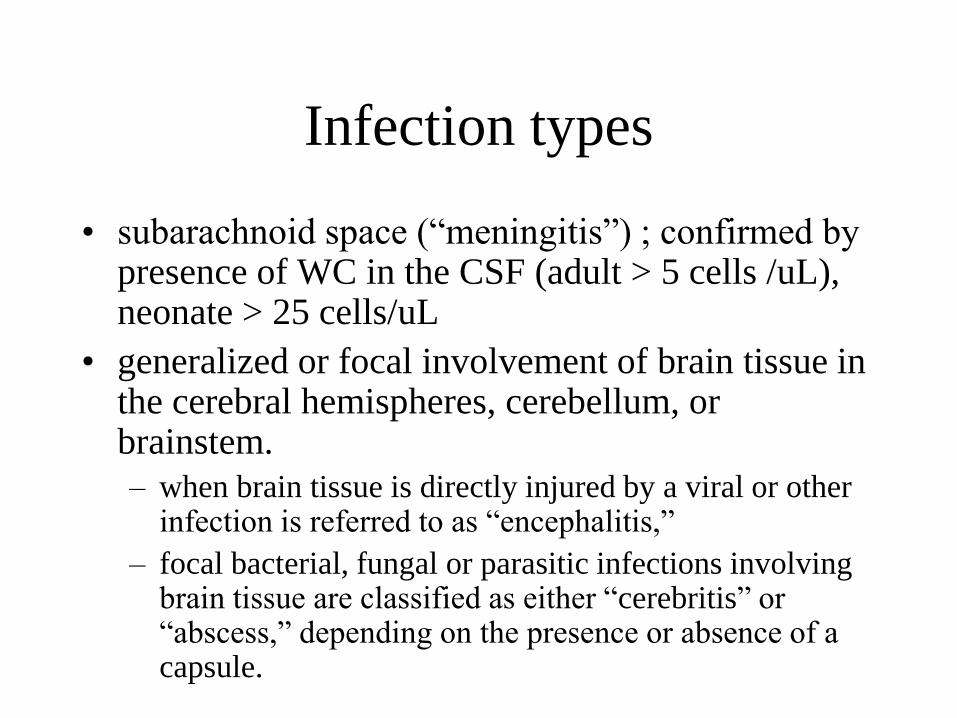
• subarachnoid space (“meningitis”) ; confirmed by presence of WC in the CSF (adult > 5 cells /uL), neonate > 25 cells/uL
• generalized or focal involvement of brain tissue in the cerebral hemispheres, cerebellum, or brainstem.
– when brain tissue is directly injured by a viral or other infection is referred to as “encephalitis,”
– focal bacterial, fungal or parasitic infections involving brain tissue are classified as either “cerebritis” or “abscess,” depending on the presence or absence of a capsule.
Clinical syndromes
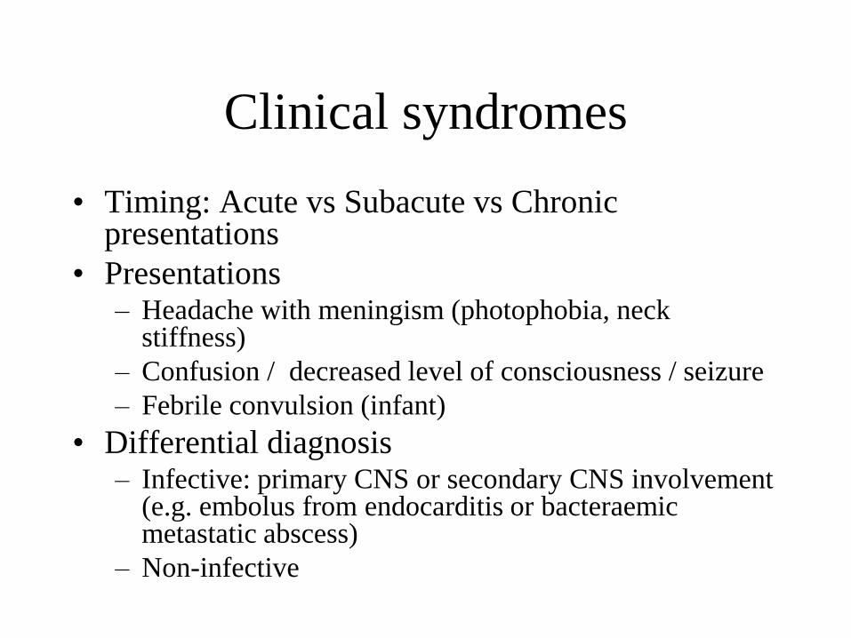
• Timing: Acute vs Subacute vs Chronic presentations
• Presentations – Headache with meningism (photophobia, neck
stiffness)
– Confusion / decreased level of consciousness / seizure
– Febrile convulsion (infant)
• Differential diagnosis – Infective: primary CNS or secondary CNS involvement
(e.g. embolus from endocarditis or bacteraemic metastatic abscess)
– Non-infective
CSF Examination
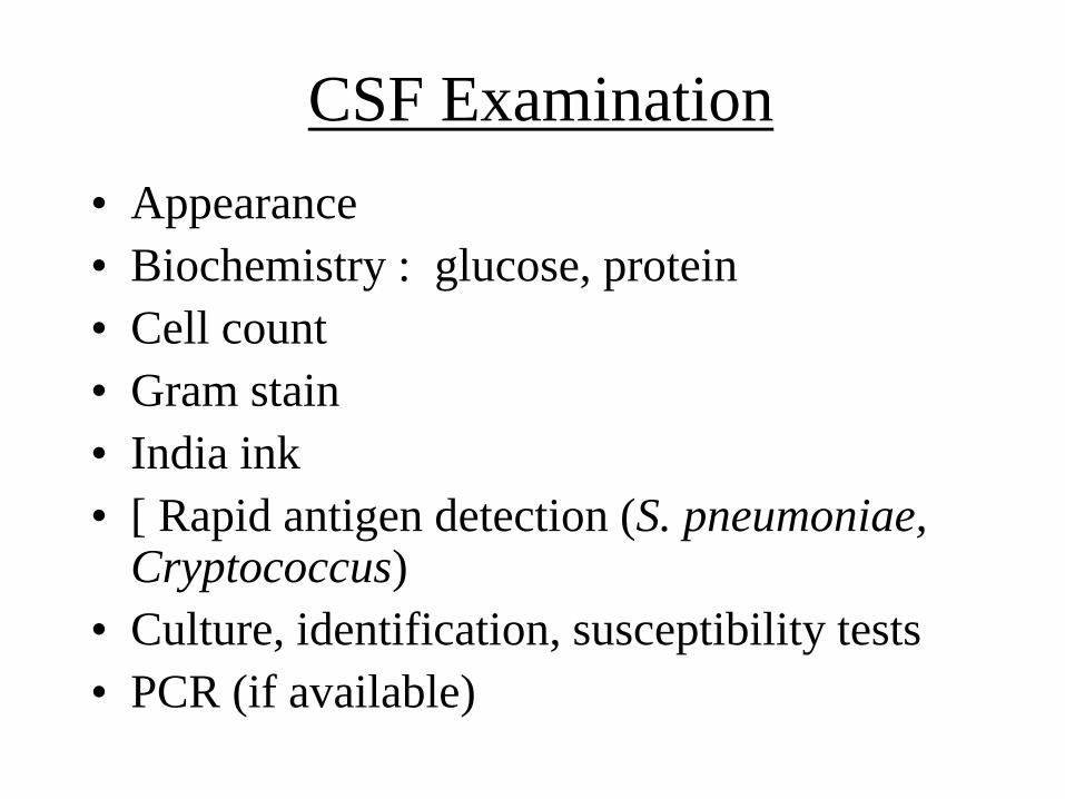
• Appearance
• Biochemistry : glucose, protein
• Cell count
• Gram stain
• India ink
• [ Rapid antigen detection (S. pneumoniae, Cryptococcus)
• Culture, identification, susceptibility tests
• PCR (if available)
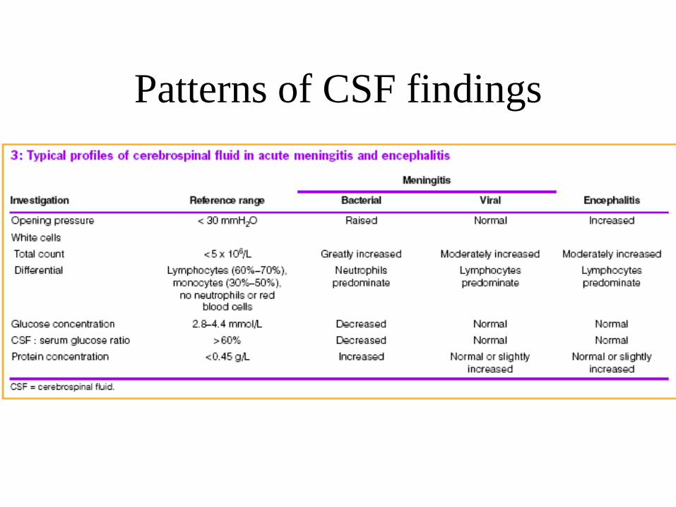
Patterns of CSF findings
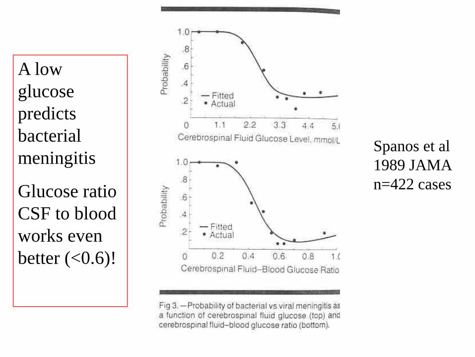
A low
glucose
predicts
bacterial
meningitis
Glucose ratio
CSF to blood
works even
better (<0.6)!
Spanos et al
1989 JAMA
n=422 cases
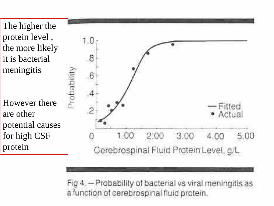
The higher the
protein level ,
the more likely
it is bacterial
meningitis
However there
are other
potential causes
for high CSF
protein
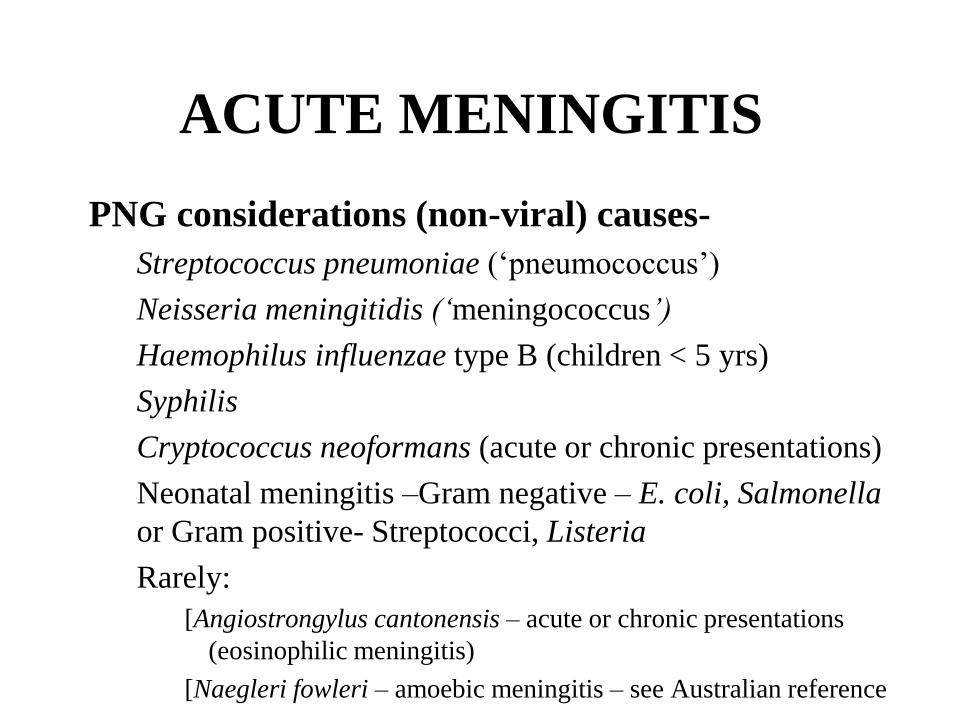
ACUTE MENINGITIS
PNG considerations (non-viral) causes-
Streptococcus pneumoniae (‘pneumococcus’)
Neisseria meningitidis (‘meningococcus’)
Haemophilus influenzae type B (children < 5 yrs)
Syphilis
Cryptococcus neoformans (acute or chronic presentations)
Neonatal meningitis –Gram negative – E. coli, Salmonella
or Gram positive- Streptococci, Listeria
Rarely:
[Angiostrongylus cantonensis – acute or chronic presentations
(eosinophilic meningitis)
[Naegleri fowleri – amoebic meningitis – see Australian reference
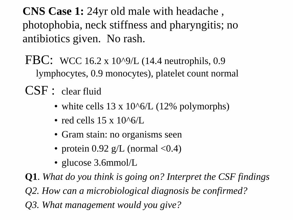
CNS Case 1: 24yr old male with headache ,
photophobia, neck stiffness and pharyngitis; no
antibiotics given. No rash.
FBC: WCC 16.2 x 10^9/L (14.4 neutrophils, 0.9
lymphocytes, 0.9 monocytes), platelet count normal
CSF : clear fluid
• white cells 13 x 10^6/L (12% polymorphs)
• red cells 15 x 10^6/L
• Gram stain: no organisms seen
• protein 0.92 g/L (normal <0.4)
• glucose 3.6mmol/L
Q1. What do you think is going on? Interpret the CSF findings
Q2. How can a microbiological diagnosis be confirmed?
Q3. What management would you give?
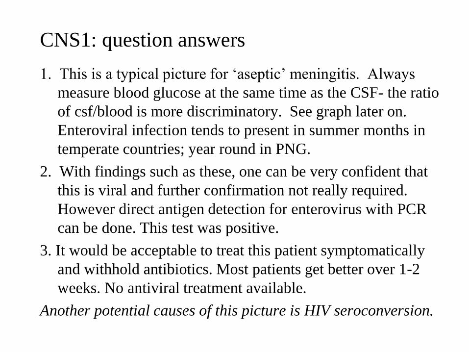
CNS1: question answers
1. This is a typical picture for ‘aseptic’ meningitis. Always
measure blood glucose at the same time as the CSF- the ratio
of csf/blood is more discriminatory. See graph later on.
Enteroviral infection tends to present in summer months in
temperate countries; year round in PNG.
2. With findings such as these, one can be very confident that
this is viral and further confirmation not really required.
However direct antigen detection for enterovirus with PCR
can be done. This test was positive.
3. It would be acceptable to treat this patient symptomatically
and withhold antibiotics. Most patients get better over 1-2
weeks. No antiviral treatment available.
Another potential causes of this picture is HIV seroconversion.
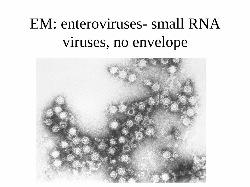
EM: enteroviruses- small RNA
viruses, no envelope
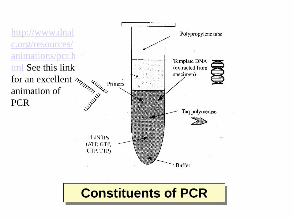
Constituents of PCR
http://www.dnal
c.org/resources/
animations/pcr.h
tml See this link
for an excellent
animation of
PCR
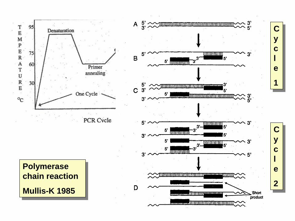
Polymerase
chain reaction
Mullis-K 1985
C
y
c
l
e
1
C
y
c
l
e
2
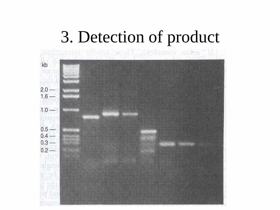
3. Detection of product

‘Real-time’
detection
‘Taqman’
probe
Binds to the
PCR product by
base pair
matching;
Reporter
molecule
removed by DNA
polymerase as it
extends the
strand
Reporter
fluoresces

CNS Case 2: 21 year old male with a 4 day history.
Initial sore throat, then myalgia and joint pain with
swelling, then severe headache for 24 hrs. No rash.
On examination: No rash, febrile and tachycardic. Mild neck
stiffness present.
FBC : WCC 22.2 x 10^9/L (15.2 neutrophils, 0.7
lymphocytes, 5.3 band forms (immature neutrophils),
platelets normal
Blood culture taken
Questions:
1. What is your differential diagnosis?
2. Would you do a Lumbar puncture?

CNS2: question answers to 1, 2
1. Acute febrile illness with severe headache and some neck
stiffness; bacterial meningitis must be suspected. Please
think of some alternatives. The high band form and very
high WCC are predictive of bacterial sepsis.
2. YES; only defer LP if there are contraindications such as
shock, local sepsis in the lumbar region, focal neurological
signs or significant decreased level of consciousness.
Always be careful to check for neck stiffness and for
Kernig’s sign. If either are present then consider an LP.
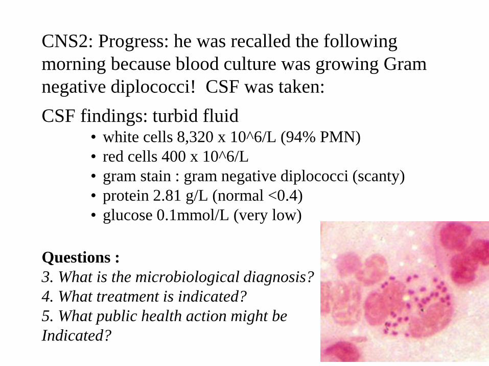
CNS2: Progress: he was recalled the following
morning because blood culture was growing Gram
negative diplococci! CSF was taken:
CSF findings: turbid fluid • white cells 8,320 x 10^6/L (94% PMN)
• red cells 400 x 10^6/L
• gram stain : gram negative diplococci (scanty)
• protein 2.81 g/L (normal <0.4)
• glucose 0.1mmol/L (very low)
Questions :
3. What is the microbiological diagnosis?
4. What treatment is indicated?
5. What public health action might be
Indicated?
CNS2: question answers 3,4, 5
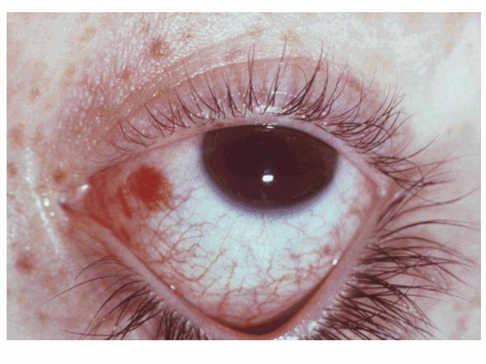
3. The diagnosis is meningococcal sepsis with meningitis. A small conjunctival haemorrhage was found in one eye and his knees, wrists and ankles were swollen due to a septic polyarthritis from meningococcus.
4. Treatment of meningococcal disease is with ceftriaxone (empirically) and later (if microbiologically confirmed) benzylpenicillin at high dose - 1.8g 4hrly intravenous. Steroids (IV dexamethasone for 4 days) not strictly necessary in meningococcal disease BUT recommended for meningitis due to Strep. pneumoniae or due to Haemophilus influenzae type B. Steroids must be given before or soon after the first antibiotic dose to work. They reduce long-term neurological sequelae (deafness etc)
5. Public health: antibiotic prophylaxis can be given to close contacts of the case to prevent them developing disease
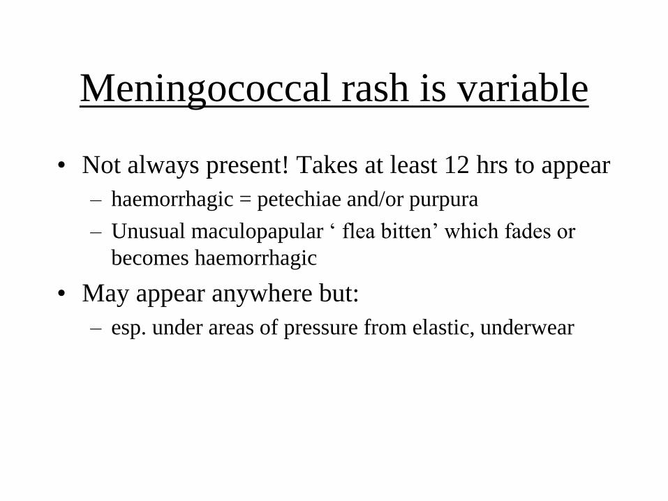
Meningococcal rash is variable
• Not always present! Takes at least 12 hrs to appear
– haemorrhagic = petechiae and/or purpura
– Unusual maculopapular ‘ flea bitten’ which fades or
becomes haemorrhagic
• May appear anywhere but:
– esp. under areas of pressure from elastic, underwear

Clinical features- bacterial
meningitis
• Headache
• Classical triad of fever, neck stiffness and altered
state of consciousness is present in 2/3. Nearly all
will have at least one of these as well
• Focal neurology- cranial nerve palsies 10-20%
• Seizures 5-10% generalised seizures
• Elderly or immune-compromised - may present
with only confusion/obtundation
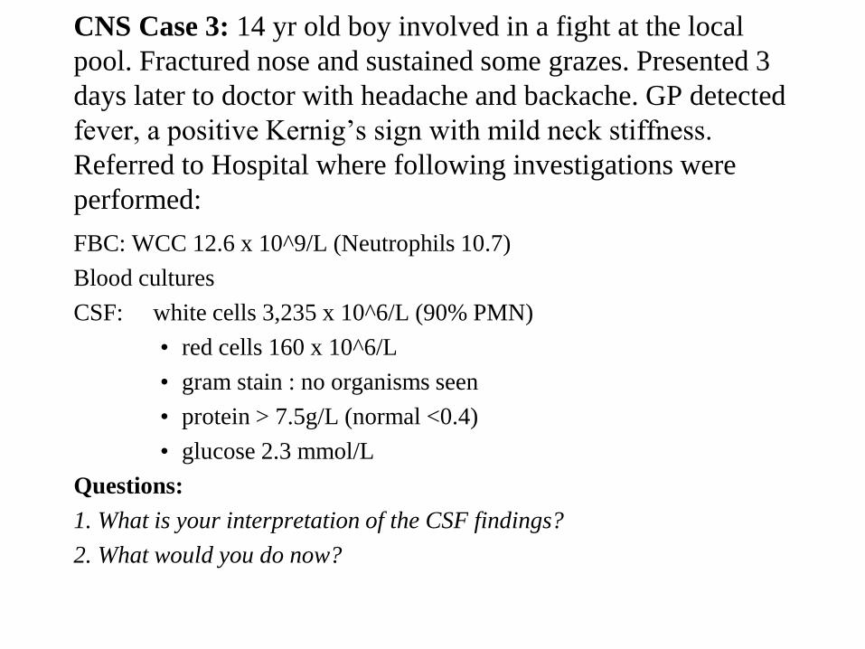
CNS Case 3: 14 yr old boy involved in a fight at the local
pool. Fractured nose and sustained some grazes. Presented 3
days later to doctor with headache and backache. GP detected
fever, a positive Kernig’s sign with mild neck stiffness.
Referred to Hospital where following investigations were
performed:
FBC: WCC 12.6 x 10^9/L (Neutrophils 10.7)
Blood cultures
CSF: white cells 3,235 x 10^6/L (90% PMN)
• red cells 160 x 10^6/L
• gram stain : no organisms seen
• protein > 7.5g/L (normal <0.4)
• glucose 2.3 mmol/L
Questions:
1. What is your interpretation of the CSF findings?
2. What would you do now?

CNS3: question answer 1
1. High CSF PMN count and protein level suggest meningitis.
However the CSF glucose level is relatively normal, given
the enormous increase in protein and cells. Also odd is the
absence of organisms on Gram stain. A Gram stain without
visible organisms may occur in early meningococcal
meningitis; however the CSF findings show a degree of
inflammation that implies late meningitis. Pre-treatment
with antibiotics might explain absence of organisms on
Gram stain but not the other findings. The absence of neck
stiffness is also inconsistent. It all doesn’t add up!
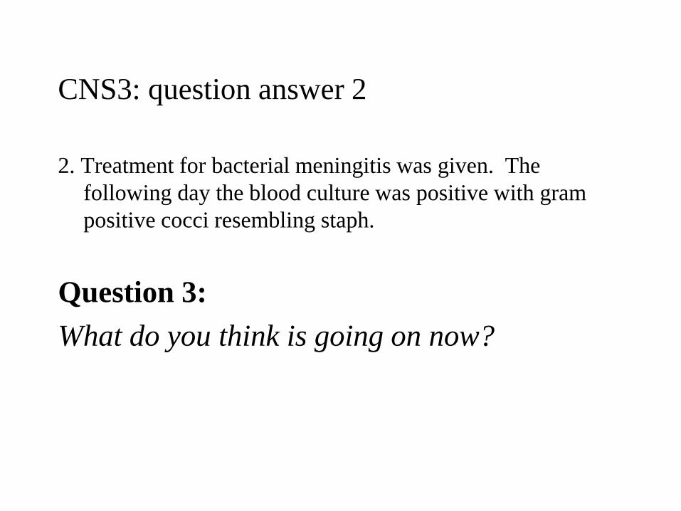
CNS3: question answer 2
2. Treatment for bacterial meningitis was given. The
following day the blood culture was positive with gram
positive cocci resembling staph.
Question 3:
What do you think is going on now?
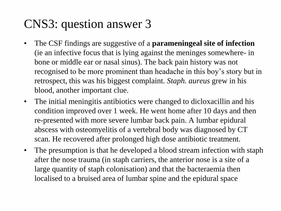
CNS3: question answer 3
• The CSF findings are suggestive of a parameningeal site of infection
(ie an infective focus that is lying against the meninges somewhere- in
bone or middle ear or nasal sinus). The back pain history was not
recognised to be more prominent than headache in this boy’s story but in
retrospect, this was his biggest complaint. Staph. aureus grew in his
blood, another important clue.
• The initial meningitis antibiotics were changed to dicloxacillin and his
condition improved over 1 week. He went home after 10 days and then
re-presented with more severe lumbar back pain. A lumbar epidural
abscess with osteomyelitis of a vertebral body was diagnosed by CT
scan. He recovered after prolonged high dose antibiotic treatment.
• The presumption is that he developed a blood stream infection with staph
after the nose trauma (in staph carriers, the anterior nose is a site of a
large quantity of staph colonisation) and that the bacteraemia then
localised to a bruised area of lumbar spine and the epidural space
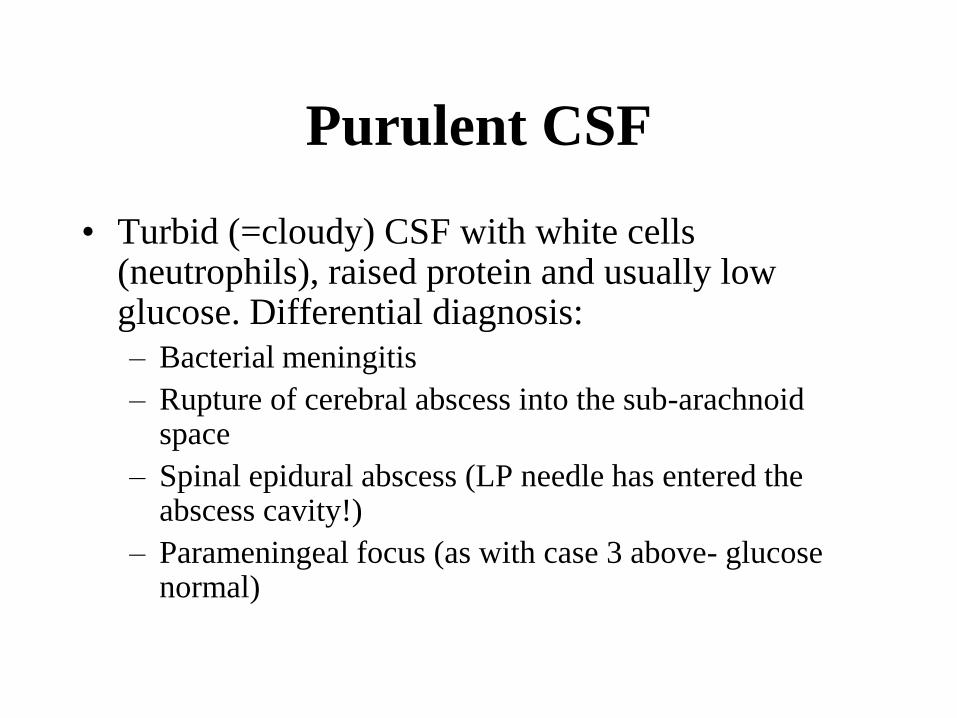
Purulent CSF
• Turbid (=cloudy) CSF with white cells (neutrophils), raised protein and usually low glucose. Differential diagnosis:
– Bacterial meningitis
– Rupture of cerebral abscess into the sub-arachnoid space
– Spinal epidural abscess (LP needle has entered the abscess cavity!)
– Parameningeal focus (as with case 3 above- glucose normal)
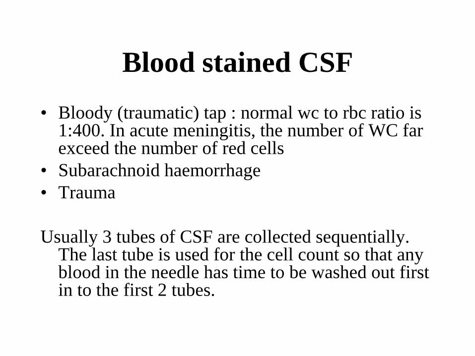
Blood stained CSF
• Bloody (traumatic) tap : normal wc to rbc ratio is 1:400. In acute meningitis, the number of WC far exceed the number of red cells
• Subarachnoid haemorrhage
• Trauma
Usually 3 tubes of CSF are collected sequentially. The last tube is used for the cell count so that any blood in the needle has time to be washed out first in to the first 2 tubes.
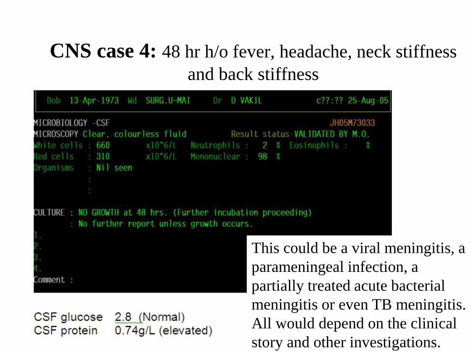
CNS case 4: 48 hr h/o fever, headache, neck stiffness
and back stiffness
This could be a viral meningitis, a
parameningeal infection, a
partially treated acute bacterial
meningitis or even TB meningitis.
All would depend on the clinical
story and other investigations.

d
Kernig and Brudzinski signs are a reliable predictor of meningitis in
infants in PNG!

Meningitis due to S. pneumoniae
• Turbid CSF usual
• [Traditional latex antigen
tests are poor – no better
than a Gram stain
• [BINAX pneumolysin-C
antigen detection card
works very well on CSF-
much more sensitive than
latex test
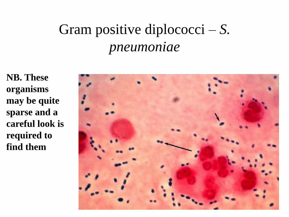
Gram positive diplococci – S.
pneumoniae
NB. These
organisms
may be quite
sparse and a
careful look is
required to
find them
Antibiotic resistance in
pneumococcus
• Screen pneumococci from CSF with 1 microgram oxacillin
disc test. Fully susceptible strains (MIC <0.12mg/L) have
an oxacillin zone >=20mm.
• Strains with reduced susceptibility to penicillin cause
penicillin to fail for treatment of meningitis; these strains
may also be tolerant to chloramphenicol causing treatment
failure
• Ceftriaxone high dose is recommended for treatment
due to the risk of penicillin resistance
• [The laboratory can check the penicillin and ceftriaxone
susceptibility in cases of pneumococcal meningitis – the E-
test is used
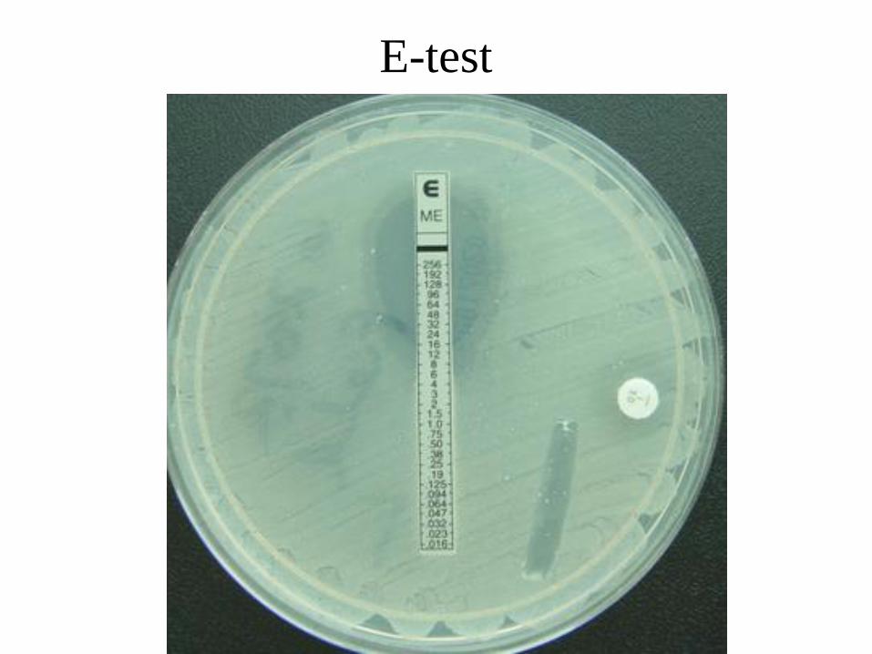
E-test
Management options: pneumococcal
meningitis where penicillin MIC is elevated
• Third generation cephalosporin (ceftriaxone)
works provided that MIC < 1mg/L for ceftriaxone
• IF E-test MIC for ceftriaxone >=1mg/L then it
may also fail
– Options are to give ceftriaxone AND either vancomycin
(iv) or rifampicin for synergy
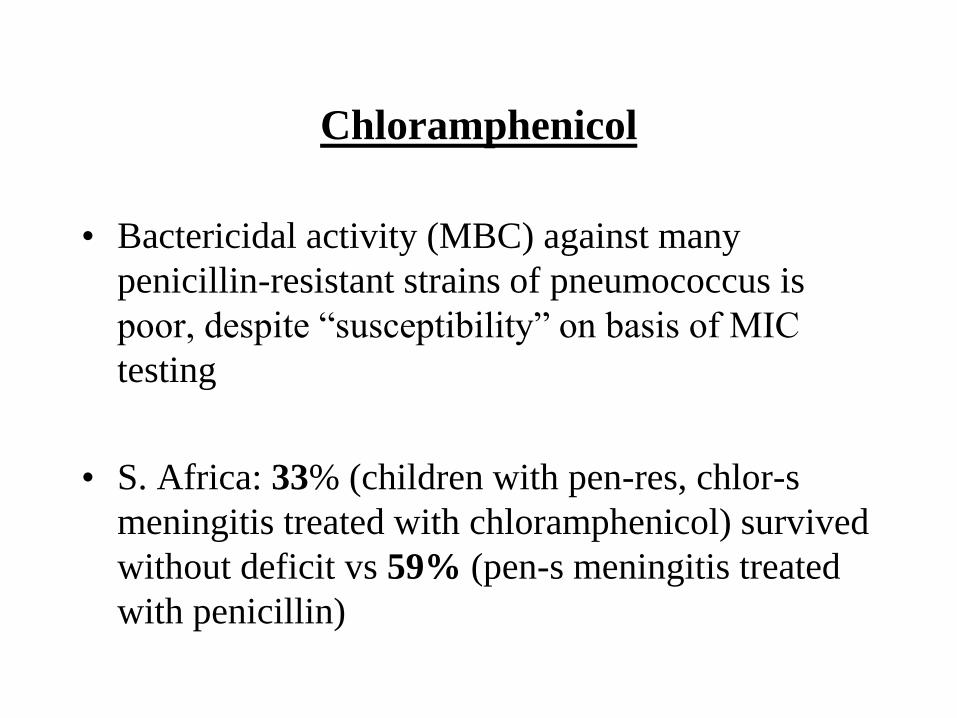
Chloramphenicol
• Bactericidal activity (MBC) against many
penicillin-resistant strains of pneumococcus is
poor, despite “susceptibility” on basis of MIC
testing
• S. Africa: 33% (children with pen-res, chlor-s
meningitis treated with chloramphenicol) survived
without deficit vs 59% (pen-s meningitis treated
with penicillin)
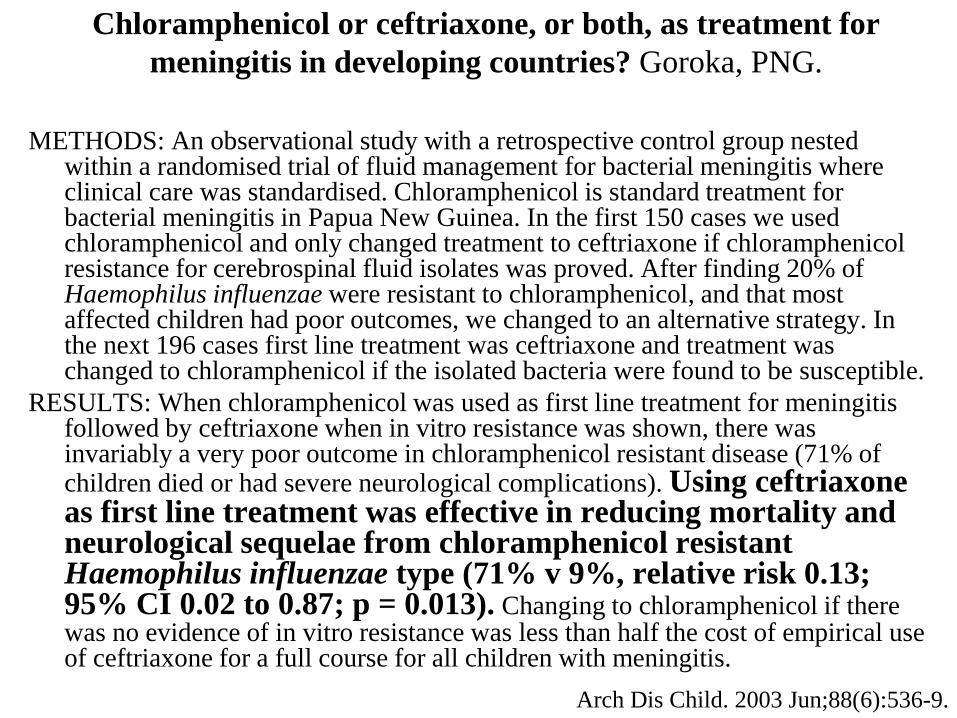
Chloramphenicol or ceftriaxone, or both, as treatment for
meningitis in developing countries? Goroka, PNG.
METHODS: An observational study with a retrospective control group nested within a randomised trial of fluid management for bacterial meningitis where clinical care was standardised. Chloramphenicol is standard treatment for bacterial meningitis in Papua New Guinea. In the first 150 cases we used chloramphenicol and only changed treatment to ceftriaxone if chloramphenicol resistance for cerebrospinal fluid isolates was proved. After finding 20% of Haemophilus influenzae were resistant to chloramphenicol, and that most affected children had poor outcomes, we changed to an alternative strategy. In the next 196 cases first line treatment was ceftriaxone and treatment was changed to chloramphenicol if the isolated bacteria were found to be susceptible.
RESULTS: When chloramphenicol was used as first line treatment for meningitis followed by ceftriaxone when in vitro resistance was shown, there was invariably a very poor outcome in chloramphenicol resistant disease (71% of children died or had severe neurological complications). Using ceftriaxone as first line treatment was effective in reducing mortality and neurological sequelae from chloramphenicol resistant Haemophilus influenzae type (71% v 9%, relative risk 0.13; 95% CI 0.02 to 0.87; p = 0.013). Changing to chloramphenicol if there was no evidence of in vitro resistance was less than half the cost of empirical use of ceftriaxone for a full course for all children with meningitis.
Arch Dis Child. 2003 Jun;88(6):536-9.

Accuracy of initial clinical diagnosis of acute
bacterial meningitis in children from a malaria-
endemic area of Papua New Guinea.
The diagnosis of acute bacterial meningitis (ABM) is challenging in resource-
limited settings where cerebral malaria and viral encephalitis are also common.
METHODS: To assess the accuracy of an initial clinical diagnosis of ABM in a
malaria-endemic area of Papua New Guinea (PNG), a retrospective chart review
of hospitalized children aged 2 months to 10 years was conducted. RESULTS: Of
the 481 eligible children, 240 had an initial clinical diagnosis of ABM that was
confirmed independently by trained research staff under standardized conditions,
with laboratory support in only 84 (17.5%; 84/481). When compared with the final
laboratory-confirmed diagnosis, an initial diagnosis of ABM had a sensitivity,
specificity, positive predictive value and negative predictive value of 76% (95%
CI 66-85%), 56% (95% CI 51- 61%), 27% (95% CI 21-33%) and 92% (95% CI
87- 95%), respectively. There was discordance between initial and final diagnosis
of ABM in 196 children; 176 initially considered to have ABM had an alternative
diagnosis, while 20 without an initial diagnosis of ABM were confirmed to have
ABM. CONCLUSION: These data show that initial misdiagnosis of ABM is
common in a malaria-endemic area of PNG. A diagnostic algorithm using
standardized assessment for meningeal irritation, coma and malaria parasitological
testing needs further evaluation in this setting.
Trans R Soc Trop Med Hyg 2014


