1 Co-management of Cataract Surgery Bill Tullo, OD, FAAO, Dipl CCL&R Vice-President of Clinical Services Cataracts • Each year about 2.8 million cataract operations are performed, making it the most common operation in the U.S. for people over age 65. Appropriateness • “Primary indication for surgery is when visual function no longer meets the patients needs and for which cataract surgery provides a reasonable likelihood of improvement or when lens opacity inhibits optimal management of posterior segment disease or the lens is causing inflammation or unmanageable glaucoma” • American Academy of Ophthalmology
Transcript
1
Co-management of Cataract Surgery
Bill Tullo, OD, FAAO, Dipl CCL&R
Vice-President of Clinical Services
Cataracts
• Each year about 2.8 million cataract operations
are performed, making it the most common
operation in the U.S. for people over age 65.
Appropriateness
• “Primary indication for surgery is when visual
function no longer meets the patients needs and
for which cataract surgery provides a reasonable
likelihood of improvement or when lens opacity
inhibits optimal management of posterior
segment disease or the lens is causing
inflammation or unmanageable glaucoma”• American Academy of Ophthalmology
2
Pre Operative Orders
Begin 4-7 days before surgery:
Topical Antibiotic
Topical NSAI
Lid Hygiene
Systemic Health Considerations
• Clearance by PCP required
• Full eval including EKG must be within 30 days of
procedure
• Some surgeons no longer require routine preoperative
medical testing
The Value of Routine Preoperative Medical Testing before Cataract Surgery: Oliver D. Schein, M.D., M.P.H., Joanne Katz, Sc.D., Eric B. Bass,
M.D., M.P.H., James M. Tielsch, Ph.D., Lisa H. Lubomski, Ph.D., Marc A. Feldman, M.D., M.P.H., Brent G. Petty, M.D., and Earl P. Steinberg, M.D.,
M.P.P. for the Study of Medical Testing for Cataract Surgery: N Engl J Med 2000; 342:168-175 January 20, 2000
Cataract Surgery
• Unilateral
• 2nd eye usually done 1-4 weeks after 1st eye
• Performed in hospital or Ambulatory Surgical
Center (ASC)
3
Cataract Surgery Preparation
• Contact Lens Removal
• D/C RGP 1 to ? Months..check stability
• Helpful to change patient to soft lens if
considering referral within 6 months
• D/C soft lens….1-2 weeks …surgeon dependent
Pre-op Medications
Topical NSAID
Topical Antibiotic
Lid hygiene
Pre-operative Testing
• Uncorrected and Best Corrected VA (monocular and binocular)
• Pinhole VA
• Binocular Status
• EOM’s
• Pupils
• Manifest Refraction
• Cycloplegic Refraction
• Slit lamp Biomicroscopy
• Tonometry
• Dilated retinal exam
• Manual Keratometry
• Topography
• Tomography
• IOL Master – A-scan
• OCT*
Informed Consent
• Risks
• Benefits
• Alternatives
• Medications
• Standard Orders
• When to Make Changes
4
RISKS
• Blindness
• Endophthalmitis
• Retinal Detachment
• Corneal
Decompensation
• CME
• Refractive Surprise
• Dryness
• NVD’s/Dysphotopsia
• Secondary Cataract
Surgery
• Anesthesia
• Topical (tetracaine, proparacaine,lidocaine)
• Advantage – low complication
• Disadvantage – no EOM akinesis, disocmfort
• Retrobulbar (lidocaine, bupivacaine)
• Advantage – total EOM akinesis
• Disadvantage – possible globe perforation (long
eyes)
Surgery
• Anesthesia
• Sub-tenon’s block
• Advantages – safe, total EOM akinesis
• Disadvantages - none
• General Anesthesia
• Advantages - No pain, total sedation
• Disadvantages - Complications, Cost
• Intracameral – added when discomfort
5
Anesthesia
• IV Versed (benzodiazepine)
• Topical proparacaine
• NPO …no food or drink after dinner day before
surgery
Incision
• Type• Scleral tunnel
• Less endothelial damage
• Easy to enlarge
• Must have retro-block
• Lower endophthalmitis*
• Location• Astigmatism, ocular
disease, ergonomics
• Size• > 4.0mm - large
• 4.0mm – 2.0mm - small
• < 2.0mm - micro
• Clear corneal• Undisturbed
conjunctiva
• No retro-block needed
• Less bleeding
• Astigmatism control
Pre-op Medications
• Pupil Dilation
• 1% cyclopentolate +
• 2.5% phenylephrine
• Dosage 1-2 drops x 10-15minutes 1 hour prior to
surgery
6
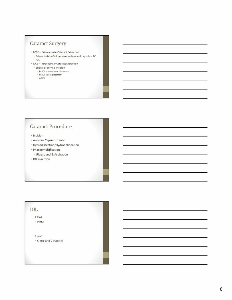
Cataract Surgery
• ECCE – Extracapsular Cataract Extraction
• Scleral incision 5-8mm remove lens and capsule – AC
IOL
• ICCE – Intracapsular Cataract Extraction
• Scleral or corneal Incision
• PC IOL intracapsular placement
• PC IOL sulcus placement
• AC IOL
Cataract Procedure
• Incision
• Anterior Capsulorrhexis
• Hydrodissection/Hydrodelineation
• Phacoemulsification
• Ultrasound & Aspiration
• IOL insertion
IOL
• 1 Part
• Plate
• 3 part
• Optic and 2 Haptics
7
Surgery
• IOL (Silicon vs. Acrylic)
• Monofocal
• DV vs. Monovision
• Spherical vs. Aspheric
• Multifocal
• Refractive
• Diffractive
• Accommodative
IOL Calculations
• Biometry – Axial Length
• A-scan
• Immersion (no corneal compression)
• IOL Master (optical no touch)
• Corneal Curvature
• Keratometry – manual
• Auto-keratometry
• Topography/Tomography
IOL Calculation - Formulas
• SRK/T
• Axial Length >22.01mm
• Holladay II
• Axial Length < 22.00mm
• K’s flatter than 42.00D
• K’s Steeper than 47.00D
• Hoffer Q (short eyes)
8
Cataract Post Operative Care
Cataract Post Operative Care
• Antibiotic
• NSAID
• Steroid
Cataract Post Operative Care
Antibiotic Medication
Most Commonly used are the 4th
generation fluoroquinolone class of
antibiotics
Resistance in older classes
Ex: Vigamox (moxifloxacin)
Zymaxid (gatifloxacin)
Besivance
(Besifloxacin)
9
Cataract Post Operative Care
Antibiotic Medication
• Dosing TID or greater
• Do Not Taper
• Typical Order:
Vigamox Oph Sol 5ml
1gtt OD TID 2 weeks
• Cipro, Levaquin, ect.
Rare (approximately 1.2 per 100,000 prescriptions) and is even
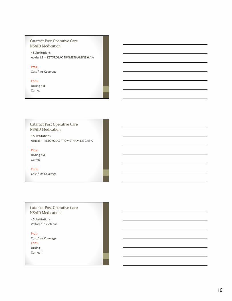
• Use of topical NSAIDs may result in keratitis. In
some susceptible patients, continued use of
topical NSAIDs may result in epithelial
breakdown, corneal thinning, corneal erosion,
corneal ulceration or corneal perforation.
• Discontinue NSAID immediately
• Use preservative free artificial tears
• Monitor closely
Topical Steroid
• Pred-forte 1%
(Prednisolone Acetate)
• Durezol 0.05%
(difluprednate)
• Lotemax (Loteprednol
Etabonate)
14
Cataract Post Operative Care
Steroid Medication
• Steroid Response
• The rise in IOP takes, on average, three weeks to
months. The decrease in IOP is also slow, taking
weeks to resolve.
• Can occur as fast as 7 days
Cataract Post Operative Care
Steroid Medication
Prednisolone Acetate 1%
Typical Order:
Pred-forte 1% Oph Sol
5ml
1gtt OD TID 2 wk then BID for 2 wk
http://www.drugs.com/pdr/images/O05103B4.jpg
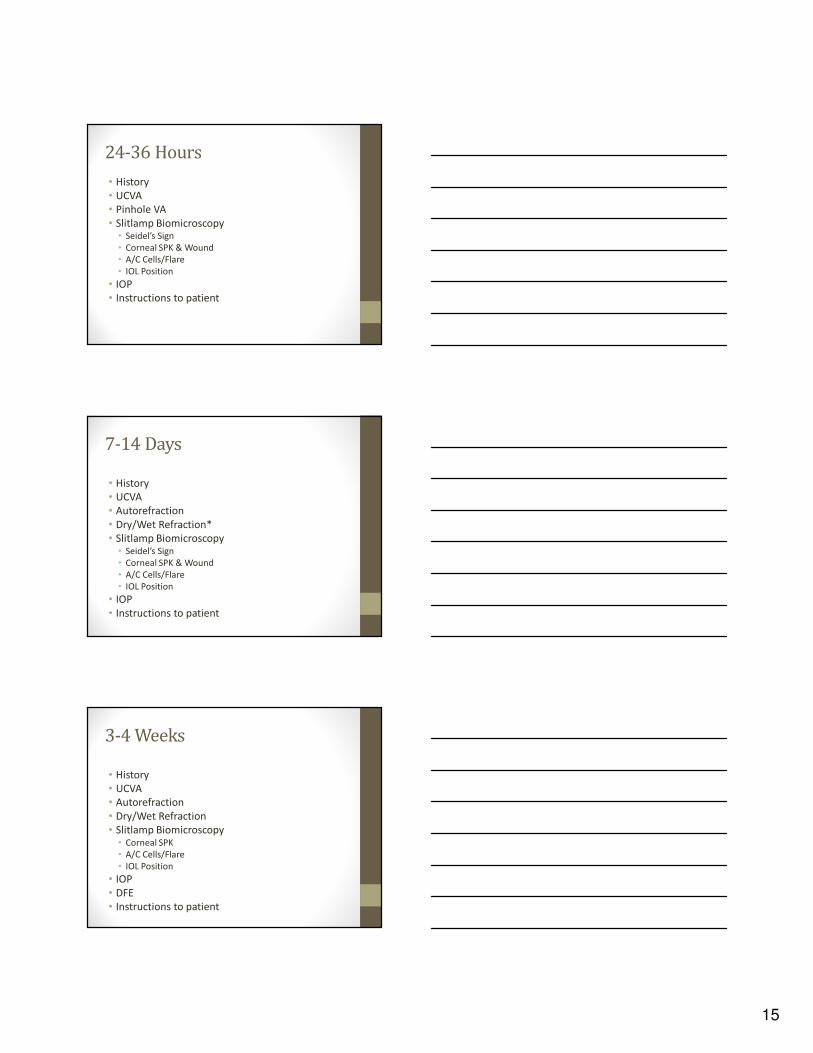
Postoperative Visits
• 24-36 Hours
• 7-14 Days
• 3-4 Weeks
• 8-12 Weeks
15
24-36 Hours
• History
• UCVA
• Pinhole VA
• Slitlamp Biomicroscopy• Seidel’s Sign
• Corneal SPK & Wound
• A/C Cells/Flare
• IOL Position
• IOP
• Instructions to patient
7-14 Days
• History
• UCVA
• Autorefraction
• Dry/Wet Refraction*
• Slitlamp Biomicroscopy• Seidel’s Sign
• Corneal SPK & Wound
• A/C Cells/Flare
• IOL Position
• IOP
• Instructions to patient
3-4 Weeks
• History
• UCVA
• Autorefraction
• Dry/Wet Refraction
• Slitlamp Biomicroscopy• Corneal SPK
• A/C Cells/Flare
• IOL Position
• IOP
• DFE
• Instructions to patient
16
8-12 Weeks
• History
• UCVA
• Autorefraction
• Dry Refraction
• Slitlamp Biomicroscopy• Corneal SPK
• A/C Cells/Flare
• IOL Position
• Capsule Clarity
• IOP
• Instructions to patient
Early Complications -
Significant
• Wound Leak – flat chamber
• Ocular Hypertension
• Endophthalmitis
• Iris Prolapse/Vitreous in wound
• IOL dislocation/vaulting
• Retinal/Choroidal Detachment
Early Complications – Less Urgent
• Wound leak – normal chamber
• Ptosis
• Diplopia
• Corneal Edema
• Hyphema
• Hypopyon
• Pupillary Capture
17
Late Complications
• Ocular Hypertension/Glaucoma
• Ptosis/Diplopia
• Corneal Edema/Decompensation
• Late Hyphema
• Chronic Uveitis
• PCO
• CME
Sutures
• Most common reason is wound burn
• Will not dissolve
• Can stay in indefinitely
• If FB sensation send back to surgeon at 1-2
months to consider removal
• Lubricate while awaiting removal decision
Cataract Post Op Pearls
Wound Leak - Hypotony
• Shallow Chamber
• Positive Seidel• Clearing of the fluorescein over the
involved area due to the leak of fluid.
• Usually noticed DOS
• May not find this until 1 wk P/O
• Really need to get this patient back to surgeon/center ASAP
18
Cataract Complications
• IOP Pressure Spike
• Types:
• Visco (Healon) / left over nucleus or cortex
• Spike first 24 hours
• Steroid responder
• Spike 1-3 weeks
• Iritis (inflammatory glaucoma)
• Spike 3-5 weeks
Cataract Post Op Pearls
Early High IOP
• 1st week• Retained visco-elastic
• Retained Lens Fragments
• Nucleus
• Retinal Specialist Consult –Vitrectomy
• Cortex
• TX:
• Monitor
• IOP management
• Return to OR
• Must R/O infectious endophthalmitis.
Cataract Post Op Pearls
• High intra-ocular pressure 1st week
• First try Alphagan P and Cosopt 2 drops 10-15 min
apart..IOP check 30-45 min
• Consider Acetazolamide 250mg (may want to consult
patient’s PCP)
• “Wound burp” nor AC paracenthesis
Pressures can be high
Eye Ache, Blur, Light Sensitivity
Monitor IOP 1 day, then weekly until stable
19
Cataract Post Op Pearls
• High intra-ocular pressure 1 -3 wks
• Steroids taken continuously for long periods of time are well known to cause a rise in intra-ocular
pressure which can pose a risk for steroid induced
glaucoma.
• About 5% of the general population are "high steroid responder", where a large and potentially dangerous rise
in eye pressure occurs after daily steroid use for 4 to 6
weeks.
• Another third of the general population may experience a
more moderate rise in eye pressure over that time frame. The remaining two-thirds of the population has a minimal
rise in eye pressure.
Cataract Post Op Pearls
Treatment of High IOP 1-3 weeks with IOP lowering drugs:
Alphagan P 0.10% TID +
Cosopt BID
Trusopt (sulfa) & Timoptic (asthma COPD CHF)
Continue until steroid is D/C
Monitor IOP 1 day, then weekly until stable
Consider sending patient back to the operating surgeon
Cataract Post Op Pearls
Late High IOP 3-5 weeks
IRITIS
Usually within first few weeks after D/C of steroid
• Light Sensitivity
• Eye Ache
• Conjunctival Injection
• AC Cells/Flare
• Blurred Vision
• Increased IOP
20
Cataract Post Op Pearls
Rebound Iritis
• Restart Pred 1% q1h-qid
• Compliance?
• Consider Durezol qid –taper slowly
• Treat IOP if elevated
• Monitor 1 day, 1 week, 2-4 weeks
• Taper slowly! 4 weeks tid to bid did not work
Cataract Post Op Pearls
Infection
• The incidence of endophthalmitis has
been reported to be between 0.13% and
0.7%.
Mamalis N, Kearsley L, Brinton E. Postoperative endophthalmitis. Curr Opin Ophthalmol.
2002;13:14–18.
Cataract Post Op Pearls
Infection
• The primary source of this intraocular infection is
considered to be bacteria from the patient's
ocular surface (cornea, conjunctiva) or adnexa
(lacrimal glands, eyelids, and extraocular
muscles). Buzard K, Liapis S. Prevention of endophthalmitis. J Cataract Refract Surg.
2004;30:1953–1959 .
21
Cataract Post Op Pearls
Infection
• The bacteria most frequently isolated are gram-
positive coagulase-negative cocci (mainly
Staphylococcus epidermidis) which account for
70% of culture-positive cases. Staphylococcus
aureus is isolated in 10% of culture-positive
cases, Streptococcus species in 9%, Enterococcus
species in 2%, and other gram-positive species in
3% of cases.Mamalis N, Kearsley L, Brinton E. Postoperative endophthalmitis. Curr Opin Ophthalmol. 2002;13:14–
18.
Cataract Post Op Pearls
Infection
• Gram-negative bacteria account for just
6% of culture-positive cases; however, an
infection with these bacteria, particularly
with Pseudomonas aeruginosa, can lead to
a devastating visual outcome.
Mamalis N, Kearsley L, Brinton E. Postoperative endophthalmitis. Curr Opin Ophthalmol.
2002;13:14–18.
Cataract Post Op Pearls
Infection
• Surgical complications, in particular a torn posterior lens capsule, can significantly increase the risk of endophthalmitis.
Kamalarajah S, Ling R, Silvestri G, Sharma NK, Cole MD, Cran G, Best RM. Presumed infectious endophthalmitis following cataract surgery in the UK: a case-control study of risk factors. Eye. 2007;21:580–586.
• The choice of intraocular lens (IOL) can affect the risk of endophthalmitis. The use of IOLs with silicone optics is associated with an increased risk of endophthalmitis, compared with that of IOLs with acrylic optics.
• The incidence of retinal detachment increases after cataract extraction, but it decreases with improved surgical technique. Postoperative posterior vitreous detachment is a major promoter of retinal detachment after cataract surgery and is related to onset of most retinal tears leading to retinal detachment.
Curr Opin Ophthalmol. 2008 May;19(3):239-42
• In some cases however, when the vitreous detaches it pulls a tear in the retina, sometimes causing a small amount of bleeding. This may appear as a shower of tiny black spots in the vision.
http://www.alberta-retina.com/PVD.html
Cataract Post Op Pearls
Posterior Vitreous Detachment
• Important to diagnose and
treated immediately
• Tear can lead to a retinal
detachment
• Tear can usually be treated
with laser. http://www.alberta-retina.com/PVD.html
By the time you first see the hemorrhage, the bleeding has already stopped. The blood will gradually disappear by itself, but it may take as long as two
weeks to absorb completely.
25
Cataract Post Op Pearls
PCO
The lens capsule is the thin, elastic-like bag that holds the intraocular lens (IOL) in position after cataract surgery. During the operation, the front (anterior) portion of the lens capsule is carefully opened and the cataract is removed. The IOL is inserted into the remaining (posterior) portion of the capsule.http://www.stlukeseye.com/conditions/PChaze.asp
Cataract Post Op Pearls
Posterior Capsule
Opacification
http://www.stlukeseye.com/conditions/PCha
ze.asp
Cataract Post Op Pearls
PCO
Posterior capsular opacification (PCO), which is the most common complication of cataract surgery occurring in up to 50% of patients by 2 to 3 years after the operation. PCO is caused by lens epithelial cells retained in the capsular bag following surgery which then proliferate, migrate and transform to myofibroblasts.
Eye. 1999 Jun;13 ( Pt 3b):489-92.
Posterior capsular opacification affects about 1 in 4 people within 5 years of having cataract surgery. The cloudiness may develop gradually over several months or years.
Schaumberg DA, et al. (1998). A systematic overview of the incidence of posterior capsule opacification. Ophthalmology, 105(7): 1213–1221.
26
Cataract Post Op Pearls
PCO
• The most common cause of posterior capsule
opacification (PCO) is proliferation and migration
of retained lens epithelial cells and their
derivatives into the visual axis. http://herkules.oulu.fi/isbn9514259793/html/c169.html
Cataract Post Op Pearls
PCO
Treatment YAG capsulotomy
“yttrium aluminum garnet”
The principle of Nd: YAG laser capsulotomy in treating
PCO is to cause photodisruption at extremely high
energy levels, thereby disintegrating tissues (Bell &
Landt 1967, Barnes & Rieckhoff 1968, Fradin et al.
1973, Aron-Rosa et al. 1981, Ficker & Steele 1985). http://herkules.oulu.fi/isbn9514259793/html/c169.html
Cataract Post Op Pearls
PCO
• This is an outpatient procedure and involves no incision.
• Using the laser beam, the physician makes an opening in the clouded capsule to let light through.
• After the procedure the patient remains in the center for an hour to be sure that pressure in the eye is not elevated.
• An eye examination for any complications should follow at 1 week.
• Nd:YAG laser posterior capsulotomy is not used to prevent clouding of the back lining of the lens capsule (posterior capsule opacification). There is no way to know who will get clouding in the back of the eye after cataract surgery. Certain lenses used in the surgery to remove the cataract may lower this risk and the need for laser surgery later.
Cataract Post Op Pearls
CME
• Cystoid Macular Edema
About 3% of patients undergoing cataract
extraction will have visual reduction due to CME
within the first postoperative year.
Coscas G, Gaudric A. Natural course of nonaphakic cystoid macular edema.
Surv Ophthalmol 1984 May;28 Suppl:471-84.
28
Cataract Post Op Pearls
CME
Visual acuity may or may not be reduced. If
reduced, vision ranges from 20/25 to 20/400
depending on the severity of the edema.
Patients may also experience metamorphopsia. http://www.revoptom.com/HANDBOOK/oct02_sec5_1.htm
Cataract Post Op Pearls
CME
• Fluorescein angiography most effectively displays
true appearance of CME, demonstrating leaky
perifoveal capillaries in the early stage with a
petalloid flower appearance in late phases. http://www.revoptom.com/HANDBOOK/oct02_sec5_1.htm
CME TREATMENT
Topical nonsteroidal medications
Acular (ketorolac, Allergan) and Voltaren (diclofenac, Novartis Ophthalmics)
Topical corticosteroid drops such as Pred Forte (prednisolone acetate, Allergan) and Lotemax (loteprednol, Bausch & Lomb).
Common dosing ranges from qid to q2h. Often a loading dose of q2h is indicated, and then rapidly dropped to qid after several days.
http://www.revoptom.com/HANDBOOK/oct02_sec5_1.htm
29
Cataract Post Op Pearls
CME
• New advances in diagnostic technology make it easier to identify CME. The Heidelberg Retinal Tomograph II and the Optical Coherence Tomography are effective and noninvasive devices that can identify CME.