Coagulopathy and Thrombosis as a Result of SevereCOVID-19 Infection: A Microvascular FocusUpendra K. Katneni1 Aikaterini Alexaki2 Ryan C. Hunt2 Tal Schiller3 Michael DiCuccio4
Paul W. Buehler1 Juan C. Ibla5 Chava Kimchi-Sarfaty2
1Department of Pediatrics, The Center for Blood Oxygen Transportand Hemostasis, University of Maryland School of Medicine,Baltimore, Maryland, United States
2Hemostasis Branch, Division of Plasma Protein Therapeutics, Officeof Tissues and Advanced Therapies, Center for Biologics Evaluation &Research, U.S. FDA, Silver Spring, Maryland, United States
3Diabetes, Endocrinology and Metabolic Disease Unit, Kaplan MedicalCenter, Rehovot, Israel
4National Center of Biotechnology Information, National Institutes ofHealth, Bethesda, Maryland, United States
5Division of Cardiac Anesthesia, Department of Anesthesiology,Perioperative and Pain Medicine, Boston Children’s Hospital andHarvard Medical School, Boston, Massachusetts, United States
Thromb Haemost 2020;120:1668–1679.
Address for correspondence Chava Kimchi-Sarfaty, HemostasisBranch, Division of Plasma Protein Therapeutics, Office of Tissues andAdvanced Therapies, Center for Biologics Evaluation & Research,US FDA, Silver Spring, MD 20993, United States(e-mail: [email protected]).
Keywords
► COVID-19► thrombosis► inflammation► ADAMTS-13► von Willebrand factor
Abstract Coronavirus disease of 2019 (COVID-19) is the clinical manifestation of the respiratoryinfection caused by severe acute respiratory syndrome coronavirus 2 (SARS-CoV-2).While primarily recognized as a respiratory disease, it is clear that COVID-19 is systemicillness impactingmultiple organ systems. One defining clinical feature of COVID-19 hasbeen the high incidence of thrombotic events. The underlying processes and riskfactors for the occurrence of thrombotic events in COVID-19 remain inadequatelyunderstood. While severe bacterial, viral, or fungal infections are well recognized toactivate the coagulation system, COVID-19-associated coagulopathy is likely to haveunique mechanistic features. Inflammatory-driven processes are likely primary driversof coagulopathy in COVID-19, but the exact mechanisms linking inflammation todysregulated hemostasis and thrombosis are yet to be delineated. Cumulative findingsof microvascular thrombosis has raised question if the endothelium and microvascula-ture should be a point of investigative focus. von Willebrand factor (VWF) and itsprotease, a disintegrin and metalloproteinase with a thrombospondin type 1 motif,member 13 (ADAMTS-13), play important role in the maintenance of microvascularhemostasis. In inflammatory conditions, imbalanced VWF-ADAMTS-13 characterizedby elevated VWF levels and inhibited and/or reduced activity of ADAMTS-13 has beenreported. Also, an imbalance between ADAMTS-13 activity and VWF antigen isassociated with organ dysfunction and death in patients with systemic inflammation.A thorough understanding of VWF-ADAMTS-13 interactions during early and advancedphases of COVID-19 could help better define the pathophysiology, guide thrombo-prophylaxis and treatment, and improve clinical prognosis.
receivedMay 14, 2020accepted after revisionJuly 14, 2020
Coronavirus diseaseof 2019 (COVID-19) is a respiratory illnesscaused by a novel coronavirus, severe acute respiratory syn-drome coronavirus 2 (SARS-CoV-2). SARS-CoV-2 is an envel-oped, positive-sense single-stranded ribonucleic acid virusbelonging to the Coronaviridae family.1 The COVID-19 out-break started inWuhan, China, in late 2019 and rapidly spreadto rest of the world. On March 11, 2020, the World HealthOrganization declared COVID-19 outbreak as pandemic. As ofJune 24, 2020, the global number of COVID-19 cases stood at9.26 million with 478,000 deaths (Source: Johns HopkinsCoronavirus Resource Center, https://coronavirus.jhu.edu/).Disease course is markedly different between individualswhile some are completely asymptomatic, others developmild symptoms including mild fever, loss of taste or smell,dry cough, sore throat, shortness of breath, andmyalgia.2–4 Insusceptible individuals, the disease progresses to pneumonia,hypoxemia, acute respiratory distress, and multiorgan dys-function that may lead to death.3 The predominance ofasymptomatic or mild infections has contributed to the rapidspread of COVID-19 compared with earlier coronavirus out-breaks of SARS andMiddle East respiratory syndrome in 2002and 2012, respectively.4,5
Consumptive Coagulopathy and the HighIncidence of Thrombosis in COVID-19 Patients
Altered coagulation is a common feature of acute systemicdiseases, specifically to those affecting primarily the respi-ratory system. Based on studies in patients with acuterespiratory distress syndrome (ARDS), the coexistence ofdisseminated intravascular coagulation (DIC) with subse-quent consumption of procoagulation proteins and plateletshas been consistently described.6 This in turn leads to theformation of microthrombi in the vascular bed of organsresulting from excess coagulation byproducts and suppres-sion of endogenous anticoagulation factors.7 The coexistenceof consumptive coagulopathy and thrombosis are the resultof a common pathologic pathway; however, the exact mech-anisms that tilts the balance toward thrombosis in COVID-19are less well understood.8 In this sense, some features of thecoagulopathy associated with COVID-19 may be not uniqueto this disease; however, the magnitude of the thromboticresponse and its impact on mortality suggests the presenceof additional mechanisms, beyond what is known for similarrespiratory acute inflammatory diseases.
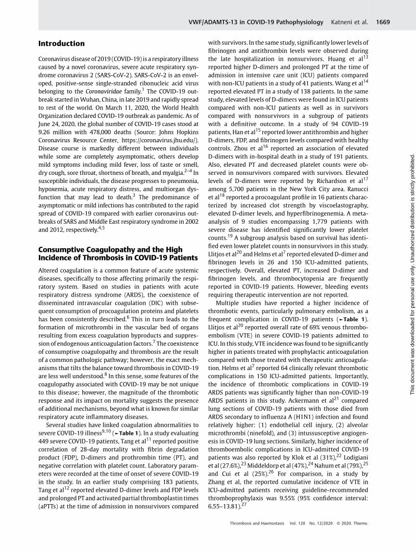
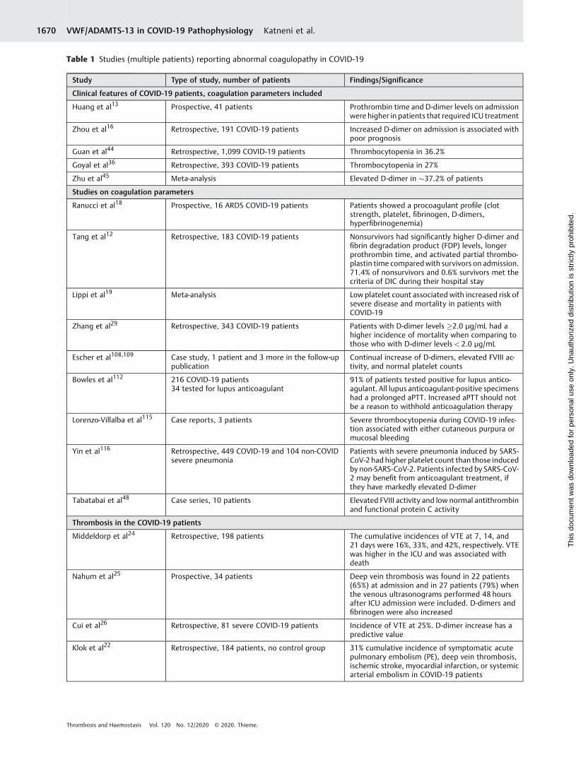
Several studies have linked coagulation abnormalities tosevere COVID-19 illness9,10 (►Table 1). In a study evaluating449 severe COVID-19 patients, Tang et al11 reported positivecorrelation of 28-day mortality with fibrin degradationproduct (FDP), D-dimers and prothrombin time (PT), andnegative correlation with platelet count. Laboratory param-eters were recorded at the time of onset of severe COVID-19in the study. In an earlier study comprising 183 patients,Tang et al12 reported elevated D-dimer levels and FDP levelsand prolonged PT and activated partial thromboplastin times(aPTTs) at the time of admission in nonsurvivors compared
with survivors. In the same study, significantly lower levels offibrinogen and antithrombin levels were observed duringthe late hospitalization in nonsurvivors. Huang et al13
reported higher D-dimers and prolonged PT at the time ofadmission in intensive care unit (ICU) patients comparedwith non-ICU patients in a study of 41 patients. Wang et al14
reported elevated PT in a study of 138 patients. In the samestudy, elevated levels of D-dimers were found in ICU patientscompared with non-ICU patients as well as in survivorscompared with nonsurvivors in a subgroup of patientswith a definitive outcome. In a study of 94 COVID-19patients, Han et al15 reported lower antithrombin and higherD-dimers, FDP, and fibrinogen levels compared with healthycontrols. Zhou et al16 reported an association of elevatedD-dimers with in-hospital death in a study of 191 patients.Also, elevated PT and decreased platelet counts were ob-served in nonsurvivors compared with survivors. Elevatedlevels of D-dimers were reported by Richardson et al17
among 5,700 patients in the New York City area. Ranucciet al18 reported a procoagulant profile in 16 patients charac-terized by increased clot strength by viscoelastography,elevated D-dimer levels, and hyperfibrinogenemia. A meta-analysis of 9 studies encompassing 1,779 patients withsevere disease has identified significantly lower plateletcounts.19 A subgroup analysis based on survival has identi-fied even lower platelet counts in nonsurvivors in this study.Llitjos et al20 andHelms et al7 reported elevatedD-dimer andfibrinogen levels in 26 and 150 ICU-admitted patients,respectively. Overall, elevated PT, increased D-dimer andfibrinogen levels, and thrombocytopenia are frequentlyreported in COVID-19 patients. However, bleeding eventsrequiring therapeutic intervention are not reported.
Multiple studies have reported a higher incidence ofthrombotic events, particularly pulmonary embolism, as afrequent complication in COVID-19 patients (►Table 1).Llitjos et al20 reported overall rate of 69% venous thrombo-embolism (VTE) in severe COVID-19 patients admitted toICU. In this study, VTE incidencewas found to be significantlyhigher in patients treated with prophylactic anticoagulationcompared with those treated with therapeutic anticoagula-tion. Helms et al7 reported 64 clinically relevant thromboticcomplications in 150 ICU-admitted patients. Importantly,the incidence of thrombotic complications in COVID-19ARDS patients was significantly higher than non-COVID-19ARDS patients in this study. Ackermann et al21 comparedlung sections of COVID-19 patients with those died fromARDS secondary to influenza A (H1N1) infection and foundrelatively higher: (1) endothelial cell injury, (2) alveolarmicrothrombi (ninefold), and (3) intussusceptive angiogen-esis in COVID-19 lung sections. Similarly, higher incidence ofthromboembolic complications in ICU-admitted COVID-19patients was also reported by Klok et al (31%),22 Lodigianiet al (27.6%),23Middeldorp et al (47%),24Nahumet al (79%),25
and Cui et al (25%).26 For comparison, in a study byZhang et al, the reported cumulative incidence of VTE inICU-admitted patients receiving guideline-recommendedthromboprophylaxis was 9.55% (95% confidence interval:6.55–13.81).27
Table 1 Studies (multiple patients) reporting abnormal coagulopathy in COVID-19
Study Type of study, number of patients Findings/Significance
Clinical features of COVID-19 patients, coagulation parameters included
Huang et al13 Prospective, 41 patients Prothrombin time and D-dimer levels on admissionwere higher in patients that required ICU treatment
Zhou et al16 Retrospective, 191 COVID-19 patients Increased D-dimer on admission is associated withpoor prognosis
Guan et al44 Retrospective, 1,099 COVID-19 patients Thrombocytopenia in 36.2%
Goyal et al36 Retrospective, 393 COVID-19 patients Thrombocytopenia in 27%
Zhu et al45 Meta-analysis Elevated D-dimer in �37.2% of patients
Studies on coagulation parameters
Ranucci et al18 Prospective, 16 ARDS COVID-19 patients Patients showed a procoagulant profile (clotstrength, platelet, fibrinogen, D-dimers,hyperfibrinogenemia)
Tang et al12 Retrospective, 183 COVID-19 patients Nonsurvivors had significantly higher D-dimer andfibrin degradation product (FDP) levels, longerprothrombin time, and activated partial thrombo-plastin time comparedwith survivors on admission.71.4% of nonsurvivors and 0.6% survivors met thecriteria of DIC during their hospital stay
Lippi et al19 Meta-analysis Low platelet count associated with increased risk ofsevere disease and mortality in patients withCOVID-19
Zhang et al29 Retrospective, 343 COVID-19 patients Patients with D-dimer levels �2.0 µg/mL had ahigher incidence of mortality when comparing tothose who with D-dimer levels< 2.0 µg/mL
Escher et al108,109 Case study, 1 patient and 3 more in the follow-uppublication
Continual increase of D-dimers, elevated FVIII ac-tivity, and normal platelet counts
Bowles et al112 216 COVID-19 patients34 tested for lupus anticoagulant
91% of patients tested positive for lupus antico-agulant. All lupus anticoagulant-positive specimenshad a prolonged aPTT. Increased aPTT should notbe a reason to withhold anticoagulation therapy
Lorenzo-Villalba et al115 Case reports, 3 patients Severe thrombocytopenia during COVID-19 infec-tion associated with either cutaneous purpura ormucosal bleeding
Yin et al116 Retrospective, 449 COVID-19 and 104 non-COVIDsevere pneumonia
Patients with severe pneumonia induced by SARS-CoV-2 had higher platelet count than those inducedby non-SARS-CoV-2. Patients infected by SARS-CoV-2 may benefit from anticoagulant treatment, ifthey have markedly elevated D-dimer
Tabatabai et al48 Case series, 10 patients Elevated FVIII activity and low normal antithrombinand functional protein C activity
Thrombosis in the COVID-19 patients
Middeldorp et al24 Retrospective, 198 patients The cumulative incidences of VTE at 7, 14, and21 days were 16%, 33%, and 42%, respectively. VTEwas higher in the ICU and was associated withdeath
Nahum et al25 Prospective, 34 patients Deep vein thrombosis was found in 22 patients(65%) at admission and in 27 patients (79%) whenthe venous ultrasonograms performed 48 hoursafter ICU admission were included. D-dimers andfibrinogen were also increased
Cui et al26 Retrospective, 81 severe COVID-19 patients Incidence of VTE at 25%. D-dimer increase has apredictive value
Klok et al22 Retrospective, 184 patients, no control group 31% cumulative incidence of symptomatic acutepulmonary embolism (PE), deep vein thrombosis,ischemic stroke, myocardial infarction, or systemicarterial embolism in COVID-19 patients
VWF/ADAMTS-13 in COVID-19 Pathophysiology Katneni et al.1670
Thi
s do
cum
ent w
as d
ownl
oade
d fo
r pe
rson
al u
se o
nly.
Una
utho
rized
dis
trib
utio
n is
str
ictly
pro
hibi
ted.
Table 1 (Continued)
Study Type of study, number of patients Findings/Significance
Zhang et al27 Prospective, 281 ICU COVID-19 patients Cumulative incidence of VTE at 28 days was 9.55%,despite all patients receiving thromboprophylaxis
Demelo-Rodríguez et al117 Prospective, 156 COVID-19 patients D-dimer levels> 1,570 ng/mL were associated withasymptomatic DVT
Grandmaison et al118 Cross-sectional study, 58 COVID-19 patients, 29 inthe ICU and 29 in the medicine ward
In the ICU, VTEs were found in 17 (58.6%) of the 29patientsIn the medicine ward, VTEs were found in 6 (20.7%)patients
Fraissé et al119 Retrospective, 92 ICU COVID-19 patients High rate of thrombotic events (TEs) in ICU COVID-19 patients highlighting the necessity for throm-boprophylaxis and TE screening. Hemorrhagicevents (HEs) were also observed in patients on full-dose anticoagulation
Jian et al114 Retrospective, 3,218 COVID-19 patients Acute stroke was the most common neuroimagingfinding, present in 1.1% of hospitalized COVID-19patients
Desborough et al121 Retrospective, 66 patients 10 patients had at least one proven episode ofthromboembolism. Major bleeding occurred inseven cases
Akel et al122 Case reports, 6 patients Patients did not have any hypercoagulable riskfactors yet presented with pulmonary embolism
Kashi et al123 Case reports, 7 patients Arterial thrombosis
Lax et al124 Prospective autopsy study, 11 deceased COVID-19patients
Death may be caused by the thrombosis observedin segmental and subsegmental pulmonary arterialvessels despite the use of prophylacticanticoagulation
Thomas et al125 Retrospective, 63 COVID-19 patients High thrombotic risk in patients with COVID-19
Gomez-Arbelaez et al126 Case reports, 4 patients Aortic thrombosis and associated ischemic com-plications in patients with severe SARS-CoV-2infection
Anticoagulation treatment in COVID-19 patients
Tang et al11 Retrospective, 449 severe COVID-19 patients, 99received heparin
Anticoagulant therapy is associated with betterprognosis in severe COVID-19 patients with sepsisinduced coagulopathy or markedly elevated D-dimer
Wang et al28 3 case reports Treatment with tissue plasminogen activator leadto improvement in the respiratory status
Ayerbe et al127 2,075 COVID-19 patients, admitted in 17 hospitalsin Spain
Heparin had been used in 1,734 patients. Heparinwas associated with lower mortality
Wang et al128 Retrospective, 1,099 COVID-19 patients High risk of venous thromboembolism, also highrisk of bleeding
Artifoni et al129 Retrospective, 62 patients 16 patients developed VTE, 7 patients developedPEVery high negative predictive value of baseline D-dimer level for VTE and PE
Russo et al130 Retrospective, 192 COVID-19 patients Preadmission antithrombotic therapy, both anti-platelet and anticoagulant, does not seem to showa protective effect in severe forms of COVID-19with ARDS at presentation and rapidly evolvingtoward death
Link between SARS-CoV-2 and thrombosis
Ackermann et al21 7 lung autopsies from COVID-19 patients and 7from ARDS
Vascular angiogenesis distinguished the pulmo-nary pathobiology of COVID-19 from that of equallysevere influenza virus infection
Maier et al131 Case studies15 COVID-19 patients with hyperviscosity
Possible causal relationship between hyperviscosi-ty and thrombotic complications in COVID-19
VWF/ADAMTS-13 in COVID-19 Pathophysiology Katneni et al. 1671
Thi
s do
cum
ent w
as d
ownl
oade
d fo
r pe
rson
al u
se o
nly.
Una
utho
rized
dis
trib
utio
n is
str
ictly
pro
hibi
ted.
A high incidence of DIC diagnosed by D-dimer, fibrinogen,and antithrombin III levels has become a focus for the initiationof anticoagulation therapy in severe COVID-19 patients,28withsome studies relying on D-dimers alone.11,29 A retrospectiveanalysis of 183 patients performed by Tang et al12 suggestedthat more than 70% of severe COVID-19 patientswho succumbto the infection demonstrate increased risk of thrombosis,further this group suggests that all of these patients meet theInternational Society on Thrombosis and Haemostasis defini-tion of DIC. Subsequently, Tang et al11 reported an equivalent28-day mortality rate (30%) in 99 patients receiving lowmolecular weight or unfractionated heparin for 7 days com-paredwith 350 nonheparin treated patients or those receivinga less than 7-day course of therapy. A case series reported byWang et al28 detailed the use and outcome following tissueplasminogen activator (tPA) in three patients with ARDS andcoagulopathy consistent withDIC. Intravenous dosingwith tPAindicatedapotentialbenefit ineachof the threecasesofCOVID-19.However, this studyalsowarns of bothunrelatedeffectsandhigh risk of severe bleeding secondary to off-label tPA use.Several of the studies in coagulopathic COVID-19 patientssuspected of DIC rely heavily on analysis of fibrin degradationand D-dimer levels, which are expected to be increased duringDIC, arterial and venous thromboses, strokes, and thromboticmicroangiopathies.30 However, D-dimers are a nonspecificindicator of thrombosis in severe COVID-19 patients withpulmonary injury. Fibrin accumulation and lysis continuouslyoccur during nonthrombotic inflammation as well as tissuenecrosis, and therefore, significant D-dimer elevations alsoaccumulate during cancers31 and infections, consistent withinflammatory processes that coincide with the progression ofsevere COVID-19-related macrophage activation syndrome.32
Therefore, we suggest that more comprehensive and robustassays be used to evaluate changes in hemostasis. For example,to date the use of thrombin, plasmin, or simultaneous throm-bin/plasmin generation assays have not been reported withinthe context of hemostasis management of COVID-19 patients.Since their introduction thrombin and plasmin generationassays have been highly informative regarding the assessmentof hemorrhage, coagulation, and fibrinolysis.33,34 Assessmentof impairment of these systems would provide a useful andappropriateguidanceneeded for andmonitoringof therapeuticinterventions in the unique coagulopathies associated withCOVID-19.33,34 Because patients are often on unfractionated
or low molecular weight heparin and plasminogen activatorinhibitor 1, vonWillebrand factor (VWF), plasminogen, fibrin-ogen, and factor VIII are all reported to be elevated in SARSinfection,35 and therefore careful modification of these assaysmay bewarranted to optimize the concentrations of added tPA,tissue factor, and thrombomodulin.
These studies present a heterogeneous picture that is diffi-cult to evaluate in the aggregate. Inclusion criteria for patientsvaried across these studies, making direct comparisonsbetween thestudiesdifficult. Further, the studies useddifferentregimens of thromboprophylaxis, which could impact out-comes. In some studies, a high proportion of patients were stillhospitalized at the end of the reporting period; conclusions andclinical courses therefore were based on incomplete informa-tion, andcompletionof thesepatients’clinicalcoursecouldalterthe final conclusions. The picture of coagulopathy in COVID-19is complex. Specific, sensitive, and temporal assessments ofcoagulation and fibrinolysis should be established and furtherwork is needed to untangle the roles of the host inflammatoryresponse, preexisting thrombotic risk, and prehospitalizationpharmacologic regimens in the optimal management of coa-gulopathy in the setting of COVID-19.
Inflammation, Liver Injury, and Hypoxia inCOVID-19 Patients
The risk of hospitalization, morbidity, and mortality fromCOVID-19 is highest for older patients with preexisting con-ditions such as hypertension, diabetes, cardiovascular disease,and obesity.13,14,16,17,36,37Acommon themeof all these comor-bidities is their association with vascular inflammation andendothelial dysfunction.38,39 Proinflammatory conditionsaffect hemostasis by blocking of fibrinolysis and induction ofprothrombotic conditions through activation of endothelialcells and innate immune cells via release of several factorsincluding tissue factor, VWF, and neutrophil extracellular traps(NETs) that promote thrombosis.40 Induction of proinflamma-tory conditions was reported in the pathophysiology of severalviral diseases including influenza and SARS.41 Increasedinflammation is commonly observed in COVID-19 patients,while severe cases are characterized by immune dysregulationand hyperinflammation, with a markedly increased seruminterleukin (IL)-6.42 Cytokine release syndrome has also beenreported in COVID-19 patients and correlates with adverse
Table 1 (Continued)
Study Type of study, number of patients Findings/Significance
Huisman et al105 12 COVID-19 patients Low ADAMTS-13 activity, increased VWF levels andfactor VIII levels
Galeano-Valle et al111 Prospective study, 24 patients Prevalence of antiphospholipid antibodies inCOVID-19 and venous thrombosis was low
Magro et al132 Case reports, 5 severe COVID-19 cases Procoagulant state is associated with systemiccomplement activation
Abbreviations: ADAMTS-13, a disintegrin and metalloproteinase with a thrombospondin type 1 motif, member 13; aPTT, activated partialthromboplastin time; ARDS, acute respiratory distress syndrome; COVID-19, coronavirus disease of 2019; DIC, disseminated intravascularcoagulation; DVT, deep vein thrombosis; FVIII, factor VIII; ICU, intensive care unit; SARS-CoV-2, severe acute respiratory syndrome coronavirus 2;VTE, venous thromboembolism; VWF, von Willebrand factor.
VWF/ADAMTS-13 in COVID-19 Pathophysiology Katneni et al.1672
Thi
s do
cum
ent w
as d
ownl
oade
d fo
r pe
rson
al u
se o
nly.
Una
utho
rized
dis
trib
utio
n is
str
ictly
pro
hibi
ted.
clinical outcomes.43 The presence of several inflammatorymarkers such as C-reactive protein, procalcitonin, ferritin,and fibrinogen are often reported in COVID-19patients13,14,16,17,36,37,44–48 (►Table 2). Further, multiple stud-ies reported elevated levels of theproinflammatorycytokine IL-6 in severe cases of COVID-1916,37,42,47,49–53 (►Table 2). Aconcurrent increase in the levels of anti-inflammatory cytokineIL-10, probably in response to overwhelming systemic inflam-mation, was also observed in several studies. The role of IL-6, inparticular, is considered central in the pathogenesis of COVID-19complications,54and therefore tocilizumab,an IL-6 inhibitor,is being used in ongoing clinical trials to prevent catastrophicinflammation.55–58
Liver injury during COVID-19 infections was described inmultiple studies, including elevated levels of alanineaminotransferase, aspartate aminotransferase, and biliru-bin.14,16,17,36,44,47 The liver is the primary source of plasmaproteins, particularly those involved in hemostasis. Thus, theoccurrence of liver injury may contribute further to derange-ments of key hemostasis proteins and contributes to coagul-opathy.59 Similarly, hypoxemia observed in COVID-19patientsinduces prothrombotic conditions through upregulation ofplasminogenactivator inhibitor and stimulationofendothelialsynthesis of procoagulants, including tissue factor andVWF.60–63 Thus, multiple clinical characteristics observed inCOVID-19 patients contribute to altered coagulation and leadto increased incidence of thrombosis.However, the earlyonsetof coagulopathy—before systemic organic effects occur—sug-gests proinflammatory conditions as the primary drivingcause of thrombotic events in COVID-19 patients.
VWF-ADAMTS-13 in Hemostasis andThrombosis
VWF and its cleaving protease, a disintegrin and metal-loproteinase with a thrombospondin type 1 motif, member
13 (ADAMTS-13), play an important role in hemostasisparticularly within the microvasculature.64 VWF is a largemultimeric glycoprotein primarily expressed by endothelialcells and platelets. Endothelial cells show both basal secre-tion and regulated release of VWF stored in Weibel–Paladebodies in response to various stimuli. On the other hand,platelets secrete VWF stored in α-granules only upon activa-tion.65 ADAMTS-13 is expressed both by hepatic stellate cellsand endothelial cells; the relative contribution of hepatic andmicrovascular expression is not clear.66 ADAMTS-13 regu-lates the biological activity of VWF by cleaving prothrom-botic ultra-large VWF multimers (> 10,000 kDa) secretedfrom endothelial cells into hemostatically active high molec-ular weight multimers (< 10,000 kDa) under shear stressconditions.67 Severe deficiency of ADAMTS-13 results inaccumulation of ultra-large VWF multimers leading tomicrovascular thrombosis and consumptive thrombocyto-penia, a condition termed thrombotic thrombocytopenicpurpura (TTP).64 In the event of vascular injury, VWF facil-itates binding of platelets to subendothelium through itsinteractions with glycoprotein Ib and collagen, therebyinducing thrombus formation.64 A reciprocal relationshipexists between VWF and ADAMTS-13 levels where elevatedcirculatory VWF antigen levels are associated with concomi-tant decrease in ADAMTS-13 activity and vice versa.68–70
Abnormal VWF-ADAMTS-13 ratios are implicated in arterialthrombosis,71 ischemic stroke,72,73 pediatric stroke,74 andperioperative thrombosis in infants.75 In addition, abnormalVWF/ADAMTS-13metabolismhas been positively associatedwith myocardial infarction in young women.76 It is worthhighlighting that in the case of perioperative thrombosis,elevated VWF even in the absence of significant deficiency ofADAMTS-13 was associated with thrombosis.75 Severehypoxia and acidosis likely caused a higher increase inVWF during cardiac surgery and were at higher risk ofthrombosis.75
Table 2 Studies reporting elevated inflammatory markers in COVID-19
Study Patient group (number of patients) comparison Elevated inflammatory markers
Huang et al13 ICU (13) vs. non-ICU (28) Procalcitonin, IL-1β, IFN-γ, IP10, and MCP1
Wang et al14 ICU (36) vs. non-ICU (102) Procalcitonin
Zhou et al16 Nonsurvivor (54) vs. survivor (137) Procalcitonin, ferritin, and IL-6
Richardson et al17 Relative to reference range (3066) Procalcitonin, ferritin, and CRP
Ruan et al37 Nonsurvivor (68) vs. survivor (82) CRP and IL-6
Giamarellos-Bourboulis et al42 Dysregulated (21) vs. intermediate state(26) of immune activation
CRP and IL-6
Chen et al47 Severe (�9) vs. moderate (�7) CRP, ferritin, IL-6, and TNF-α
Han et al49 COVID-19 patients (102) vs. controls (45) CRP, IL-6, TNF-α, and IFN-γ
Du et al50 Mild pneumonia (124) vs. no pneumonia(54) (pediatric patients)
Procalcitonin, IL-6, TNF-α, and IFN-γ
Wang et al52 SpO2 �90% (� 36) vs.SpO2< 90% (�7)
CRP and IL-6
Tan et al53 Severe (25) vs. mild/moderate 31) CRP and IL-6
Tabatabai et al48 Relative to reference range (10) Fibrinogen, CRP, and ferritin
VWF/ADAMTS-13 in COVID-19 Pathophysiology Katneni et al. 1673
Thi
s do
cum
ent w
as d
ownl
oade
d fo
r pe
rson
al u
se o
nly.
Una
utho
rized
dis
trib
utio
n is
str
ictly
pro
hibi
ted.
Elevated levels of VWF are found in several inflammatoryandmetabolic disorders including diabetes, obesity, and sicklecelldisease.77 Inpatientswithsystemic inflammatory responsesyndrome, activeVWFpredicted28-daymortality.78VWF is anacute-phase responseprotein releasedbyactivatedendothelialcells in response to inflammatory stimuli.77 Inflammatorycytokines, IL-8 and tumor necrosis factor-α induced the releaseof VWF from human umbilical vein endothelial cells.79 VWFreleased in inflammationbinds toNETs released fromactivatedneutrophils and recruits platelets and leukocytes to promotethrombosis.77 ADAMTS-13 deficiency in inflammatory condi-tions was demonstrated to promote VWF-dependent leuko-cyte adhesion and extravasation in mice.80
In patientswith systemic inflammation, ADAMTS-13 activitydecreases proportional to the inflammatory response; animbalance between ADAMTS-13 activity and VWF antigen isassociated with organ dysfunction and death.81,82 Dysregulatedhost response to infection including inflammation can result inseptic shock. In septic shock, ADAMTS-13 activity was signifi-cantly lower83–85andelevated ratioofVWFpropeptide (VWFpp)that is secretedalongwithultra-largeVWFmultimers in tobloodstream and ADAMTS-13 was associated with disease severity.86
In patients with DIC, ADAMTS-13 activity decreased with DICscore87 and VWFpp/ADAMTS-13 ratiowas significantly elevatedin nonsurvivors compared with survivors.88 An interestingobservation is that smoking, which is associated with adverseoutcomes in COVID-19 patients,89was also found to be associat-edwith decreased plasma ADAMTS-13 levels in a study of 3,244individuals.90 Increased expression of angiotensin-convertingenzyme2, theentryreceptor forSARS-CoV-2, in thesmall airwayepithelia of smokers was suggested as the potential mechanismfor increased risk of severe COVID-19 in smokers.91 Smoking isalso associated with increased inflammatory markers.92
The imbalance between ADAMTS-13 and VWF in height-ened inflammation could be a result of inhibition and/ordeficiency of ADAMTS-13 activity.93 The inhibition of VWFcleavage by ADAMTS-13 in inflammatory conditions wassuggested to bemediated by several mechanisms: (1) throm-bospondin-1 released from α-granules of activated plateletsby binding to the A2-A3 domain of VWF94,95; (2) α-defensinsreleased from neutrophils by binding to the A2 domain ofVWF96; and (3) oxidation of Met 1606 residue in theADAMTS-13 cleavage site of VWF.97 Moreover, nonphysio-logical high concentrations of IL-6 have been shown toinhibit cleavage of VWF by ADAMTS-13 in vitro under shearflow conditions.79 Granulocyte elastases, plasmin, andthrombin that are elevated in inflammatory conditions lowerADAMTS-13 activity through its proteolytic cleavage.98,99
VWF-ADAMTS-13 Interactions in COVID-19
Despite playing an important role in the maintenance of hemo-stasis and the occurrence of micro- and macrovascular throm-bosis, VWF-ADAMTS-13 interactions have not received muchinvestigative attention in the evaluation of COVID-19 patho-physiology, specifically in relation to elevated incidence of VTE.Importantly, reduced ADAMTS-13 activity has been shown tocorrelate with increased inflammation in multiple sys-
tems,100–102 while IL-6 has been shown to inhibit the cleavageof ultra-large VWF strings by ADAMTS-13 under flowing con-ditions.79,103 The authors could find only five studies evaluatingboth VWF and ADAMTS-13 levels in COVID-19 patients inliterature104–108 (►Table 3). Majority of these studies reportedlower ADAMTS-13 activity concurrent with higher VWF inCOVID-19 patients.104–107 In one of these studies, Bazzanet al104 reported lower ADAMTS-13 levels in 88 COVID-19patients compared with healthy controls (48.71� 18.7% vs.healthy control, 108� 9.1%; normal value 60–130%). Withinpatient cohort, lower ADAMTS-13 and higher VWF levels werefound in nonsurvivors (9/88) comparedwith survivors. Further,lower than 30% ADAMTS-13 activitywere significantly associat-edwithmortality in survivoranalysis.Huismanetal105observedlow ADAMTS-13 activity levels (0.48� 0.14 IU/mL against areference range of 0.61–1.31) in parallel with elevated VWFantigen and activity (� fourfold) in 12 ICU-admitted patients. Asimilar reduction in ADAMTS-13 and increased VWF levels wasalso reportedbyAdamet al106andLatimer et al107 in 4 adult and1 pediatric patients, respectively. On the other hand, Escheret al108 observed normal to lower-normal ADAMTS-13 levelsconcurrentlywith> 2.5-fold increase in VWFantigen and activ-ity in 3 ICU-admitted patients. Two other studies7,109 reportedVWF measurements alone, observing> threefold increase inboth VWF antigen and activity. From the limited number ofstudies so far, it appears that COVID-19 infection may becharacterized by markedly elevated VWF levels and belownormal ADAMTS-13 activity. However, the current literature islimited by the small number of studies and variable timing ofVWF/ADAMTS-13 measurements in relation to disease onset.Further evaluation of VWFandADAMTS-13 interactions in largepatient cohorts are warranted to more confidently understandtheir contributions to COVID-19 pathogenesis.
A secondary mechanism potentially contributing toADAMTS-13 deficiency relates to the antiphospholipid anti-body generation during SARS-CoV-2 infection.7,110–112 Anti-phospholipid antibodies have been inconsistently reported inall cases of COVID 19,7,111,112 but strongly associated toprolong aPTT as reported by Bowles et al.112 Patients withantiphospholipidsyndromehavebeen foundtohaveabnormalADAMTS-13 plasmatic activity further increasing the risk ofthrombosis.113 The exactmechanisms bywhich antiphospho-lipid antibodies interfere with ADAMTS-13 cleaving activityare unclear. We speculate that antiphospholipid antibodiesgenerated during active SARS-CoV-2 infection can potentiallybind the spacer domain of ADAMTS-13 interfering with therecognition and proteolysis of VWF. Such a mechanism issimilar to the binding of autoantibodies against ADAMTS-13present in TTP resulting in clinical thrombosis.114
Based on the limited available data,we propose amechanis-tic model in which: (1) SARS-CoV-2 causes endothelial activa-tion and damage leading to overwhelming VWF release and (2)proinflammatory mediators or antibodies during the severephase ofCOVID-19 result in reduced cleavageofhighmolecularweight VWF by ADAMTS-13, ultimately leading to thrombosis,see►Fig. 1. This concept should be confirmed by large patientcohorts that encompass mild and severe clinical courses ofCOVID-19 disease. Amechanistic understanding of thrombosis
VWF/ADAMTS-13 in COVID-19 Pathophysiology Katneni et al.1674
Thi
s do
cum
ent w
as d
ownl
oade
d fo
r pe
rson
al u
se o
nly.
Una
utho
rized
dis
trib
utio
n is
str
ictly
pro
hibi
ted.
during COVID-19 infection is greatly needed to better guidethromboprophylaxis and treatment. The extent towhich VWF-ADAMTS-13 interactions contribute to the pathophysiology ofCOVID-19 should be an important investigative focus.
FundingThis work was partly supported by funds from the Hemo-stasis Branch/Division of Plasma Protein Therapeutics/Office of Tissues and Advanced Therapies/Center forBiologics Evaluation and Research of the U.S. Food andDrug Administration. This researchwas also supported bythe Intramural Research Program of the National Libraryof Medicine at the NIH.
Conflict of interestNone declared.
References1 Lu R, Zhao X, Li J, et al. Genomic characterisation and epidemiol-
ogy of 2019 novel coronavirus: implications for virus origins andreceptor binding. Lancet 2020;395(10224):565–574
2 Epidemiology Working Group for NCIP Epidemic Response,Chinese Center for Disease Control and Prevention. The epide-miological characteristics of an outbreak of 2019 novel corona-virus diseases (COVID-19) in China [in Chinese]. Zhonghua LiuXing Bing Xue Za Zhi 2020;41(02):145–151
3 Singhal T. A review of coronavirus disease-2019 (COVID-19).Indian J Pediatr 2020;87(04):281–286
Table 3 Studies reporting ADAMTS-13 and VWF levels in COVID-19
Study Patient group (number of patients) comparison Findings/Significance
Bazzan et al104 Nonsurvivor (9) vs. survivor (79) Lower ADAMTS-13 and elevated VWF levels in nonsurvivorscompared with survivors. After survival analysis, lower than 30%ADAMTS-13 levels were significantly associated with higher mortality
Huisman et al105 Relative to reference range (12) Lower ADAMTS-13 and elevated VWF levels
Adam et al106 Relative to reference range (4) Lower ADAMTS-13 and elevated VWF levels
Latimer et al107 Relative to reference range(1 pediatric patient)
Lower ADAMTS-13 and elevated VWF levels
Escher et al108,109 Case study, 1 patient and3 more in the follow-uppublication
Massive elevation of VWF and normal to lower-normal ADAMTS-13activity. COVID-19 coagulopathy may be a distinct entity of highlyprothrombotic alterations most probably an endothelial disease
Helms et al7 Relative to referencerange (150)
Elevated VWF levels
Abbreviations: ADAMTS-13, a disintegrin and metalloproteinase with a thrombospondin type 1 motif, member 13; COVID-19, coronavirus disease of2019; VWF, von Willebrand factor.
Fig. 1 von Willebrand factor (VWF)-a disintegrin and metalloproteinase with a thrombospondin type 1 motif, member 13 (ADAMTS-13) metabolism ininflammation. (A) During normal homeostasis, ADAMTS-13 regulates the activity of VWF by cleaving prothrombotic ultra-large VWF multimers releasedfrom endothelial cells in to hemostatically active high molecular weight multimers. (B) In inflammatory disorders, proinflammatory cytokines (e.g.,interleukin [IL]-8 and tumor necrosis factor [TNF]-α) stimulate excess release of VWF stored inWeibel–Palade bodies of endothelial cells. VWF interacts withneutrophil extracellular traps (NETs) released from neutrophils to provide a scaffold for platelet adhesion and thrombus formation. (C) In inflammation,cleavage of VWF by ADAMT-S13 is prevented by multiple mechanisms that either inhibit or reduce the proteolytic activity of ADAMTS-13.
VWF/ADAMTS-13 in COVID-19 Pathophysiology Katneni et al. 1675
Thi
s do
cum
ent w
as d
ownl
oade
d fo
r pe
rson
al u
se o
nly.
Una
utho
rized
dis
trib
utio
n is
str
ictly
pro
hibi
ted.
4 Rothe C, Schunk M, Sothmann P, et al. Transmission of 2019-nCoV infection from an asymptomatic contact in Germany. NEngl J Med 2020;382(10):970–971
5 Petrosillo N, Viceconte G, Ergonul O, Ippolito G, Petersen E.COVID-19, SARS and MERS: are they closely related? Clin Micro-biol Infect 2020;26(06):729–734
6 Gando S, Fujishima S, Saitoh DJapanese Association for AcuteMedicine (JAAM) Focused Outcomes Research in EmergencyCare in Acute Respiratory Distress Syndrome, Sepsis and Trauma(FORECAST) Study Group, et al; The significance of disseminatedintravascular coagulation on multiple organ dysfunction duringthe early stage of acute respiratory distress syndrome. ThrombRes 2020;191:15–21
7 Helms J, Tacquard C, Severac FCRICS TRIGGERSEP Group (ClinicalResearch in Intensive Care and Sepsis Trial Group for GlobalEvaluation and Research in Sepsis), et al; High risk of thrombosisin patients with severe SARS-CoV-2 infection: a multicenterprospective cohort study. Intensive Care Med 2020;46(06):1089–1098
8 Boral BM, Williams DJ, Boral LI. Disseminated intravascularcoagulation. Am J Clin Pathol 2016;146(06):670–680
9 Willyard C. Coronavirus blood-clot mystery intensifies. Nature2020;581(7808):250
10 Thachil J, Tang N, Gando S, et al. ISTH interim guidance onrecognition and management of coagulopathy in COVID-19. JThromb Haemost 2020;18(05):1023–1026
11 Tang N, Bai H, Chen X, Gong J, Li D, Sun Z. Anticoagulanttreatment is associated with decreased mortality in severecoronavirus disease 2019 patients with coagulopathy. J ThrombHaemost 2020;18(05):1094–1099
12 Tang N, Li D, Wang X, Sun Z. Abnormal coagulation parametersare associated with poor prognosis in patients with novelcoronavirus pneumonia. J Thromb Haemost 2020;18(04):844–847
13 Huang C, Wang Y, Li X, et al. Clinical features of patients infectedwith 2019 novel coronavirus in Wuhan, China. Lancet 2020;395(10223):497–506
14 Wang D, Hu B, Hu C, et al. Clinical characteristics of 138hospitalized patients with 2019 novel coronavirus-infectedpneumonia in Wuhan, China. JAMA 2020;323(11):1061–1069
15 Han H, Yang L, Liu R, et al. Prominent changes in blood coagula-tion of patients with SARS-CoV-2 infection. Clin Chem Lab Med2020;58(07):1116–1120
16 Zhou F, Yu T, Du R, et al. Clinical course and risk factors formortality of adult inpatients with COVID-19 in Wuhan, China: aretrospective cohort study. Lancet 2020;395(10229):1054–1062
17 Richardson S, Hirsch JS, NarasimhanMand theNorthwell COVID-19 Research Consortium, et al; Presenting characteristics,comorbidities, and outcomes among 5700 patients hospitalizedwith COVID-19 in the New York City area. JAMA 2020;323(20):2052–2059
18 RanucciM, Ballotta A, Di DeddaU, et al. The procoagulant patternof patients with COVID-19 acute respiratory distress syndrome. JThromb Haemost 2020;18(07):1747–1751
19 Lippi G, Plebani M, Henry BM. Thrombocytopenia is associatedwith severe coronavirus disease 2019 (COVID-19) infections: ameta-analysis. Clin Chim Acta 2020;506:145–148
20 Llitjos JF, Leclerc M, Chochois C, et al. High incidence of venousthromboembolic events in anticoagulated severe COVID-19patients. J Thromb Haemost 2020;18(07):1743–1746
21 AckermannM, Verleden SE, KuehnelM, et al. Pulmonary vascularendothelialitis, thrombosis, and angiogenesis in Covid-19. N EnglJ Med 2020;383(02):120–128
22 Klok FA, Kruip MJHA, van der Meer NJM, et al. Incidence ofthrombotic complications in critically ill ICU patients withCOVID-19. Thromb Res 2020;191:145–147
23 Lodigiani C, Iapichino G, Carenzo LHumanitas COVID-19 TaskForce, et al; Venous and arterial thromboembolic complications
in COVID-19 patients admitted to an academic hospital in Milan,Italy. Thromb Res 2020;191:9–14
24 Middeldorp S, Coppens M, van Haaps TF, et al. Incidence ofvenous thromboembolism in hospitalized patients with COVID-19. J Thromb Haemost 2020
25 Nahum J, Morichau-Beauchant T, Daviaud F, et al. Venous throm-bosis among critically ill patients with coronavirus disease 2019(COVID-19). JAMA Netw Open 2020;3(05):e2010478
26 Cui S, Chen S, Li X, Liu S, Wang F. Prevalence of venous throm-boembolism in patients with severe novel coronavirus pneumo-nia. J Thromb Haemost 2020;18(06):1421–1424
27 Zhang C, Zhang Z, Mi J, et al. The cumulative venous thrombo-embolism incidence and risk factors in intensive care patientsreceiving the guideline-recommended thromboprophylaxis.Medicine (Baltimore) 2019;98(23):e15833
28 Wang J, Hajizadeh N, Moore EE, et al. Tissue plasminogenactivator (tPA) treatment for COVID-19 associated acute respi-ratory distress syndrome (ARDS): a case series. J Thromb Hae-most 2020;18(07):1752–1755
29 Zhang L, Yan X, Fan Q, et al. D-dimer levels on admission topredict in-hospital mortality in patients with Covid-19. JThromb Haemost 2020;18(06):1324–1329
30 Urban K, Kirley K, Stevermer JJ. PURLs: it’s time to use an age-based approach to D-dimer. J Fam Pract 2014;63(03):155–158
31 Schäfer M, Werner S. Cancer as an overhealing wound: an oldhypothesis revisited. Nat Rev Mol Cell Biol 2008;9(08):628–638
32 Merad M, Martin JC. Pathological inflammation in patients withCOVID-19: a key role for monocytes and macrophages. Nat RevImmunol 2020;20(06):355–362
33 Hemker HC, Giesen P, Al Dieri R, et al. Calibrated automatedthrombin generation measurement in clotting plasma. Patho-physiol Haemost Thromb 2003;33(01):4–15
34 Simpson ML, Goldenberg NA, Jacobson LJ, Bombardier CG, Hath-awayWE, Manco-Johnson MJ. Simultaneous thrombin and plas-min generation capacities in normal and abnormal states ofcoagulation and fibrinolysis in children and adults. Thromb Res2011;127(04):317–323
35 Wu YP, Wei R, Liu ZH, et al. Analysis of thrombotic factors insevere acute respiratory syndrome (SARS) patients. ThrombHaemost 2006;96(01):100–101
36 Goyal P, Choi JJ, Pinheiro LC, et al. Clinical characteristics ofCovid-19 in New York City. N Engl J Med 2020;382(24):2372–2374
37 Ruan Q, Yang K, Wang W, Jiang L, Song J. Clinical predictors ofmortality due to COVID-19 based on an analysis of data of 150patients from Wuhan, China. Intensive Care Med 2020;46(05):846–848
38 Petrie JR, Guzik TJ, Touyz RM. Diabetes, hypertension, andcardiovascular disease: clinical insights and vascular mecha-nisms. Can J Cardiol 2018;34(05):575–584
39 Milan-Mattos JC, Anibal FF, Perseguini NM, et al. Effects ofnatural aging and gender on pro-inflammatory markers. Braz JMed Biol Res 2019;52(09):e8392
40 Engelmann B, Massberg S. Thrombosis as an intravascular effec-tor of innate immunity. Nat Rev Immunol 2013;13(01):34–45
41 Jamilloux Y, Henry T, Belot A, et al. Should we stimulate orsuppress immune responses in COVID-19? Cytokine and anti-cytokine interventions. Autoimmun Rev 2020;19(07):102567
42 Giamarellos-Bourboulis EJ, Netea MG, Rovina N, et al. Compleximmune dysregulation in COVID-19 patients with severe respi-ratory failure. Cell Host Microbe 2020;27(06):992–1000.e3
43 Zhang C, Wu Z, Li JW, Zhao H, Wang GQ. Cytokine releasesyndrome in severe COVID-19: interleukin-6 receptor antago-nist tocilizumab may be the key to reduce mortality. Int JAntimicrob Agents 2020;55(05):105954
44 Guan WJ, Ni ZY, Hu YChina Medical Treatment Expert Group forCovid-19, et al; Clinical characteristics of coronavirus disease2019 in China. N Engl J Med 2020;382(18):1708–1720
VWF/ADAMTS-13 in COVID-19 Pathophysiology Katneni et al.1676
Thi
s do
cum
ent w
as d
ownl
oade
d fo
r pe
rson
al u
se o
nly.
Una
utho
rized
dis
trib
utio
n is
str
ictly
pro
hibi
ted.
45 Zhu J, Ji P, Pang J, et al. . Clinical characteristics of 3,062 COVID-19patients: a meta-analysis. J Med Virol 2020. Doi: 10.1002/jmv.25884
46 Connors JM, Levy JH. COVID-19 and its implications for throm-bosis and anticoagulation. Blood 2020;135(23):2033–2040
47 Chen G, Wu D, GuoW, et al. Clinical and immunological featuresof severe and moderate coronavirus disease 2019. J Clin Invest2020;130(05):2620–2629
48 Tabatabai A, Rabin J, Menaker J, et al. Factor VIII and functionalprotein C activity in critically ill patients with coronavirusdisease 2019: a case series. A A Pract 2020;14(07):e01236
49 Han H, Ma Q, Li C, et al. Profiling serum cytokines in COVID-19patients reveals IL-6 and IL-10 are disease severity predictors.Emerg Microbes Infect 2020;9(01):1123–1130
50 Du H, Dong X, Zhang JJ, et al. Clinical characteristics of 182pediatric COVID-19 patients with different severities and aller-gic status. Allergy 2020
51 Liu J, Li S, Liu J, et al. Longitudinal characteristics of lympho-cyte responses and cytokine profiles in the peripheral blood ofSARS-CoV-2 infected patients. EBioMedicine 2020;55:102763
52 Wang Z, Yang B, Li Q,Wen L, Zhang R. Clinical features of 69 caseswith coronavirus disease 2019 in Wuhan, China. Clin Infect Dis2020:ciaa272
53 TanM, Liu Y, Zhou R, et al. Immunopathological characteristics ofcoronavirus disease 2019 cases in Guangzhou, China. Immunol-ogy 2020;160(03):261–268
54 Chen X, Zhao B, Qu Y, et al. Detectable serum SARS-CoV-2 viralload (RNAaemia) is closely correlated with drastically elevatedinterleukin 6 (IL-6) level in critically ill COVID-19 patients. ClinInfect Dis 2020:ciaa449
55 Tocilizumab in COVID-19 Pneumonia (TOCIVID-19). Available at:https://ClinicalTrials.gov/show/NCT04317092. Accessed July 29,2020
56 Tocilizumab for Prevention of Respiratory Failure in PatientsWith Severe COVID-19 Infection. Available at: https://Clinical-Trials.gov/show/NCT04377659. Accessed July 29, 2020
57 Efficacy of Early Administration of Tocilizumab in COVID-19 Patients.Available at: https://ClinicalTrials.gov/show/NCT04346355. AccessedJuly 29, 2020
58 Tocilizumab in the Treatment of Coronavirus Induced Disease(COVID-19). Available at: https://ClinicalTrials.gov/show/NCT04335071. Accessed July 29, 2020
59 Premkumar M, Saxena P, Rangegowda D, et al. Coagulationfailure is associated with bleeding events and clinical outcomeduring systemic inflammatory response and sepsis in acute-on-chronic liver failure: an observational cohort study. Liver Int2019;39(04):694–704
60 Mojiri A, Nakhaii-Nejad M, Phan WL, et al. Hypoxia results inupregulation and de novo activation of von Willebrand factorexpression in lung endothelial cells. Arterioscler Thromb VascBiol 2013;33(06):1329–1338
61 Matsuura Y, Yamashita A, Iwakiri T, et al. Vascular wall hypoxiapromotes arterial thrombus formation via augmentation ofvascular thrombogenicity. Thromb Haemost 2015;114(01):158–172
62 Ogawa S, Clauss M, Kuwabara K, et al. Hypoxia induces endothe-lial cell synthesis of membrane-associated proteins. Proc NatlAcad Sci U S A 1991;88(21):9897–9901
63 Fearns C, Loskutoff DJ. Induction of plasminogen activatorinhibitor 1 gene expression in murine liver by lipopolysaccha-ride. Cellular localization and role of endogenous tumor necrosisfactor-alpha. Am J Pathol 1997;150(02):579–590
64 Katneni UK, Ibla JC, Hunt R, Schiller T, Kimchi-Sarfaty C. vonWillebrand factor/ADAMTS-13 interactions at birth: implica-tions for thrombosis in the neonatal period. J Thromb Haemost2019;17(03):429–440
65 Bryckaert M, Rosa JP, Denis CV, Lenting PJ. Of von Willebrandfactor and platelets. Cell Mol Life Sci 2015;72(02):307–326
66 Zheng XL. ADAMTS13 and von Willebrand factor in thromboticthrombocytopenic purpura. Annu Rev Med 2015;66:211–225
67 Stockschlaeder M, Schneppenheim R, Budde U. Update on vonWillebrand factor multimers: focus on high-molecular-weightmultimers and their role in hemostasis. Blood Coagul Fibrinoly-sis 2014;25(03):206–216
68 Mannucci PM, Capoferri C, Canciani MT. Plasma levels of vonWillebrand factor regulate ADAMTS-13, its major cleaving pro-tease. Br J Haematol 2004;126(02):213–218
69 Reiter RA, Knöbl P, Varadi K, Turecek PL. Changes in von Wille-brand factor-cleaving protease (ADAMTS13) activity after infu-sion of desmopressin. Blood 2003;101(03):946–948
70 Reiter RA, Varadi K, Turecek PL, Jilma B, Knöbl P. Changes inADAMTS13 (von-Willebrand-factor-cleaving protease) activityafter induced release of von Willebrand factor during acutesystemic inflammation. Thromb Haemost 2005;93(03):554–558
71 Sonneveld MA, de Maat MP, Leebeek FW. Von Willebrand factorand ADAMTS13 in arterial thrombosis: a systematic review andmeta-analysis. Blood Rev 2014;28(04):167–178
72 Bongers TN, deMaatMP, van GoorML, et al. High vonWillebrandfactor levels increase the risk of first ischemic stroke: influenceof ADAMTS13, inflammation, and genetic variability. Stroke2006;37(11):2672–2677
73 Sonneveld MA, de Maat MP, Portegies ML, et al. Low ADAMTS13activity is associated with an increased risk of ischemic stroke.Blood 2015;126(25):2739–2746
74 Lambers M, Goldenberg NA, Kenet G, et al. Role of reducedADAMTS13 in arterial ischemic stroke: a pediatric cohort study.Ann Neurol 2013;73(01):58–64
75 Hunt R, Hoffman CM, Emani S, et al. Elevated preoperative vonWillebrand factor is associatedwith perioperative thrombosis ininfants and neonates with congenital heart disease. J ThrombHaemost 2017;15(12):2306–2316
76 Andersson HM, Siegerink B, Luken BM, et al. High VWF, lowADAMTS13, and oral contraceptives increase the risk of ischemicstroke and myocardial infarction in young women. Blood 2012;119(06):1555–1560
77 Gragnano F, Sperlongano S, Golia E, et al. The role of vonWillebrand factor in vascular inflammation: from pathogenesisto targeted therapy. Mediators Inflamm 2017;2017:5620314
78 Hyseni A, KempermanH, de LangeDW, Kesecioglu J, de Groot PG,Roest M. Active vonWillebrand factor predicts 28-day mortalityin patients with systemic inflammatory response syndrome.Blood 2014;123(14):2153–2156
79 Bernardo A, Ball C, Nolasco L, Moake JF, Dong JF. Effects ofinflammatory cytokines on the release and cleavage of theendothelial cell-derived ultralarge von Willebrand factor multi-mers under flow. Blood 2004;104(01):100–106
80 Chauhan AK, Kisucka J, Brill A, Walsh MT, Scheiflinger F, WagnerDD. ADAMTS13: a new link between thrombosis and inflamma-tion. J Exp Med 2008;205(09):2065–2074
81 Bockmeyer CL, Claus RA, BuddeU, et al. Inflammation-associatedADAMTS13 deficiency promotes formation of ultra-large vonWillebrand factor. Haematologica 2008;93(01):137–140
82 Reuken PA, Kussmann A, Kiehntopf M, et al. Imbalance of vonWillebrand factor and its cleaving protease ADAMTS13 duringsystemic inflammation superimposed on advanced cirrhosis.Liver Int 2015;35(01):37–45
83 Kremer Hovinga JA, Zeerleder S, Kessler P, et al. ADAMTS-13, vonWillebrand factor and related parameters in severe sepsis andseptic shock. J Thromb Haemost 2007;5(11):2284–2290
84 Bongers TN, Emonts M, de Maat MP, et al. Reduced ADAMTS13 inchildren with severe meningococcal sepsis is associated withseverity andoutcome. ThrombHaemost 2010;103(06):1181–1187
85 Karim F, Adil SN, Afaq B, Ul Haq A. Deficiency of ADAMTS-13 inpediatric patients with severe sepsis and impact on in-hospitalmortality. BMC Pediatr 2013;13(01):44
86 Fukushima H, Nishio K, Asai H, et al. Ratio of von Willebrandfactor propeptide to ADAMTS13 is associated with severity ofsepsis. Shock 2013;39(05):409–414
87 Hyun J, Kim HK, Kim JE, et al. Correlation between plasmaactivity of ADAMTS-13 and coagulopathy, and prognosis indisseminated intravascular coagulation. Thromb Res 2009;124(01):75–79
88 Habe K, Wada H, Ito-Habe N, et al. Plasma ADAMTS13, vonWillebrand factor (VWF) and VWF propeptide profiles inpatients with DIC and related diseases. Thromb Res 2012;129(05):598–602
89 Vardavas CI, Nikitara K. COVID-19 and smoking: a systematicreview of the evidence. Tob Induc Dis 2020;18:20
90 Ma Q, Jacobi PM, Emmer BT, et al. Genetic variants in ADAMTS13aswell as smoking aremajor determinants of plasmaADAMTS13levels. Blood Adv 2017;1(15):1037–1046
91 Leung JM, Yang CX, Tam A, et al. ACE-2 expression in the smallairway epithelia of smokers and COPD patients: implications forCOVID-19. Eur Respir J 2020;55(05):2000688
92 BarigyeO. Smoking and inflammation. PLoSMed2005;2(06):e19893 Schwameis M, Schörgenhofer C, Assinger A, Steiner MM, Jilma B.
VWF excess and ADAMTS13 deficiency: a unifying pathome-chanism linking inflammation to thrombosis in DIC,malaria, andTTP. Thromb Haemost 2015;113(04):708–718
94 Wang A, Liu F, Dong N, et al. Thrombospondin-1 and ADAMTS13competitively bind to VWF A2 and A3 domains in vitro. ThrombRes 2010;126(04):e260–e265
95 Bonnefoy A, Daenens K, Feys HB, et al. Thrombospondin-1controls vascular platelet recruitment and thrombus adherencein mice by protecting (sub)endothelial VWF from cleavage byADAMTS13. Blood 2006;107(03):955–964
96 Pillai VG,Bao J, Zander CB, et al. Humanneutrophil peptides inhibitcleavageofvonWillebrand factorbyADAMTS13:apotential linkofinflammation to TTP. Blood 2016;128(01):110–119
97 Chen J, Fu X, Wang Y, et al. Oxidative modification of vonWillebrand factor by neutrophil oxidants inhibits its cleavageby ADAMTS13. Blood 2010;115(03):706–712
98 Crawley JT, Lam JK, Rance JB, Mollica LR, O’Donnell JS, Lane DA.Proteolytic inactivation of ADAMTS13 by thrombin and plasmin.Blood 2005;105(03):1085–1093
99 OnoT,Mimuro J, Madoiwa S, et al. Severe secondary deficiencyofvonWillebrand factor-cleaving protease (ADAMTS13) in patientswith sepsis-induced disseminated intravascular coagulation: itscorrelation with development of renal failure. Blood 2006;107(02):528–534
100 Liu C, Zhao L, Zhao J, Xu Q, Song Y,Wang H. Reduced ADAMTS-13level negatively correlates with inflammation factors in plasmaof acute myeloid leukemia patients. Leuk Res 2017;53:57–64
101 Takaya H, Kawaratani H, KuboT, et al. Platelet hyperaggregabilityis associated with decreased ADAMTS13 activity and enhancedendotoxemia in patients with acute cholangitis. Hepatol Res2018;48(03):E52–E60
102 Takaya H, Yoshiji H, Kawaratani H, et al. Decreased activity ofplasma ADAMTS13 are related to enhanced cytokinemia andendotoxemia in patients with acute liver failure. Biomed Rep2017;7(03):277–285
104 Bazzan M, Montaruli B, Sciascia S, Cosseddu D, Norbiato C,Roccatello D. Low ADAMTS 13 plasma levels are predictors ofmortality in COVID-19 patients. Intern Emerg Med 2020
105 Huisman A, Beun R, Sikma M, Westerink J, Kusadasi N. Involve-ment of ADAMTS13 and von Willebrand factor in thromboem-bolic events in patients infected with SARS-CoV-2. Int J LabHematol 2020
106 Adam EH, Zacharowski K, MiesbachW. A comprehensive assess-ment of the coagulationprofile in critically ill COVID-19 patients.Thromb Res 2020;194:42–44
107 Latimer G, Corriveau C, DeBiasi RL, et al. Cardiac dysfunction andthrombocytopenia-associated multiple organ failure inflamma-tion phenotype in a severe paediatric case of COVID-19. LancetChild Adolesc Health 2020;4(07):552–554
108 Escher R, Breakey N, Lämmle B. ADAMTS13 activity, von Wille-brand factor, factor VIII and D-dimers in COVID-19 inpatients.Thromb Res 2020;192:174–175
109 Escher R, Breakey N, Lämmle B. Severe COVID-19 infectionassociatedwith endothelial activation. Thromb Res 2020;190:62
110 Lee SJ, Kim JE, Han KS, Kim HK. Thrombotic risk of reducedADAMTS13 activity in patients with antiphospholipid antibod-ies. Blood Coagul Fibrinolysis 2016;27(08):907–912
111 Galeano-Valle F, Oblitas CM, Ferreiro-Mazón MM, et al. Anti-phospholipid antibodies are not elevated in patients with severeCOVID-19 pneumonia and venous thromboembolism. ThrombRes 2020;192:113–115
112 Bowles L, Platton S, Yartey N, et al. Lupus anticoagulant andabnormal coagulation tests in patients with Covid-19. N Engl JMed 2020;383(03):288–290
113 Austin SK, Starke RD, Lawrie AS, Cohen H, Machin SJ, Mackie IJ.The VWF/ADAMTS13 axis in the antiphospholipid syndrome:ADAMTS13 antibodies and ADAMTS13 dysfunction. Br J Haema-tol 2008;141(04):536–544
114 Jian C, Xiao J, Gong L, et al. Gain-of-function ADAMTS13 variantsthat are resistant to autoantibodies against ADAMTS13 inpatients with acquired thrombotic thrombocytopenic purpura.Blood 2012;119(16):3836–3843
115 Lorenzo-Villalba N, Zulfiqar AA, Auburtin M, et al. Thrombocy-topenia in the course of COVID-19 infection. Eur J Case Rep InternMed 2020;7(06):001702
116 Yin S, Huang M, Li D, Tang N. Difference of coagulation featuresbetween severe pneumonia induced by SARS-CoV2 and non-SARS-CoV2. J Thromb Thrombolysis 2020. Doi: 10.1007/s11239-020-02105-8
117 Demelo-Rodríguez P, Cervilla-Muñoz E, Ordieres-Ortega L, et al.Incidence of asymptomatic deep vein thrombosis in patientswith COVID-19 pneumonia and elevated D-dimer levels. ThrombRes 2020;192:23–26
118 Grandmaison G, Andrey A, Périard D, et al. Systematic screeningfor venous thromboembolic events in COVID-19 pneumonia. THOpen 2020;4(02):e113–e115
119 Fraissé M, Logre E, Pajot O, Mentec H, Plantefève G, Contou D.Thrombotic and hemorrhagic events in critically ill COVID-19patients: a French monocenter retrospective study. Crit Care2020;24(01):275
120 Bastiani G, Valle MT. Determination of factor XII in bloodproducts and correction of its deficiency after plasma trans-fusion in a case [in Italian]. Haematologica 1979;64(05):635–640
121 Desborough MJR, Doyle AJ, Griffiths A, Retter A, Breen KA, HuntBJ. Image-proven thromboembolism in patients with severeCOVID-19 in a tertiary critical care unit in the United Kingdom.Thromb Res 2020;193:1–4
122 Akel T, Qaqa F, Abuarqoub A, Shamoon F. Pulmonary embolism: acomplication of COVID 19 infection. Thromb Res 2020;193:79–82
123 Kashi M, Jacquin A, Dakhil B, et al. Severe arterial thrombosisassociated with Covid-19 infection. Thromb Res 2020;192:75–77
124 Lax SF, Skok K, Zechner P, et al. . Pulmonary arterial thrombosis inCOVID-19with fatal outcome: results from a prospective, single-center, clinicopathologic case series. Ann Intern Med 2020;173(05):350–361
125 ThomasW, Varley J, JohnstonA, et al. Thrombotic complications ofpatients admitted to intensive care with COVID-19 at a teachinghospital in the United Kingdom. Thromb Res 2020;191:76–77
126 Gomez-Arbelaez D, Ibarra-Sanchez G, Garcia-Gutierrez A,Comanges-Yeboles A, Ansuategui-Vicente M, Gonzalez-Fajardo
VWF/ADAMTS-13 in COVID-19 Pathophysiology Katneni et al.1678
Thi
s do
cum
ent w
as d
ownl
oade
d fo
r pe
rson
al u
se o
nly.
Una
utho
rized
dis
trib
utio
n is
str
ictly
pro
hibi
ted.
JA. COVID-19-related aortic thrombosis: a report of four cases.Ann Vasc Surg 2020:S0890-5096(20)30438-6
127 Ayerbe L, Risco C, Ayis S. The association between treatmentwithheparin and survival in patients with Covid-19. J ThrombThrombolysis 2020;50(02):298–301
128 Wang T, Chen R, Liu C, et al. Attention should be paid to venousthromboembolism prophylaxis in the management of COVID-19.Lancet Haematol 2020;7(05):e362–e363
129 Artifoni M, Danic G, Gautier G, et al. Systematic assessment ofvenous thromboembolism in COVID-19 patients receivingthromboprophylaxis: incidence and role of D-dimer as predic-tive factors. J Thromb Thrombolysis 2020;50(01):211–216
130 Russo V, Di Maio M, Attena E, et al. Clinical impact of pre-admission antithrombotic therapy in hospitalized patients withCOVID-19: a multicenter observational study. Pharmacol Res2020;159:104965
131 Maier CL, Truong AD, Auld SC, Polly DM, Tanksley CL, Duncan A.COVID-19-associated hyperviscosity: a link between inflamma-tion and thrombophilia? Lancet 2020;395(10239):1758–1759
132 Magro C, Mulvey JJ, Berlin D, et al. Complement associatedmicrovascular injury and thrombosis in the pathogenesis ofsevere COVID-19 infection: a report of five cases. Transl Res2020;220:1–13