Cognition in the Vegetative State Martin M. Monti Department of Psychology and Department of Neurosurgery, University of California, Los Angeles, Los Angeles, California 90095; email: [email protected]Annu. Rev. Clin. Psychol. 2012. 8:431–54 First published online as a Review in Advance on January 3, 2012 The Annual Review of Clinical Psychology is online at clinpsy.annualreviews.org This article’s doi: 10.1146/annurev-clinpsy-032511-143050 Copyright c 2012 by Annual Reviews. All rights reserved 1548-5943/12/0427-0431$20.00 Keywords disorders of consciousness (DOC), coma, vegetative state (VS), minimally conscious state (MCS), functional magnetic resonance imaging (fMRI), electroencephalography (EEG) Abstract Awake but not aware: This puzzling dissociation of the two central elements of consciousness defines the vegetative state. Traditionally, this condition has been believed to imply a brain with preserved hy- pothalamic and brainstem autonomic functions but with no capacity for cortical cognitive processes. As is discussed in this review, over a 20-year span neuroimaging techniques have clearly demonstrated that this characterization of patients in a vegetative state is incorrect. Contrary to the initial belief, the “vegetative” brain can retain several high-level aspects of cognitive functions, across sensory modalities, in- cluding language processing and learning dynamics. Nonetheless, the residual cognitive functions observed in vegetative patients might re- flect intact but functionally disconnected cortical modules that do not give rise to the subjective feeling of phenomenological awareness. 431 Annu. Rev. Clin. Psychol. 2012.8:431-454. Downloaded from www.annualreviews.org by University of California - Los Angeles (Young Research Library) on 06/01/12. For personal use only.
Transcript
CP08CH17-Monti ARI 12 March 2012 9:3
Cognition in theVegetative StateMartin M. MontiDepartment of Psychology and Department of Neurosurgery, University of California,Los Angeles, Los Angeles, California 90095; email: [email protected]
Annu. Rev. Clin. Psychol. 2012. 8:431–54
First published online as a Review in Advance onJanuary 3, 2012
The Annual Review of Clinical Psychology is onlineat clinpsy.annualreviews.org
This article’s doi:10.1146/annurev-clinpsy-032511-143050
disorders of consciousness (DOC), coma, vegetative state (VS),minimally conscious state (MCS), functional magnetic resonanceimaging (fMRI), electroencephalography (EEG)
Abstract
Awake but not aware: This puzzling dissociation of the two centralelements of consciousness defines the vegetative state. Traditionally,this condition has been believed to imply a brain with preserved hy-pothalamic and brainstem autonomic functions but with no capacityfor cortical cognitive processes. As is discussed in this review, overa 20-year span neuroimaging techniques have clearly demonstratedthat this characterization of patients in a vegetative state is incorrect.Contrary to the initial belief, the “vegetative” brain can retain severalhigh-level aspects of cognitive functions, across sensory modalities, in-cluding language processing and learning dynamics. Nonetheless, theresidual cognitive functions observed in vegetative patients might re-flect intact but functionally disconnected cortical modules that do notgive rise to the subjective feeling of phenomenological awareness.
431
Ann
u. R
ev. C
lin. P
sych
ol. 2
012.
8:43
1-45
4. D
ownl
oade
d fr
om w
ww
.ann
ualr
evie
ws.
org
by U
nive
rsity
of
Cal
ifor
nia
- L
os A
ngel
es (
You
ng R
esea
rch
Lib
rary
) on
06/
01/1
2. F
or p
erso
nal u
se o
nly.
CP08CH17-Monti ARI 12 March 2012 9:3
Coma: a conditionof unarousableunresponsiveness
Vegetative state(VS): a condition ofwakefulness in theabsence of awareness
Minimally consciousstate (MCS):a condition in whichpatients can exhibitinconsistent butreproducible signs of(self ) awareness
Everybody knows what consciousness is: it iswhat vanishes every night when we fall intodreamless sleep and reappears when we wakeup or when we dream. It is also all we areand all we have: lose consciousness and, as faras you are concerned, your own self and theentire world dissolve into nothingness.
(Tononi, 2008, p. 216)
What is consciousness? This question has longbeen proprietary to philosophical inquiry. To-day, however, the issue of how consciousnessarises from the interplay of billions of neurons isperhaps one of the most exciting and challeng-ing frontiers of scientific inquiry. In particular,
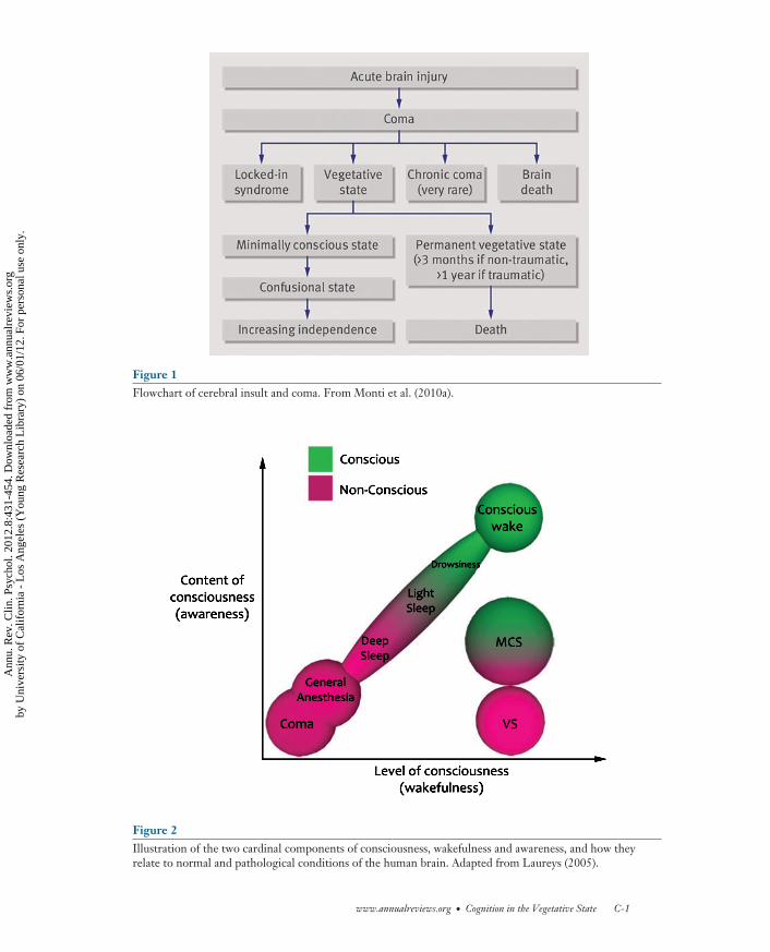
the endeavor to model and quantify the pres-ence of consciousness in the human brain, aswell as the effort to delineate how much of hu-man cognition depends on its presence, are keyto understanding some of the most mysteriousconditions of the human brain: disorders ofconsciousness (DOC). This term refers to a setof related disorders, including coma, the veg-etative state (VS), and the minimally consciousstate (MCS), in which an individual’s con-sciousness is altered in a transient or permanentfashion due to severe acquired or developedbrain injury (Monti et al. 2010a). As a conse-quence of great progresses in intensive (andgeneral) medical care, an increasing numberof patients survive severe brain damage. Someof these patients go on to make significant re-covery; others, as illustrated in Figure 1, entereither transiently or permanently a conditionof DOC (or other associated neurologicalconditions).
One of the most challenging aspects of DOCis the fact that without a direct understandingof consciousness and a means to reliably assessits presence, determining whether an indi-vidual other than ourselves is conscious mustnecessarily rely on indirect measures (cf. Monti& Owen 2010). This constraint bears veryfar-reaching implications. On the one hand,despite increasingly sophisticated behavioralprotocols to assess consciousness at the bedside,indirect measures have consistently been shownto be suboptimal and often prone to error(Andrews et al. 1996, Childs et al. 1993,Schnakers et al. 2009). On the other hand, thelack of a direct understanding of consciousnessseverely constrains our ability to developeffective interventions and treatments aimed at“reigniting” it (Elliott & Walker 2005). Simi-larly, our limited understanding of how muchcognition is possible at the lower boundariesof consciousness (Ropper 2010) severely con-strains our ability to understand the psychologyof DOC. These issues ultimately lie at theheart of the unique legal and ethical challengesassociated with these conditions (Fins et al.2008, Fisher & Appelbaum 2010, Jennett2005)—often rendered even more complicated
432 Monti
Ann
u. R
ev. C
lin. P
sych
ol. 2
012.
8:43
1-45
4. D
ownl
oade
d fr
om w
ww
.ann
ualr
evie
ws.
org
by U
nive
rsity
of
Cal
ifor
nia
- L
os A
ngel
es (
You
ng R
esea
rch
Lib
rary
) on
06/
01/1
2. F
or p
erso
nal u
se o
nly.
CP08CH17-Monti ARI 12 March 2012 9:3
by the use of inconsistent terminology ( Jennett2002) and incorrect media reporting (Racineet al. 2008).
The remainder of this review is organizedinto four sections. First, I present disorders ofconsciousness, one at a time, focusing on basicconcepts and definitions. Second, I briefly dis-cuss the challenges and complexities associatedwith assessing residual cognitive processing andawareness in patients with DOC, as well as thepotential role of modern neuroimaging tech-niques in this context. Third, I review selectedcontributions to the study of cognitive process-ing in VS and MCS patients, focusing in par-ticular on functional neuroimaging approaches.Finally, I briefly conclude by outlining the viewthat residual cortical processing in vegetativepatients reflects functionally disconnected cog-nitive modules that are isolated from the rest ofthe brain and thus do not give rise to the sub-jective feeling of phenomenological awareness.
WHAT ARE DISORDERSOF CONSCIOUSNESS?
Consciousness is often conceptualized as en-compassing two cardinal elements (Laureys2005): wakefulness and awareness. Wakefulnessrefers to the level of consciousness, whereasawareness refers to its contents. Different statesof the human brain, both normal and patho-logical, can be described with respect to thesetwo elements. As illustrated in Figure 2,wakefulness and awareness generally vary to-gether: both are absent (or low) in coma andgeneral anesthesia, return jointly in the pro-gression from deep sleep to light sleep anddrowsiness, and are present during normal (i.e.,healthy) wake. Occasionally, the two cardinalelements of consciousness dissociate, produc-ing very puzzling conditions. The oneiric expe-riences that often characterize rapid eye move-ment (REM) sleep, for example, illustrate ascenario in which (some kind of) awarenessis present in the absence of wakefulness. AsI discuss below, the reverse dissociation, inwhich wakefulness is present in the absence ofawareness, defines the vegetative state.
For convenience, in what follows I discussimpairments of consciousness as a set of discreteentities. However, it is now increasingly clearthat DOC are best understood as representinga continuous spectrum (Andrews 1996, Laureys& Boly 2008).
Coma
Coma describes a transient condition ofunresponsiveness in which patients presentwith their eyes (continuously) closed, do notrespond to attempts to arouse them, and exhibitno evidence of awareness of self or of theirsurrounding (Posner et al. 2007). As portrayedin Figure 2, patients in a state of coma thuslack both awareness and wakefulness. Thesepatients typically either recover or progressto a vegetative state within four weeks, withprolonged coma being an exceedingly rareoutcome (Royal College of Physicians 1996).It should be noted that, by virtue of enteringa pharmacologically induced coma, individualsundergoing general anesthesia appear to enter(transiently and reversibly) a similar condition(cf. Brown et al. 2010).
When patients “awaken” from a state ofcoma without recovering awareness, they aresaid to progress to a vegetative state ( Jennett &Plum 1972). This condition of “wakefulness inthe absence of awareness” is defined by threemain clinical features: (a) alternating periods ofeye opening and closing, giving the appearanceof sleep-wake cycles (whether this featureactually reflects genuine circadian rhythms iscurrently unclear; cf. Bekinschtein et al. 2009b,Cologan et al. 2010, Landsness et al. 2011);(b) complete lack of any sign of awareness ofthe self or the environment; and (c) partial orcomplete preservation of hypothalamic andbrainstem autonomic functions (Multi-SocietyTask Force on PVS 1994a, Royal College ofPhysicians 1996). According to the guidelinesset by the Multi-Society Task Force on PVSin 1994, a vegetative state is considered to bepersistent when it lasts longer than one month.After traumatic brain injuries, a vegetative stateis considered to be permanent when it lasts
www.annualreviews.org • Cognition in the Vegetative State 433
Ann
u. R
ev. C
lin. P
sych
ol. 2
012.
8:43
1-45
4. D
ownl
oade
d fr
om w
ww
.ann
ualr
evie
ws.
org
by U
nive
rsity
of
Cal
ifor
nia
- L
os A
ngel
es (
You
ng R
esea
rch
Lib
rary
) on
06/
01/1
2. F
or p
erso
nal u
se o
nly.
CP08CH17-Monti ARI 12 March 2012 9:3
Permanentvegetative state(PVS): a vegetativestate that lasts longerthan three months fornontraumatic injuriesand longer than oneyear for traumaticinjuries
Persistent vegetativestate: a vegetativestate that lasts longerthan one month
LIS: locked-insyndrome
longer than one year. In contrast, a vegetativestate acquired following nontraumatic braininjury is considered permanent after threemonths (Multi-Society Task Force on PVS1994a). Although both permanent vegetativestates and persistent vegetative states are oftenabbreviated to PVS, it has been suggested thatto avoid confusion the abbreviation shouldbe used exclusively to indicate a permanentvegetative state (Laureys et al. 2000a).
Vegetative State
The term vegetative state is currently themost widely used label; nonetheless, othernomenclatures have also been employed (par-ticularly in Europe) to describe patients in asimilar condition, including apallic syndrome,permanent/irreversible coma, coma vigile,postcomatose unawareness, and vegetative sur-vival, among others (for a review, see Jennett2002). As used by Jennett & Plum (1972), theterm vegetative was chosen to describe the factthat in these patients, basic vegetative nervousfunctions, including thermoregulation, respira-tion, and sleep-wake cycles, are preserved, butin the absence of any sensation and thought.Nonetheless, because of the perceived demean-ing and discriminatory subhuman connota-tion of the term, some have proposed alter-nate labels, including wakeful unconscious state(Sazbon & Groswasser 1991) and unresponsivewakefulness syndrome (Laureys et al. 2010). Toavoid any potential future confusion while theclinical and scientific community evaluates theuse of these alternative nomenclatures, in thisreview we maintain the label vegetative state.
Minimally Conscious State
Although some patients remain in a vegetativestate permanently, others emerge to a min-imally conscious state (MCS) (Giacino et al.2002). As illustrated in Figure 2, the MCS isdefined as a condition in which patients appearnot only to be wakeful (like vegetative-statepatients) but also exhibit inconsistent butreproducible signs of awareness of themselves
or their surroundings. This state of minimalawareness is at times intermittent, alternatingwith periods of prolonged unresponsiveness.According to the guidelines mentioned above,once a patient is considered to be in a PVS, thechances of emerging to an MCS are exceed-ingly small. Nonetheless, the prognosis of ir-reversible unconsciousness implied in the termPVS is necessarily probabilistic in nature (cf.Multi-Society Task Force on PVS 1994b, RoyalCollege of Physicians 1996), as confirmed bya small number of documented late recoveries(e.g., Childs & Mercer 1996). The MCS can bepermanent or preface emergence to a state ofsevere disability (which is said to exist when apatient recovers meaningful object use or con-sistent communication; cf. Giacino et al. 2002).
Other Related Conditions
Finally, it is worth mentioning two additionalconditions, brain death and locked-in syndrome(LIS), which, although not typically consideredimpairments of consciousness, are possible out-comes from the state of postinjury acute coma(see Figure 1). Brain death represents a con-dition of irreversible loss of all functions ofthe brain, including the brainstem (Am. Acad.Neurol. 1995), as manifested by absolute un-responsiveness to stimulation, absence of allspontaneous muscle activity and brainstem re-flexes, and an isoelectric electroencephalogramfor 30 minutes, in the absence of hypothermiaor intoxication by central nervous system de-pressants. The LIS, also referred to as pseu-docoma, is a condition in which patients areboth awake and aware but are either unable toproduce any motor response (complete LIS) orseverely limited in their behavioral repertoire,typically restricted to vertical eye movementsor blinking (partial LIS; Bauer et al. 1979).
ASSESSING AWARENESS ANDCOGNITION IN DISORDERSOF CONSCIOUSNESS
How do we ever know that someone, otherthan ourselves, is conscious? In the absence
434 Monti
Ann
u. R
ev. C
lin. P
sych
ol. 2
012.
8:43
1-45
4. D
ownl
oade
d fr
om w
ww
.ann
ualr
evie
ws.
org
by U
nive
rsity
of
Cal
ifor
nia
- L
os A
ngel
es (
You
ng R
esea
rch
Lib
rary
) on
06/
01/1
2. F
or p
erso
nal u
se o
nly.
CP08CH17-Monti ARI 12 March 2012 9:3
of a direct means of establishing or quantify-ing the presence of consciousness in the hu-man brain, its assessment must necessarily relyon indirect strategies. The differential diagno-sis of brain-injured patients thus relies on thedetection (or failure to do so) of behavioralindicators revealing the presence of wakeful-ness and/or awareness (cf. Monti et al. 2009b).The return of wakefulness, for example, whichmarks the transition from coma to VS, is un-ambiguously indexed by the return of periodsof eye-opening. The return of awareness, on theother hand, which marks the transition from VSand MCS, is more elusive. Currently, the as-sessment of awareness is mainly based on care-ful (albeit subjective) behavioral observation bytrained personnel. Spontaneous and elicited be-havior in response to multisensory stimulationis recorded in accordance with specific proto-cols (e.g., Glasgow Coma Scale, Teasdale &Jennett 1974; Coma Recovery Scale-Revised,Giacino et al. 2004; for an overview, see Giacinoet al. 2009, Majerus et al. 2005). Regardless ofthe specific procedure used, the examination isfocused on uncovering signs of (a) awareness ofthe self or the environment; (b) sustained, re-producible, purposeful or voluntary responseto visual, olfactory, auditory, tactile, or nox-ious stimuli; and (c) comprehension of languageor expression. If any such evidence is found,the patient is considered to be (minimally)aware (i.e., MCS). Conversely, if no evidenceof awareness is found, a VS diagnosis is made.
In the past 25 years, an increasing numberof studies, including retrospective clinicalaudits (Andrews 1996, Childs et al. 1993)as well as direct comparison of alternativeclinical assessment protocols (Gill-Thwaites1997, Schnakers et al. 2006), have shown thatmisdiagnosis of minimally conscious patientsas being in a vegetative state might be as highas 40%. Several issues underlie this alarminglyhigh rate of misdiagnosis; however, most ofthem are expressions of a crucial flaw embeddedin the logic of assessing the “lack of awareness”(Monti et al. 2009b): Absence of evidence (ofawareness) is interpreted as evidence of absence(of awareness). A VS diagnosis thus ultimately
relies on a null result, an approach that is proneto false negatives (Owen & Coleman 2008).What if a patient were conscious but unable toconvey this fact via a clearly recognizable behav-ioral response? To illustrate this issue, considerFigure 3. The horizontal plane of the graphdepicts the same two components of con-sciousness illustrated in Figure 2. On theelevation axis we add a third dimension, whichcaptures the ability of a patient to producevoluntary motor behavior. As for the othertwo dimensions, we mark a conventional pointalong the elevation axis (highlighted by thegrey plane) separating “behavioral” individualscapable of producing voluntary responses from“nonbehavioral” individuals unable to producevoluntary output. When awareness is absent(e.g., coma, anesthesia, vegetative state, anddeep sleep) individuals are unable to producevoluntary output (i.e., they are nonbehavioral).Conversely, aware and awake individuals aretypically able to perform voluntary responses(i.e., behavioral). Standard clinical testing basedon behavioral responsiveness can thus separatethese two classes of individuals. However, ifany pathology impairs the ability of a patient torespond to stimulation, behavioral assessmentsare bound to return false-negative resultsand thus underestimate the patient’s level ofresidual brain function. Physical disabilities,for example, might entirely mask the presenceof residual cognition and awareness in MCSpatients. In a large retrospective study, 100%of misdiagnosed patients were indeed found tosuffer from motor disabilities (Andrews 1996).Sensory impairments might also have a similareffect (cf. Childs et al. 1993, Gill-Thwaites1997), particularly if the impairment is in thevisual domain (Andrews 1996). In addition,different clinical assessment protocols might bedifferently able to detect behaviors consistentwith the presence of consciousness (Schnakerset al. 2006). Finally, the characteristic inconsis-tent behavior typical of MCS patients makes itdifficult to interpret their responses, and theiroften protracted periods of unawareness makesit difficult to interpret their failure to respond(Royal College of Physicians 1996).
www.annualreviews.org • Cognition in the Vegetative State 435
Ann
u. R
ev. C
lin. P
sych
ol. 2
012.
8:43
1-45
4. D
ownl
oade
d fr
om w
ww
.ann
ualr
evie
ws.
org
by U
nive
rsity
of
Cal
ifor
nia
- L
os A
ngel
es (
You
ng R
esea
rch
Lib
rary
) on
06/
01/1
2. F
or p
erso
nal u
se o
nly.
CP08CH17-Monti ARI 12 March 2012 9:3
PET: positronemission tomography
rCBF: regionalcerebral blood flow
Effectively, as illustrated in Figure 3,there exists a subset of aware patients (i.e., thesection of MCS patients that falls below thegrey plane) who are impossible to distinguishfrom VS patients on the basis of currentclinical assessments. For this de facto categoryof nonresponsive MCS patients, a VS diagnosisis technically appropriate, since they show no(behavioral) sign of awareness, but it does notaccurately reflect their state of (minimal) con-sciousness. It is in this context that noninvasiveneuroimaging techniques, by observing brainresponses instead of behavioral responses,might provide a crucial supplement to bedsideclinical assessments (cf. Monti & Owen 2010).
COGNITION IN DISORDERSOF CONSCIOUSNESS
As classically defined, the (persistent) VS im-plies a uniform and chronic loss of expres-sion of forebrain function (as assessed behav-iorally; Jennett & Plum 1972) and thereforethe absence of any sign of a functioning mind(Royal College of Physicians 1996). In the past15 to 20 years, however, novel evidence hasclearly shown that despite profound structuraland functional damage, several aspects of cogni-tive processing can be preserved in VS patients.In what follows, we focus on the contribution ofneuroimaging techniques to our understandingof brain function in VS and MCS patients.
A central issue that is addressed throughoutthe following discussion is the evaluationof what can be inferred on the basis of theexisting neuroimaging data. Pragmatically,where behavior is limited or absent, neu-roimaging may be the only dependent variableaccessible to study cognition in this patientgroup. Nonetheless, assessing the presenceof cognitive processes on the sole basis ofbrain data is an inductive (i.e., probabilistic),rather than deductive (i.e., certain), inference(Henson 2005, Poldrack 2006). Therefore, itis crucial to interpret the data with respect tothe specificity of the cognitive paradigms em-ployed (for discussion on the point, see Owenet al. 2006), the degree to which inferences
reflect results expected on the basis of priorliterature as opposed to posthoc explanationsof “surprising” activations (Poldrack 2006),and the use of complementary models to testthe inferences drawn from the experiments inthis population (e.g., anesthesia, Davis et al.2007; see Language Processing section below).
Somatosensation and Nociception
Can patients in a vegetative state perceive pain?This difficult question is at the center of manyof the clinical, legal, and ethical discussions sur-rounding disorders of consciousness (cf. Finset al. 2008, McQuillen 1991, Rifkinson-Mann2003, Whyte 2008). However, as is the case forother aspects of cognitive processing in this pa-tient population, when self-report is not avail-able, it is difficult to make inferences aboutwhether an individual perceives pain or experi-ences suffering. Furthermore, according to theMulti-Society Task Force on PVS (1994b), nei-ther behavioral nociceptive responses, such asflexor or extensor spasms and withdrawal ofextremities following painful stimulation (e.g.,pinprick), nor grimacing and crying behav-ior necessarily imply the perception of pain.Therefore, the subjective and private nature ofthe experience of pain as well as our incom-plete understanding of its neural substrate havelong invited discussion (Klein 1997, Schnakers& Zasler 2007) and development of novel clin-ical tools (Schnakers et al. 2010).
Laureys et al. (2002a) employed positronemission tomography (PET) to measurechanges in regional cerebral blood flow (rCBF)during high-intensity electrical stimulation ofthe median nerve in a set of 15 persistent VSpatients. Despite the overall reduction of brainmetabolism, painful stimulation activated sub-cortical regions, in the midbrain and thala-mus, as well as primary somatosensory cor-tex. Nonetheless, no activation was detectedin the hierarchically higher-order cortices ob-served in a group of healthy volunteers, in-cluding secondary somatosensory cortex, in-sula, anterior cingulate, and posterior parietalcortices. In addition, in the VS patients, primary
436 Monti
Ann
u. R
ev. C
lin. P
sych
ol. 2
012.
8:43
1-45
4. D
ownl
oade
d fr
om w
ww
.ann
ualr
evie
ws.
org
by U
nive
rsity
of
Cal
ifor
nia
- L
os A
ngel
es (
You
ng R
esea
rch
Lib
rary
) on
06/
01/1
2. F
or p
erso
nal u
se o
nly.
CP08CH17-Monti ARI 12 March 2012 9:3
somatosensory cortex appeared to be function-ally disconnected from higher-order associativeareas. A similar finding was reported for a groupof anoxic patients fulfilling criteria for a perma-nent VS diagnosis by Kassubek et al. (2003). Asfor the patients described by Laureys and col-leagues (2002a), primary sensory cortex hyper-perfusion was detected in response to painfulstimulation. In addition, significant responseswere also observed in other regions of the painmatrix, in bilateral posterior insula and ante-rior cingulate, suggesting that to some extentresidual cortical processing of noxious stimulican propagate beyond the first cortical relay.Schiff et al. (2005) employed a (nonnoxious)tactile stimulation paradigm to assess the levelof residual somatosensory processing availablein two MCS patients. Bilateral hand stimula-tion elicited activations across several areas typ-ically implicated in somatosensory processing(primary and secondary) as well as higher-levelassociation areas (e.g., parietal and prefrontalcortices), suggesting partial preservation of dis-tributed networks for processing of somatosen-sory information in patients demonstrating in-consistent signs of consciousness. Taking a stepfurther, Boly et al. (2008) compared the rCBFresponse to painful median nerve stimulationof five MCS patients to that observed in thehealthy volunteers and VS patients describedin Laureys et al. (2002a). Overall, no signifi-cant difference was observed between MCS pa-tients and healthy volunteers across all the re-gions of the pain matrix. The two groups didexhibit quantitative differences, but it is unclearwhether these reflected a genuine lower-levelrCBF response in MCS patients or the unevensample sizes across the two groups (i.e., 15 ver-sus five for healthy volunteers and MCS pa-tients, respectively). Despite a similar sample-size imbalance, direct comparison of VS andMCS patients revealed significantly greater ac-tivations throughout the whole pain matrixfor the latter group. Furthermore, the levelof functional connectivity between primary so-matosensory and lateral fronto-parietal corticesin the MCS group was comparable to that ob-served in healthy volunteers, in contrast to the
lack of connectivity observed in the VS patients(Laureys et al. 2002a).
These findings suggest that the sensory-discriminative component of pain, putativelyhoused in primary and secondary somatosen-sory cortices, can be preserved beyond theboundary of unconsciousness (due to severebrain injury), perhaps subsisting as a residualcognitive module dissociated from the higher-order cortices that would be necessary to pro-duce awareness (Schiff et al. 2002). On theother hand, the affective and motivational com-ponent of pain, supposed to rely on anteriorcingulate cortex and prefrontal areas, appearsto be absent in VS patients. In contrast, con-scious (albeit minimally) patients demonstratea quasi-normal metabolic response to noxiousstimuli, suggesting they possess the process-ing resources necessary to generate the subjec-tive feeling of pain. However, it is importantto stress that the exact functional significanceof activations in the so-called pain matrix, aswell as their specificity to noxious stimulation,is currently debated (Mouraux et al. 2011). Thisuncertainty prevents interpreting activations inthese areas as unequivocal evidence of pain-specific cortical processing. Overall, then, thequestions of whether VS patients actually retainanything similar to the subjective experience ofdiscomfort and pain reported by healthy vol-unteers remains at present outside the reach ofneuroimaging studies.
Vision
From a clinical standpoint, visual processingabilities in patients surviving severe brain injuryhave received much attention. According tocurrent guidelines, the ability to maintainfixation or orient gaze voluntarily (e.g., visualpursuit) is sufficient to distinguish MCS fromVS patients (Giacino et al. 2002). Furthermore,visual tracking is often one of the first ob-servable signs of recovery (Multi-Society TaskForce on PVS 1994a, Shiel et al. 2000). Con-sequently, clinical assessment protocols thatdo not explicitly test for residual visual abilitiessuffer from higher rates of misdiagnoses
www.annualreviews.org • Cognition in the Vegetative State 437
Ann
u. R
ev. C
lin. P
sych
ol. 2
012.
8:43
1-45
4. D
ownl
oade
d fr
om w
ww
.ann
ualr
evie
ws.
org
by U
nive
rsity
of
Cal
ifor
nia
- L
os A
ngel
es (
You
ng R
esea
rch
Lib
rary
) on
06/
01/1
2. F
or p
erso
nal u
se o
nly.
CP08CH17-Monti ARI 12 March 2012 9:3
fMRI: functionalmagnetic resonanceimaging
(Schnakers et al. 2006), and patients who sufferfrom visual impairments are often misdiag-nosed (Andrews et al. 1996). The degree ofvisual information processing and (neural) rep-resentation of visual information that may beavailable at the lower boundaries of conscious-ness, however, has received less attention.
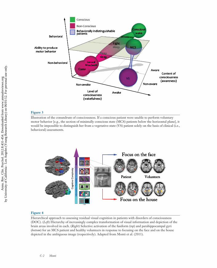
In a series of pioneering PET studies,Menon et al. (1998) and Owen et al. (2002)investigated metabolic response to visual stim-ulation in a persistent VS patient. Surprisingly,simple visual stimuli and pictures of familiarfaces elicited a metabolic response comparableto that observed in healthy volunteers (inprimary visual cortex and the fusiform gyrus).In 2006, Giacino and collaborators reportedsignificant rCBF changes in primary visualcortex in response to simple pattern flashes(as compared to no stimulation) in each of fivepersistent VS patients (Giacino et al. 2006).In a functional magnetic resonance imaging(fMRI) adaptation of this approach, the authorsalso reported significant activations in primaryand higher-level visual cortices (e.g., fusiformgyrus) in response to more complex stimuli(i.e., pictures of familiar and unfamiliar faces,hands, and landscapes) for an MCS patient. Incontrast to the neuroimaging result, the patientfailed to show any evidence of object recog-nition on standardized bedside assessment ofvisual function. Giacino et al. (2009) employeda similar paradigm to assess the degree of cog-nitive function in an anoxic patient exhibitingcharacteristically infrequent, inconsistent, andqualitatively ambiguous signs of consciousness.Simple patterns of light stimulation, pictures offaces, and pictures of landscapes all elicited acti-vation in the medial sections of occipital cortex.In addition, pictures of faces and landscapesselectively recruited regions of the fusiform andparahippocampal gyri, respectively, consistentwith high-level processing of the stimuli.
Taken together, these results show thatpatients with DOC can retain some level ofvisual processing. Furthermore, they illustratethe wide discrepancy that may exist betweenobservable behavior and the underlying neu-rophysiologic processes believed to support
cognitive processing (Giacino et al. 2009).However, passive paradigms comparing brainresponse to different stimuli (e.g., light ver-sus dark, pictures of faces versus scrambledversions of the same images) only allowinferring metabolic integrity of (some aspectof ) neurocognitive systems. Whether patientsconsciously perceived the images cannot be de-termined (cf. Monti & Owen 2010), for it is wellestablished that brain activation in high-levelvisual areas (e.g., fusiform gyrus) can bedetected in the absence of any subjectiveconscious perception (Dehaene et al. 2006). Toaddress this issue, Monti et al. (2011) developeda 3T fMRI hierarchical approach assessing bothpassive and active aspects of visual cognition(Figure 4). In a series of steps, they testedseveral levels of visual cognition, includingresponse to light, color, motion, organizedcontours, and discrimination of different cate-gories of objects (i.e., faces versus houses). Inaddition, at the last step of this battery, Montiet al. (2011) presented a set of figures, each con-sisting of a face and a house overlaid on top ofeach other (see Figure 4). During this sectionof the experiment, participants were promptedto alternate periods of focusing on the houseto periods of focusing on the face. Importantly,each figure was presented twice, once underthe instruction to focus on the face and onceunder the instruction to focus on the house.Consequently, any difference detected betweenthe two periods can only be interpreted as re-flecting different (voluntary) mental processes.When an MCS patient was assessed on the pas-sive levels of the battery, brain activations wereobserved in the same occipital and temporalregions seen in a set of healthy volunteers.In addition, in the active level of the battery,metabolic response in the fusiform gyrus wasup-regulated in the periods in which the patientwas instructed to focus on the faces as comparedto the periods in which the patient was requiredto focus on the houses (Figure 4). The conversepattern was observed in the parahippocampalgyrus. These results mirror the activationsseen in healthy volunteers performing thesame task. In contrast to the fMRI result, no
438 Monti
Ann
u. R
ev. C
lin. P
sych
ol. 2
012.
8:43
1-45
4. D
ownl
oade
d fr
om w
ww
.ann
ualr
evie
ws.
org
by U
nive
rsity
of
Cal
ifor
nia
- L
os A
ngel
es (
You
ng R
esea
rch
Lib
rary
) on
06/
01/1
2. F
or p
erso
nal u
se o
nly.
CP08CH17-Monti ARI 12 March 2012 9:3
evidence of object discrimination/recognitionor command-following was evident duringstandard clinical assessments (similarly to thecase described in Giacino et al. 2009). Thisresult shows that where appropriate paradigmsare employed (cf. Monti & Owen 2010), itis possible to infer not only the presence ofresidual (automatic) bottom-up processing,but also the presence of consciously mediatedtop-down processes, such as deploying visualattention in response to verbal commands.
Despite this series of intriguing results, itis important to stress that at present there isno systematic group study addressing the de-gree of residual bottom-up and top-down vi-sual cognition available in VS and MCS pa-tients and evaluating the discrepancy betweenthe visual behavior that is observable at thebedside and that is observable with functionalneuroimaging.
Audition
To date, audition has been the most exploitedmodality in studying residual cognitive pro-cesses in DOC patients. The advantage enjoyedby this modality rests on at least two practi-cal/experimental considerations. First, listen-ing to sounds through a headphone requiresvery little cooperation on the part of a subject,even for complex stimuli. Second, the effective-ness of auditory stimulations does not dependon anything other than integrity of the auditorysystem itself. Visual stimulation, in contrast, re-quires motor behaviors that are often compro-mised in these patients (e.g., maintenance ofeye-opening, fixation, and pursuit) for anythingmore complex than light flashes.
Laureys et al. (2000b) recorded rCBFchanges in response to simple auditory clicksin a sample of five persistent VS patients.In healthy volunteers and patients alike, thestimuli elicited significant activity in bilateralprimary auditory cortices, including the trans-verse temporal and the superior temporal gyri.Healthy volunteers also exhibited activationsbeyond primary cortices, in the lateral sectionsof the superior temporal gyrus and the superior
temporal sulcus contralateral to the stimula-tion. No such response was apparent for thepatients. Furthermore, VS patients exhibitedsignificantly less functional connectivity be-tween cortical regions than did volunteers.Reductions in functional connectivity wereapparent between the primary auditory cortexcontralateral to the stimulation, its homol-ogous region in the opposite hemisphere,and posterior temporal and parietal regionscontralateral to the stimulation. A similarreduction of functional connectivity was alsoapparent between the superior temporal sulcuscontralateral to the stimulation and areas previ-ously associated with higher-order processingof auditory stimuli (e.g., hippocampus andcingulate cortex; see the discussion section inLaureys et al. 2000b). This finding, mirroredin other modalities (cf. Laureys et al. 2002b),suggested a limited engagement of auditoryprocesses in VS patients confined to subcorticaland primary cortical relays only. Employingthe same paradigm, Boly et al. (2004) assessedrCBF changes in 15 VS and five MCS patients.All volunteers and patients exhibited bilateralresponses in primary cortical areas. Activationin higher-order processing regions, however,was observed only in healthy volunteers andMCS patients. Direct comparison of activationlevels for VS and MCS patients failed to showsignificant differences. Nonetheless, the twogroups differed significantly in the degree offunctional connectivity between secondaryauditory regions and areas in the posteriorsuperior temporal and middle temporal gyrias well as regions of the inferior, middle, andsuperior frontal gyri. These results integratethe findings of Laureys et al. (2000b) in twoways. On the one hand, they extend to agreater sample the finding of limited corticalactivation and functional connectivity observedin VS patients. On the other hand, they showphysiological differences mirroring the stateof consciousness of patients. Due to the strongsample imbalance (five MCS versus 15 VS), it isdifficult to interpret the null result, with respectto activation levels, between the two groups.Nonetheless, is noteworthy that a qualitative
www.annualreviews.org • Cognition in the Vegetative State 439
Ann
u. R
ev. C
lin. P
sych
ol. 2
012.
8:43
1-45
4. D
ownl
oade
d fr
om w
ww
.ann
ualr
evie
ws.
org
by U
nive
rsity
of
Cal
ifor
nia
- L
os A
ngel
es (
You
ng R
esea
rch
Lib
rary
) on
06/
01/1
2. F
or p
erso
nal u
se o
nly.
CP08CH17-Monti ARI 12 March 2012 9:3
EEG: electroen-cephalography
ERP: event-relatedpotential
change in state (i.e., minimally conscious ver-sus nonconscious) was clearly captured by thedegree of residual connectivity between hi-erarchically organized cortical relays. As wediscuss below, the functional disconnection ofneurocognitive modules might play a key rolein the failure of residual information processesto generate the subjective experience of per-ception (Giacino et al. 2006, Schiff et al. 2002).
A different question relates to which as-pects of auditory information processing under-lie the observed metabolic responses to simplesounds. Jones et al. (2000) employed electroen-cephalography (EEG) to assess the degree ofearly-stage analysis of sounds that is preservedin patients with severe brain injury. In a sam-ple of 22 postcomatose patients, ranging fromnonresponsive/VS to communicative, theyrecorded auditory-evoked potentials (AEPs)triggered by changes in tone pitch, timber, andin tone sequences. Surprisingly, only one pa-tient exhibited no AEPs to any of the condi-tions. Twelve patients exhibited clear responses[e.g., N1 and mismatch negativity (MMN) po-tentials], whereas the remaining patients exhib-ited less-clear evidence of appropriate informa-tion processing. These findings suggest that theactivations uncovered in the PET studies dis-cussed above might entail (to a minimum) pro-cesses related to the analysis of pitch and timberas well as the comparison of incoming soundsto an image or template of the previous ones(e.g., echoic memory; Jones et al. 2000). In ad-dition, for most patients, the level of auditoryresponsiveness observed with EEG matchedthat observed in bedside clinical testing. In fourcases, however, the two sources of evidencewere in disagreement, including two in whichAEPs were present in the absence of behavioralresponses and two exhibiting the reverse pat-tern. (The issue of noncorrespondence betweenbehavioral and neuroimaging responses is dis-cussed in the Volition section below.)
Kotchoubey et al. (2005) tested the notionthat only MCS patients retain some level ofinformation processing in higher sensory andassociation areas. Employing a combinationof stimuli (e.g., sine waves, harmonic chords,
words, sentences) and experimental proce-dures (e.g., oddball and MMN paradigms),Kotchoubey et al. (2005) assessed several neu-rophysiological indices of cortical processing[i.e., event-related potentials (ERPs)]. Impor-tantly, different ERPs occur at different laten-cies from the target event, presumably reflect-ing increasingly deep aspects of informationprocessing (e.g., primary undifferentiated au-ditory cortical responses in the 60 to 100 msrange; deep processing of high-level aspectsof a stimulus in the 300 to 500 ms range).As expected, for the quasi totality of VS pa-tients with nonpathological resting EEG slow-ing (i.e., background EEG activity smaller than4 Hz), early aspects of primary undifferenti-ated auditory cortical responses (in the 60 to140 ms range) were observed, together withthe preattentive MMN component (cf. Joneset al. 2000). However, in contrast to the idea oflimited cortical processing proposed in Laureyset al. (2000b) and Boly et al. (2004), a significantnumber of VS (and MCS) patients also exhib-ited late components, in the 400 to 600 ms in-terval, elicited by the semantic features of stim-uli (e.g., semantic relatedness of pairs of words).Remarkably, although VS patients exhibitedlower frequency of preserved higher-level in-formation processing than did MCS patients,the difference disappeared when the two groupswere matched for background EEG activity. Infact, the major differences were observed be-tween VS patients with very severe backgroundEEG disturbances and VS patients with moder-ate disturbances only. Where the resting EEGwas below 4 Hz, no response was observed be-yond the very early components. As discussedby Kotchoubey et al. (2005), this finding stressesthe centrality of the thalamo-cortical gatingsystem in mediating neural mechanisms ofperception.
Overall, these studies (among several others)show that several complex aspects of auditoryinformation processing can be preserved in VSpatients despite not giving rise to the subjectivefeeling of perception.
Progressing beyond the automatic aspects ofauditory processing, Bekinschtein et al. (2009a)
440 Monti
Ann
u. R
ev. C
lin. P
sych
ol. 2
012.
8:43
1-45
4. D
ownl
oade
d fr
om w
ww
.ann
ualr
evie
ws.
org
by U
nive
rsity
of
Cal
ifor
nia
- L
os A
ngel
es (
You
ng R
esea
rch
Lib
rary
) on
06/
01/1
2. F
or p
erso
nal u
se o
nly.
CP08CH17-Monti ARI 12 March 2012 9:3
employed a clever two-level auditory violationparadigm to assess the presence of subjectivephenomenal awareness (of auditory stimuli) ina cohort of four VS and four MCS patients. Inparticular, they measured brain response at thescalp for “local” violations (i.e., a deviant tonefollowing a repeating standard tone) and for“global” violations (i.e., a deviant sequence oftones following a repeating standard sequenceof tones). As observed in previous studies,local violations are known to elicit automaticauditory processes (cf. Kotchoubey et al. 2005).Conversely, detection of global violationsdepends upon several additional cognitiveprocesses that are believed to require thepresence of consciousness. In particular, globalviolations require the ability to extract a rule(i.e., the standard sequence) and maintain itthrough time, as new sequences are delivered.As expected, the two kinds of violations eliciteddistinct spatio-temporal ERP patterns, withthe global violation occurring significantlylater in time. Crucially, the latter componentwas shown to disappear when individuals weredistracted by a concurrent task or by simplymind-wandering, confirming the link betweensubjective awareness of the global structureof the task and the presence of the late ERPresponse. Consistent with previous findings, allMCS patients and three VS patients exhibiteda significant response to local violations. NoVS patient, however, exhibited a responseto global violations, whereas three out offour MCS patients did, which suggests thatviolation of global regularities can indeed serveas a marker of conscious auditory processing.
Language Processing
The study of language processing in this pop-ulation is driven by several different motiva-tions. From a scientific standpoint, languageis a central aspect of the human mind. Fromthe clinical viewpoint, language comprehensionand restored communicative ability are diag-nostic milestones (implying transition from VSto MCS and emergence from MCS, respec-tively; cf. Giacino et al. 2002).
Pioneering the use of hierarchical exper-imental designs in this patient group, Owenet al. (2002, 2005b) employed PET andfMRI to assess increasingly complex levelsof auditory and language processing in twoVS patients. In both cases, the comparison ofsound epochs to periods of silence revealedextensive rCBF increases in the bilateral supe-rior temporal planes, consistent with previousreports (Laureys et al. 2000b). As depicted inFigure 5, both patients also exhibited signifi-cant rCBF changes, in the superior and middletemporal gyri, in response to hearing shortsentences (as compared to an unintelligible ver-sion of the same stimuli obtained by disruptingthe spectral and temporal properties of speechwhile preserving the duration, amplitude, andoverall spectral composition of the original).This result, which closely matches the activa-tions seen in healthy volunteers, demonstratesthat in the two patients, residual auditory andlinguistic processes were sufficient to discrim-inate patterns of sounds organized accordingto the rules of a natural language (i.e., English)from patterns of sound that lack this form oforganization. At the top level of this hierar-chy, Owen et al. (2005b) employed fMRI tocompare, in one patient, brain response to sen-tences containing high-ambiguity words (e.g.,“There were dates and pears in the fruit bowl”)to sentences containing only low-ambiguitywords (e.g., “There was beer and cider onthe kitchen shelf”). In healthy volunteers, thissubtraction has been shown to recruit leftlateralized posterior sections of the inferiortemporal gyrus and the inferior frontal gyrus.In the patient, however, significant activationwas only detected within the posterior temporalregions. Nonetheless, these findings imply thatVS patients may possess sufficiently preservedlanguage-related processes to enable detectionof speech as a specifically organized form ofsound sequences and to enable detection (andpresumably selection) of context-appropriatemeaning.
Employing a similar approach, Schiff et al.(2005) assessed responses to simple narrativesand a backward-played version of the same
www.annualreviews.org • Cognition in the Vegetative State 441
Ann
u. R
ev. C
lin. P
sych
ol. 2
012.
8:43
1-45
4. D
ownl
oade
d fr
om w
ww
.ann
ualr
evie
ws.
org
by U
nive
rsity
of
Cal
ifor
nia
- L
os A
ngel
es (
You
ng R
esea
rch
Lib
rary
) on
06/
01/1
2. F
or p
erso
nal u
se o
nly.
CP08CH17-Monti ARI 12 March 2012 9:3
stimuli in two MCS patients. Intelligibleand nonintelligible (i.e., backward) narrativesequally recruited primary auditory areas in thetransverse section of the superior temporalgyrus and in language-sensitive regions in thesuperior and middle temporal gyri. Althoughweak, some activations in response to forwardnarratives were also observed in inferiorfrontal and posterior parietal cortices, furtherconfirming that information processing canoccur beyond primary sensory areas. Extendingthis experimental approach to a larger cohortof patients, Fernandez-Espejo et al. (2008)compared brain activity in response to sounds,specifically to speech sounds, in a cohort ofthree VS and three MCS patients. In four pa-tients (two VS and two MCS), the comparisonof all sounds (i.e., narratives and unintelligiblenarratives) versus epochs of silence revealedactivations in primary auditory cortices andin the superior and middle temporal gyri.Consistent with the results of Schiff and Owen,language-specific activations (uncovered bycomparing intelligible and unintelligible nar-ratives) were also detected in middle temporalgyrus in one MCS patient and in middle andsuperior temporal gyri in one VS patient. Theprevalence of high-level processing of soundsand speech was examined in a large cohort ofpatients (22 VS and 19 MCS) by Coleman andcolleagues (Coleman et al. 2007, 2009). Usingthe hierarchical approach described in Owenet al. (2005a), 41% and 84% of patients (VSand MCS, respectively) demonstrated appro-priate superior temporal activity in response tosounds. In 32% and 63% of cases, significantspeech-specific activity was also detected (insuperior temporal cortex). Finally, 9% of VSand 10% of MCS patients exhibited (limited)selective response in inferior frontal and/orposterior temporal regions to high-ambiguitysentences.
Do these findings imply that patientscomprehended the sentences? Davis et al.(2007) addressed this very issue by assessingbrain responses to nonspeech and (ambiguousand nonambiguous) speech stimuli in sedatedhealthy volunteers. Bilateral superior temporal
activations in response to sounds (in gen-eral) and speech sounds (specifically) weredetected at all levels of sedation. Conversely,light amounts of sedation were sufficientto obliterate any brain response specific tohigh-ambiguity sentences. Importantly, thedecreased level of activation in inferior frontaland posterior temporal regions correlatedsignificantly with postexperiment recall of the(intelligible) stimuli. This finding thus linksobserved brain response to actual encoding ofthe verbal materials and suggests that states ofdecreased consciousness (at least as inducedby anesthetic agents) affect the higher-levelprocesses involved in computing the meaningof sentences and encoding them into memory.Conversely, the lower-level aspects of speechprocessing appear to remain intact even asconsciousness fades (consistent with the braininjury data; e.g., Kotchoubey et al. 2005).
Perrin et al. (2006) employed EEG to ex-plore semantic processing of self-related stimuli(e.g., the subject’s own name) in DOC patients.As expected, for all six MCS patients (as wellas four LIS patients), hearing one’s own nameelicited a significant positive deflection (i.e.,P3 wave) as compared with hearing other firstnames. The P3 potential is known to respond tostimulus saliency in general (e.g., acoustic rar-ity); however, because all names were presentedwith equal probability, in this circumstance it ismore likely to reflect the semantic saliency ofone’s own name. Out of four VS patients, twoexhibited a significant P3 response, consistentwith other reports (e.g., Kotchoubey et al. 2005,Owen et al. 2005b). Although this response cer-tainly entails semantic processing of the stim-uli, the P3 is known to occur without consciousperception (e.g., in subliminal presentations).Indeed, the P3 response did not differentiate VSfrom MCS (or LIS) patients. Similar findingshave also been reported with longer latency po-tentials [i.e., a potential with negative deflectionaround 400 ms (N400)] in response to sentencescontaining semantic anomalies (e.g., “The cof-fee is too hot to fly”; Schoenle & Witzke 2004).
Finally, aspects of speech prosody, includingemotional prosody and affective processing,
442 Monti
Ann
u. R
ev. C
lin. P
sych
ol. 2
012.
8:43
1-45
4. D
ownl
oade
d fr
om w
ww
.ann
ualr
evie
ws.
org
by U
nive
rsity
of
Cal
ifor
nia
- L
os A
ngel
es (
You
ng R
esea
rch
Lib
rary
) on
06/
01/1
2. F
or p
erso
nal u
se o
nly.
CP08CH17-Monti ARI 12 March 2012 9:3
have also received some attention. Theseaspects of language processing are particularlyinteresting because they appear to be modularwith respect to other components of language(e.g., semantic and syntactic interpretation; cf.Kotchoubey et al. 2009, p. 154). Furthermore,emotionally salient stimuli have been reportedto increase the chances of eliciting (brain) re-sponses in comatose patients (Signorino et al.1995). Affective processing in a 16-year-oldpersistent VS patient was examined by deJong et al. (1997). As compared to nonspeechsounds, personally relevant narratives readby the patient’s mother elicited significantactivation in right middle temporal cortex (theleft hemisphere was heavily lesioned), as wellas the anterior midline sections of the cingulategyrus, and right precentral gyrus. Althoughthese activations are consistent with affectiveprocessing of narratives, the use of nonwordsas a baseline condition does not allow usto exclude that they reflect a more generallanguage-specific response. This issue wascontrolled for in a later case report of an MCSpatient (Bekinschtein et al. 2005). Using fMRI,the brain response to the voice of the patient’smother was compared with the brain responseto an unfamiliar voice. As compared withsilence, the latter stimulus elicited activationsin superior temporal regions as well as in moreanterior regions (in the insula), consistent withprevious findings (e.g., Schiff et al. 2005). Ascompared with the unfamiliar voice, listeningto the mother’s voice elicited additional strongactivations in the amygdala and insula, spread-ing to the inferior frontal gyrus (among otherareas). In contrast to the de Jong et al. (1997)study, the experimental design employed byBekinschtein and colleagues allows interpret-ing activations in emotion-related areas asreflecting a selective affective response to thefamiliar stimulus. In a larger cohort study,Kotchoubey et al. (2009) focused on a negativeERP putatively regarded as an electrophysi-ological marker for recognition of changes inemotional prosody (i.e., N300). Using an odd-ball paradigm, they contrasted the response toone (deviant) exclamation of awe interspersed
among four (standard) exclamations of joy. Insix out of 27 patients, significant ERPs wereobserved in response to the affective oddball(i.e., the sad stimulus). Although patients witha clear temporal lobe lesion exhibited a signif-icant lower proportion of N300 responses, nodifference was detected between VS and MCSpatients, a finding that is in line with severalprevious reports demonstrating preservationof a large part of automatic processes acrossthe boundary of (minimal) consciousness.
Taken together, these findings clearly indi-cate that several high-level aspects of language,including semantic and affective processing, canremain in VS patients. However, it should bestressed that these positive findings demon-strate the ability of a brain to discriminate thepresence/absence of a given target feature (e.g.,affective tone) but not that the patient had anyconscious experience of the feature itself (e.g.,the emotional content of utterances).
Learning and Memory
Kotchoubey et al. (2006) first assessed thepresence of preserved cortical dynamics con-sistent with elementary forms of learning in agroup of 33 VS patients. The design includeda sequential presentation of ten simple tonesat 400 ms intervals. Sequences were repeated,at 5 s intervals, in blocks of 10, and the wholeblock was then repeated 10 times. Across thegroup of VS patients, decreases in the ampli-tude of the negative AEP typically observedabout 100 ms poststimulus (i.e., N1) supportthe idea of at least two forms of preservedelementary learning. On the one hand, withineach single series, the N1 response decreasedin amplitude from the first tone presentation tothe tenth, indicating short-term habituation.On the other hand, a significant decrease inN1 amplitude was also observed across runs(i.e., from the first sequence of ten tones to thelast), indicating longer-term habituation. Thisresult suggests that in VS patients, auditorycortices can retain the ability to learn toselectively ignore repeated irrelevant stimuli.Nonetheless, the processes reflected in the N1
www.annualreviews.org • Cognition in the Vegetative State 443
Ann
u. R
ev. C
lin. P
sych
ol. 2
012.
8:43
1-45
4. D
ownl
oade
d fr
om w
ww
.ann
ualr
evie
ws.
org
by U
nive
rsity
of
Cal
ifor
nia
- L
os A
ngel
es (
You
ng R
esea
rch
Lib
rary
) on
06/
01/1
2. F
or p
erso
nal u
se o
nly.
CP08CH17-Monti ARI 12 March 2012 9:3
response are known to be automatic in nature.A higher-level form of learning, straddling theboundary between learning and consciousness,was recently explored by Bekinschtein et al.(2009c) using electromyographic recordings.In a sample of 13 VS and seven MCS pa-tients, they employed a trace conditioningparadigm to assess the presence of learningprocesses. In this paradigm, two auditorytones are presented, with one of the two beingassociated, after a temporal delay, with anair-puff to the cornea. Crucially, as comparedwith classical conditioning, trace conditioningrequires an active cognitive bridging of thetone (conditioned stimulus) and the air-puff(unconditioned stimulus) across a temporaldelay. This bridging has been shown to requirea widespread cortical and cerebellar network(including the hippocampus and prefrontalcortex) and awareness of the contingencybetween the two stimuli. Across healthy volun-teers and patients, three different patterns wereobserved. Half of the volunteers (eight out of16), two VS patients, and one MCS patientexhibited clear learning of the contingencybetween one of the tones and the air-puff asshown by increasing peri-ocular muscle activityfollowing the tone paired with the air-puff.Furthermore, the muscle activity during thetemporal delay increased in close proximityto the air-puff delivery. At the same time, nosignificant anticipatory muscle activity wasrecorded after the tone that was not associatedwith the air-puff. At a more lenient criterion,five additional controls, two VS patients, andone MCS patient also exhibited a differencebetween muscle activity in the late and earlystages of the temporal delay. A subset ofparticipants (one control, four VS, and threeMCS) exhibited some amount of nonspecificlearning. For them, significant muscle activitywas observed in the late stages of the temporaldelay, but for both the paired and unpairedtones. The remaining seven patients (five VS,two MCS) and three healthy volunteers failedto show any evidence of learning. Interestingly,four out of the five VS nonlearners had non-traumatic brain injury etiologies, consistent
with their overall poorer prognosis (cf. Montiet al. 2010a). Groupwise, VS and MCS patientsdid not show significant differences, bothequally exhibiting less overall learning and lessspecific learning in comparison with healthyvolunteers. Nonetheless, for the reasonsmentioned above, the finding of preservedtrace-conditioning learning in patients withDOC is remarkable and is likely to indexthe presence of some level of consciousness.Indeed, when the same experiment was per-formed on a set of 12 volunteers undergoinganesthesia, only one participant exhibitedsome level of learning (though only whenemploying the more lenient test criterion).Finally, a remarkable finding of Bekinschteinand colleagues (2009c) is the close relationshipbetween clinical improvement (within a six-month to two-year follow-up period) and thepresence of trace conditioning. Clinical im-provements were observed for all but three ofthe patients exhibiting some level of learning.Even more significant, not one of the patientsfailing to exhibit any sort of learning showedclinical improvements. Overall, these resultscan either be interpreted as demonstratinga more sensitive approach to uncoveringthe presence of consciousness in severelybrain-injured patients or, to a minimum, as ameans to detect integrity of distributed circuitsthat are necessary for a brain to recover at leastminimal consciousness. As a footnote, it shouldbe stressed that, in contrast to all the paradigmsdesigned to assess the presence of awarenessthat I discuss in the following paragraphs, theapproach of Bekinschtein et al. (2009c) hasthe very desirable quality of not requiringverbally mediated instructions. This featuremight allow this approach to detect tracesof awareness even in patients with aphasia,a condition that is likely to affect a subset ofDOC patients (Majerus et al. 2009).
Executive Functions and Attention
In a modified version of the subject’s own nameparadigm discussed above, Schnakers et al.(2008) tested the ability of 22 patients (eight
444 Monti
Ann
u. R
ev. C
lin. P
sych
ol. 2
012.
8:43
1-45
4. D
ownl
oade
d fr
om w
ww
.ann
ualr
evie
ws.
org
by U
nive
rsity
of
Cal
ifor
nia
- L
os A
ngel
es (
You
ng R
esea
rch
Lib
rary
) on
06/
01/1
2. F
or p
erso
nal u
se o
nly.
CP08CH17-Monti ARI 12 March 2012 9:3
VS and 14 MCS) to voluntarily direct atten-tion to a target name interspersed among a setof nontarget names. During the passive com-ponent of the task, ERPs were recorded whilepatients listened to a series of names, includingtheir own. In the active component of the task,patients were instructed to count, at differenttimes, either the number of repetitions of theirown name or the number of repetitions of atarget unfamiliar name. Consistent with previ-ous results, passive listening to one’s own nameelicited, in MCS patients and volunteers alike, asignificant P3 deflection. In contrast to the re-sults reported by Perrin et al. (2006), however,no P3 was observed for any of the VS patients.In addition, as compared to passive listening,active counting elicited significantly greater P3waves in nine MCS patients (five in responseto actively listening for their own name andfour in response to actively listening for a targetother name). Despite greater P3 latencies in theactive condition, a likely sign of slower infor-mation processing consequent to severe braininjury, MCS patients and volunteers did notexhibit significantly different response ampli-tudes, which suggests that a subset of patientsis able to voluntarily allocate top-down atten-tion. Making exclusive use of nonintrinsicallysalient stimuli, Monti et al. (2009a) assessedglobal state changes correlating with the ac-tive maintenance of information through timewhile monitoring for incoming information. Intheir design, an MCS patient (as well as a set ofhealthy controls) was prompted to either pas-sively listen to a set of neutral words or to countthe occurrences of a randomly chosen targetword (different for every block). The two con-ditions were matched in all perceptual respects,ensuring that any difference between the twocould only reflect the process of actively holdingin mind information through time, somethingthat is considered to require conscious aware-ness (Dehaene & Naccache 2001). Consistentwith the results of Schnakers et al. (2008), thecomparison of active counting epochs and pas-sive listening uncovered significant activity ina subset of fronto-parietal regions typically re-cruited by tasks requiring executive functions
and considered to be a crucial component ofthe neural basis of consciousness (for greaterdiscussion on the point, see Monti et al. 2009a).These two studies demonstrate the high level ofexecutive and attentional functions that can bepreserved in MCS patients. Indeed, in both ap-proaches, differential activity across active andpassive tasks is difficult to explain without as-suming a conscious decision on the part ofthe patient to assign, in a top-down fashion,saliency to otherwise neutral words, to activelymaintain a target word in working memory, andto monitor incoming stimuli. As expected, how-ever, this kind of active cognitive process canbe seen in some MCS patients but not in VSpatients.
Volition and Communication
In a landmark paper, Owen et al. (2006)described the case of a persistent VS patientwho, despite showing no signs of conscious-ness during standard clinical assessments,could unequivocally demonstrate a state ofconsciousness by willfully modulating her ownbrain activity in response to verbal commands.In this paradigm, the patient is instructed toimagine playing tennis (motor imagery), toimagine walking around the rooms of her house(spatial imagery), or simply to relax (baseline).Crucially, throughout the three conditions, nostimulation is delivered beyond a one-secondverbal cue, at the beginning of each 30-secondepoch, instructing the patient on what taskto engage in (i.e., “tennis,” “house,” and“relax,” for motor imagery, spatial imagery,and baseline epochs, respectively). As shownin Figure 6, when epochs of motor imagerywere compared to rest epochs, activations wereobserved in the same medial frontal regionsactive in healthy volunteers performing thesame task. Conversely, when spatial imageryepochs were compared to rest, activations weredetected in the parahippocampal gyri for boththe patient and healthy volunteers. This result,in stark contrast to the patient’s persistent VSdiagnosis, necessarily implied that she musthave been, to some extent, conscious.
www.annualreviews.org • Cognition in the Vegetative State 445
Ann
u. R
ev. C
lin. P
sych
ol. 2
012.
8:43
1-45
4. D
ownl
oade
d fr
om w
ww
.ann
ualr
evie
ws.
org
by U
nive
rsity
of
Cal
ifor
nia
- L
os A
ngel
es (
You
ng R
esea
rch
Lib
rary
) on
06/
01/1
2. F
or p
erso
nal u
se o
nly.
CP08CH17-Monti ARI 12 March 2012 9:3
Could this activation reflect automatic re-sponses to the one-second verbal cues? As dis-cussed in Owen et al. (2007), this is unlikelyfor several reasons. First, each epoch of im-agery (and rest) lasted 30 seconds, requiringbrain responses to be protracted in time in or-der to reach statistical significance. The au-tomatic responses generated by word stimuliare known to occur on a much shorter timescale (within the first few hundred milliseconds;Hauk et al. 2004) and in language-sensitive ar-eas, not in the supplementary motor area orthe parahippocampal gyri. Furthermore, whenthe same cues were delivered to healthy volun-teers who had not been instructed to respond byengaging in mental imagery, no activation wasdetected. Similarly, no activation was detectedwhen cues were replaced with more evocativesentences (ending with the cue word; e.g., “Theman played tennis,” “The man walked aroundhis house”; Owen et al. 2007). The patient de-scribed by Owen et al. (2006) thus illustratesexactly the circumstance depicted in Figure 3:Minimally conscious patients who cannot per-form sufficiently clear motor responses (i.e., thesection of the MCS sphere below the grey hor-izontal plane) are indistinguishable from VSpatients on the basis of clinical (i.e., behav-ioral) assessments alone. It is this very kindof patient who is at risk of being misdiag-nosed and who would maximally benefit fromnon-muscle-dependent assessments (includingfMRI, EEG, MEG, and electromyography).
In a study of 23 VS patients (among oth-ers), Monti et al. (2010b) reported four patientswho produced significant responses to the im-agery tasks described above. Despite the fMRIresults, for two of these patients, repeated bed-side clinical examinations consistently yield noobservable sign of awareness. For the remainingtwo, thorough clinical assessments could un-cover inconsistent but detectable responses. Onthe one hand, this finding further confirms thelink between behavioral nonresponsiveness andVS misdiagnosis. On the other hand, it high-lights the importance of thorough bedside as-sessments. Taking advantage of one patient’sability to produce brain responses to command,
Monti et al. (2010b) employed the imageryparadigm as a strategy for the patient to respondto simple binary questions. The patient wasasked a simple binary question (e.g., “Is your fa-ther’s name Alexander?”) and was instructed toengage in motor imagery to convey an affirma-tive response and in spatial imagery to convey anegative response. For five of the six questions,the pattern of activations detected matched thefactually correct answer. (No significant activ-ity was detected in the sixth question.) Im-portantly, response epochs were all cued withthe neutral word “answer”; it was the patient’stask to decide which imagery was to be per-formed to correctly respond. This small modi-fication of the paradigm definitively puts to restany uncertainty concerning the possibility thatthe activations observed in Owen et al. (2006)and here could be automatic responses trig-gered by the semantics of the words tennis andhouse.
Despite the success of active fMRIparadigms in uncovering signs of awareness inpatients who appear vegetative at the bedside,negative results are typically reported in thevast majority of cases. How should thesebe interpreted? For some patients, negativefindings might genuinely reflect a state of un-consciousness. Other patients, instead, mightbe conscious but lack some cognitive processnecessary to comprehend a set of instructionsor perform a specific task. However, in somecases the failure to detect any activationmight reflect genuine false negatives. Thiscircumstance was recently described by Bardinet al. (2011). A patient who had emergedfrom MCS produced no detectable activationin the imagery task described above, despiteher ability to behaviorally report, outside theMRI machine, her active engagement in thetask. In a second case, an LIS patient couldproduce significant activations in response tothe imagery command but could not employthis strategy to communicate, despite beingable to do so at the bedside. The reversedissociation, with fMRI results exceeding whatwas observed at the bedside, was reported foran MCS patient who could communicate using
446 Monti
Ann
u. R
ev. C
lin. P
sych
ol. 2
012.
8:43
1-45
4. D
ownl
oade
d fr
om w
ww
.ann
ualr
evie
ws.
org
by U
nive
rsity
of
Cal
ifor
nia
- L
os A
ngel
es (
You
ng R
esea
rch
Lib
rary
) on
06/
01/1
2. F
or p
erso
nal u
se o
nly.
CP08CH17-Monti ARI 12 March 2012 9:3
a neuroimaging strategy (as in Monti et al.2010b), but not at the bedside. Finally, for thethree remaining patients, fMRI and behavioralresults were in agreement.
Overall, dissociations between what isobservable at the bedside and what is observ-able in neuroimaging tests can be as infor-mative (where positive neuroimaging resultsare observed in the absence of positive behav-ioral results) as they are problematic (wherenegative neuroimaging results are observed inthe presence of positive behavioral findings).Nonetheless, false negatives are a consistentproblem known to occur in low-level EEGstudies (e.g., Jones et al. 2000; see Audition sec-tion above) as well as in behavioral assessments(e.g., Schnakers et al. 2006).
In light of these and other factors (e.g., MRIversus EEG portability), John et al. (2011)used source localization methods to explore anEEG version of the imagery paradigm. Despitea pathological EEG profile (e.g., excesses ofslow delta and theta oscillations, with markedlydecreased alpha and beta activity), imaginingsinging and performing mental arithmetic re-vealed different (probable) sources of electricalactivity recorded at the scalp. In particular,singing imagery elicited mostly bilateral medialand frontal activations, whereas mental calcu-lation recruited (among others) left lateralizedinferior frontal and inferior temporal regionsas well as right lateralized temporo-parietalregions. Despite the fact that the activations forthe two tasks were not quantitatively compared,each pattern closely matched the activations ob-served in a healthy control performing the sametasks. Although this is a single case report, andin fact the first report of an anoxic VS patientdemonstrating awareness via brain imagingmethods, this result highlights the potentialof quantitative EEG measures. In a systematiclarge-scale study, Cruse et al. (2011) employedclassification algorithms to distinguish foci ofimagery-induced synchronization and desyn-chronization of electrical activity recorded atthe scalp. In their paradigm, a set of 16 VS pa-tients (and 12 volunteers) performed imaginarymovements of their right hand and of their
feet. For three out of 16 diagnosed VS patients,a support vector machine algorithm couldrecognize and distinguish spatio-temporalpatterns in the EEG for the two tasks with sig-nificant accuracy (comparable to that observedin healthy volunteers; 61%, 71%, and 78%for each patient, respectively). Importantly,the classifier was at chance when attemptingto classify precue epochs (i.e., 500 ms prior toeach cue prompting the patients to engage inthe imagery task), indicating that the findingis specific to the periods of imagery execution.In addition, the classifier was also at chancewhen attempting to classify spatio-temporalpatterns observed in healthy volunteers lis-tening to the same auditory cues but withouthaving been instructed to engage in mentalimagery. This finding discounts the possibilitythat the observed activations and successfulclassification results depended on automaticprocesses elicited by the semantics of thecues.
Overall, this series of studies confirms thatan important share of patients with a VS di-agnosis (17% and 19% in Monti et al. 2010band Cruse et al. (2011), respectively) can be ob-served to respond to command, and even en-gage in basic communication, using neuroimag-ing techniques.
Self-Awareness
The presence of self-awareness in DOCpatients is an important clinical milestone (cf.Giacino et al. 2002). However, assessing thepresence of self-awareness and self-reflectionin patients who are unable to express theirthoughts is extremely challenging (Laureyset al. 2007). Nonetheless, as described above,some studies have focused on brain response toself-relevant stimuli (e.g., Perrin et al. 2006).In an fMRI version of the subject’s-own-nametask, Di et al. (2007) reported that five outof seven VS patients (as well as four out offour MCS patients) exhibited primary auditoryactivity in response to their own name. In addi-tion, in two VS patients (and all MCS patients),activations in higher-level posterior temporal
www.annualreviews.org • Cognition in the Vegetative State 447
Ann
u. R
ev. C
lin. P
sych
ol. 2
012.
8:43
1-45
4. D
ownl
oade
d fr
om w
ww
.ann
ualr
evie
ws.
org
by U
nive
rsity
of
Cal
ifor
nia
- L
os A
ngel
es (
You
ng R
esea
rch
Lib
rary
) on
06/
01/1
2. F
or p
erso
nal u
se o
nly.
CP08CH17-Monti ARI 12 March 2012 9:3
cortices were also detected. Unfortunately, itis difficult to assess the specificity of these ac-tivations to self-related processing consideringthat epochs of silence (or rather, scanner noise)served as a baseline comparison. Nonetheless,it is noteworthy that the two VS patients ex-hibiting significant activation beyond primarycortices recovered some level of consciousnessby three months after the MRI session. In asimilar fMRI experiment, Qin et al. (2010)reported significant activations for six out ofseven VS patients in one or more of threemedial prefrontal regions previously linked toprocessing self-related stimuli (in caudal androstral sections of the anterior cingulate gyrus,the ACC, and the SMA; for an overview, seeLaureys et al. 2007). The same regions werealso found to be active in all four tested MCSpatients. As for the study by Di and colleagues(2007), the lack of a baseline task preventsinterpreting the findings as clear evidence ofselective processing of self-related materials.However, the areas they assessed were shown inhealthy volunteers to be selectively engaged bythe subject’s own name, and not other names,particularly when spoken by a familiar voice(see experiments I and II in Qin et al. 2010). Inaddition, the activation in the caudal section ofthe ACC was significantly different for VS andMCS patients and thus significantly correlatedwith behavioral measures of consciousness.Finally, the only two VS patients exhibitingsignificant activations in this specific area pro-gressed to MCS by three months after the MRIsession, consistent with the report by Di et al.(2007).
Overall, these initial results suggest thatsome aspect of self-related processing may wellbe preserved in MCS patients and that whenmarkers of self-related processing are observedin VS patients they might herald some levelof recovery. It is nonetheless important tostress that the selectiveness of these brain re-sponses to self-related stimuli is still to be es-tablished in DOC patients. Similarly, it is alsoto be established whether they bear any sub-stantial connection to the subjective feeling ofself-awareness.
DISCUSSION: A FUNCTIONALLYDISCONNECTED BRAIN?
One of the most consistent findings in VS pa-tients is a strong decrease of brain metabolism(typically around 40% of the values ob-served in healthy volunteers; De Volder et al.1990, Laureys et al. 2002b, Levy et al. 1987,Tommasino et al. 1995). Nonetheless, it is nowclear that, albeit in the absence of consciousawareness, the vegetative brain can sustain com-plex aspects of information processing acrosssensory modalities. The specific kind and extentof information processing that may be availablein a given patient, however, is likely to be pri-marily dependent upon the specific pattern ofbrain injury. So far, the study of residual cogni-tion and consciousness in this patient cohort hascentered on single-case reports and central ten-dencies, leaving a significant amount of within-category variance (e.g., within the group of VSpatients) largely untackled. In this respect, fu-ture development of truly multimodal tech-nology, where structural analysis informs andmerges with functional results, will be a nec-essary step toward explaining patient-specificvariability.
How can we interpret the level of residualfunction observed in VS patients and its failureto generate phenomenological awareness? Onepossibility is to allow for residual informationprocessing to extend to primary sensory cor-tices only (e.g., Boly et al. 2008, Laureys et al.2002b) without it propagating to higher-leveland polimodal integration areas that mightbe necessary for conscious experience (cf.Dehaene et al. 2006). The evidence, however,suggests that this might not be a correct char-acterization of the “vegetative” brain. Indeed,fMRI and EEG studies have well demonstratedthe presence of activations beyond primary cor-tices and ERP components believed to requirecoordinated activity across cortical regions(e.g., P3 and N400). An alternative hypothesisis that residual cognition in VS patients mightreflect isolated cognitive modules that, in theabsence of global integration, do not generateconscious experience (cf. Kotchoubey et al.
448 Monti
Ann
u. R
ev. C
lin. P
sych
ol. 2
012.
8:43
1-45
4. D
ownl
oade
d fr
om w
ww
.ann
ualr
evie
ws.
org
by U
nive
rsity
of
Cal
ifor
nia
- L
os A
ngel
es (
You
ng R
esea
rch
Lib
rary
) on
06/
01/1
2. F
or p
erso
nal u
se o
nly.
CP08CH17-Monti ARI 12 March 2012 9:3
2005, Schiff et al. 2002). This possibilitywould account for the brain responses andisolated behavioral fragments often observedin VS patients, as in the unique case of avegetative patient who randomly producedoccasional single words (Schiff & Plum 1999).VS patients might thus suffer from a functional“disconnection syndrome” (cf. Giacino et al.2006, Laureys et al. 2000b, Schiff et al. 2002).This condition of decreased informationintegration might be the crucial factor under-lying the absence of consciousness (Tononi2004, 2008). Along these lines, for example,Vanhaudenhuyse et al. (2010) have shownthat VS patients exhibit reduced resting-stateconnectivity, as compared to MCS patients andhealthy volunteers, in proportion to the degreeof consciousness impairment. Which specificaspect of brain circuitry underlies disruptedconnectivity, however, remains unclear. In arecent report, Boly et al. (2011) demonstratedsystematic differences in top-down connec-tivity across MCS and VS patients. In thelatter group, the backward connections fromfrontal to superior temporal cortices weresignificantly impaired, implicating a specificrole for top-down effective connectivity inconsciousness. A second aspect of brain cir-cuitry that has been proposed to play a centralrole in DOC is the thalamo-cortical axis, asshown by the case of a VS patient with rela-tively preserved cortico-cortical connectivity(i.e., default mode network) but impairedthalamo-cortical connectivity (Boly et al.2009). Furthermore, Laureys et al. (2000c)reported, in a single patient, restoration ofthalamo-cortical connectivity upon recoveryof awareness. Indeed, both postmortem andin vivo examinations have shown that VS
patients exhibit widespread thalamic damageand atrophy (Adams et al. 2000, Fernandez-Espejo et al. 2010). The specific pattern ofconnections of the central portion of thethalami with fronto-parietal cortical systems,as well as brainstem attentional capture mech-anisms, might thus be crucial to supportingthe large-scale cerebral dynamics associatedwith goal-directed behavior and consciousness(Schiff 2008). Damage to these structuresand circuits might thus effectively disconnecta structurally intact cortex and impair con-sciousness even in the presence of functionallyintact (bottom-up) modules of informationprocessing.
CONCLUSION: UNCONSCIOUSBUT NOT “JUST” VEGETATIVE
How vegetative is the vegetative brain? Takingstock of nearly 20 years of research, it is nolonger possible to conceive of the vegeta-tive brain as an apallic brain (i.e., a-pallium,“without a cortex”) capable of sustaining only“vegetative” neural functions. Indeed, despitesevere reductions in brain metabolism andprofound structural damage, some aspects ofcognitive information processing can remain.Nonetheless, residual information processingis likely to reflect functionally disconnectedcognitive modules that do not give rise tophenomenological awareness.
Although unconscious, the vegetative brainis clearly not just vegetative, perhaps warrant-ing the search for a new, scientifically updatednomenclature that may reflect not only whatis observable behaviorally but also the level ofresidual brain function (cf. Kotchoubey 2005,Laureys et al. 2010).
SUMMARY POINTS
1. The VS is a condition of the human brain in which patients who survive severe braininjury appear to be awake but show no signs of awareness of the self or the environment.
2. The VS is traditionally believed to reflect a condition of preserved brainstem and hy-pothalamic functions in the absence of any cortical information processing.
www.annualreviews.org • Cognition in the Vegetative State 449
Ann
u. R
ev. C
lin. P
sych
ol. 2
012.
8:43
1-45
4. D
ownl
oade
d fr
om w
ww
.ann
ualr
evie
ws.
org
by U
nive
rsity
of
Cal
ifor
nia
- L
os A
ngel
es (
You
ng R
esea
rch
Lib
rary
) on
06/
01/1
2. F
or p
erso
nal u
se o
nly.
CP08CH17-Monti ARI 12 March 2012 9:3
3. Reliance on behavioral assessments of awareness and cognitive function might underes-timate the level of residual processing available in VS patients.
4. Functional neuroimaging techniques have shown that several aspects of brain functionmay remain in VS patients, including auditory, visual, somatosensory, and linguisticprocessing.
5. Residual aspects of brain processing in these patients likely reflect functionally discon-nected cognitive modules that do not generate phenomenological awareness.
FUTURE ISSUES
1. What aspects of brain processing constitute the necessary and sufficient correlates ofconscious experience?
2. What is the clinical status of patients who appear to be nonresponsive (i.e., VS) duringclinical/behavioral assessments but can demonstrate signs of awareness in neuroimagingstudies?
3. Can sensory, pharmacological, or neuromodulatory interventions help restore conscious-ness after severe brain injury?
4. What role will scientific advances in neuroimaging play in routine clinical assessments?
5. To what extent can brain-computer interfaces be employed to assist patients with disor-ders of consciousness?
DISCLOSURE STATEMENT
The author is not aware of any affiliations, memberships, funding, or financial holdings that mightbe perceived as affecting the objectivity of this review.
ACKNOWLEDGMENTS
This work was funded in part by the James S. McDonnell Foundation Scholar Award.
LITERATURE CITED
Adams JH, Graham DI, Jennett B. 2000. The neuropathology of the vegetative state after an acute brain insult.Brain 123(Pt. 7):1327–38
Am. Acad. Neurol. 1995. Practice parameters for determining brain death in adults (summary statement). TheQuality Standards Subcommittee of the American Academy of Neurology. Neurology 45:1012–14
Andrews K. 1996. International working party on the management of the vegetative state: summary report.Brain Inj. 10:797–806
Andrews K, Murphy L, Munday R, Littlewood C. 1996. Misdiagnosis of the vegetative state: retrospectivestudy in a rehabilitation unit. BMJ 313:13–16
Bardin JC, Fins JJ, Katz DI, Hersh J, Heier LA, et al. 2011. Dissociations between behavioural and functionalmagnetic resonance imaging-based evaluations of cognitive function after brain injury. Brain 134:769–82
Bauer G, Gerstenbrand F, Rumpl E. 1979. Varieties of the locked-in syndrome. J. Neurol. 221:77–91Bekinschtein TA, Dehaene S, Rohaut B, Tadel F, Cohen L, Naccache L. 2009a. Neural signature of the
conscious processing of auditory regularities. Proc. Natl. Acad. Sci. USA 106:1672–77
450 Monti
Ann
u. R
ev. C
lin. P
sych
ol. 2
012.
8:43
1-45
4. D
ownl
oade
d fr
om w
ww
.ann
ualr
evie
ws.
org
by U
nive
rsity
of
Cal
ifor
nia
- L
os A
ngel
es (
You
ng R
esea
rch
Lib
rary
) on
06/
01/1
2. F
or p
erso
nal u
se o
nly.
CP08CH17-Monti ARI 12 March 2012 9:3
Bekinschtein TA, Golombek DA, Simonetta SH, Coleman MR, Manes FF. 2009b. Circadian rhythms in thevegetative state. Brain Inj. 23:915–19
Bekinschtein TA, Shalom DE, Forcato C, Herrera M, Coleman MR, et al. 2009c. Classical conditioning inthe vegetative and minimally conscious state. Nat. Neurosci. 12:1343–49
Bekinschtein T, Tiberti C, Niklison J, Tamashiro M, Ron M, et al. 2005. Assessing level of consciousness andcognitive changes from vegetative state to full recovery. Neuropsychol. Rehabil. 15:307–22
Boly M, Faymonville ME, Peigneux P, Lambermont B, Damas P, et al. 2004. Auditory processing in severelybrain injured patients: differences between the minimally conscious state and the persistent vegetativestate. Arch. Neurol. 61:233–38
Boly M, Faymonville ME, Schnakers C, Peigneux P, Lambermont B, et al. 2008. Perception of pain in theminimally conscious state with PET activation: an observational study. Lancet Neurol. 7:1013–20
Boly M, Garrido MI, Gosseries O, Bruno MA, Boveroux P, et al. 2011. Preserved feedforward but impairedtop-down processes in the vegetative state. Science 332:858–62
Boly M, Tshibanda L, Vanhaudenhuyse A, Noirhomme Q, Schnakers C, et al. 2009. Functional connectivityin the default network during resting state is preserved in a vegetative but not in a brain dead patient.Hum. Brain Mapp. 30:2393–400
Brown EN, Lydic R, Schiff ND. 2010. General anesthesia, sleep, and coma. N. Engl. J. Med. 363:2638–50Childs NL, Mercer WN. 1996. Brief report: late improvement in consciousness after post-traumatic vegetative
state. N. Engl. J. Med. 334:24–25Childs NL, Mercer WN, Childs HW. 1993. Accuracy of diagnosis of persistent vegetative state. Neurology
43:1465–67Coleman MR, Davis MH, Rodd JM, Robson T, Ali A, et al. 2009. Towards the routine use of brain imaging
to aid the clinical diagnosis of disorders of consciousness. Brain 132:2541–52Coleman MR, Rodd JM, Davis MH, Johnsrude IS, Menon DK, et al. 2007. Do vegetative patients retain
aspects of language comprehension? Evidence from fMRI. Brain 130:2494–507Cologan V, Schabus M, Ledoux D, Moonen G, Maquet P, Laureys S. 2010. Sleep in disorders of consciousness.
Sleep Med. Rev. 14:97–105Cruse D, Chennu S, Chatelle C, Bekinschtein TA, Fernandez-Espejo D, et al. 2011. Bedside detection
of awareness in the vegetative state: a cohort study. Lancet. In press. http://www.thelancet.com/journals/lancet/article/PIIS0140-6736%2811%2961224-5/abstract
Davis MH, Coleman MR, Absalom AR, Rodd JM, Johnsrude IS, et al. 2007. Dissociating speech perceptionand comprehension at reduced levels of awareness. Proc. Natl. Acad. Sci. USA 104:16032–37
Dehaene S, Changeux JP, Naccache L, Sackur J, Sergent C. 2006. Conscious, preconscious, and subliminalprocessing: a testable taxonomy. Trends Cogn. Sci. 10:204–11
Dehaene S, Naccache L. 2001. Towards a cognitive neuroscience of consciousness: basic evidence and aworkspace framework. Cognition 79:1–37
de Jong BM, Willemsen AT, Paans AM. 1997. Regional cerebral blood flow changes related to affective speechpresentation in persistent vegetative state. Clin. Neurol. Neurosurg. 99:213–16
De Volder AG, Goffinet AM, Bol A, Michel C, de Barsy T, Laterre C. 1990. Brain glucose metabolism inpostanoxic syndrome. Positron emission tomographic study. Arch. Neurol. 47:197–204
Di HB, Yu SM, Weng XC, Laureys S, Yu D, et al. 2007. Cerebral response to patient’s own name in thevegetative and minimally conscious states. Neurology 68:895–99
Elliott L, Walker L. 2005. Rehabilitation interventions for vegetative and minimally conscious patients.Neuropsychol. Rehabil. 15:480–93
Fernandez-Espejo D, Junque C, Cruse D, Bernabeu M, Roig-Rovira T, et al. 2010. Combination of diffusiontensor and functional magnetic resonance imaging during recovery from the vegetative state. BMC Neurol.10:77
Fernandez-Espejo D, Junque C, Vendrell P, Bernabeu M, Roig T, et al. 2008. Cerebral response to speech invegetative and minimally conscious states after traumatic brain injury. Brain Inj. 22:882–90
Fins JJ, Illes J, Bernat JL, Hirsch J, Laureys S, Murphy E. 2008. Neuroimaging and disorders of consciousness:envisioning an ethical research agenda. Am. J. Bioeth. 8:3–12
Fisher CE, Appelbaum PS. 2010. Diagnosing consciousness: neuroimaging, law, and the vegetative state.J. Law Med. Ethics 38:374–85
www.annualreviews.org • Cognition in the Vegetative State 451
Giacino JT, Ashwal S, Childs N, Cranford R, Jennett B, et al. 2002. The minimally conscious state: definitionand diagnostic criteria. Neurology 58:349–53
Giacino JT, Hirsch J, Schiff N, Laureys S. 2006. Functional neuroimaging applications for assessment andrehabilitation planning in patients with disorders of consciousness. Arch. Phys. Med. Rehabil. 87:S67–76
Giacino JT, Kalmar K, Whyte J. 2004. The JFK Coma Recovery Scale-Revised: measurement characteristicsand diagnostic utility. Arch. Phys. Med. Rehabil. 85:2020–29
Giacino JT, Schnakers C, Rodriguez-Moreno D, Kalmar K, Schiff N, Hirsch J. 2009. Behavioral assessmentin patients with disorders of consciousness: gold standard or fool’s gold? Prog. Brain Res. 177:33–48
Gill-Thwaites H. 1997. The Sensory Modality Assessment Rehabilitation Technique—a tool for assessmentand treatment of patients with severe brain injury in a vegetative state. Brain Inj. 11:723–34
Hauk O, Johnsrude I, Pulvermuller F. 2004. Somatotopic representation of action words in human motor andpremotor cortex. Neuron 41:301–7
Henson R. 2005 What can functional neuroimaging tell the experimental psychologist? Q. J. Exp. Psychol. A58(2):193–233
Jennett B. 2002. The Vegetative State: Medical Facts, Ethical and Legal Dilemmas. London: Cambridge Univ.Press
Jennett B. 2005. Thirty years of the vegetative state: clinical, ethical and legal problems. Prog. Brain Res.150:537–43
Jennett B, Plum F. 1972. Persistent vegetative state after brain damage. A syndrome in search of a name. Lancet1:734–37
John ER, Halper JP, Lowe RS, Merkin H, Defina P, Prichep LS. 2011. Source imaging of QEEG as a methodto detect awareness in a person in vegetative state. Brain Inj. 25:426–32
Jones SJ, Pato MV, Sprague L, Stokes M, Munday R, Haque N. 2000. Auditory evoked potentials to spectro-temporal modulation of complex tones in normal subjects and patients with severe brain injury. Brain123(Pt. 5):1007–16
Kassubek J, Juengling FD, Els T, Spreer J, Herpers M, et al. 2003. Activation of a residual cortical networkduring painful stimulation in long-term postanoxic vegetative state: a 15O-H2O PET study. J. Neurol.Sci. 212:85–91
Klein M. 1997. Perception of pain in the persistent vegetative state? Eur. J. Pain 1:165–67; discussion 167–68Kotchoubey B. 2005. Apallic syndrome is not apallic: Is vegetative state vegetative? Neuropsychol. Rehabil.
15:333–56Kotchoubey B, Jetter U, Lang S, Semmler A, Mezger G, et al. 2006. Evidence of cortical learning in vegetative
state. J. Neurol. 253:1374–76Kotchoubey B, Kaiser J, Bostanov V, Lutzenberger W, Birbaumer N. 2009. Recognition of affective prosody
in brain-damaged patients and healthy controls: a neurophysiological study using EEG and whole-headMEG. Cogn. Affect. Behav. Neurosci. 9:153–67
Kotchoubey B, Lang S, Mezger G, Schmalohr D, Schneck M, et al. 2005. Information processing in severedisorders of consciousness: vegetative state and minimally conscious state. Clin. Neurophysiol. 116:2441–53
Landsness E, Bruno MA, Noirhomme Q, Riedner B, Gosseries O, et al. 2011. Electrophysiological correlatesof behavioural changes in vigilance in vegetative state and minimally conscious state. Brain 134:2222–32
Laureys S. 2005. The neural correlate of (un)awareness: lessons from the vegetative state. Trends Cogn. Sci.9:556–59
Laureys S, Antoine S, Boly M, Elincx S, Faymonville ME, et al. 2002a. Brain function in the vegetative state.Acta Neurol. Belg. 102:177–85
Laureys S, Boly M. 2008. The changing spectrum of coma. Nat. Clin. Pract. Neurol. 4:544–46Laureys S, Celesia GG, Cohadon F, Lavrijsen J, Leon-Carrion J, et al. 2010. Unresponsive wakefulness
syndrome: a new name for the vegetative state or apallic syndrome. BMC Med. 8:68Laureys S, Faymonville ME, Berre J. 2000a. Rapid response: Permanent vegetative state and
persistent vegetative state are not interchangeable terms. BMJ. http://www.bmj.com/content/321/7266/916.1?tab=responses
Laureys S, Faymonville ME, Luxen A, Lamy M, Franck G, Maquet P. 2000c. Restoration of thalamocorticalconnectivity after recovery from persistent vegetative state. Lancet 355:1790–91
Laureys S, Faymonville ME, Peigneux P, Damas P, Lambermont B, et al. 2002b. Cortical processing ofnoxious somatosensory stimuli in the persistent vegetative state. Neuroimage 17:732–41
Laureys S, Perrin F, Bredart S. 2007. Self-consciousness in non-communicative patients. Conscious. Cogn.16:722–41; discussion 742–45
Levy DE, Sidtis JJ, Rottenberg DA, Jarden JO, Strother SC, et al. 1987. Differences in cerebral blood flowand glucose utilization in vegetative versus locked-in patients. Ann. Neurol. 22:673–82
Majerus S, Bruno MA, Schnakers C, Giacino JT, Laureys S. 2009. The problem of aphasia in the assessmentof consciousness in brain-damaged patients. Prog. Brain Res. 177:49–61
Majerus S, Gill-Thwaites H, Andrews K, Laureys S. 2005. Behavioral evaluation of consciousness in severebrain damage. Prog. Brain Res. 150:397–413
McQuillen MP. 1991. Can people who are unconscious or in the “vegetative state” perceive pain? Issues LawMed. 6:373–83
Menon DK, Owen AM, Williams EJ, Minhas PS, Allen CM, et al. 1998. Cortical processing in persistentvegetative state. Lancet 352:200
Monti MM, Coleman MR, Owen AM. 2009a. Executive functions in the absence of behavior: functionalimaging of the minimally conscious state. Prog. Brain Res. 177:249–60
Monti MM, Coleman MR, Owen AM. 2009b. Neuroimaging and the vegetative state: resolving the behavioralassessment dilemma? Ann. N. Y. Acad. Sci. 1157:81–89
Monti MM, Laureys S, Owen AM. 2010a. The vegetative state. BMJ 341:c3765Monti MM, Owen AM. 2010. Behavior in the brain using functional neuroimaging to assess residual cognition
and awareness after severe brain injury. J. Psychophysiol. 24:76–82Monti MM, Pickard JD, Owen AM. 2011. Visual cognition in disorders of consciousness: from V1 to top-down
attention. Hum. Brain Mapp. In pressMonti MM, Vanhaudenhuyse A, Coleman MR, Boly M, Pickard JD, et al. 2010b. Willful modulation of brain
activity in disorders of consciousness. N. Engl. J. Med. 362:579–89Mouraux A, Diukova A, Lee MC, Wise RG, Iannetti GD. 2011. A multisensory investigation of the functional
significance of the “pain matrix.” Neuroimage 54:2237–49Multi-Society Task Force on PVS. 1994a. Medical aspects of the persistent vegetative state (1). N. Engl. J.
Med. 330:1499–508Multi-Society Task Force on PVS. 1994b. Medical aspects of the persistent vegetative state (2). N. Engl. J.
Med. 330:1572–79Owen AM, Coleman MR. 2008. Functional neuroimaging of the vegetative state. Nat. Rev. Neurosci. 9:235–43Owen AM, Coleman MR, Boly M, Davis MH, Laureys S, Pickard JD. 2006. Detecting awareness in the
vegetative state. Science 313:1402Owen AM, Coleman MR, Boly M, Davis MH, Laureys S, et al. 2007. Response to comments on “detecting
awareness in the vegetative state.” Science 315:1221Owen AM, Coleman MR, Menon DK, Berry EL, Johnsrude IS, et al. 2005a. Using a hierarchical approach
to investigate residual auditory cognition in persistent vegetative state. Prog. Brain Res. 150:457–71Owen AM, Coleman MR, Menon DK, Johnsrude IS, Rodd JM, et al. 2005b. Residual auditory function in
persistent vegetative state: a combined PET and fMRI study. Neuropsychol. Rehabil. 15:290–306Owen AM, Menon DK, Johnsrude IS, Bor D, Scott SK, et al. 2002. Detecting residual cognitive function in
persistent vegetative state. Neurocase 8:394–403Perrin F, Schnakers C, Schabus M, Degueldre C, Goldman S, et al. 2006. Brain response to one’s own name
in vegetative state, minimally conscious state, and locked-in syndrome. Arch. Neurol. 63:562–69Poldrack RA. 2006. Can cognitive processes be inferred from neuroimaging data? Trends Cogn. Sci. 10:59–63Posner JB, Saper CB, Schiff ND, Plum F. 2007. The Diagnosis of Stupor and Coma. New York: Oxford Univ.
PressQin P, Di H, Liu Y, Yu S, Gong Q, et al. 2010. Anterior cingulate activity and the self in disorders of
consciousness. Hum. Brain Mapp. 31:1993–2002Racine E, Amaram R, Seidler M, Karczewska M, Illes J. 2008. Media coverage of the persistent vegetative
state and end-of-life decision-making. Neurology 71:1027–32
www.annualreviews.org • Cognition in the Vegetative State 453
Ann
u. R
ev. C
lin. P
sych
ol. 2
012.
8:43
1-45
4. D
ownl
oade
d fr
om w
ww
.ann
ualr
evie
ws.
org
by U
nive
rsity
of
Cal
ifor
nia
- L
os A
ngel
es (
You
ng R
esea
rch
Lib
rary
) on
06/
01/1
2. F
or p
erso
nal u
se o
nly.
CP08CH17-Monti ARI 12 March 2012 9:3
Rifkinson-Mann S. 2003. Legal consequences and ethical dilemmas of pain perception in persistent vegetativestates. J. Health Law 36:523–48
Ropper AH. 2010. Cogito ergo sum by MRI. N. Engl. J. Med. 362:648–49Royal College of Physicians. 1996. The permanent vegetative state. Review by a working group convened by
the Royal College of Physicians and endorsed by the Conference of Medical Royal Colleges and theirfaculties of the United Kingdom. J. R. Coll. Phys. Lond. 30:119–21
Schiff ND. 2008. Central thalamic contributions to arousal regulation and neurological disorders of con-sciousness. Ann. N. Y. Acad. Sci. 1129:105–18
Schiff ND, Plum F. 1999. Cortical function in the persistent vegetative state. Trends Cogn. Sci. 3:43–44Schiff ND, Ribary U, Moreno DR, Beattie B, Kronberg E, et al. 2002. Residual cerebral activity and behavioural
fragments can remain in the persistently vegetative brain. Brain 125:1210–34Schiff ND, Rodriguez-Moreno D, Kamal A, Kim KHS, Giacino JT, et al. 2005. fMRI reveals large-scale
network activation in minimally conscious patients. Neurology 64:514–23Schnakers C, Chatelle C, Majerus S, Gosseries O, Val MD, Laureys S. 2010. Assessment and detection of
pain in noncommunicative severely brain-injured patients. Expert Rev. Neurother. 10:1725–31Schnakers C, Giacino J, Kalmar K, Piret S, Lopez E, et al. 2006. Does the FOUR score correctly diagnose
the vegetative and minimally conscious states? Ann. Neurol. 60:744–45; author reply 745Schnakers C, Perrin F, Schabus M, Majerus S, Ledoux D, et al. 2008. Voluntary brain processing in disorders
of consciousness. Neurology 71:1614–20Schnakers C, Vanhaudenhuyse A, Giacino J, Ventura M, Boly M, et al. 2009. Diagnostic accuracy of the vege-
tative and minimally conscious state: clinical consensus versus standardized neurobehavioral assessment.BMC Neurol. 9:35
Schnakers C, Zasler ND. 2007. Pain assessment and management in disorders of consciousness. Curr. Opin.Neurol. 20:620–26
Schoenle PW, Witzke W. 2004. How vegetative is the vegetative state? Preserved semantic processing in VSpatients—evidence from N400 event-related potentials. Neurorehabilitation 19:329–34
Shiel A, Horn SA, Wilson BA, Watson MJ, Campbell MJ, McLellan DL. 2000. The Wessex Head InjuryMatrix (WHIM) main scale: a preliminary report on a scale to assess and monitor patient recovery aftersevere head injury. Clin. Rehabil. 14:408–16
Signorino M, D’Acunto S, Angeleri F, Pietropaoli P. 1995. Eliciting P300 in comatose patients. Lancet 345:255–56
Teasdale G, Jennett B. 1974. Assessment of coma and impaired consciousness: a practical scale. Lancet 304:81–84
Tommasino C, Grana C, Lucignani G, Torri G, Fazio F. 1995. Regional cerebral metabolism of glucose incomatose and vegetative state patients. J. Neurosurg. Anesthesiol. 7:109–16
Tononi G. 2004. An information integration theory of consciousness. BMC Neurosci. 5:42Tononi G. 2008. Consciousness as integrated information: a provisional manifesto. Biol. Bull. 215:216–42Vanhaudenhuyse A, Noirhomme Q, Tshibanda LJF, Bruno MA, Boveroux P, et al. 2010. Default network
connectivity reflects the level of consciousness in non-communicative brain-damaged patients. Brain133:161–71
Whyte J. 2008. Clinical implications of the integrity of the pain matrix. Lancet Neurol. 7:979–80
454 Monti
Ann
u. R
ev. C
lin. P
sych
ol. 2
012.
8:43
1-45
4. D
ownl
oade
d fr
om w
ww
.ann
ualr
evie
ws.
org
by U
nive
rsity
of
Cal
ifor
nia
- L
os A
ngel
es (
You
ng R
esea
rch
Lib
rary
) on
06/
01/1
2. F
or p
erso
nal u
se o
nly.
CP08CH17-Monti ARI 12 March 2012 9:3
Figure 1Flowchart of cerebral insult and coma. From Monti et al. (2010a).
Figure 2Illustration of the two cardinal components of consciousness, wakefulness and awareness, and how theyrelate to normal and pathological conditions of the human brain. Adapted from Laureys (2005).
www.annualreviews.org • Cognition in the Vegetative State C-1
Ann
u. R
ev. C
lin. P
sych
ol. 2
012.
8:43
1-45
4. D
ownl
oade
d fr
om w
ww
.ann
ualr
evie
ws.
org
by U
nive
rsity
of
Cal
ifor
nia
- L
os A
ngel
es (
You
ng R
esea
rch
Lib
rary
) on
06/
01/1
2. F
or p
erso
nal u
se o
nly.
CP08CH17-Monti ARI 12 March 2012 9:3
Figure 3Illustration of the conundrum of consciousness. If a conscious patient were unable to perform voluntarymotor behavior [e.g., the section of minimally conscious state (MCS) patients below the horizontal plane], itwould be impossible to distinguish her from a vegetative-state (VS) patient solely on the basis of clinical (i.e.,behavioral) assessments.
Figure 4Hierarchical approach to assessing residual visual cognition in patients with disorders of consciousness(DOC). (Left) Hierarchy of increasingly complex transformation of visual information and depiction of thebrain areas involved in each. (Right) Selective activation of the fusiform (top) and parahippocampal gyri(bottom) for an MCS patient and healthy volunteers in response to focusing on the face and on the housedepicted in the ambiguous image (respectively). Adapted from Monti et al. (2011).
C-2 Monti
Ann
u. R
ev. C
lin. P
sych
ol. 2
012.
8:43
1-45
4. D
ownl
oade
d fr
om w
ww
.ann
ualr
evie
ws.
org
by U
nive
rsity
of
Cal
ifor
nia
- L
os A
ngel
es (
You
ng R
esea
rch
Lib
rary
) on
06/
01/1
2. F
or p
erso
nal u
se o
nly.
CP08CH17-Monti ARI 12 March 2012 9:3
Figure 5Areas of significant brain response in a group of healthy volunteers and the two VS patients described inOwen et al. (2002, 2005b). Adapted from Monti & Owen (2010).
Figure 6Brain activations for the motor and spatial imagery tasks for healthy volunteers and a patient with a VSdiagnosis. From Owen et al. (2006), reprinted with permission from AAAS.
www.annualreviews.org • Cognition in the Vegetative State C-3