Biosynthesis of Collagen Presented by: Guided by: Nausheer Pagarkar Dr S.Y Bhedasgaonkar P.G Student Professor & HOD Dept of Periodontics Dept of Periodontics VPDC & H VPDC & H Sangli Sangli
Transcript
Biosynthesis of Collagen
Presented by: Guided by:
Nausheer Pagarkar Dr S.Y Bhedasgaonkar
P.G Student Professor & HOD
Dept of Periodontics Dept of Periodontics
VPDC & H VPDC & H
Sangli Sangli
CONTENTS INTRODUCTIONSTRUCTURE OF COLLAGEN TYPES OF COLLAGENCHEMICAL COMPOSITION OF
COLLAGENFIBROBLASTSYNTHESIS OF COLLAGEN COLLAGEN IN ORAL TISSUESDEGRADATION AND REMODELLING
OF COLLAGEN
SUMMARYCONCLUSION REFERENCES
IntroductionCollagen is the most abundant protein in mammals and
accounts for 25-30% of their protein content.
Collagen is the main fibrous component of skin, bone ,tendon, cartilage and teeth.
It comprises about 90% of the organic matrix of the bone
It seems collagen appeared early in evolution and its composition and structure have changed relatively little suggesting its design has reached optimum efficiency.
The name ‘collagen’ comes from Greek meaning ‘glue producer’. When collagen is heated in water , it gradually breaks down to produce soluble derived protein i.e. gelatin or animal glue.
The collagen molecule is a rigid rod like structure that resists stretching.
Fibers made up of collagen have a high tensile strength.
Therefore this protein is an important structural component in tissues such as the periodontal ligament and muscle tendons in which the mechanical forces need to be transmitted without loss.
Apart from their structural role collagens can also influence cell shape, differentiation and many other cellular activities.
STRUCTURE OF COLLAGEN
As a group of proteins collagens contain a number of characteristic features that distinguish them from other matrix molecules.
All collagens are composed of 3 polypeptide alpha chains coiled around each other to form the triple helix configuration. The individual polypeptide chains of collagen each contain app. 1000 amino acid residues
The collagen molecule is long (280nm) and narrow(1.35nm)
The α chains are left handed helices that wrap around each other into a right handed rope like triple helical rod.
Each such helix is around 1.4 nanometers in diameter and 300 nanometers in length
The triple helix may be of a continuous stretch or it may be interrupted by non collagenous elements.
Glycine occupies every third position in the repeating amino acid sequence
Gly – X – Y where X and Y are amino acids other than
glycine.Glycine is essential for the triple helical
conformation because larger amino acids will not fit in the center of the triple helix.
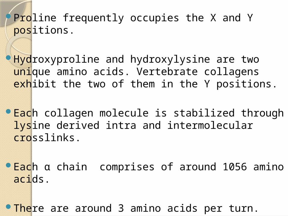
Proline frequently occupies the X and Y positions.
Hydroxyproline and hydroxylysine are two unique amino acids. Vertebrate collagens exhibit the two of them in the Y positions.
Each collagen molecule is stabilized through lysine derived intra and intermolecular crosslinks.
Each α chain comprises of around 1056 amino acids.
There are around 3 amino acids per turn.
In the α chain of type I collagen there are 338 Gly – X – Y triplets repeated in a sequence. The additional 32 amino acids flank the long triplet sequence at each end. They are known as telopeptides.
There is both an amino terminal ( -NH2 ) and a carboxy terminal (-COOH ) telopeptide.
The structure of collagen typically depends on high amounts of proline and hydroxyproline in the α chains.
Both these amino acids differ from other α-amino acids of proteins because they are amino acids with a rigid cyclical structure. This rigid structure prevents the rotation of the polypeptide chain backbone.
Chemical composition of collagen
The most abundant constituent is glycine, which, instead of a hydrocarbon side chain , has a second hydrogen atom attached to the backbone carbon atom. Glycine makes up almost 1/3rd of the collagen molecule,every third residue is glycine.
Collagen contains two amino acids,hydroxy proline and hydroxy lysine , that are not found in other animal tissue proteins. Hydroxy proline is present as 1 in 10 residues while Hydroxylysine is present as 1 in 200 residues.
Proline and hydroxy proline together account for 2/9th of the residues Thus the total amino acid content is higher than in most proteins.
Biosynthesis of collagensCollagen is synthesized mainly in: a)Mesenchymal cells and their
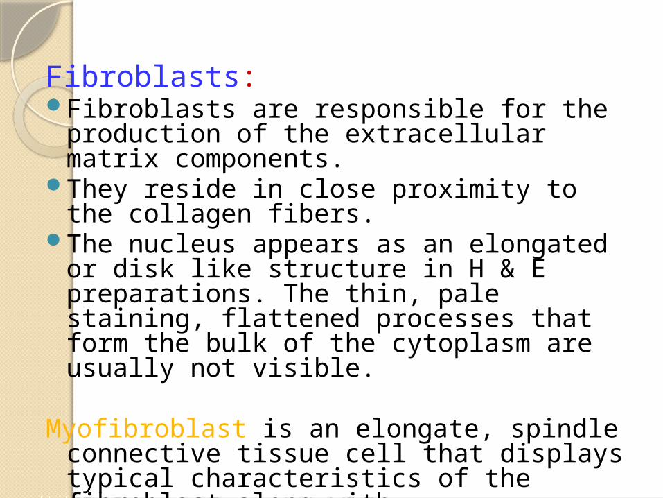
production of the extracellular matrix components.
They reside in close proximity to the collagen fibers.
The nucleus appears as an elongated or disk like structure in H & E preparations. The thin, pale staining, flattened processes that form the bulk of the cytoplasm are usually not visible.
Myofibroblast is an elongate, spindle connective tissue cell that displays typical characteristics of the fibroblast along with characteristics of smooth muscle cells.
Fibroblast in periodontal CT:
The fibroblast is the predominant cell type in the soft CT of the periodontium and consequently plays a central role in normal function and pathologic alterations.
Fibroblasts synthesize and maintain diverse group of CT matrices throughout the periodontium and exhibit motility and contractility, functions which help shape structural organization of tissue regeneration and development.
They are usually identified by cigar shaped or stellate morphology, presence of vimentin intermediate filaments and synthesis of matrix collagens and fibronectin.
Fibroblasts of healthy and diseased periodontal tissues are heterogeneous and show diversity of synthetic products, rates of product synthesis, responses to regulatory molecules, cellular turnover rates and morphologic features.
Tissue culture studies reveal that fibroblasts are attached tightly to matrix elements and that these attachments function in cellular movement.
Mechanisms of fibroblast attachments can be divided into two classes:
First class of mechanism requires cell membrane receptors for fibril proteins, and glycoproteins, which are attached to the fibrils.
Second class appears to involve the covalent association with specific integral plasma membrane proteins.
SYNTHESIS OF COLLAGEN
The entire process of collagen biosynthesis can be
best understood under the following stages
Gene expressionTranslational and post translational events
or intracellular steps in collagen synthesisExtracellular collagen biosynthetic eventsRegulation of synthesis
The 3 polypeptide chains of collagen molecule are formed separately under the direction of their respective genes.
The initial RNA transcript is processed to mRNA and it gives rise to the primary polypeptide chains in the ribosomes .
The polypeptide chain formed initially is a helical molecule with two non-helical extensions one at the NH2 and the other at the –COOH terminal end. The –NH2 terminal extension has a leader or signal sequence that directs the entry of the molecule into the rough endoplasmic reticulum
Hydroxylation: Hydroxyproline and Hydroxylysine are formed in the RER by the hydroxylation of prolyl and lysyl residues. This is an essential step in biosynthesis of collagen for it stabilises the molecules. Requirements for hydroxylation are:
1. Specific enzymes :prolyl hydroxylase and lysyl hydroxylase
2. α-ketoglutarate
3. Ferrous ions
4. Molecular oxygen
5. Ascorbic acid (Vitamin C)
Glycosylation of hydroxylysine: The enzyme galactosyl transferase catalyzes the addition of galactose to a hydroxylysyl residue . Glucosyl transferase catalyzes the further addition of glucose. Manganese(Mn) is required as a cofactor. Has a role in fibril organization
Formation of procollagen :Following hydroxylation and glycosylation, three polypeptide chains form a triple helix .
Secretion of procollagen: Procollagen passes into the Golgi complex before its secretion into the interstitial spaces.
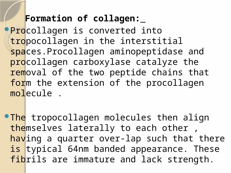
Formation of collagen: Procollagen is converted into tropocollagen
in the interstitial spaces.Procollagen aminopeptidase and procollagen carboxylase catalyze the removal of the two peptide chains that form the extension of the procollagen molecule .
The tropocollagen molecules then align themselves laterally to each other , having a quarter over-lap such that there is typical 64nm banded appearance. These fibrils are immature and lack strength.
Cross-linkage of fibrils to form fibres: There is oxidative deamination of specific lysyl or hydroxylysyl residues to form aldehydes The reaction is catalyzed by lysyl oxidase. The aldehyde cross-linkage precursors that are so produced either form Schiff bases with the neighbouring amino groups on nearby chains or aldol condensation groups with neighbouring aldehydes.
Cross-linkage is a slow process and the tensile strength of collagen steadily increases over a long period
The formation and secretion of the collagen molecule take about 35 to 60 minutes (known as the transit time)
Degradation of collagen and other matrix elements is a key component of normal tissue remodeling including ovulation, embryo implantation and involution of the uterus and mammary glands.
Nevertheless extracellular matrix degradation also contributes to pathologic alterations, for example matrix degradation is the major cause of connective tissue destruction in periodontitis, rheumatoid arthritis and other chronic inflammatory diseases. Enzymes capable of degrading the matrix aid in the tumor invasion by metastatizing cancer cells.
It is primarily mediated by collagenases though several other enzymes are involved in degradation of matrix components. These enzymes have specifically evolved to hydrolyze collagens because the triple helical collagen structure is resistant to most common proteinases. The collagenases belong to a family of enzymes called
Matrix metalloproteinases (MMPs’).
They are 13 in number each one having a 21kd catalytic domain that contains Zn+2 binding site and reuqires Ca +2 as a stabilizer
Based on their substrate specificity Woessner et al 1991, Mignatti et al 1996 have classified them into the following types
CollagenasesGelatinasesStromelysinsMatrilysins
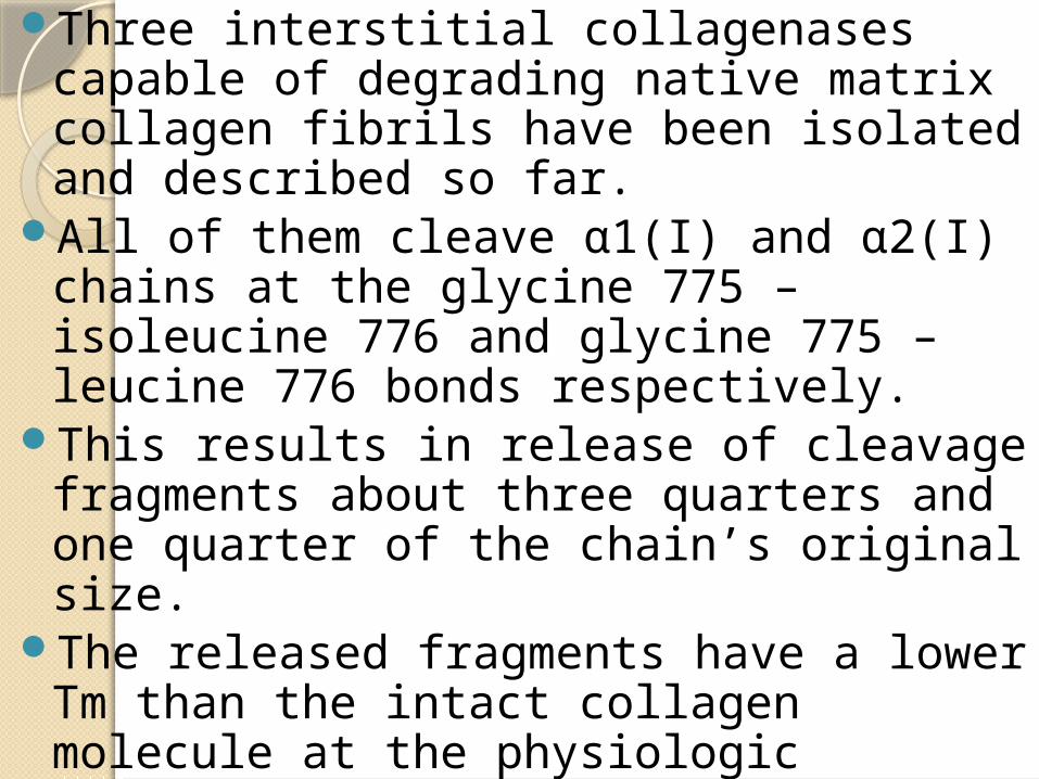
Three interstitial collagenases capable of degrading native matrix collagen fibrils have been isolated and described so far.
All of them cleave α1(I) and α2(I) chains at the glycine 775 – isoleucine 776 and glycine 775 – leucine 776 bonds respectively.
This results in release of cleavage fragments about three quarters and one quarter of the chain’s original size.
The released fragments have a lower Tm than the intact collagen molecule at the physiologic temperature ,therefore become denatured and are subsequently denatured by other proteinases.
Collagenase 1 / MMP-1 / Fibroblast type collagenase
It is produced by a variety of human epithelial and mesenchymal cells including keratinocytes,fibroblasts and macrophages.
It can hydrolyze type I, II, III, VI, VIII and X collagen and gelatin ( Birkedal-Hansen et al 1993).
It hydrolyzes type III collagen molecules faster when compared to type I.
Collagenase 2 / PMN leukocyte collagenase / MMP-8
It hydrolyzes both type I and type III collagen.It degrades type I faster than type III collagen.
It is found only in the specific granules of polymorphonuclear neutrophil cells.
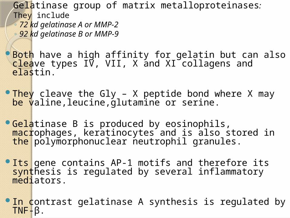
Gelatinase group of matrix metalloproteinases: They include◦ 72 kd gelatinase A or MMP-2◦ 92 kd gelatinase B or MMP-9
Both have a high affinity for gelatin but can also cleave types IV, VII, X and XI collagens and elastin.
They cleave the Gly – X peptide bond where X may be valine,leucine,glutamine or serine.
Gelatinase B is produced by eosinophils, macrophages, keratinocytes and is also stored in the polymorphonuclear neutrophil granules.
Its gene contains AP-1 motifs and therefore its synthesis is regulated by several inflammatory mediators.
In contrast gelatinase A synthesis is regulated by TNF-β.
◦ Stromelysins They have broad specificity with the ability to
degrade proteoglycans, basement membrane, laminin and fibronectin in addition to the collagens.


![ID 1 SESSION 4.pptx [Autoguardado].pptx](https://static.documents.pub/doc/80x56/55cf8c675503462b138c00e6/id-1-session-4pptx-autoguardadopptx.jpg)


![[MS-PPTX]: PowerPoint (.pptx) Extensions to the Office ...interoperability.blob.core.windows.net/files/MS-PPTX/[MS-PPTX... · 1 / 76 [MS-PPTX] — v20140428 PowerPoint (.pptx) Extensions](https://static.documents.pub/doc/80x56/5ae7f6357f8b9a6d4f8ed3b3/ms-pptx-powerpoint-pptx-extensions-to-the-office-ms-pptx1-76-ms-pptx.jpg)


![[MS-PPTX]: PowerPoint (.pptx) Extensions to the Office ...MS-PPTX].pdfPowerPoint (.pptx) Extensions to the Office Open XML File FormatFile Size: 4MBPage Count: 145](https://static.documents.pub/doc/80x56/5ed5954ddb0f8b20f04b0446/ms-pptx-powerpoint-pptx-extensions-to-the-office-ms-pptxpdf-powerpoint.jpg)


