310
| Date post: | 16-Aug-2015 |
| Category: |
Health & Medicine |
| Upload: | scu-hospital |
| View: | 64 times |
| Download: | 3 times |
Color Atlas of
Vascular Tumors and
Vascular Malformations

Color Atlas of
Vascular Tumors and
Vascular Malformations
Odile Enjolras, MDAPHP, Consultations
des Angiomes,
Hopital Lariboisiere,
Service de Neuroradiologie;
and Hopital d’Enfants
Armand Trousseau,
Service de Chirurgie
Maxillofaciale et de
Chirurgie Plastique
(Paris, France)
Michel Wassef, MDAPHP, Hopital Lariboisiere,
Service d’Anatomie
Pathologique,
Universite Paris 7 Faculte
de Medecine
(Paris, France)
Rene Chapot, MDService de
Neuroradiologie
Hopital Universitaire Dupuytren
(Limoges, France)
CAMBRIDGE UNIVERSITY PRESS
Cambridge, New York, Melbourne, Madrid, Cape Town, Singapore, Sao Paulo
Cambridge University Press
32 Avenue of the Americas, New York, NY 10013-2473, USA
www.cambridge.org
Information on this title: www.cambridge.org/9780521848510
� Cambridge University Press 2007
This publication is in copyright. Subject to statutory exception
and to the provisions of relevant collective licensing agreements,
no reproduction of any part may take place without
the written permission of Cambridge University Press.
First published 2007
Printed in India by Replika
A catalog record for this publication is available from the British Library.
Library of Congress Cataloging in Publication Data
Enjolras, Odile, 1940�
Color atlas of vascular tumors and vascular malformations / Odile
Enjolras, Michel Wassef, Rene Chapot.
p. ; cm.
Includes bibliographical references and index.
ISBN-13: 978-0-521-84851-0 (hardback)
ISBN-10: 0-521-84851-2 (hardback)
1. Blood-vessels�Tumors�Atlases. 2. Blood-vessels�
Abnormalities�Atlases. I. Wassef, Michel, 1949� . II. Chapot, Rene,
1967� . III. Title.
[DNLM: 1. Vascular Neoplasms�Atlases. 2. Arteriovenous
Malformations�Atlases. WG 17 585c 2006]
RC280.B56E55 2006
616.99’413�dc22
2006027036
ISBN 978-0-521-84851-0 hardback
Every effort has been made in preparing this publication to provide accurate and up-to-date
information that is in accord with accepted standards and practice at the time of
publication. Although case histories are drawn from actual cases, every effort has been
made to disguise the identities of the individuals involved. Nevertheless, the authors,
editors, and publishers can make no warranties that the information contained herein is
totally free from error, not least because clinical standards are constantly changing through
research and regulation. The authors, editors, and publishers therefore disclaim all
liability for direct or consequential damages resulting from the use of material contained
in this publication. Readers are strongly advised to pay careful attention to information
provided by the manufacturer of any drugs or equipment that they plan to use.
Cambridge University Press has no responsibility for the persistence or accuracy of URLS
for external or third-party Internet Web sites referred to in this publication and does not
guarantee that any content on such Web sites is, or will remain, accurate or
appropriate.

This Atlas is dedicated to our much-loved patients and their families, who trusted us
through exceptionally difficult times

Contents
Acknowledgments page ix
Introduction: ISSVA Classification 1
PART I: INVESTIGATIONS AND RADIOLOGICAL TOOLS 13
Conventional X-Rays 15
Ultrasonography in Combination with Doppler 15
Computed Tomography (CT) 16
Magnetic Resonance Imaging (MRI) 16
Conventional Vascular Imaging 17
PART II: VASCULAR TUMORS 19
II.A Infantile Hemangioma (IH) 21
II.B Other Vascular Tumors 78
II.B.1 Congenital Hemangiomas: RICH, NICH, and Missing Links 78
II.B.2 Tufted Angioma, Kaposiform Hemangioendothelioma,
Kasabach�Merritt Phenomenon (KMP) 101
PART III: VASCULAR MALFORMATIONS 123
III.A Capillary Malformations (CM) 125
III.A.1 Common Capillary Malformations: Port-wine
Stains (PWS) 125
III.A.2 Capillary Malformations and Associations 127
III.A.3 Syndromic Capillary Malformations 128
III.A.4 Telangiectasia and Syndromes with Telangiectasia 133
III.A.5 Angiokeratomas 135
III.B Venous Malformations (VM) 168
III.B.1 Common Venous Malformations 168
v i i
III.B.2 Syndromic Venous Malformations, Nosology 173
III.C Lymphatic Malformations (LM) 224
III.C.1 Common Lymphatic Malformations 224
III.C.2 Syndromic Lymphatic Malformations and Lymphedemas 227
III.D Arteriovenous Malformations (AVM) 255
III.D.1 Common Arteriovenous Malformations 255
III.D.2 Syndromic Arteriovenous Malformations 258
PART IV: CONCLUSION 287
Index 291
C O N T E N T S
v i i i
Acknowledgments
Figures were provided by:
. Consultation des Angiomes, Department of Neuroradiology and Department
of Pathology, Hopital Lariboisiere, Assistance Publique Hopitaux de Paris,
Faculte de Medecine Denis Diderot, Universite Paris 7, France.
. Consultation des Angiomes, Department of Orofacial and Plastic Surgery,
Hopital d’Enfants Armand Trousseau, Assistance Publique Hopitaux de Paris,
Faculte de Medecine Pierre et Marie Curie, Universite Paris 6, France.
. Department of Dermatology, Hopital Tarnier/Cochin, Assistance Publique
Hopitaux de Paris, Faculte de Medecine Rene Descartes, Universite Paris 5,
France.
We also thank colleagues who provided illustrations and contributed to patient care:
. Our colleagues from the Multidisciplinary Team for Vascular Anomalies,
APHP Lariboisiere Hospital at Pr Jean-Jacques Merland � Neuroradiology
Department, Universite Paris 7, 75010 Paris, France: Dr. Annouk
Bisdorff (Interventional Radiologist), Dr. Francoise Lemarchand-Venencie
(Dermatologist and Laser Surgeon), Dr. Benoit Faucon (ENT and Plastic
Surgeon), Dr. Didier Salvan (ENT and Plastic Surgeon), Dr. Michel Borsik
(ENT and Plastic Surgeon), Dr. Dominique Deffrennes (ENT and Plastic
Surgeon), Dr. George-Marie Breviere (Cardiologist and Pediatrician),
Professsor Ludovic Drouet (Hematologist), Mrs. Maya Malet (Psychologist).
. Our colleagues from the Multidisciplinary Pediatric Vascular Clinics, APHP
Armand Trousseau Children’s Hospital at Pr Marie Paule Vazquez � Orofacial
and Plastic Department, Universite Paris 6, INSERM U714, 75012 Paris,
France: Dr. Veronique Soupre (Orofacial and Plastic Surgeon), Dr. Jacques
Buis (Orofacial and Plastic Surgeon), Dr. Virginie Fayard (Dermatologist
and Laser Surgeon), Dr. Arnaud Picard (Orofacial and Plastic Surgeon),
i x
Dr. Frederic Zazurca (Orofacial and Plastic Surgeon), Dr. Patrick Diner
(Orofacial and Plastic Surgeon), Dr. Sonia Ariche-Maman (Radiologist),
Mrs. Pascale Gavelle (Psychologist).
In addition we thank:
. Professor John B. Mulliken (Plastic Surgeon, Children’s Hospital, Harvard
Medical School, Boston, USA); Dr. Patrice Josset (Pathologist, APHP-Armand
Trousseau Children’s Hospital Paris 75012, France); Dr. Claude Laurian
(Vascular Surgeon, Department of Vascular Surgery, Hopital Saint Joseph,
Paris); Dr. E. Mazoyer (Hematologist, Department of Hemobiology, Hopital
Avicenne, APHP Paris, France); Dr. Gilles Roger (ENT and Plastic Surgeon,
APHP-Armand Trousseau Children’s Hospital, Paris 75012, France);
Dr. C. Chiron (Department of Pediatric Neurology, Necker-Enfants Malades
Hospital, APHP, Paris); Dr. Didier Bessis (Dermatologist, Saint Eloi Hospital,
CHU Montpellier, France); Professor Catherine Adamsbaum (Radiologist,
APHP Saint Vincent de Paul Hospital, Paris 75014, France); Dr. M. Pelisse
(Dermatologist, Tarnier-Cochin Hospital, APHP Paris, France); Dr. Paul Rieu
(Pediatric Surgeon, Department of Pediatric Surgery, St. Radboud Hospital,
Nijmegen, The Netherlands); Professor Metin Tovi (Neuroradiologist,
Karolinska Institute, Stockholm, Sweden); Professor Maureen Rogers
(Pediatric Dermatologist, Westmead Children’s Hospital, Sydney, Australia);
Dr. Eulalia Baselga (Pediatric Dermatologist, Hospital de la Santa Creu I
San Pau, Barcelona, Spain); Professor Susan B. Mallory (Dermatologist,
Washington University School of Medicine, St. Louis, USA); Dr. Aicha Salhi
(Dermatologist, Ain Nadja Hospital, CHU Alger, Algeria).
A C K N O W L E D G M E N T S
x

Introduction: ISSVA
Classification

The International Society for the Study of Vascular Anomalies (ISSVA) was
born in 1992 after 16 years of biennial international workshops. Interdisciplinary
and international collaboration has been the guiding principle of the ISSVA,
with a primary goal of improving our understanding and management of these
lesions. This continuing workshop has taken place every two years in various
countries around the world.
Multiple nomenclatures for ‘‘angiomas’’ or ‘‘vascular birthmarks’’ have
long been an important obstacle to communication amongst the various medical
specialists (pediatricians, dermatologists, surgeons, radiologists, angiologists,
ophthalmologists, ENT surgeons, pathologists, etc.) involved in the management
of these patients (13).
During discussions among members of the workshop it was decided to discard
the old terms ‘‘angioma’’ and ‘‘birthmark.’’ A very basic classification system was
adopted by the ISSVA during its 1996 workshop, to give us a common language.
We now distinguish two main types of vascular anomalies: vascular
tumors (the most common type is infantile hemangioma, but other rare vascular
tumors occur in children as well as in adults) and vascular malformations (10).
This system is based on the founding biological investigation of Mulliken
and Glowacki published in 1982, which provided the groundwork for a proper
identification of vascular birthmarks (16). Vascular tumors have been differ-
entiated from vascular malformations based on their clinical appearance,
radiological and pathological features (21), and biological behavior. The suffix
‘‘oma’’ (used in the term ‘‘angioma’’) means proliferation of a tumor, and thus the
words ‘‘angioma,’’ ‘‘hemangioma,’’ ‘‘lymphangioma’’ are erroneous when used
for vascular malformations (10, 16).
Vascular tumors grow by cellular (mainly endothelial) hyperplasia: the very
common infantile hemangioma is in reality a benign vascular tumor. In contrast,
vascular malformations have a quiescent endothelium and are considered to be
localized defects of vascular morphogenesis, likely caused by dysfunction in
pathways regulating embryogenesis and vasculogenesis (Table 1). Vascular tumors
3
can regress or persist depending on their type. Vascular malformations never
regress, they persist throughout life. Most of them have commensurate growth
during childhood, and some worsen over time if not treated (11, 17).
Differentiating between vascular tumors and malformations is essential as not
only their clinical, radiological and pathologic features and their morbidity, but
also their management are quite different.
In addition to separation between vascular tumors and vascular malforma-
tions, a subdivision of vascular malformations, based on hemodynamics and on
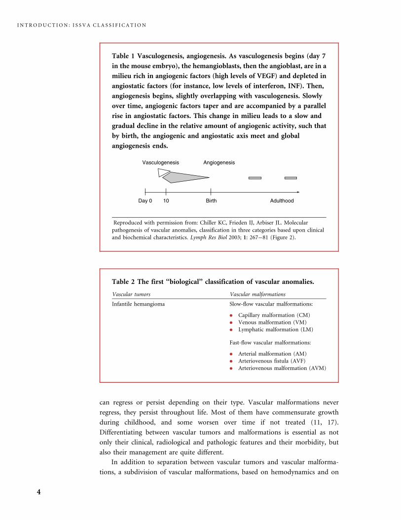
Table 1 Vasculogenesis, angiogenesis. As vasculogenesis begins (day 7
in the mouse embryo), the hemangioblasts, then the angioblast, are in a
milieu rich in angiogenic factors (high levels of VEGF) and depleted in
angiostatic factors (for instance, low levels of interferon, INF). Then,
angiogenesis begins, slightly overlapping with vasculogenesis. Slowly
over time, angiogenic factors taper and are accompanied by a parallel
rise in angiostatic factors. This change in milieu leads to a slow and
gradual decline in the relative amount of angiogenic activity, such that
by birth, the angiogenic and angiostatic axis meet and global
angiogenesis ends.
Reproduced with permission from: Chiller KC, Frieden IJ, Arbiser JL. Molecular
pathogenesis of vascular anomalies, classification in three categories based upon clinical
and biochemical characteristics. Lymph Res Biol 2003; 1: 267�81 (Figure 2).
Table 2 The first ‘‘biological’’ classification of vascular anomalies.
Vascular tumors Vascular malformations
Infantile hemangioma Slow-flow vascular malformations:
. Capillary malformation (CM)
. Venous malformation (VM)
. Lymphatic malformation (LM)
Fast-flow vascular malformations:
. Arterial malformation (AM)
. Arteriovenous fistula (AVF)
. Arteriovenous malformation (AVM)
I N T R O D U C T I O N : I S S V A C L A S S I F I C A T I O N
4
predominant anomalous channels, was created (10, 11, 21). Vascular malforma-
tions are either slow-flow or fast-flow, and they are subcategorized into capillary
malformation (CM), venous malformation (VM), lymphatic malformation (LM),
and arteriovenous malformation (AVM) (Tables 1�4). This is quite important,
since their management, with regard to both diagnosis (Table 5) and treatment
(Table 6), will also be quite different depending on their subtype (5�9, 17, 21).
Some patients have complex-combined vascular malformations, defined as capi-
llary venous malformation (CVM), capillary lymphatic malformation (CLM),
capillary lymphatic venous malformation (CLVM), lymphatic venous malforma-
tion (LVM), capillary arteriovenous malformation (C-AVM), or lymphatic arte-
riovenous malformation (L-AVM). Many of these syndromes are still labeled using
eponymous terminology (Table 7).
Since 1982, a number of biological investigations have confirmed obvious
differences between vascular tumors and malformations. Markers of cellular
proliferation, such as cell nuclear antigen, type IV collagenase, vascular endothelial
growth factor (VEGF), and basic fibroblast growth factor (bFGF), are elevated in
proliferating hemangiomas, and not in vascular malformations (19). Serum levels
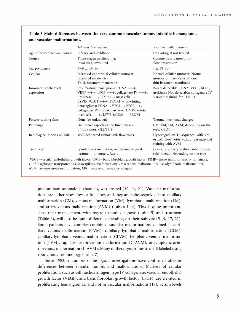
Table 3 Main differences between the very common vascular tumor, infantile hemangioma,
and vascular malformations.
Infantile hemangioma Vascular malformations
Age of occurrence and course Infancy and childhood Everlasting if not treated
Course Three stages: proliferating,
involuting, involuted
Commensurate growth or
slow progression
Sex prevalence 3�9 girls/1 boy 1 girl/1 boy
Cellular Increased endothelial cellular turnover.
Increased mastocytes.
Thick basement membrane
Normal cellular turnover. Normal
number of mastocytes. Normal
thin basement membrane
Immunohistochemical
expression
Proliferating hemangioma: PCNA þþþ,
VEGF þþþ, bFGF þþþ, collagenase IV þþþ,
urokinase þþ, TIMP-1 -, mast cells �,
LYVE-1/CD31 þþþ, PROX1 � Involuting
hemangioma: PCNA -, VEGF þ, bFGF þþ,
collagenase IV -, urokinase þþ, TIMP-1þþþ,
mast cells þþþ, LYVE-1/CD31 �, PROX1 �
Barely detectable: PCNA, VEGF, bFGF,
urokinase Not detectable: collagenase IV
Variable staining for TIMP 1
Factors causing flare None (or unknown) Trauma, hormonal changes
Pathology Distinctive aspects of the three phases
of the tumor. GLUT1 þ
CM, VM, LM, AVM, depending on the
type. GLUT1 �
Radiological aspects on MRI Well-delineated tumor with flow voids Hypersignal on T2-sequences with VM
or LM. Flow voids without parenchymal
staining with AVM
Treatment Spontaneous involution, or pharmacological
treatment, or surgery, lasers
Lasers, or surgery and/or embolization/
sclerotherapy depending on the type
VEGF¼vascular endothelial growth factor; bFGF¼basic fibroblast growth factor; TIMP¼tissue inhibitor matrix proteinase;
GLUT1¼glucose transporter 1; CM¼capillary malformation; VM¼venous malformation; LM¼lymphatic malformation;
AVM¼arteriovenous malformation; MRI¼magnetic resonance imaging.
I N T R O D U C T I O N : I S S V A C L A S S I F I C A T I O N
5

Table 4 Updated ISSVA classification of vascular anomalies.
Vascular tumors Vascular malformations
. Infantile hemangiomas
. Congenital hemangiomas (RICH and NICH)
. Tufted angioma (with or without
Kasabach�Merritt syndrome)
. Kaposiform hemangioendothelioma (with or without
Kasabach�Merritt syndrome)
. Spindle cell hemangioendothelioma
. Other, rare hemangioendotheliomas (epithelioid,
composite, retiform, polymorphous, Dabska tumor,
lymphangioendotheliomatosis, etc.)
. Dermatologic acquired vascular tumors (pyogenic
granuloma, targetoid hemangioma, glomeruloid
hemangioma, microvenular hemangioma, etc.)
Slow-flow vascular malformations:
. Capillary malformation (CM)
Port-wine stain
Telangiectasia
Angiokeratoma
. Venous malformation (VM)
Common sporadic VM
Bean syndrome
Familial cutaneous and mucosal venous
malformation (VMCM)
Glomuvenous malformation (GVM)
(glomangioma)
Maffucci syndrome
. Lymphatic malformation (LM)
Fast-flow vascular malformations:
. Arterial malformation (AM)
. Arteriovenous fistula (AVF)
. Arteriovenous malformation (AVM)
Complex-combined vascular malformations:
. CVM, CLM, LVM, CLVM,
AVM-LM, CM-AVM
C¼capillary; V¼venous; L¼lymphatic; AV¼arteriovenous; M¼malformation. RICH¼rapidly involuting congenital
hemangioma; NICH¼noninvoluting congenital hemangioma.
Table 5 Diagnostic imaging devices and the various vascular anomalies.
Infantile
hemangioma
CM VM LM AVM
Ultrasonography/Doppler þþþ þþ þþ þþ þþþ
Plain radiographs � � þþ (phleboliths,
bone)
þ/� (bone) þ (bone)
MRI, MRA, MRV þþ � þþþ þþþ þþ
CT þ � þ þ þ
Angio-CT scans � � þ � þþ
Lymphoscintigraphy � � � þ �
Biopsy þ þ þ þ þ
Angiography � _ þ � þþþ
MRI¼magnetic resonance imaging; MRA¼magnetic resonance angiography; MRV¼magnetic resonance venography;
CT¼computed tomography; CM¼capillary malformation; VM¼venous malformation; LM¼lymphatic malformation;
AVM¼arteriovenous malformation.
I N T R O D U C T I O N : I S S V A C L A S S I F I C A T I O N
6

of VEGF are significantly higher in proliferating hemangiomas than in involuting
hemangiomas, vascular malformations, and normal controls (23).
The origin of endothelial cells within the common hemangiomas of infancy
has been discussed since it was established that they express GLUT1, merosin,
Lewis Y antigen, and FCg receptor II, during the three stages of hemangioma life
(proliferating, involuting, and involuted stages) (18). These markers are also pre-
sent on endothelial cells of placenta microvessels. These proteins are not expressed
on endothelial cells of vascular malformations: the placenta-like microvascular
phenotype is lacking in all types of vascular malformations (18). As GLUT1
positivity is lost in hemangioma cultures further experiments would determine if
hemangioma endothelial cells actually originate from placenta or if both heman-
gioma endothelial cells and placenta endothelial cells simply share a similarly
immature phenotype.
LYVE-1/CD 31 double staining gave positive results in proliferating heman-
gioma and not in involuting hemangioma, while PROX-1 was negative in both
phases of hemangioma, and Dadras et al. concluded that these infantile tumors
are arrested in an early developmental vascular differentiation state (8) (Table 3).
New, mainly immunohistological, data let us update and complete the ISSVA
classification (Table 4).
In roughly half of cases a hemangioma regresses to result in normal-appearing
skin; however, it has long been observed that some involuted hemangiomas
develop into a prominent fibro-fatty residuum. According to Bischoff (4) and Yu
et al. (22) mesenchymal stem cells with adipogenic potential are present in pro-
liferating hemangioma, and these cells probably contribute to this adipogenesis.
Table 6 Main therapeutic strategies depending on the type of vascular
anomaly.
Modality Vascular tumors Vascular malformations
Pharmacological therapies
(glucocorticosteroids, interferon
alpha 2a or 2b, vincristine,
cyclophosphamide,
bleomycine, etc.)
þþþ þ/�
Lasers (FPDL, Nd-YAG,
Diode, etc.)
þ CM þþþ
VM and LM þ
Surgical excision/resection þþ þþ
Direct puncture sclerotherapy � VM and LM þþþ
AVM þ
Arterial superselective
embolization
þ/� (liver hemangiomas,
hemangiomas with
congestive cardiac failure
AVM þþþ
VM þ/�
FPDL¼flashlamp pulsed dye laser; CM¼capillary malformation; VM¼venous
malformation; LM¼lymphatic malformation; AVM¼arteriovenous malformation.
I N T R O D U C T I O N : I S S V A C L A S S I F I C A T I O N
7
Various theories concerning the pathogenesis of hemangioma have been
developed (3). Some suggest an intrinsic defect of hemangioma endothelial cells
(hem ECs): the clonality of hem ECs has been demonstrated and a somatic
mutation in a single progenitor cell has been hypothesized as the cause of
hemangioma. The intrinsic theory is reinforced by the demonstration of loss of
heterozygosity in 5q and by paradoxical response to endostatin of cultured hem
ECs (3). Other theories suggest that hemangioma endothelial cells respond to
extrinsic defects present in the local environment. These are based on various
experiments: release of VEGF from in vitro cultured proliferating hemangioma was
found (1), and alteration of expression of interferon-b in the epidermis overlying
proliferating hemangioma, but not in the keratinocytes distant to the heman-
gioma, was demonstrated (2).
A balance between intrinsic and extrinsic factors, and between stimulators
and inhibitors of angiogenesis, might account for the rapid growth and slow
subsequent involution of infantile hemangiomas (3, 12).
It is currently hypothesized that infantile hemangiomas are primarily the
consequence of excess angiogenesis (‘‘hemangiogenesis’’), while vascular malfor-
mations could be the result of errors in vessel remodeling (6). It has long been
unclear whether true angiogenesis occurs in some vascular malformations that
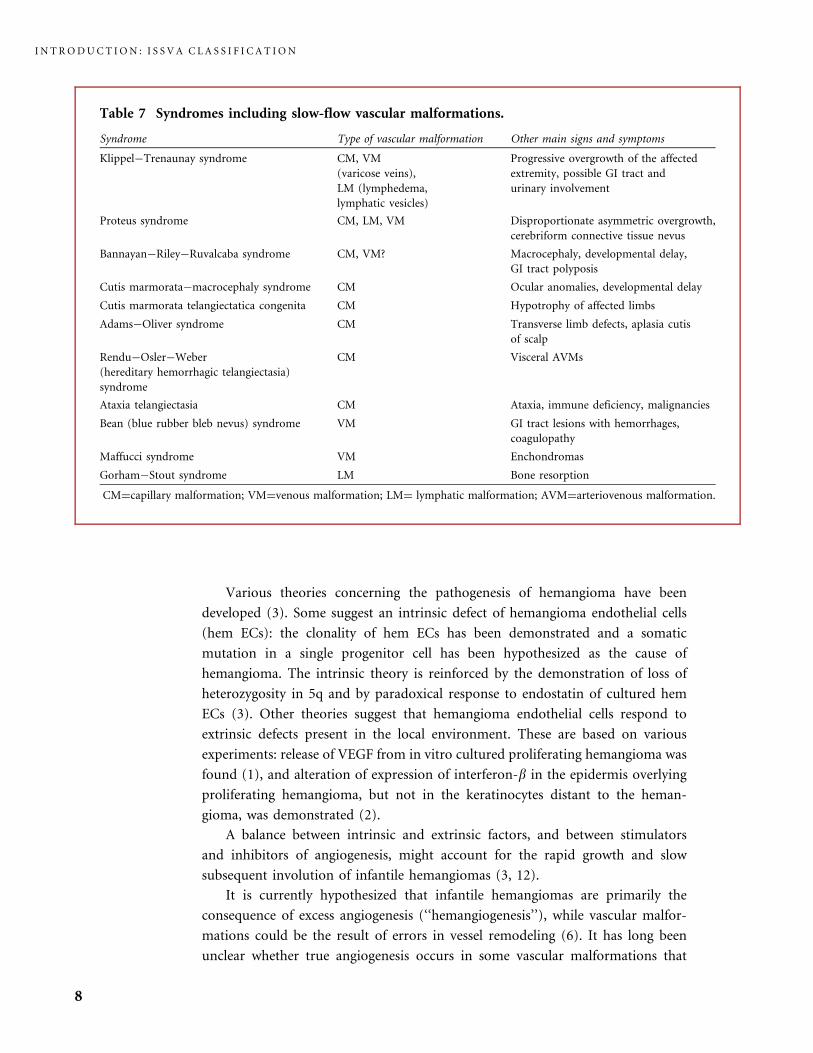
Table 7 Syndromes including slow-flow vascular malformations.
Syndrome Type of vascular malformation Other main signs and symptoms
Klippel�Trenaunay syndrome CM, VM
(varicose veins),
LM (lymphedema,
lymphatic vesicles)
Progressive overgrowth of the affected
extremity, possible GI tract and
urinary involvement
Proteus syndrome CM, LM, VM Disproportionate asymmetric overgrowth,
cerebriform connective tissue nevus
Bannayan�Riley�Ruvalcaba syndrome CM, VM? Macrocephaly, developmental delay,
GI tract polyposis
Cutis marmorata�macrocephaly syndrome CM Ocular anomalies, developmental delay
Cutis marmorata telangiectatica congenita CM Hypotrophy of affected limbs
Adams�Oliver syndrome CM Transverse limb defects, aplasia cutis
of scalp
Rendu�Osler�Weber
(hereditary hemorrhagic telangiectasia)
syndrome
CM Visceral AVMs
Ataxia telangiectasia CM Ataxia, immune deficiency, malignancies
Bean (blue rubber bleb nevus) syndrome VM GI tract lesions with hemorrhages,
coagulopathy
Maffucci syndrome VM Enchondromas
Gorham�Stout syndrome LM Bone resorption
CM¼capillary malformation; VM¼venous malformation; LM¼ lymphatic malformation; AVM¼arteriovenous malformation.
I N T R O D U C T I O N : I S S V A C L A S S I F I C A T I O N
8

exhibit a clear propensity to thicken over the years, or expand, or even multiply.
An example can be found with the lifelong increasing number of venous lesions in
Bean syndrome (also known as blue rubber bleb nevus syndrome). Another
example is the lethal, inexorably expanding, unalleviated course of some visceral
thoracic and abdominal microcystic lymphatic malformations. New findings
indicate that vascular malformations may also be angiogenesis-dependent
disorders: urinary high-molecular-weight matrix metalloproteinases (hMW
MMPs) and bFGF levels are elevated not only in vascular tumors but also in
some vascular malformations, such as lymphatic or lymphatico-venous malforma-
tions and arteriovenous malformations (15). It is noticeable that this urinary
increase in bFGF and hMW MMPs parallels the extent and progression of the
vascular anomaly in patients with expanding, unremitting vascular malformations,
while urinary VEGF levels do not (15).
Fewer data are available concerning the pathogenesis of vascular malforma-
tions, compared with what is currently known about infantile hemangioma. The
excess of proteolytic enzymes like the hMW MMPs probably parallels the tissue
remodeling observed in diffuse and expanding vascular malformations, such as
some AVM or some LM, and the work of Marler et al. suggests that drugs targeting
bFGF or MMPs might be an adequate therapeutic strategy for these patients (15).
The existence of inherited forms of vascular malformations, although rare, has
permitted a new insight into the complex process of vasculogenesis and the
molecular pathways physiologically involved in vascular malformations (7). As
genetic defects are being identified in various types of vascular malformations
(VM, glomuvenous malformation, familial lymphedema, arteriovenous-capillary
malformation), the objective is to understand how such gene alterations, and
modifications in signaling pathways (Table 8) result in abnormal vascular
channels, with changes in embryonic blood or lymphatic vessels remodeling,
ending in the familial forms of vascular malformations (3, 6, 20).
Molecular biology may completely change our approach to the classification of
the various vascular anomalies (20). However, as we do not know whether the
biological mechanisms of the sporadic vascular malformations, the most frequent
ones, are similar to those of inherited forms, it is currently highly speculative to
propose a shift to a genetic classification.
In addition, current progress in the understanding of the pathogenesis of
angiogenesis-dependent vascular anomalies offers novel targets for their treatment.
As an example, the knowledge of the enzyme defect in Fabry disease has resulted
in enzyme replacement therapy with agalsidase alpha treatment, and this has
changed the prognosis of this severe familial vascular disease (14). Future therapies
for other types of vascular anomalies should be tailored to their specific defects
once they are identified.
Treatments for the various vascular anomalies have become more specifically
adapted over the last 30 years. Some treatments appeared to have more risks than
benefits and were discarded. This was the case for the various types of ionizing
radiation therapy. Therapeutic embolization through the arterial route and
I N T R O D U C T I O N : I S S V A C L A S S I F I C A T I O N
9
sclerotherapy through direct puncture of the lesion now have clear indications for
use. Surgical procedures have been adapted and customized by both plastic and
vascular surgeons. The development of laser technology, since the early 1960s, has
resulted in major progress in the treatment of capillary malformations, with better
clinical results as the devices have been improved. When successful, early laser
treatment of port wine stains provides better results than the surgical treatments
previously performed, and they allow the children to develop a positive self-image,
reducing the subconscious psychological impact of the CM. A great deal of
progress has been achieved in the field of vascular anomalies, but much still
remains to be accomplished, in particular to improve our knowledge of their
pathogenesis and the results of therapy.
References
1 Berard M, Ortega N, Carrier JL, Peyri N, Wassef M, Enjolras O, Drouet L. Plouet J.
Vascular endothelial growth factor confers a growth advantage in vitro and in vivo to
stromal cells cultured from neonatal hemangiomas. Am J Pathol 1997; 150: 1315�26.
2 Bielenberg DR, Bucana CD, Sanchez R. Progressive growth of infantile cutaneous
hemangiomas is directly correlated with hyperplasia and angiogenesis of adjacent
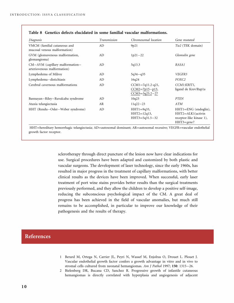
Table 8 Genetics defects elucidated in some familial vascular malformations.
Diagnosis Transmission Chromosomal location Gene mutated
VMCM (familial cutaneous and
mucosal venous malformation)
AD 9p21 Tie2 (TEK domain)
GVM (glomuvenous malformation,
glomangioma)
AD 1p21�22 Glomulin gene
CM�AVM (capillary malformation�
arteriovenous malformation)
AD 5q13.3 RASA1
Lymphedema of Milroy AD 5q34�q35 VEGFR3
Lymphedema�distichiasis AD 16q24 FOXC2
Cerebral cavernous malformations AD CCM1¼7q11.2-q21,
CCM2¼7p15�p13,
CCM3¼3q25.2�27
CCM1:KRIT1,
ligand de Krev/Rap1a
Bannayan�Riley�Ruvalcaba syndrome AD 10q23 PTEN
Ataxia telangiectasia AR 11q22�23 ATM
HHT (Rendu�Osler�Weber syndrome) AD HHT1¼9q33,
HHT2¼12q13,
HHT3¼5q31.5�32
HHT1¼ENG (endoglin),
HHT2¼ALK1(activin
receptor-like kinase 1),
HHT3¼gene ?
HHT¼hereditary hemorrhagic telangiectasia; AD¼autosomal dominant; AR¼autosomal recessive; VEGFR¼vascular endothelial
growth factor receptor.
I N T R O D U C T I O N : I S S V A C L A S S I F I C A T I O N
10

epidermis and inversely correlated with expression of endogenous angiogenesis inhibitor,
IFN-beta. Int J Oncol 1999; 14: 401�8.
3 Bischoff J. Monoclonal expansion of endothelial cells in hemangioma: an intrinsic defect
with extrinsic consequences? Trends Cardiovasc Med 2002; 12: 220�4.
4 Bischoff J. in: Infantile hemangiomas: current knowledge, future directions. Proceedings
of a Research Workshop on Infantile Hemangioma. Bethesda Maryland, April 7�9, 2005.
Pediatr Dermatol 2005; 22: 383�406.
5 Burrows PE, Laor T, Paltiel H, Robertson RL. Diagnostic imaging in the evaluation of
vascular birthmarks. Dermatol Clin 1998; 16: 455�88.
6 Chiller KG, Frieden IJ, Arbiser JL. Molecular pathogenesis of vascular anomalies.
Classification into three categories based upon clinical and biochemical characteristics.
Lymphatic Res and Biol 2003; 1: 267�82.
7 Cohen MM. Vasculogenesis, angiogenesis, hemangiomas and vascular malformations.
Am J Med Genet 2002; 108: 265�74.
8 Dadras SS, North PE, Bertoncini J, Mihm MC, Detmar M. Infantile hemangiomas are
arrested in an early vascular differentiation state. Mod Pathol 2004; 17: 1068�79.
9 Dubois J, Garel L. Imaging and therapeutic approach of hemangiomas and vascular
malformations in the pediatric age group. Pediatr Radiol 1999; 29: 879�93.
10 Enjolras O, Mulliken JB. Vascular tumors and vascular malformations (new issues).
Adv Dermatol 1997; 13: 375�423.
11 Enjolras O, Riche MC. Atlas des Hemangiomes et Malformations Vasculaires Superficielles.
Paris: Medsi-McGraw-Hill ; 1990.
12 Folkman J. Fundamental concepts of the angiogenic process. Curr Mol Med 2003; 3:
643�51.
13 Hand JL, Frieden IJ. Vascular birthmarks of infancy: resolving nosologic confusion.
Am J Med Genet 2002; 108: 257�64.
14 Hoffmann B, Garciade Lorenzo A, Mehta A, Beck M, Widmer U, Ricci R. FOS European
Investigators. Effects of enzyme replacement therapy on pain and health related quality of
life in patients with Fabry disease: data from FOS (Fabry Outcome Survey). J Med Genet
2005; 42: 247�52.
15 Marler JJ, Fishman SJ, Kilroy SM, Fang J, Upton J, Mulliken JB, Burrows PE, Zurakowski
D, Folkman J, Moses MA. Increased expression of urinary matrix metalloproteinases
parallels the extent and activity of vascular anomalies. Pediatrics 2005; 116: 38�45.
16 Mulliken JB, Glowacki J. Hemangiomas and vascular malformations in infants and children: a
classification based on endothelial characteristics. Plast Reconstr Surg 1982; 69: 412�22.
17 (Mulliken JB, Young AE, eds.) Vascular Birthmarks: Hemangiomas, & Malformations.
Philadelphia: WB Saunders, 1988.
18 North PE, Waner M, Mizeracki A, Mrak RE, Nicholas R, Kincannon J, Suen JY, Mihm
MC Jr. A unique microvascular phenotype shared by juvenile hemangiomas and human
placenta. Arch Dermatol 2001; 137: 559�70.
19 Takahashi K, Mulliken JB, Kozakewich HPW, Rogers RA, Folkman J, Ezekowitz RA.
Cellular markers that distinguish the phases of hemangioma during infancy and
childhood. J Clin Invest 1994; 93: 2357�64.
20 Vikkula M, Boon LM, Mulliken JB. Molecular genetics of vascular anomalies. Matrix Biol
2001; 20: 327�35.
21 Wassef M, Enjolras O. Les malformations vasculaires superficielles: classification
et histopathologie. Ann Pathol 1999; 19: 253�64.
22 Yu Y, Fuhr J, Boye E, Gyorffy S, et al., Mesenchymal stem cells and adipogenesis
in hemangioma involution. Stem Cells 2006; epub ahead of print.
23 Zhang L, Lin X, Wand W, Zhuang X, Dong J, Qi Z, Hu Q. Circulating level of vascular
endothelial growth factor in differentiating hemangioma from vascular malformation
patients. Plast Reconstr Surg 2005; 116: 200�4.
R E F E R E N C E S
11


PART I
Investigations and
Radiological Tools

Various imaging tools are available for the diagnosis of vascular malformations
(1�5). Techniques must be adapted to the clinical findings and to the aim of
imaging, which may be diagnosis, pre-therapeutic assessment, or follow-up with or
without treatment.
Conventional X-Rays
These are usually of little interest and are normal in most situations. Venous
malformations may be diagnosed if phleboliths are seen on plain radiographs.
Bone distortion is only seen in large malformations with an important soft tissue
mass effect. Some diffuse venous malformations in the limbs match up with fragile,
thinner, curved bones, and sometimes lytic lesions, and a risk of pathologic
fracture. Occasionally, an arteriovenous malformation involves a bone and either
the intraosseous nidus, or large draining venous channels, after the nidus, create
lytic bony lesions.
Ultrasonography in Combination with Doppler
This scan is frequently used as the primary diagnostic tool (4). It often permits
distinction between tumors and malformations. It also allows a vascular malfor-
mation to be identified and pinpoints the type of lesion. It shows whether the
lesion is cystic or tissular, demonstrates the presence or absence of flow, and thus
15

differentiates between fast-flow and slow-flow malformations. Angioarchitecture
and vessel density may be analyzed but reliability is often poor. Peak flow velocities
and arterial output may also be measured in AVMs. In a patient with an AVM
in the head and neck or in an extremity, comparing the arterial output on the
normal side to that on the contralateral vascular abnormal side (e.g. both carotid,
or both humeral, or both femoral arterial outputs, depending on the site of the
AVM), is indispensable to get an idea of the prognosis, and particularly of possible
cardiac failure. These techniques are particularly useful for the noninvasive follow-
up of AVMs.
Computed Tomography (CT)
This is of limited interest, even after iodinated contrast injection, only allowing us
to decide if a lesion is highly vascularized or not. Precise description and diagnosis
of soft tissue lesions remain weak, except in macrocystic lymphatic malformations
where the cysts are clearly depicted. The presence of phleboliths may direct us
towards a diagnosis of venous malformation as these round calcifications develop
on thromboses linked to the slow flow. Bony displacement or alteration can also
be seen due to chronic compression in VMs and LMs. Transcranial connections
are also identified by CT in head and neck VMs. CT scan angiography, with 3-D
reconstruction, however, may superbly map the enlarged vascular channels in an
arteriovenous malformation.
Magnetic Resonance Imaging (MRI)
This is the best diagnostic tool, allowing optimal analysis of soft tissue masses and
adequate diagnosis, differentiating tissular from cystic lesions, and showing fast
and slow circulating vessels. As an example, MRI is indispensable in the diagnosis
of peri-ocular hemangioma (3). Venous and lymphatic malformations have a
characteristic pattern, being hyperintense on spin echo T2-weighted sequences,
and optimally seen on fat suppression sequences. Fat suppression T1-weighted
sequences with gadolinium injection show an intense enhancement in infantile
hemangioma tumors, whereas the enhancement is variable and progressive on
dynamic sequences in venous malformations. Gadolinium contrast injection
permits differential diagnosis between VM and LM. LMs can be differentiated
from VMs as they show only enhancement at the margins of the cysts, while VMs
I N V E S T I G A T I O N S A N D R A D I O L O G I C A L T O O L S
16

are usually clearly stained. MRI is not only useful for identification and diagnosis
of the lesion, but is also mandatory before treatment to delineate the extent of the
lesion and depict the relationship between the vascular malformation and
neighboring vessels and nerves. In fast circulating vessels, they will appear as flow
voids on most sequences. MR-angiography may then be performed, confirming
the diagnosis of fast circulating vessels, but it remains insufficient for precisely
depicting the AVM nidus and analyzing the angioarchitecture.
Conventional Vascular Imaging
These techniques are mostly not indicated for the diagnosis of a vascular
malformation, except for fast-flow vascular lesions.
Indirect phlebography is usually of little interest and should not be used
systematically in VMs as opacification of a venous malformation is inconsistently
seen. It is of some interest in some diffuse extremity VMs, before a therapeutic
decision is made.
Direct percutaneous phlebography is valuable for depicting a VM and to show
the draining pattern. However, it should only be used as a pre-therapeutic step,
immediately before sclerotherapy, and not as a diagnostic tool.
Conventional angiography has few indications in slow-flow vascular malfor-
mations as it will show a variable blush with a nonspecific pattern. Angiography
remains, however, indispensable for the diagnosis and pre-therapeutic assessment
of an AVM, the characteristic feature of which is an early venous drainage.
Angiography also allows us to analyze the angioarchitecture of the AVM, to
precisely identify its location, and to depict the arterial suppliers and draining
veins and its relationship to the normal surrounding arteries and veins.
Angiography is specially indicated to establish the diagnosis in quiescent AVMs,
simulating a capillary malformation, where it may be important not to miss the
diagnosis before proposing a treatment that may trigger the growth of a dormant
AVM, such as pulsed dye laser treatment (5).
References
1 Burrows PE, Laor T, Paltiel H, Robertson RL. Diagnostic imaging in the evaluation of
vascular birthmarks. Dermatol Clin 1998; 16: 455�88.
2 Dubois J, Garel L. Imaging and therapeutic approach of hemangiomas and vascular
malformations in the pediatric age group. Pediatr Radiol 1999; 29: 879�93.
R E F E R E N C E S
17

3 Millischer-Bellaiche AE, Enjolras O, Andre Ch, Bursztyn J, Kalifa G, Adamsbaum C.
Les hemangiomes palpebraux du nourrisson. J Radiol 2004; 85: 2019�28.
4 Paltiel H J, Burrows P E, Kozakewich H P, Zurakowski D, Mulliken JB. Soft-tissue
vascular anomalies: utility of US for diagnosis. Radiology 2000; 214: 747�54.
5 Wu JK, Bisdorff A, Gelbert F, Enjolras O, Burrows PE, Mulliken JB. Auricular
arteriovenous malformation: evaluation, management, and outcome. Plast Reconstr Surg
2005; 115: 985�95.
I N V E S T I G A T I O N S A N D R A D I O L O G I C A L T O O L S
18
PART II
Vascular Tumors


CHAPTER II.A
Infantile Hemangioma (IH)
Introduction
Infantile hemangioma (IH) is also known as ‘‘strawberry mark’’ and ‘‘immature
hemangioma;’’ the other names, ‘‘capillary hemangioma’’ and ‘‘cavernous heman-
gioma,’’ have long caused confusion with vascular malformations. IH is a very
frequent benign vascular tumor that grows rapidly in an infant over a period of a
few weeks or months after birth (the proliferating phase). Then it slowly and
constantly regresses over some years (the involuting phase), to leave nearly normal
skin, or skin and shape changes (the involuted phase). This third stage is rarely
reached at the age of 1 or 2 years, and is most commonly attained around 5 or
6 years, and sometimes not before 10 years. No such tumor occurs in an adolescent
or adult; thus, using the wording ‘‘hemangioma’’ or ‘‘capillary hemangioma’’ for
a vascular tumor appearing in an adolescent or an adult is misleading.
IHs affect about 10% of children. Dark-skinned infants have a lower incidence
than fair-skinned infants. Transcervical chorionic villus sampling increases the
risk of IH in the newborn, but not amniocentesis.
The incidence of hemangioma is increased in premature infants of very low
birth weight (under 1000 g) (4). A group of US Pediatric Dermatologists, the
Hemangioma Investigator Group, confirmed this finding in a prospective study
presented at the NIH-sponsored research workshop held in April 2005 at the
Bethesda Campus (41). They also highlighted an increased incidence in cases of
high maternal age for a first baby, multiple gestation, placental abnormalities,
placenta praevia, or preeclampsia.
Numerous factors inducing postnatal growth of hemangioma and subsequent
involution have been documented (9, 41, 99, 95), but the very first event initiating
the lesion itself remains unidentified, as the reasons for growth starting soon after
birth, and for the shift from proliferation to involution after a few months of
proliferation remain unknown (9).
It has been shown that IH has a distinctive placenta-like microvascular
phenotype (82) that is stable in vivo and lost in culture.
21
Two hypotheses currently rely on this finding:
1. the hemangioma could result from a somatic mutation occurring in a
regulatory gene in a progenitor endothelial cell ending in an immature
placental endothelial phenotype; and
2. it could originate from the clonal expansion of embole of placental
endothelial cells (82).
IHs are subcategorized into three groups: superficial, deep, and the ‘‘mixed’’
type which is both superficial and deep. They are single or multiple, or dis-
seminated (the miliary type or disseminated neonatal hemangiomatosis (DNH)).
An IH is of variable size: from a small dot to a diffuse plaque-like or bossed tumor
covering the face, part of the trunk, or an extremity. Color depends on the dermal
extent: a very superficial IH has the brightest red color, a deeply growing IH gives
a bluish shade and telangiectasia to the overlying skin, or is situated under
normally colored skin.
IH predominantly affects the skin (ubiquitously), the oral and genital mucous
membrane, the orbit, the airway, and the parotid. Specific locations, such as
the lids, nose or lips, have distinctive aspects and complications. It has been
suggested that facial hemangiomas develop in a nonrandom distribution (104).
Visceral involvement is uncommon. Muscles are not affected but IH may infiltrate
the fascia between muscles (in the literature venous malformations of muscles
are too often incorrectly labeled intramuscular hemangiomas). Bones are not
affected (the so-called hemangioma of bones in the literature corresponds mainly
to bony venous malformation). A very rare facial bony tumor present at birth
may mimic IH on pathology, but it is GLUT1 negative (OE, unpublished data).
GLUT1 is a highly selective immunohistochemical marker for IH, of major value
when the pathological diagnosis is somewhat doubtful (81, 82).
The three phases of an IH, proliferation, spontaneous involution, and
involuted, sometimes with sequelae, are of variable duration depending on
the patient. In visceral locations they follow the same three-phase course as
superficial IH.
A majority of IHs (80 to 90%) are small and not dangerous, and may be
left to recede spontaneously. However, location has a crucial role in determining
possible risks (26). IHs that are alarming due to size, site, volume, function-
threatening location (eyelid and orbit, airway, etc.) or that are life-threatening
(massive tumor, ulceration and subsequent infection, visceral location, recur-
rent hemorrhages, congestive heart failure), will require active therapeutic
management.
Infants with DNH are at greater risk of visceral involvement. The liver is the
most common location; however, many patients with diffuse liver IH do not have
skin lesions or only a few lesions (41). IHs can develop in many other visceral
sites: GI tract, pancreas, kidney, lung, heart, meninges, brain, etc. A majority of
skin and liver DNHs follow a benign self-limiting course, and are assessed by
regular clinical and ultrasonographic follow-up; involution of both superficial
I N F A N T I L E H E M A N G I O M A ( I H )
22
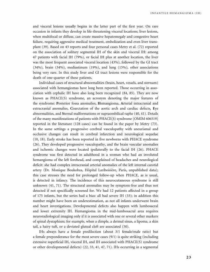
and visceral lesions usually begins in the latter part of the first year. On rare
occasion in infants they develop in life-threatening visceral locations; liver lesions,
when multifocal or diffuse, can create massive hepatomegaly and congestive heart
failure, requiring aggressive medical treatment, embolization and even liver trans-
plant (39). Based on 43 reports and four personal cases Metry et al. (72) reported
on the association of solitary segmental IH of the skin and visceral IH: among
47 patients with facial IH (79%), or facial IH plus at another location, the liver
was the most frequent associated visceral location (43%), followed by the GI tract
(34%), brain (34%), mediastinum (19%), and lung (15%), other associations
being very rare. In this study liver and GI tract lesions were responsible for the
death of one-quarter of these patients.
Individual cases of structural abnormalities (brain, heart, vessels, and sternum)
associated with hemangiomas have long been reported. Those occurring in asso-
ciation with cephalic IH have also long been recognized (84, 85). They are now
known as PHACE(S) syndrome, an acronym denoting the major features of
the syndrome: Posterior fossa anomalies, Hemangioma, Arterial intracranial and
extracranial anomalies, Coarctation of the aortic arch and cardiac defects, Eye
abnormalities, and Sternal malformations or supraombilical raphe (40, 41). Details
of the many manifestations of patients with PHACE(S) syndrome (OMIM 606519)
reported in the literature (128 cases) can be found in the paper by Metry (73).
In the same settings a progressive cerebral vasculopathy with aneurismal and
occlusive changes can result in cerebral infarction and neurological sequelae
(10, 18). Early stroke has been reported in five newborns with PHACE syndrome
(26). They developed progressive vasculopathy, and the brain vascular anomalies
and ischemic changes were located ipsilaterally to the facial IH (26). PHACE
syndrome was first detected in adulthood in a woman who had an involuted
hemangioma of the left forehead, and complained of headaches and neurological
deficit: she had complex intracranial arterial anomalies of the left internal carotid
artery (Dr. Monique Boukobza, Hopital Lariboisiere, Paris, unpublished data);
this case stresses the need for prolonged follow-up when PHACE, as is usual,
is detected in infancy. The incidence of this neurocutaneous syndrome is still
unknown (41, 71). The structural anomalies may be symptom-free and thus not
detected if not specifically screened for. We had 12 patients affected in a group
of 175 infants, but the series had a bias: all had severe IH (33); in addition this
number might have been an underestimation, as not all infants underwent brain
and heart investigations. Developmental defects also happen with lumbosacral
and lower extremity IH. Hemangioma in the mid-lumbosacral area requires
neuroradiological imaging only if it is associated with one or several other markers
of spinal dysraphism; for example, when a dimple, a dermal sinus, a lipoma, a skin
tail, a hairy tuft, or a deviated gluteal cleft are associated (50).
IHs always have a female predilection (about 3/1 female/male ratio) but
a female preponderance for the most severe cases (9/1) is quite striking (including
extensive superficial IH, visceral IH, and IH associated with PHACE(S) syndrome
or other developmental defects) (22, 33, 41, 47, 71). IHs occurring in a segmental
I N F A N T I L E H E M A N G I O M A ( I H )
23
morphology carry a higher risk of complications (22, 74). The cause of PHACE(S)
syndrome is unknown and the female predominance leads to the hypothesis of
an X-linked defect surviving by mosaıcism with lethality in males (16, 41).
Concerning the association of a vascular tumor, the IH, with structural
malformations Bauland et al. hypothesized either developmental field anomalies
or single gene defects (9).
Pathology
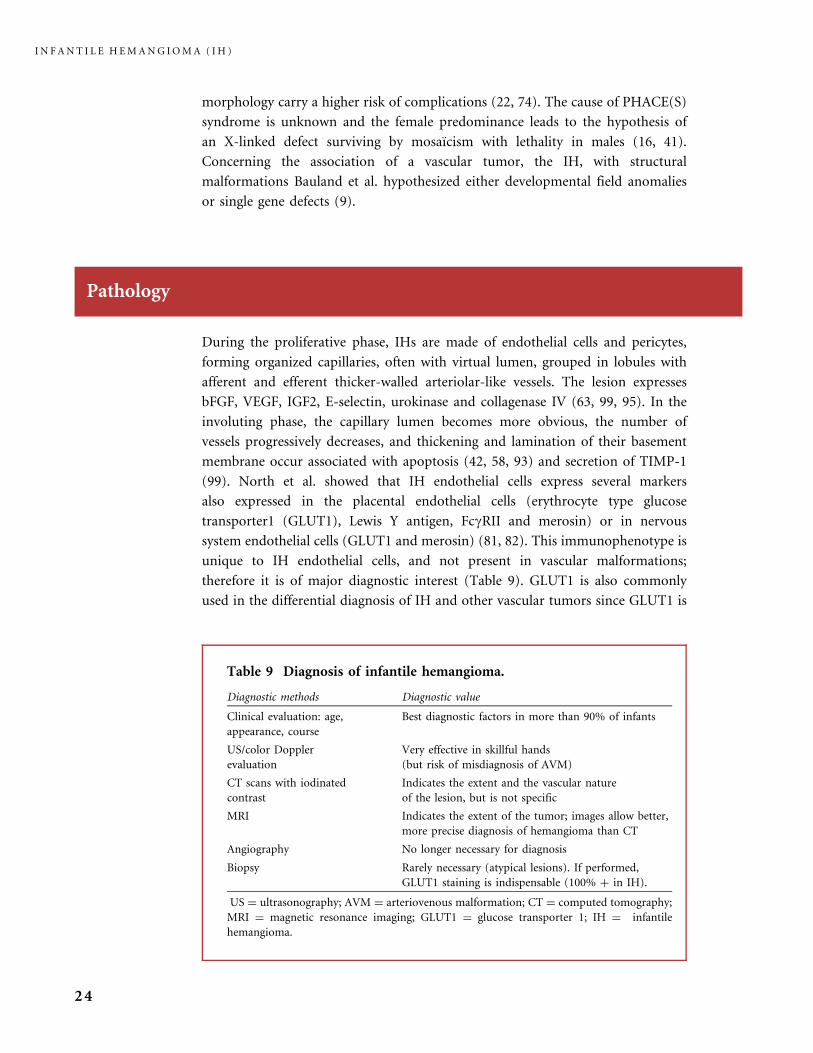
During the proliferative phase, IHs are made of endothelial cells and pericytes,
forming organized capillaries, often with virtual lumen, grouped in lobules with
afferent and efferent thicker-walled arteriolar-like vessels. The lesion expresses
bFGF, VEGF, IGF2, E-selectin, urokinase and collagenase IV (63, 99, 95). In the
involuting phase, the capillary lumen becomes more obvious, the number of
vessels progressively decreases, and thickening and lamination of their basement
membrane occur associated with apoptosis (42, 58, 93) and secretion of TIMP-1
(99). North et al. showed that IH endothelial cells express several markers
also expressed in the placental endothelial cells (erythrocyte type glucose
transporter1 (GLUT1), Lewis Y antigen, FcgRII and merosin) or in nervous
system endothelial cells (GLUT1 and merosin) (81, 82). This immunophenotype is
unique to IH endothelial cells, and not present in vascular malformations;
therefore it is of major diagnostic interest (Table 9). GLUT1 is also commonly
used in the differential diagnosis of IH and other vascular tumors since GLUT1 is
Table 9 Diagnosis of infantile hemangioma.
Diagnostic methods Diagnostic value
Clinical evaluation: age,
appearance, course
Best diagnostic factors in more than 90% of infants
US/color Doppler
evaluation
Very effective in skillful hands
(but risk of misdiagnosis of AVM)
CT scans with iodinated
contrast
Indicates the extent and the vascular nature
of the lesion, but is not specific
MRI Indicates the extent of the tumor; images allow better,
more precise diagnosis of hemangioma than CT
Angiography No longer necessary for diagnosis
Biopsy Rarely necessary (atypical lesions). If performed,
GLUT1 staining is indispensable (100% þ in IH).
US ¼ ultrasonography; AVM ¼ arteriovenous malformation; CT ¼ computed tomography;
MRI ¼ magnetic resonance imaging; GLUT1 ¼ glucose transporter 1; IH ¼ infantile
hemangioma.
I N F A N T I L E H E M A N G I O M A ( I H )
24
positive in 100% of IH endothelial cells and negative in the other infantile vascular
tumors, including congenital hemangiomas, tufted angioma, and kaposiform
hemangioendothelioma.
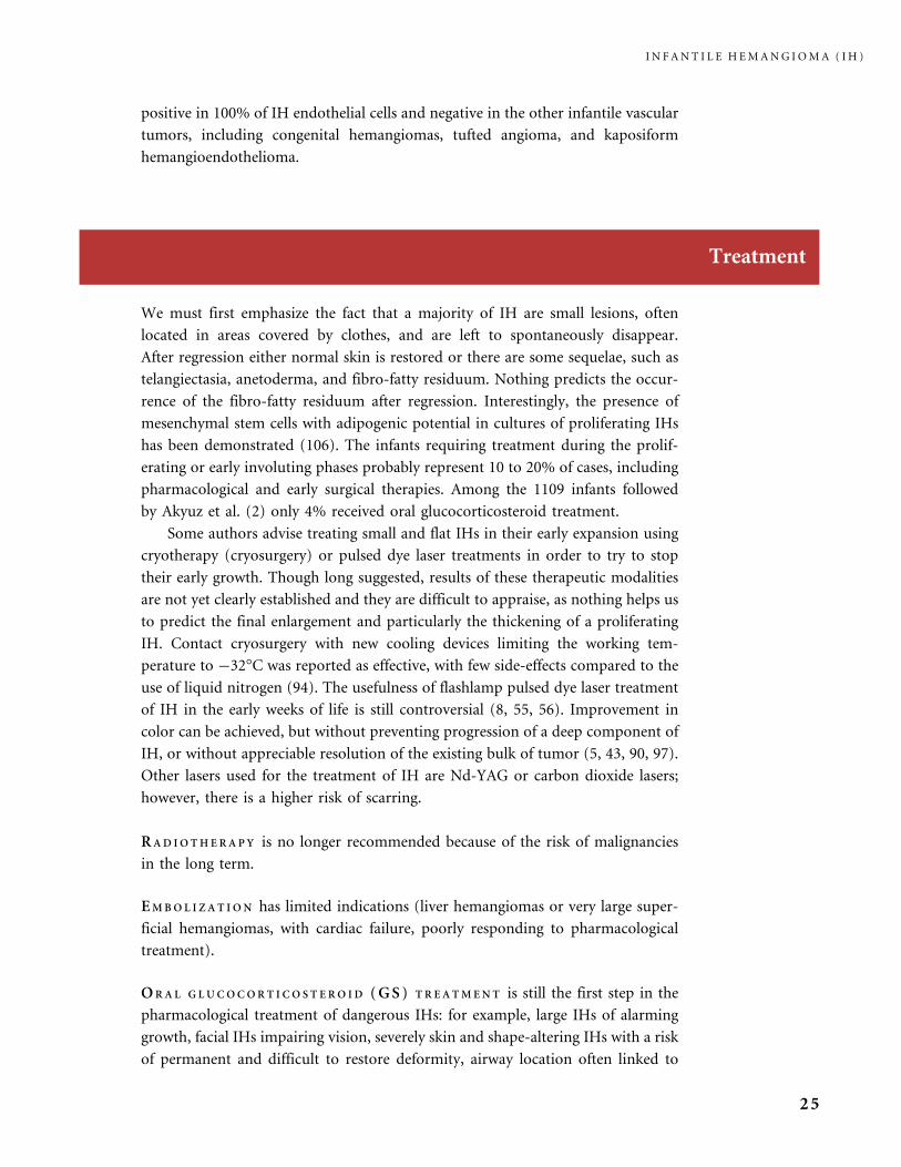
Treatment
We must first emphasize the fact that a majority of IH are small lesions, often
located in areas covered by clothes, and are left to spontaneously disappear.
After regression either normal skin is restored or there are some sequelae, such as
telangiectasia, anetoderma, and fibro-fatty residuum. Nothing predicts the occur-
rence of the fibro-fatty residuum after regression. Interestingly, the presence of
mesenchymal stem cells with adipogenic potential in cultures of proliferating IHs
has been demonstrated (106). The infants requiring treatment during the prolif-
erating or early involuting phases probably represent 10 to 20% of cases, including
pharmacological and early surgical therapies. Among the 1109 infants followed
by Akyuz et al. (2) only 4% received oral glucocorticosteroid treatment.
Some authors advise treating small and flat IHs in their early expansion using
cryotherapy (cryosurgery) or pulsed dye laser treatments in order to try to stop
their early growth. Though long suggested, results of these therapeutic modalities
are not yet clearly established and they are difficult to appraise, as nothing helps us
to predict the final enlargement and particularly the thickening of a proliferating
IH. Contact cryosurgery with new cooling devices limiting the working tem-
perature to �32�C was reported as effective, with few side-effects compared to the
use of liquid nitrogen (94). The usefulness of flashlamp pulsed dye laser treatment
of IH in the early weeks of life is still controversial (8, 55, 56). Improvement in
color can be achieved, but without preventing progression of a deep component of
IH, or without appreciable resolution of the existing bulk of tumor (5, 43, 90, 97).
Other lasers used for the treatment of IH are Nd-YAG or carbon dioxide lasers;
however, there is a higher risk of scarring.
RAD IO TH E RA P Y is no longer recommended because of the risk of malignancies
in the long term.
EMBO L I Z A T I ON has limited indications (liver hemangiomas or very large super-
ficial hemangiomas, with cardiac failure, poorly responding to pharmacological
treatment).
ORAL G LUCOCOR T I CO S T E RO I D (GS ) T R E A TMEN T is still the first step in the
pharmacological treatment of dangerous IHs: for example, large IHs of alarming
growth, facial IHs impairing vision, severely skin and shape-altering IHs with a risk
of permanent and difficult to restore deformity, airway location often linked to
I N F A N T I L E H E M A N G I O M A ( I H )
25
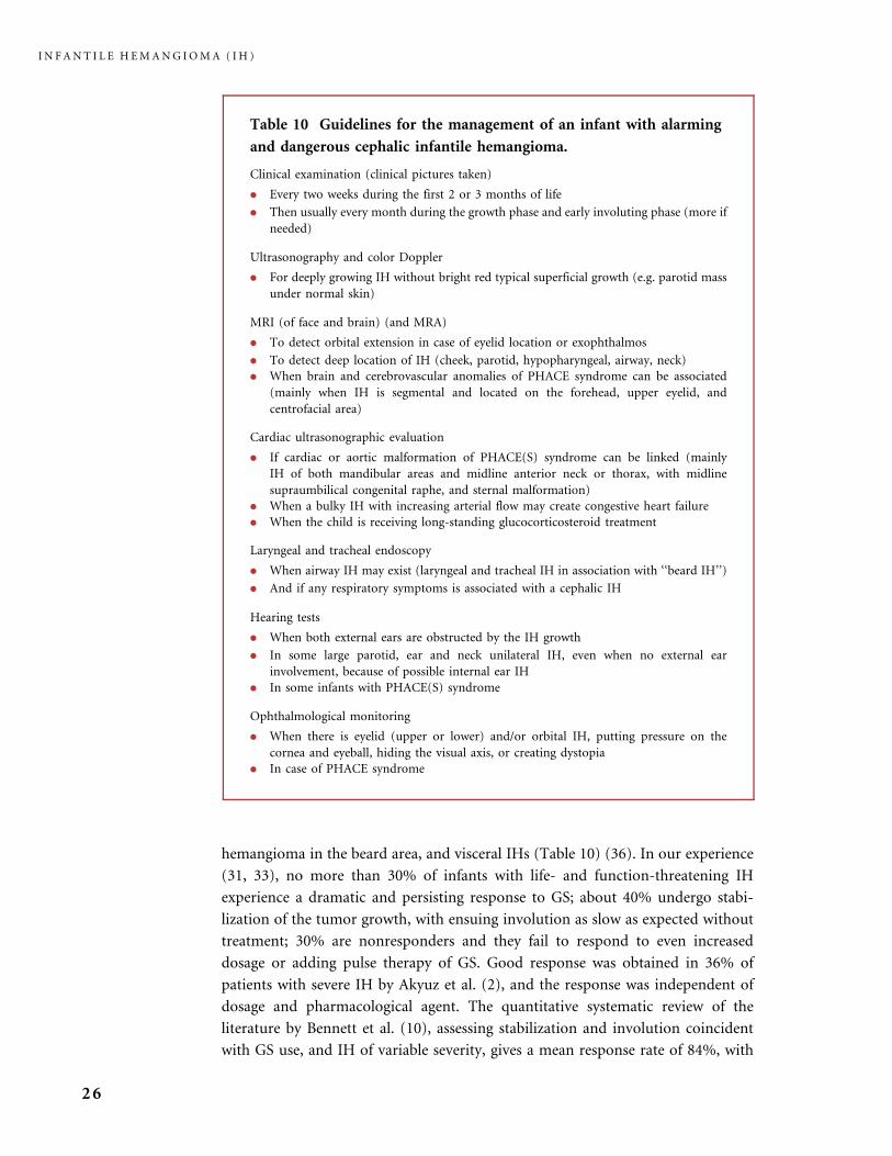
hemangioma in the beard area, and visceral IHs (Table 10) (36). In our experience
(31, 33), no more than 30% of infants with life- and function-threatening IH
experience a dramatic and persisting response to GS; about 40% undergo stabi-
lization of the tumor growth, with ensuing involution as slow as expected without
treatment; 30% are nonresponders and they fail to respond to even increased
dosage or adding pulse therapy of GS. Good response was obtained in 36% of
patients with severe IH by Akyuz et al. (2), and the response was independent of
dosage and pharmacological agent. The quantitative systematic review of the
literature by Bennett et al. (10), assessing stabilization and involution coincident
with GS use, and IH of variable severity, gives a mean response rate of 84%, with
Table 10 Guidelines for the management of an infant with alarming
and dangerous cephalic infantile hemangioma.
Clinical examination (clinical pictures taken)
. Every two weeks during the first 2 or 3 months of life
. Then usually every month during the growth phase and early involuting phase (more if
needed)
Ultrasonography and color Doppler
. For deeply growing IH without bright red typical superficial growth (e.g. parotid mass
under normal skin)
MRI (of face and brain) (and MRA)
. To detect orbital extension in case of eyelid location or exophthalmos
. To detect deep location of IH (cheek, parotid, hypopharyngeal, airway, neck)
. When brain and cerebrovascular anomalies of PHACE syndrome can be associated
(mainly when IH is segmental and located on the forehead, upper eyelid, and
centrofacial area)
Cardiac ultrasonographic evaluation
. If cardiac or aortic malformation of PHACE(S) syndrome can be linked (mainly
IH of both mandibular areas and midline anterior neck or thorax, with midline
supraumbilical congenital raphe, and sternal malformation)
. When a bulky IH with increasing arterial flow may create congestive heart failure
. When the child is receiving long-standing glucocorticosteroid treatment
Laryngeal and tracheal endoscopy
. When airway IH may exist (laryngeal and tracheal IH in association with ‘‘beard IH’’)
. And if any respiratory symptoms is associated with a cephalic IH
Hearing tests
. When both external ears are obstructed by the IH growth
. In some large parotid, ear and neck unilateral IH, even when no external ear
involvement, because of possible internal ear IH
. In some infants with PHACE(S) syndrome
Ophthalmological monitoring
. When there is eyelid (upper or lower) and/or orbital IH, putting pressure on the
cornea and eyeball, hiding the visual axis, or creating dystopia
. In case of PHACE syndrome
I N F A N T I L E H E M A N G I O M A ( I H )
26
an apparent dose�response relationship, and with rebound in 36%. Prednisone or
prednisolone are the GS habitually prescribed (starting dose usually 2�3 mg/kg/
day). Betamethasone is also prescribed with a dosage of 0.15 to 0.25 mg/kg/day.
There are various regimens for oral GS treatment of IH. We use GS given by the
oral route in a single morning dose, with an initial dose maintenance for as long as
8 weeks in most cases. Sometimes an even longer initial period is required. Then
slow tapering of the dose is performed over 2 to 3 months, in order to prevent
rebound growth of the tumor and to allow adrenal suppression to recover. Careful
monitoring of the child is required. Nearly inevitable side-effects of GS include
irritability, insomnia, gastric irritation and increased reflux, Cushingoid face with
hairiness, and growth suppression. Growth curves normalize after 2 years of age
(13). In the literature, hypertension has been underestimated or not assessed at all,
even in studies reporting on prolonged and very high daily doses of GS.
Hypertension developed more quickly in patients who were given a higher initial
dose of GS (13, 46, 100). Other rare complications of GS are: hypertrophic
cardiomyopathy (91, 100), cataract, infection (one report of pneumocystis carinii
pneumonia (6)), osteoporosis with protracted GS treatment, and prolonged
adrenal suppression. Infants with more pronounced growth suppression might
be at higher risk of adrenal suppression (41). Careful monitoring of the infant is
recommended for the developement of side-effects.
Neurodevelopmental impairment was reported in preterm infants who
received early postnatal dexamethasone treatment for lung disease (105); no
such adverse effect has been appraised in infants treated for IH; however, this
requires precise further evaluation.
I N T RA L E S I ON A L G LUCOCOR T I CO S T E RO I D treatment is sometimes preferred
for rapidly growing nodular IH, for example in the cheek, tip of the nose, forehead,
or lip. There is a risk of cutaneous atrophy and hypochromia, both transient (21).
Periocular injections of corticosteroid in IH, introduced by Kushner in 1982, yield
distinctive complications. They carry rare but severe risks, including blindness
linked to retrograde flow migration of GS particles in the central retinal artery
(probably dependent on high injection pressure), ulceration, lid necrosis,
sclerodermiform linear atrophy, hypopigmentation, and perforation of the globe
(29, 30, 65, 98, 101). Adrenal suppression was also noticed but not as often as with
oral GS treatment. Response rates seem similar to those with oral GS treatment.
TOP I C A L COR T I CO S T E RO I D has been used, mainly on superficial IH, with as yet
unclear results (16, 45).
Failure of GS treatment for alarming, endangering, function- or life-threatening
hemangiomas requires alternative therapy.
I N T E R F E RON A L PHA 2A OR 2 B ( IFN) was first employed in the early 1990s
(37). Daily subcutaneous injection of 3 million units/m2 is usually prescribed for
6 to 12 months. Results are good, particularly on the bulk of the tumor in our
experience, in the vast majority of patients (20, 37, 44, 67,). The main indications
I N F A N T I L E H E M A N G I O M A ( I H )
27
have been sight-threatening IH (52) and airway IH (69) as well as any life-
threatening IH (26). However, we now limit its prescription because, beside the
well-known and reversible side effects (flu-like symptoms, alteration of hemato-
logical, liver and thyroid parameters, and possibly seizures or personality changes),
a distinctive neurological complication (spastic diplegia) has been reported in
these infants (7, 28, 34, 37). Infants receiving IFN must be closely monitored for
any neurological change, and the treatment must be stopped if the monthly
neurological examination documents some anomalous sign. A meta-analysis con-
firmed that this unwanted, yet poorly understood effect, occurred only in infants
receiving IFN for a vascular tumor: 6.1% of 441 children with hemangioma or
another vascular tumor developed either spastic diplegia (SD, 11 cases) or mild
motor developmental disturbance (MDD, 18 cases), while none of 2140 children
receiving IFN for chronic hepatitis had SD or MDD; the authors advocate pre-
scribing IFN as a last resort and, if possible, in children older than one year (76).
V INCR I S T I N E (VCR), a vinca-alcaloid, was introduced as an alternative to IFN
for dangerous corticoresistant skin and visceral (airway, orbit, liver) IHs. VCR is
prescribed once a week, by IV injection, at a dosage of 1 mg/m2, or lower (0.75mg/
m2) if the infant’s body weight is less than 5 kg. In our experience VCR is effective,
but the number of necessary injections may vary from 5 to 25. It was particularly
rapidly successful as first-line therapy in a newborn affected with multifocal liver
IH and congestive heart failure (35). Adams also reported good results in patients
with complicated superficial or visceral IH who received VCR because of signi-
ficant side-effects of their GS treatment inability to diminish GS, or no response to
GS (1). Short-term side-effects reported with VCR included constipation and
abdominal pain, worsening of esophageal reflux ileus, peripheral neuropathy,
alopecia, hematological toxicity; long-term side-effects are very limited.
Intralesional bleomycin treatment has recently been introduced in the
treatment of unsafe hemangioma; it looks very effective (89). However, a protocol
has not yet been clearly established to avoid the risk of pulmonary fibrosis.
Prospective trials are still needed to define the best first- and second-line
pharmacological therapy for dangerous IH.
SURG I C A L T R E A TMENT has a role during three periods in the life of an IH:
surgery in emergency for some complication (for example an impossible-to-stop
hemorrhage from an ulceration), early excision during the proliferating phase or
at the beginning of the involuting process, and late repair of residual after-effects
(101). Early surgery during the proliferating phase is used in some locations (sight-
threatening eyelid-deep IH, Cyrano-nose IH), as well as for some ‘‘pendulum’’ IH,
for IH distorting an adjacent structure if a cosmetically acceptable surgical scar can
be expected, and for some complications (extremely painful ulceration with no
propensity to heal with medical care and dressings) (24, 41, 79, 101).
Surgical procedures are chosen to match particular locations, for example
laryngeal surgery for airway lesions (103). Airway IH is probably the most frequent
I N F A N T I L E H E M A N G I O M A ( I H )
28

visceral IH. In a large review of the outcome of treatments of subglottic heman-
gioma, including 116 patients from three centers, 77% of those who received GS
treatment did not respond adequately; CO2 laser applications gave either good or
minimum benefit with a high risk of stenosis; open approach laryngotracheoplasty
in a single stage modality was recommended for patients who have a subglottic
lesion causing more than 70% subglottic narrowing, those with bilateral or
circumferential lesion, and in nonresponders to GS treatment (92). A literature
review of 372 infants highlighted the claim that oral GS are poorly effective in
symptomatic airway IH, with only one-quarter of patients responding (12).
Late surgical removal of damaged, expanded, lax skin and fibrofatty residuum,
and reconstruction of structural consequences of IH take place in the late involut-
ing or involuted phase. For example, a lip IH often results in distortion requiring
progressive harmonization of the contour of the mouth (101). Flashlamp pulsed
dye laser treatment clears residual telangiectasia. Laser resurfacing may somewhat
improve the appearance of irregularly pigmented wrinkled skin of the involuted
stage.
I N F A N T I L E H E M A N G I O M A ( I H )
29
Figures
INFANTILE HEMANGIOMA (IH)
Pathology
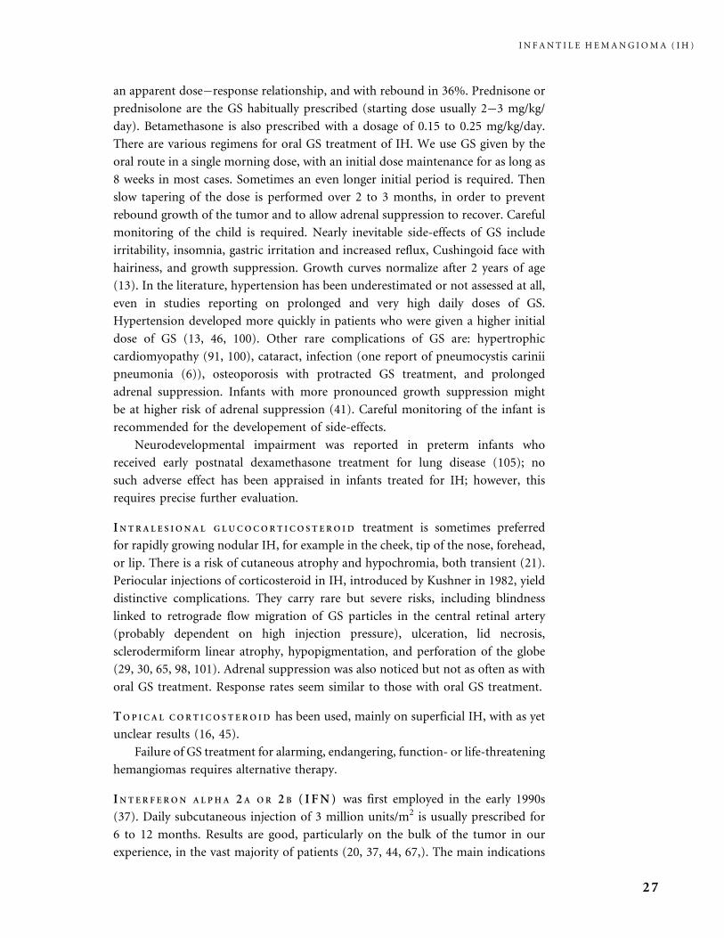
During the proliferation phase, IHs are made up of densely
packed capillaries, often with virtual lumen, grouped in
distinct or more confluent lobules.
Many of the lobules are associated with afferent and
efferent arterial-type vessels with a well-defined muscular
media (a). The capillaries are made up of an internal layer
of endothelial cells surrounded by a layer of pericytes;
some anisocaryosis and mitosis are frequently seen (b).
I N F A N T I L E H E M A N G I O M A ( I H )
30
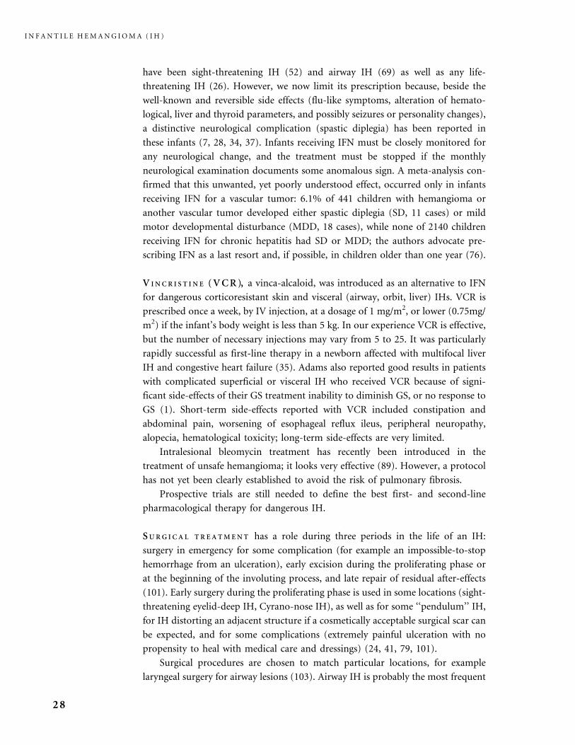
Pathology
During the proliferation phase, cellular density and the
absence of open lumen may obscure the vascular nature
of the lesion (a). Reticulin stains highlight the regular
vascular architecture of the lesion (b).
At the end of the proliferation phase, and during the
involuting phase, the vascular lumen are open and the
vascular nature of the lesion is obvious. Afferent or
efferent arteries are still present.
I N F A N T I L E H E M A N G I O M A ( I H )
31
Pathology
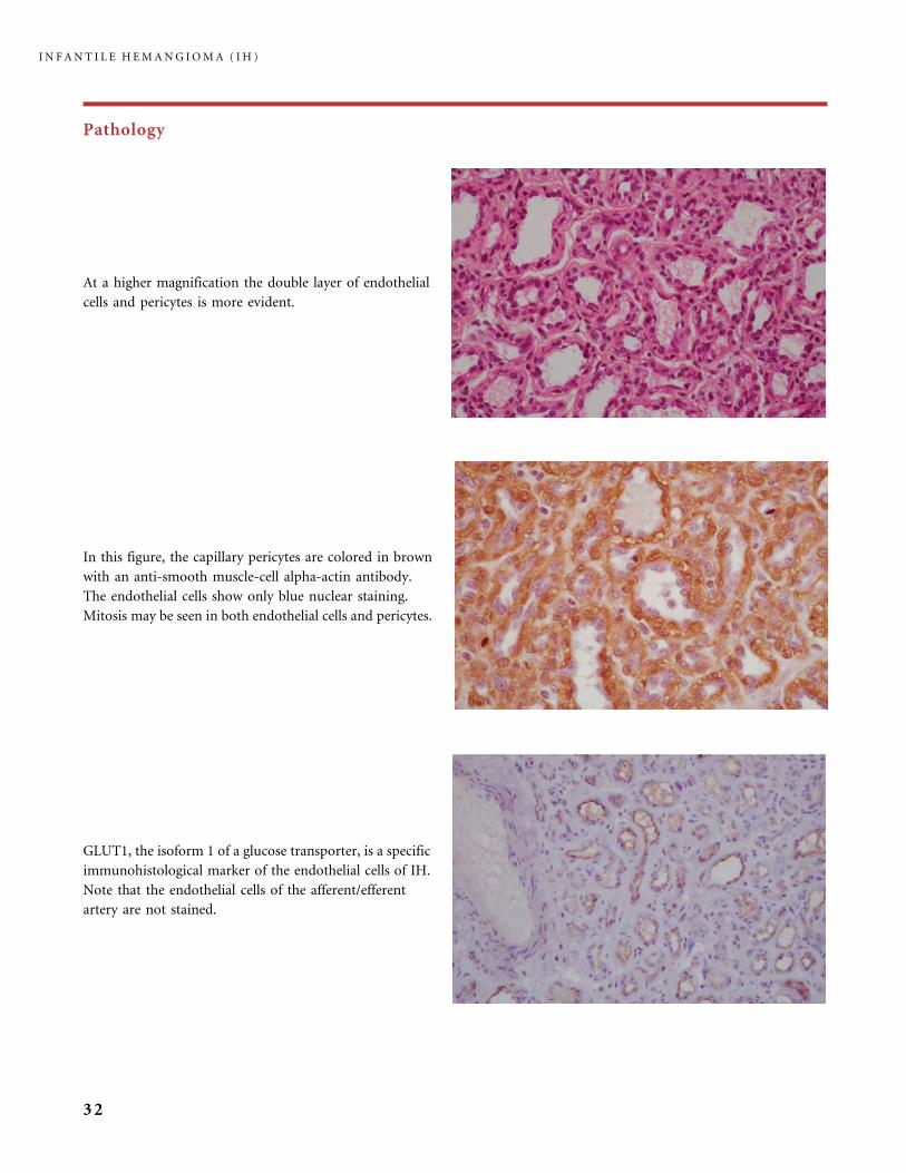
GLUT1, the isoform 1 of a glucose transporter, is a specific
immunohistological marker of the endothelial cells of IH.
Note that the endothelial cells of the afferent/efferent
artery are not stained.
At a higher magnification the double layer of endothelial
cells and pericytes is more evident.
In this figure, the capillary pericytes are colored in brown
with an anti-smooth muscle-cell alpha-actin antibody.
The endothelial cells show only blue nuclear staining.
Mitosis may be seen in both endothelial cells and pericytes.
I N F A N T I L E H E M A N G I O M A ( I H )
32
Pathology
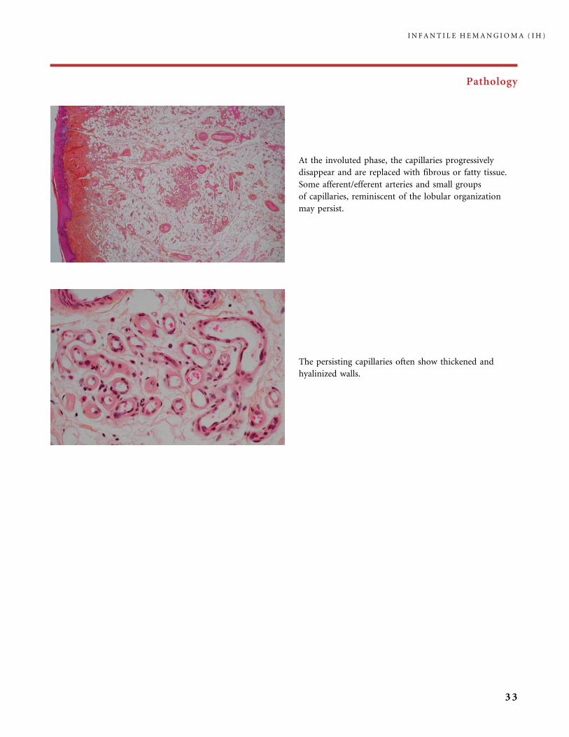
At the involuted phase, the capillaries progressively
disappear and are replaced with fibrous or fatty tissue.
Some afferent/efferent arteries and small groups
of capillaries, reminiscent of the lobular organization
may persist.
The persisting capillaries often show thickened and
hyalinized walls.
I N F A N T I L E H E M A N G I O M A ( I H )
33

Pathology
The capillary lobules may enclose normal fat cells (a), sweat glands (b), pilo-sebaceous follicles (c), or minor salivary gland
acini. Small nerves may be permeated by the capillary proliferation (d). In an IH, this must not be considered as a sign of
malignancy.
I N F A N T I L E H E M A N G I O M A ( I H )
34

Clinical Aspects, Investigations, and Treatment
An infantile hemangioma (IH) grows as superficial, crimson red, mammillated tumor, the ‘‘strawberry mark’’ (a); or as
a bump under normal or bluish or slightly telangiectatic skin, the subcutaneous ‘‘deep hemangioma’’ (b); or as a deep
and superficial mixed hemangioma, with a bluish deeper expansion secondarily developed and growing beyond the red
component (c). According to Nakayama (80) who reviewed 1247 patients with IH, subcutaneous hemangiomas make up
only 3.1% of cases.
I N F A N T I L E H E M A N G I O M A ( I H )
35
Clinical Aspects, Investigations, and Treatment
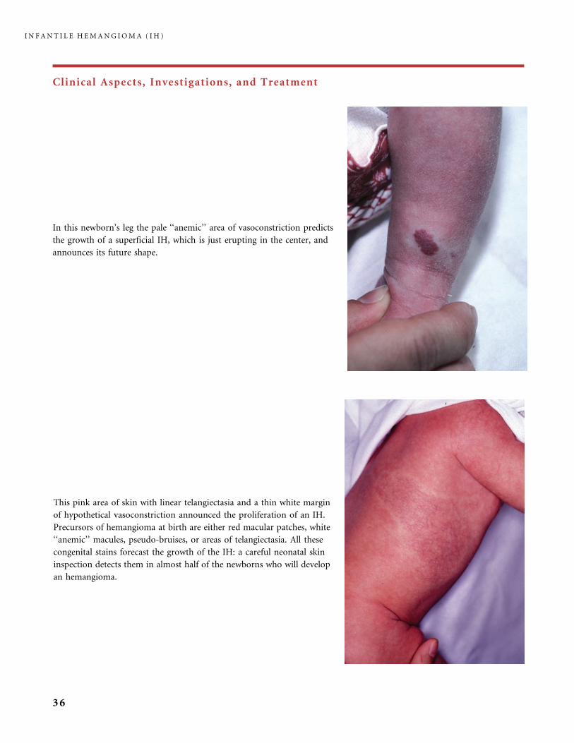
In this newborn’s leg the pale ‘‘anemic’’ area of vasoconstriction predicts
the growth of a superficial IH, which is just erupting in the center, and
announces its future shape.
This pink area of skin with linear telangiectasia and a thin white margin
of hypothetical vasoconstriction announced the proliferation of an IH.
Precursors of hemangioma at birth are either red macular patches, white
‘‘anemic’’ macules, pseudo-bruises, or areas of telangiectasia. All these
congenital stains forecast the growth of the IH: a careful neonatal skin
inspection detects them in almost half of the newborns who will develop
an hemangioma.
I N F A N T I L E H E M A N G I O M A ( I H )
36

Clinical Aspects, Investigations, and Treatment
Precursors of IH at birth, like this telangiectatic stain (a)
or this red stain mimicking a CM (c), prefigure quite
well the size and shape of the upcoming proliferating IH
but nothing allows us to predict the final volume of the
tumor. In the female infant in (a) the IH remained
superficial (b), while in the other (c) who also had early
respiratory distress from airway IH, a bulky bilateral
mandibular tumor grew (d). Larger lesions often have
a longer growth phase of 12 to 24 months.
I N F A N T I L E H E M A N G I O M A ( I H )
37
Clinical Aspects, Investigations, and Treatment
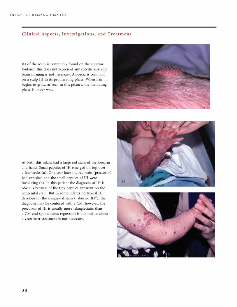
At birth this infant had a large red stain of the forearm
and hand. Small papules of IH emerged on top over
a few weeks (a). One year later the red stain (precursor)
had vanished and the small papules of IH were
involuting (b). In this patient the diagnosis of IH is
obvious because of the tiny papules apparent on the
congenital stain. But in some infants no typical IH
develops on the congenital stain (‘‘aborted IH’’): the
diagnosis may be confused with a CM; however, the
precursor of IH is usually more telangiectatic than
a CM and spontaneous regression is attained in about
a year; laser treatment is not necessary.
IH of the scalp is commonly found on the anterior
fontanel: this does not represent any specific risk and
brain imaging is not necessary. Alopecia is common
on a scalp IH in its proliferating phase. When hair
begins to grow, as seen in this picture, the involuting
phase is under way.
I N F A N T I L E H E M A N G I O M A ( I H )
38
Clinical Aspects, Investigations, and Treatment
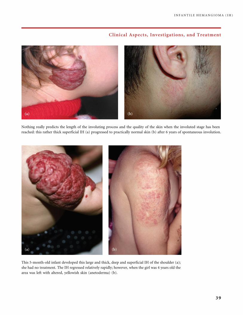
Nothing really predicts the length of the involuting process and the quality of the skin when the involuted stage has been
reached: this rather thick superficial IH (a) progressed to practically normal skin (b) after 6 years of spontaneous involution.
This 5-month-old infant developed this large and thick, deep and superficial IH of the shoulder (a);
she had no treatment. The IH regressed relatively rapidly; however, when the girl was 4 years old the
area was left with altered, yellowish skin (anetoderma) (b).
I N F A N T I L E H E M A N G I O M A ( I H )
39

Clinical Aspects, Investigations, and Treatment
Multiple small painful ulcers arose on this large and thick IH of the hand and forearm during its proliferating phase. It was
demonstrated that nerves are most numerous in growing hemangiomas (59): one can hypothesize that these nerves may
contribute to the sharp pain suffered by the infant when an ulcerated hemangioma is exposed to air or physical contact.
Corticosteroid treatment (CS) and hydrocolloid dressings helped the ulcers healing in this infant. After 1 month of
treatment white macules of involution had developed (a) but the lesion was still thick and folded. Four years later the skin
folds had fully receded. Multiple scars were noticeable on the forearm, as a consequence of the many ulcers, and some
telangiectasia remained on the dorsum of the hand (b).
Nothing predicts the occurrence of a fibro-fatty residuum with slack skin after regression of IH. Interestingly, Yu et al.
demonstrated the presence of mesenchymal stem cells with adipogenic potential in cultures of proliferating IHs (106).
I N F A N T I L E H E M A N G I O M A ( I H )
40
Clinical Aspects, Investigations, and Treatment
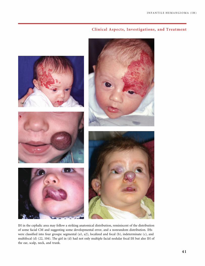
IH in the cephalic area may follow a striking anatomical distribution, reminiscent of the distribution
of some facial CM and suggesting some developmental error, and a nonrandom distribution. IHs
were classified into four groups: segmental (a1, a2), localized and focal (b), indeterminate (c), and
multifocal (d) (22, 104). The girl in (d) had not only multiple facial nodular focal IH but also IH of
the ear, scalp, neck, and trunk.
I N F A N T I L E H E M A N G I O M A ( I H )
41
Clinical Aspects, Investigations, and Treatment
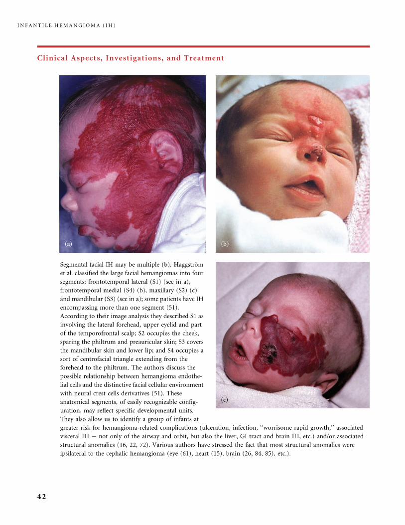
Segmental facial IH may be multiple (b). Haggstrom
et al. classified the large facial hemangiomas into four
segments: frontotemporal lateral (S1) (see in a),
frontotemporal medial (S4) (b), maxillary (S2) (c)
and mandibular (S3) (see in a); some patients have IH
encompassing more than one segment (51).
According to their image analysis they described S1 as
involving the lateral forehead, upper eyelid and part
of the temporofrontal scalp; S2 occupies the cheek,
sparing the philtrum and preauricular skin; S3 covers
the mandibular skin and lower lip; and S4 occupies a
sort of centrofacial triangle extending from the
forehead to the philtrum. The authors discuss the
possible relationship between hemangioma endothe-
lial cells and the distinctive facial cellular environment
with neural crest cells derivatives (51). These
anatomical segments, of easily recognizable config-
uration, may reflect specific developmental units.
They also allow us to identify a group of infants at
greater risk for hemangioma-related complications (ulceration, infection, ‘‘worrisome rapid growth,’’ associated
visceral IH � not only of the airway and orbit, but also the liver, GI tract and brain IH, etc.) and/or associated
structural anomalies (16, 22, 72). Various authors have stressed the fact that most structural anomalies were
ipsilateral to the cephalic hemangioma (eye (61), heart (15), brain (26, 84, 85), etc.).
I N F A N T I L E H E M A N G I O M A ( I H )
42
Clinical Aspects, Investigations, and Treatment
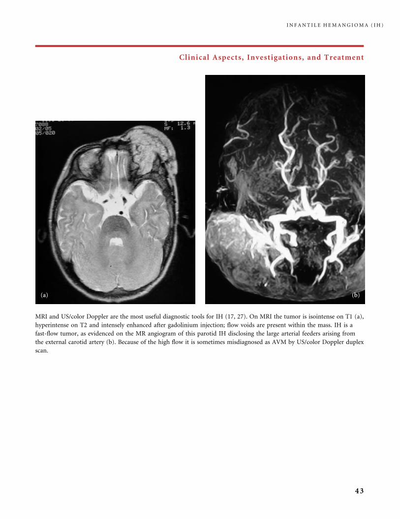
MRI and US/color Doppler are the most useful diagnostic tools for IH (17, 27). On MRI the tumor is isointense on T1 (a),
hyperintense on T2 and intensely enhanced after gadolinium injection; flow voids are present within the mass. IH is a
fast-flow tumor, as evidenced on the MR angiogram of this parotid IH disclosing the large arterial feeders arising from
the external carotid artery (b). Because of the high flow it is sometimes misdiagnosed as AVM by US/color Doppler duplex
scan.
I N F A N T I L E H E M A N G I O M A ( I H )
43
Clinical Aspects, Investigations, and Treatment
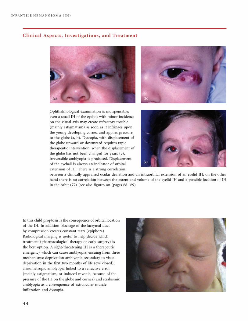
Ophthalmological examination is indispensable:
even a small IH of the eyelids with minor incidence
on the visual axis may create refractory trouble
(mainly astigmatism) as soon as it infringes upon
the young developing cornea and applies pressure
to the globe (a, b). Dystopia, with displacement of
the globe upward or downward requires rapid
therapeutic intervention: when the displacement of
the globe has not been changed for years (c),
irreversible amblyopia is produced. Displacement
of the eyeball is always an indicator of orbital
extension of IH. There is a strong correlation
between a clinically appraised ocular deviation and an intraorbital extension of an eyelid IH; on the other
hand there is no correlation between the extent and volume of the eyelid IH and a possible location of IH
in the orbit (77) (see also figures on (pages 68�69).
In this child proptosis is the consequence of orbital location
of the IH. In addition blockage of the lacrymal duct
by compression creates constant tears (epiphora).
Radiological imaging is useful to help decide which
treatment (pharmacological therapy or early surgery) is
the best option. A sight-threatening IH is a therapeutic
emergency which can cause amblyopia, ensuing from three
mechanisms: deprivation amblyopia secondary to visual
deprivation in the first two months of life (eye closed);
anisometropic amblyopia linked to a refractive error
(mainly astigmatism, or induced myopia, because of the
pressure of the IH on the globe and cornea) and strabismic
amblyopia as a consequence of extraocular muscle
infiltration and dystopia.
I N F A N T I L E H E M A N G I O M A ( I H )
44
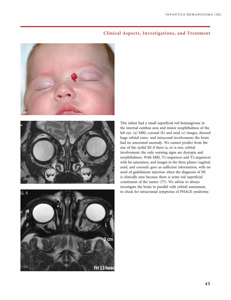
Clinical Aspects, Investigations, and Treatment
This infant had a small superficial red hemangioma in
the internal canthus area and minor exophthalmos of the
left eye. (a) MRI, coronal (b) and axial (c) images, showed
huge orbital extra- and intraconal involvement; the brain
had no associated anomaly. We cannot predict from the
size of the eyelid IH if there is, or is not, orbital
involvement; the only warning signs are dystopia and
exophthalmos. With MRI, T1-sequences and T2-sequences
with fat saturation, and images in the three planes (sagittal,
axial, and coronal) gave us sufficient information, with no
need of gadolinium injection when the diagnosis of IH
is clinically sure because there is some red superficial
constituent of the tumor (77). We advise to always
investigate the brain in parallel with orbital assessment,
to check for intracranial symptoms of PHACE syndrome.
I N F A N T I L E H E M A N G I O M A ( I H )
45
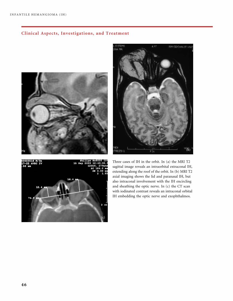
Clinical Aspects, Investigations, and Treatment
Three cases of IH in the orbit. In (a) the MRI T2
sagittal image reveals an intraorbital extraconal IH,
extending along the roof of the orbit. In (b) MRI T2
axial imaging shows the lid and paranasal IH, but
also intraconal involvement with the IH encircling
and sheathing the optic nerve. In (c) the CT scan
with iodinated contrast reveals an intraconal orbital
IH embedding the optic nerve and exophthalmos.
I N F A N T I L E H E M A N G I O M A ( I H )
46
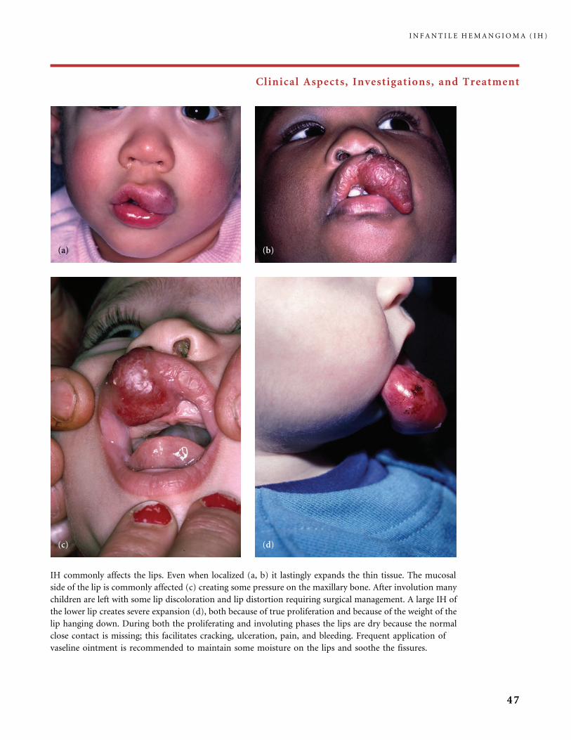
Clinical Aspects, Investigations, and Treatment
IH commonly affects the lips. Even when localized (a, b) it lastingly expands the thin tissue. The mucosal
side of the lip is commonly affected (c) creating some pressure on the maxillary bone. After involution many
children are left with some lip discoloration and lip distortion requiring surgical management. A large IH of
the lower lip creates severe expansion (d), both because of true proliferation and because of the weight of the
lip hanging down. During both the proliferating and involuting phases the lips are dry because the normal
close contact is missing; this facilitates cracking, ulceration, pain, and bleeding. Frequent application of
vaseline ointment is recommended to maintain some moisture on the lips and soothe the fissures.
I N F A N T I L E H E M A N G I O M A ( I H )
47
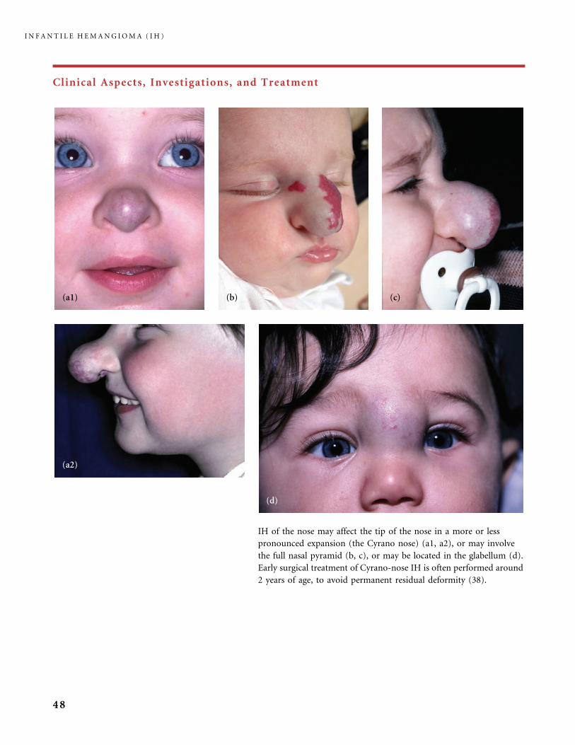
Clinical Aspects, Investigations, and Treatment
IH of the nose may affect the tip of the nose in a more or less
pronounced expansion (the Cyrano nose) (a1, a2), or may involve
the full nasal pyramid (b, c), or may be located in the glabellum (d).
Early surgical treatment of Cyrano-nose IH is often performed around
2 years of age, to avoid permanent residual deformity (38).
I N F A N T I L E H E M A N G I O M A ( I H )
48

Clinical Aspects, Investigations, and Treatment
An IH susceptible to ulceration is an IH with a superficial
bright red component (deep hemangioma under
apparently normal skin usually do not ulcerate) and
this can be a side-effect of a local treatment (cryotherapy,
lasers). In this infant three ulcers were caused by
cryotherapy applied during the proliferating phase.
After healing of the necrotic areas the patient developed
three bad white scars, while the part of the IH left to
spontaneously regress was clearly achieving a better
quality of skin.
Hemangioma affecting the breast area may be small,
or large, as in this girl whose lesion encompasses the
mammary bud. In our experience, the risk of developing
a small ‘‘Amazon’’ breast at puberty has often been
overestimated and the breast may grow normally. If excess
skin has been left after involution, we never offer surgical
repair before the full growth of the breast, to avoid damage
to the fragile arterial feeders of the mammary bud. If
necessary, cosmetic repair is considered after pubertal
breast growth.
Large thoracic and arm IH may induce congestive heart
failure (CHF) due to the high flow through the numerous
arterial feeders. Monitoring of cardiac function is
recommended. Medical treatment of CHF is necessary,
in addition to the pharmacological treatment of the
tumor. Occasionally, embolization through the arterial
route using particles helps alleviate the cardiac overload.
I N F A N T I L E H E M A N G I O M A ( I H )
49

Clinical Aspects, Investigations, and Treatment
Spontaneous ulceration of hemangioma is quite common
in the buttocks. This girl had an ulcerated IH of the left
buttock and extreme pain when urine and stools came
into contact with it. She had a first procedure of
flashlamp pulsed dye laser treatment (a); 20 days later
healing was nearly complete (b), a second laser applica-
tion was made and rapid healing obtained (c) (operator:
Dr. Virginie Fayard, Paris, France). Reported benefits of
laser treatment for ulcerated hemangioma range from
rapid relief of pain, rapid healing, accelerated involution,
or no response at all (23). The most common dressings
applied to ulcerated IHs are the hydrocolloid dressings,
or the hydrocellular dressings in case of important fluid
outflow; both protect the wound and reduce pain.
Some authors use antimicrobial ointments or zinc oxide
paste. Becaplermin gel (Regranex�), a recombinant
platelet-derived growth factor used in the cure of diabetic
ulcers, has been reported to be effective for severely
ulcerated IH not responding to conventional local
treatment and systemic therapy (75).
I N F A N T I L E H E M A N G I O M A ( I H )
50
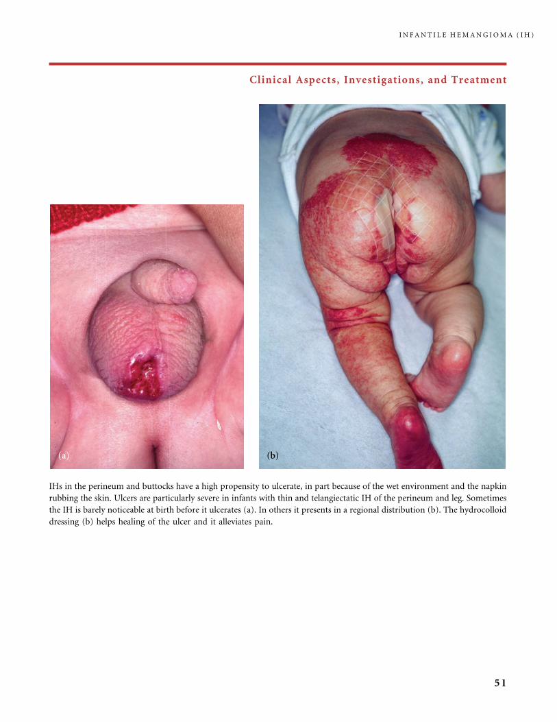
Clinical Aspects, Investigations, and Treatment
IHs in the perineum and buttocks have a high propensity to ulcerate, in part because of the wet environment and the napkin
rubbing the skin. Ulcers are particularly severe in infants with thin and telangiectatic IH of the perineum and leg. Sometimes
the IH is barely noticeable at birth before it ulcerates (a). In others it presents in a regional distribution (b). The hydrocolloid
dressing (b) helps healing of the ulcer and it alleviates pain.
I N F A N T I L E H E M A N G I O M A ( I H )
51
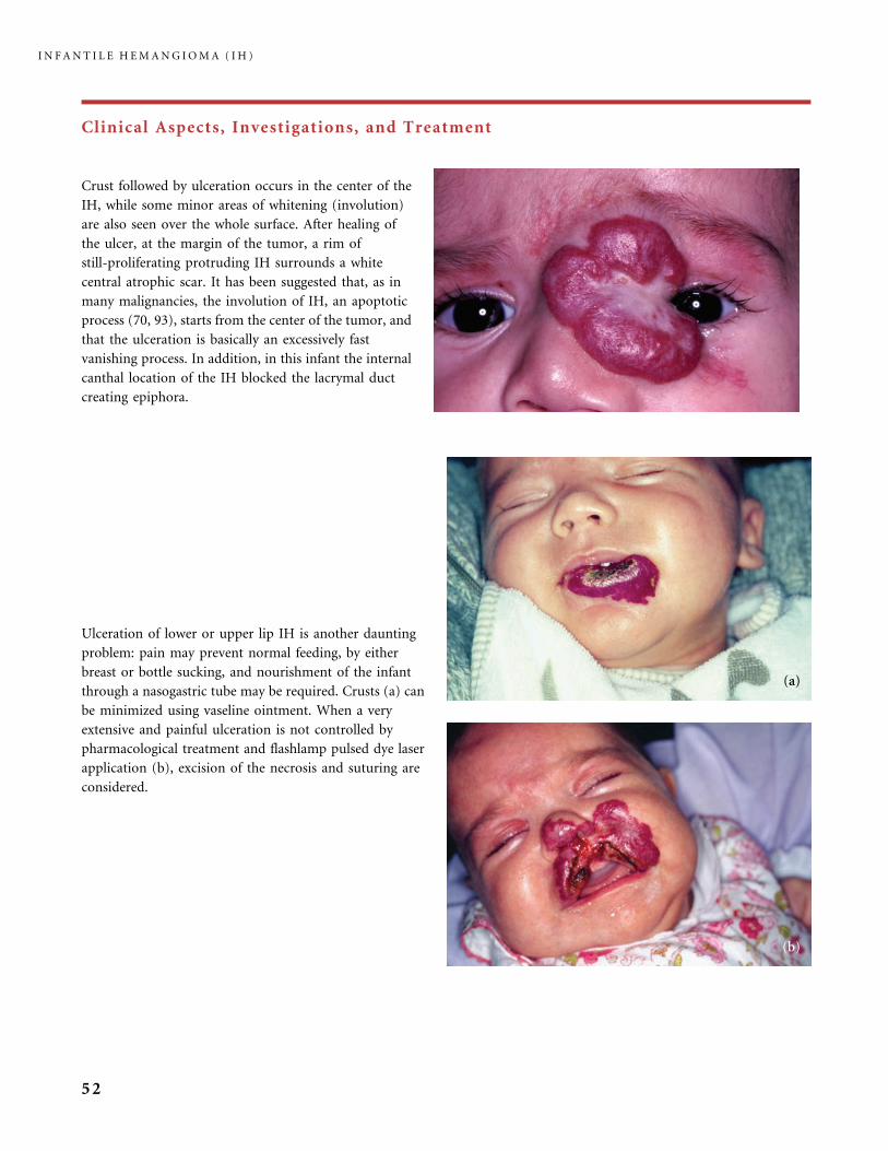
Clinical Aspects, Investigations, and Treatment
Ulceration of lower or upper lip IH is another daunting
problem: pain may prevent normal feeding, by either
breast or bottle sucking, and nourishment of the infant
through a nasogastric tube may be required. Crusts (a) can
be minimized using vaseline ointment. When a very
extensive and painful ulceration is not controlled by
pharmacological treatment and flashlamp pulsed dye laser
application (b), excision of the necrosis and suturing are
considered.
Crust followed by ulceration occurs in the center of the
IH, while some minor areas of whitening (involution)
are also seen over the whole surface. After healing of
the ulcer, at the margin of the tumor, a rim of
still-proliferating protruding IH surrounds a white
central atrophic scar. It has been suggested that, as in
many malignancies, the involution of IH, an apoptotic
process (70, 93), starts from the center of the tumor, and
that the ulceration is basically an excessively fast
vanishing process. In addition, in this infant the internal
canthal location of the IH blocked the lacrymal duct
creating epiphora.
I N F A N T I L E H E M A N G I O M A ( I H )
52

Clinical Aspects, Investigations, and Treatment
This newborn was transferred to us with a misdiagnosis of CM; the lesion was in fact a precursor of
a bilateral mandibular, ear and neck IH (a). ‘‘Beard’’ IH is known to carry a high risk of associated
airway IH (36, 83). At that early stage, a pharyngeal and a nonobstructive circumferential laryngeal
subglottic IH was observed on fiberoptic laryngoscopy. She received high dosage of glucocorti-
costeroid (GS), but after 1 month she became tachypneic with a stridor even at rest. At 7 weeks of
age she had a single-stage laryngotracheoplasty: open-approach excision of the airway lesion and
graft of auricular cartilage for augmentation of the subglottis (operator: Dr. Gilles Roger, Hopital
Armand Trousseau, Paris, France). Respiratory outcome was excellent. Despite ongoing GS
treatment, the superficial IH grew to create a bilateral massive parotid tumor with multiple large
painful ulcerations in the neck (b). GS were tapered and stopped when she was 3 months old. Then,
she received interferon alpha 2b treatment over 10 months, without unwanted side-effects. Healing
of the ulcers was obtained after 4 weeks. Rapid shrinkage of the bilateral tumor occurred, resulting
in lax skin with widespread scars. The involuted stage was reached at 14 months of age (c) and
surgical procedures (four facelift procedures and a neck CO2 laser resurfacing) began at 24 months
of age. At 7 years (d) she is a bright happy girl who copes with the residual leucomelanodermic scars
on her neck and lower face, undergoing various procedures of laser treatment.
I N F A N T I L E H E M A N G I O M A ( I H )
53
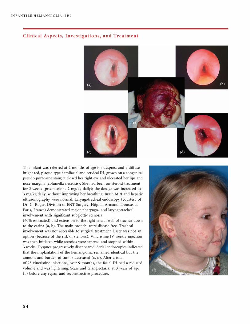
Clinical Aspects, Investigations, and Treatment
This infant was referred at 2 months of age for dyspnea and a diffuse
bright red, plaque-type hemifacial and cervical IH, grown on a congenital
pseudo port-wine stain; it closed her right eye and ulcerated her lips and
nose margins (columella necrosis). She had been on steroid treatment
for 2 weeks (prednisolone 2 mg/kg daily); the dosage was increased to
3 mg/kg daily, without improving her breathing. Brain MRI and hepatic
ultrasonography were normal. Laryngotracheal endoscopy (courtesy of
Dr. G. Roger, Division of ENT Surgery, Hopital Armand Trousseau,
Paris, France) demonstrated major pharyngo- and laryngotracheal
involvement with significant subglottic stenosis
(60% estimated) and extension to the right lateral wall of trachea down
to the carina (a, b). The main bronchi were disease free. Tracheal
involvement was not accessible to surgical treatment. Laser was not an
option (because of the risk of stenosis). Vincristine IV weekly injection
was then initiated while steroids were tapered and stopped within
3 weeks. Dyspnea progressively disappeared. Serial endoscopies indicated
that the implantation of the hemangioma remained identical but the
amount and burden of tumor decreased (c, d). After a total
of 23 vincristine injections, over 9 months, the facial IH had a reduced
volume and was lightening. Scars and telangiectasia, at 3 years of age
(f ) before any repair and reconstructive procedure.
I N F A N T I L E H E M A N G I O M A ( I H )
54
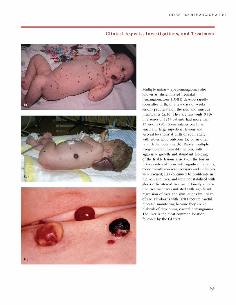
Clinical Aspects, Investigations, and Treatment
Multiple miliary-type hemangiomas also
known as disseminated neonatal
hemangiomatosis (DNH) develop rapidly
soon after birth; in a few days or weeks
lesions proliferate on the skin and mucous
membranes (a, b). They are rare: only 0.4%
in a series of 1247 patients had more than
17 lesions (80). Some infants combine
small and large superficial lesions and
visceral locations at birth or soon after,
with either good outcome (a) or an often
rapid lethal outcome (b). Rarely, multiple
pyogenic-granuloma-like lesions, with
aggressive growth and abundant bleeding
of the friable lesions arise (96): the boy in
(c) was referred to us with significant anemia;
blood transfusion was necessary and 12 lesions
were excised; IHs continued to proliferate in
the skin and liver, and were not stabilized with
glucocorticosteroid treatment. Finally vincris-
tine treatment was initiated with significant
regression of liver and skin lesions by 1 year
of age. Newborns with DNH require careful
repeated monitoring because they are at
highrisk of developing visceral hemangiomas.
The liver is the most common location,
followed by the GI tract.
I N F A N T I L E H E M A N G I O M A ( I H )
55
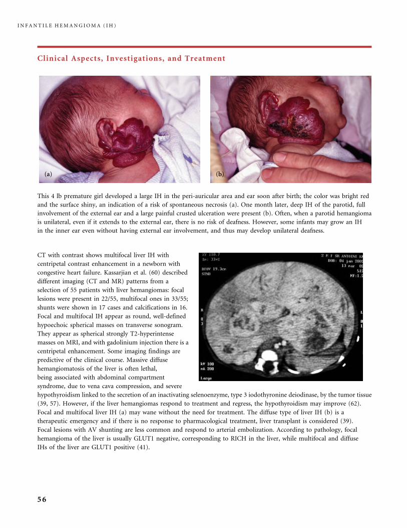
Clinical Aspects, Investigations, and Treatment
This 4 lb premature girl developed a large IH in the peri-auricular area and ear soon after birth; the color was bright red
and the surface shiny, an indication of a risk of spontaneous necrosis (a). One month later, deep IH of the parotid, full
involvement of the external ear and a large painful crusted ulceration were present (b). Often, when a parotid hemangioma
is unilateral, even if it extends to the external ear, there is no risk of deafness. However, some infants may grow an IH
in the inner ear even without having external ear involvement, and thus may develop unilateral deafness.
CT with contrast shows multifocal liver IH with
centripetal contrast enhancement in a newborn with
congestive heart failure. Kassarjian et al. (60) described
different imaging (CT and MR) patterns from a
selection of 55 patients with liver hemangiomas: focal
lesions were present in 22/55, multifocal ones in 33/55;
shunts were shown in 17 cases and calcifications in 16.
Focal and multifocal IH appear as round, well-defined
hypoechoic spherical masses on transverse sonogram.
They appear as spherical strongly T2-hyperintense
masses on MRI, and with gadolinium injection there is a
centripetal enhancement. Some imaging findings are
predictive of the clinical course. Massive diffuse
hemangiomatosis of the liver is often lethal,
being associated with abdominal compartment
syndrome, due to vena cava compression, and severe
hypothyroidism linked to the secretion of an inactivating selenoenzyme, type 3 iodothyronine deiodinase, by the tumor tissue
(39, 57). However, if the liver hemangiomas respond to treatment and regress, the hypothyroidism may improve (62).
Focal and multifocal liver IH (a) may wane without the need for treatment. The diffuse type of liver IH (b) is a
therapeutic emergency and if there is no response to pharmacological treatment, liver transplant is considered (39).
Focal lesions with AV shunting are less common and respond to arterial embolization. According to pathology, focal
hemangioma of the liver is usually GLUT1 negative, corresponding to RICH in the liver, while multifocal and diffuse
IHs of the liver are GLUT1 positive (41).
I N F A N T I L E H E M A N G I O M A ( I H )
56

Clinical Aspects, Investigations, and Treatment
Children at risk of PHACE syndrome should receive neurological, ophthalmological and cardiac assessment (71). This
girl had a deep IH of the upper eyelid and minor IH staining of the temporal skin and lower lip (a). MRI showed signs
of PHACE syndrome with posterior fossa malformation; hypoplasia of hemi-cerebellum and a complex anomaly of the
cerebellar vermis. MRI T2-sequence (b) and T1-sequence with gadolinium injection (c) display eyelid, temporal, and full
orbital involvement. Eye abnormalities are an important part of PHACE syndrome. Some ocular abnormalities result
from the presence of the periocular hemangioma and they vary from strabismus, refractive error and amblyopia, to
proptosis or ptosis. Others correspond to ocular structural defects, such as optic nerve atrophy, coloboma, optic disc
excavation or congenital cataract, and these are part of the PHACE syndrome (64).
A girl who was developmentally normal at 1 year of age,
had a right facial parotid IH, right microtia. PHACE
syndrome was diagnosed because of various intracranial
arterial anomalies detected on MRI/MRA imaging
(courtesy of Dr. I.J. Frieden and Dr. C. Dowd, UCSF,
San Francisco, USA). She had a significantly diminished
in caliber right internal carotid artery, compared to the left,
and fenestration or duplication of the anterior commu-
nicating artery. PHACE (OMIM 606519) is an acronym for
a neurocutaneous syndrome encompassing the following
features: posterior fossa brain malformations, hemangio-
mas, arterial anomalies, cardiac anomalies, and eye
abnormalities. Ventral developmental defects such as
sternal cleft or supraombilical raphe may also be present (PHACE(S) syndrome) (40, 41, 85). Diagnosis is made when one
or more associated anomalies are present in addition to hemangioma. Most patients do not have all the associated features.
Cerebrovascular anomalies consist of persistent embryonic arteries, agenesis or hypoplasia of major arteries and dilatation
and tortuosity of others. Patients may be asymptomatic or can have seizures, stroke, developmental delay, and headaches
(18, 26, 71, 86).
I N F A N T I L E H E M A N G I O M A ( I H )
57
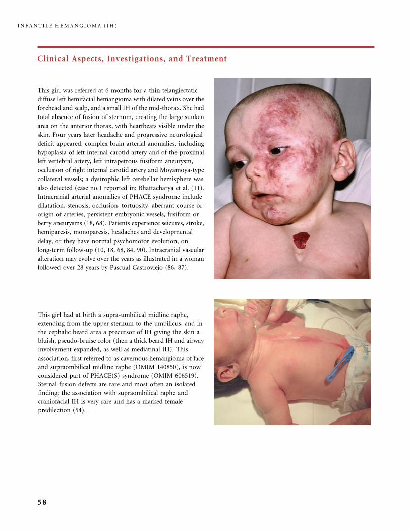
Clinical Aspects, Investigations, and Treatment
This girl was referred at 6 months for a thin telangiectatic
diffuse left hemifacial hemangioma with dilated veins over the
forehead and scalp, and a small IH of the mid-thorax. She had
total absence of fusion of sternum, creating the large sunken
area on the anterior thorax, with heartbeats visible under the
skin. Four years later headache and progressive neurological
deficit appeared: complex brain arterial anomalies, including
hypoplasia of left internal carotid artery and of the proximal
left vertebral artery, left intrapetrous fusiform aneurysm,
occlusion of right internal carotid artery and Moyamoya-type
collateral vessels; a dystrophic left cerebellar hemisphere was
also detected (case no.1 reported in: Bhattacharya et al. (11).
Intracranial arterial anomalies of PHACE syndrome include
dilatation, stenosis, occlusion, tortuosity, aberrant course or
origin of arteries, persistent embryonic vessels, fusiform or
berry aneurysms (18, 68). Patients experience seizures, stroke,
hemiparesis, monoparesis, headaches and developmental
delay, or they have normal psychomotor evolution, on
long-term follow-up (10, 18, 68, 84, 90). Intracranial vascular
alteration may evolve over the years as illustrated in a woman
followed over 28 years by Pascual-Castroviejo (86, 87).
This girl had at birth a supra-umbilical midline raphe,
extending from the upper sternum to the umbilicus, and in
the cephalic beard area a precursor of IH giving the skin a
bluish, pseudo-bruise color (then a thick beard IH and airway
involvement expanded, as well as mediatinal IH). This
association, first referred to as cavernous hemangioma of face
and supraombilical midline raphe (OMIM 140850), is now
considered part of PHACE(S) syndrome (OMIM 606519).
Sternal fusion defects are rare and most often an isolated
finding; the association with supraombilical raphe and
craniofacial IH is very rare and has a marked female
predilection (54).
I N F A N T I L E H E M A N G I O M A ( I H )
58
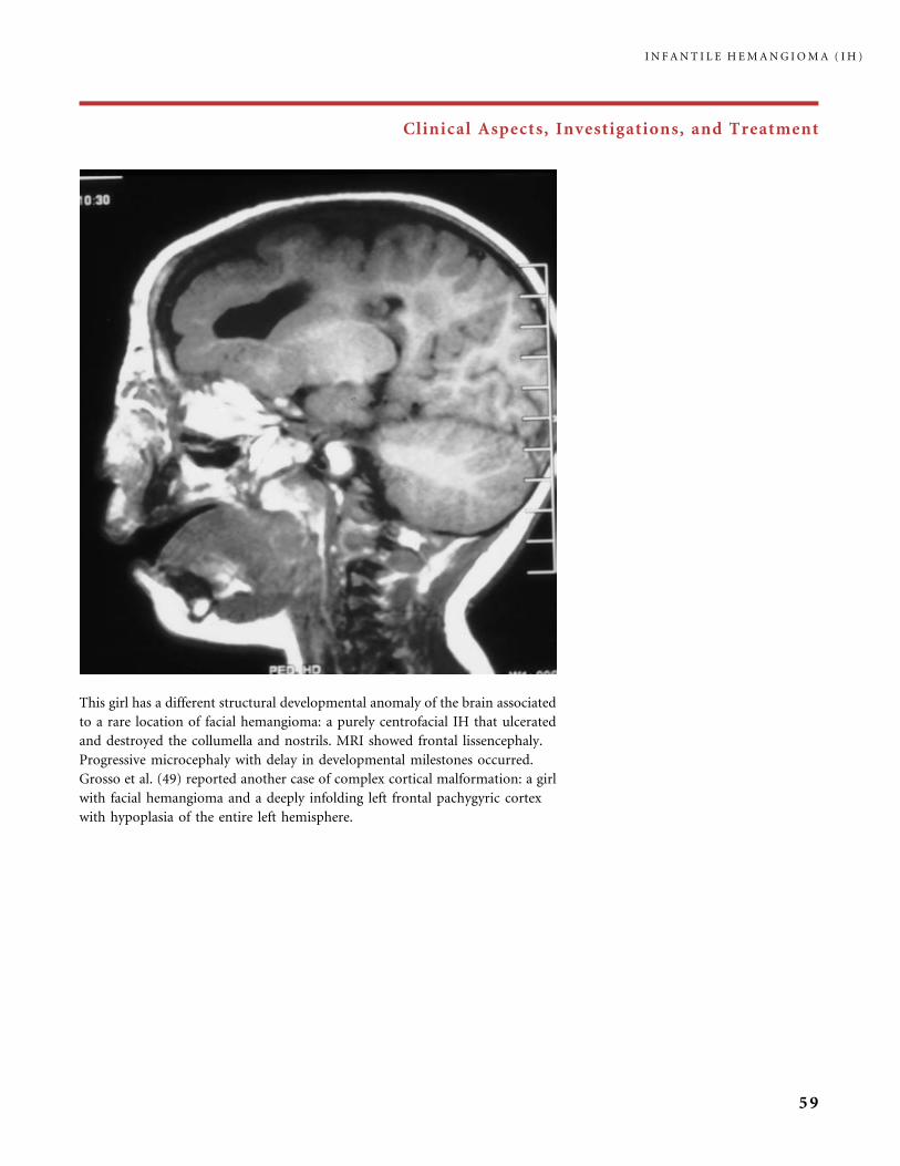
Clinical Aspects, Investigations, and Treatment
This girl has a different structural developmental anomaly of the brain associated
to a rare location of facial hemangioma: a purely centrofacial IH that ulcerated
and destroyed the collumella and nostrils. MRI showed frontal lissencephaly.
Progressive microcephaly with delay in developmental milestones occurred.
Grosso et al. (49) reported another case of complex cortical malformation: a girl
with facial hemangioma and a deeply infolding left frontal pachygyric cortex
with hypoplasia of the entire left hemisphere.
I N F A N T I L E H E M A N G I O M A ( I H )
59
Clinical Aspects, Investigations, and Treatment
IH of the buttocks, perineum and lower extremity, proliferating in a diffuse regional
distribution, are sometimes associated with abnormal genitalia, imperforate anus, and
anomalous urinary tract (14). The boy in (b) (courtesy of Dr. Bessis, Saint Eloi
Hospital, Montpellier, France) had ambiguous external genitalia. The girl in (a), with
perineal ulceration, had incomplete bladder exstrophy and a low position of the navel.
Spinal dysraphism, as illustrated by the MRI scan in (c), is present if spinal IH occurs
in conjunction with other cutaneous markers of dysraphism (3, 50). The acronym
PELVIS was recently created to underline the association of Perineal hemangioma,
External genitalia malformations, Lipomyelomeningocele, Vesicorenal abnormalities,
Imperforate anus, and Skin tag (14a). Some infants have deeply invasive IH without
true associated dysraphic anomalies: the infant in (d) remained asymptomatic,
although she had developed a deep IH in the nape after birth, and systematic MRI had
revealed mediastinal, paraspinal, and intraspinal extension of the IH, a difficult
diagnosis as already reported (53).
I N F A N T I L E H E M A N G I O M A ( I H )
60
Clinical Aspects, Investigations, and Treatment
Glucocorticosteroid (GS) treatment
remains the first option for
dangerous IH. In this girl, the
sight-impairing IH (a) involved the
upper eyelid as well as the orbit
on MRI. There was no PHACE
syndrome. The right visual axis was
occluded. Oral GS treatment induced
rapid cessation of proliferation and
reopening of the eye in 1 month (b).
She had a total of 4 months of
treatment including the tapering
phase, with good tolerance. No
rebound growth occurred at the end
of the treatment. Excellent cosmetic
outcome was achieved at the last
follow-up when she was 3 years old.
However, astigmatism of the right eye
required patching and glasses. Infants
receiving systemic GS must have close
monitoring including frequent blood
pressure measurement. Cardiac
left ventricular hypertrophy may
also be detected by US cardiac
evaluation, without any relationship
with hypertension. Adrenal suppres-
sion seems to correlate with higher
daily doses and total dose received.
I N F A N T I L E H E M A N G I O M A ( I H )
61

Clinical Aspects, Investigations, and Treatment
This infant developed an IH soon after birth that completely masked the eye. Because of the risk of deprivation amblyopia,
she received corticosteroid treatment during the first weeks of life without any improvement (a). MRI evaluation showed an
upper eyelid and orbital intraconal IH and microphthalmos, cataract, and hypoplasia of the optic nerve (b), allowing the
diagnosis of PHACE syndrome. No other associated anomaly was present. Structural eye anomalies of PHACE syndrome
encompass optic nerve hypoplasia, microphtalmos and cataract (as in this infant), glaucoma, coloboma, morning glory and
peripapillary excavation, or optic atrophy (66). It was decided to treat this girl with interferon alpha 2a, 3 million units/m2
a day for 6 months (lyophilized sterile powder of IFNa 2a). She achieved excellent improvement with no adverse effects, but
she still had an apparent ptosis of the eyelid because of the tiny globe (c). However, due to the volume attained by the orbital
hemangioma at the end of its proliferating phase, the orbit had reached a correct size. The abnormal eye was enucleated and
a prosthetic eye was provided, with excellent cosmetic results (d).
I N F A N T I L E H E M A N G I O M A ( I H )
62
Clinical Aspects, Investigations, and Treatment
This extensive left hemifacial, scalp, and neck IH with a laryngeal IH was not improved with high-dose
glucocorticosteroid (GS) treatment, including megadoses. On the contrary, at 3 months of age necrosis of
the lower lip and of the left ear auricle occurred, the IH was still growing, while her general health was
deteriorating. No PHACES syndrome was detected. At 7 months of age, only a mild fading of the superficial
part of the IH was obtained; but the left eye was partly masked and a parotid mass had developed (b), she
was fed through a nasogastric tube, and was enduring extreme pain, not diminished by morphine, from
increasing multiple deep ulcerations in the cheek and neck. Interferon alpha 2a (IFN) treatment was then
introduced and GS slowly tapered. She had a dramatic response to IFN. After 4 months all ulcers had healed,
the eye was wide open and the left parotid mass had shrunk. Eight months later (c) she was in good health,
in the late involuting stage of her IH. When 21-months old her IH reached the involuted stage. Diffuse
scars were noticeable on the lower part of her left cheek and neck (d), which required further therapeutic
procedures, including surgical repair. The scars were much less significant on the forehead and eyelid.
Astigmatism of the left eye required orthoptic management.
I N F A N T I L E H E M A N G I O M A ( I H )
63
Clinical Aspects, Investigations, and Treatment
This monozygotic twin girl (the other twin did not develop IH) had a red
stain of the left hemiface at birth. She came to us at 3 months of age (a) with
this huge, ulcerated, purple facial and scalp IH. The family doctor having
said that this ‘‘goes spontaneously’’ the parents were waiting. We initiated
oral glucocorticosteroid (GS) treatment but it was poorly effective, giving
only slight whitening one month later (b). Pain linked to ulcers was extreme
and not soothed by morphine. Interferon alpha 2a (IFN (lyophilized sterile
powder)) treatment was then introduced and GS stopped over a period of 1
month. INF was continued for 13 months. The results were excellent, with
re-opening of the eye and fading of the tumor ((c) appearance after 4
months of IFN; (d) appearance at the end of IFN treatment at 18 months of
age). No neurological side-effects occurred. At 4 years of age the IH had
fully vanished but the skin was badly wrinkled and yellowish. A first surgical
procedure brought normal skin from the neck to the mandibular area (e)
but the repair of the damaged skin of the eyelids will be a tricky task.
I N F A N T I L E H E M A N G I O M A ( I H )
64

Clinical Aspects, Investigations, and Treatment
This girl had a sight-threatening IH, still growing under glucocorticosteroid treatment: in (a) she had received
prednisone 2 mg/kg/day for 3 weeks; it was changed to betamethasone 0.20 mg/kg/day but growth of the IH
continued; (b) is the appearance after a total of 6 weeks of steroids, with the tumor nearly closing the right visual
field, and filling the floor of the orbit on MRI. No PHACE syndrome was present. Tapering of steroid was decided on
and she received vincristine treatment (a total of 20 weekly IV injections, over 4½ months). A clear improvement
with reopening of the eye was visible after the sixth injection (c). The treatment was well tolerated, except for
increased esophageal reflux and infection of the central line. Two years later, the IH was in the involuted stage (d).
Surgical repair of excess skin could begin at 3 years, while vision was still improving with glasses and patching.
I N F A N T I L E H E M A N G I O M A ( I H )
65
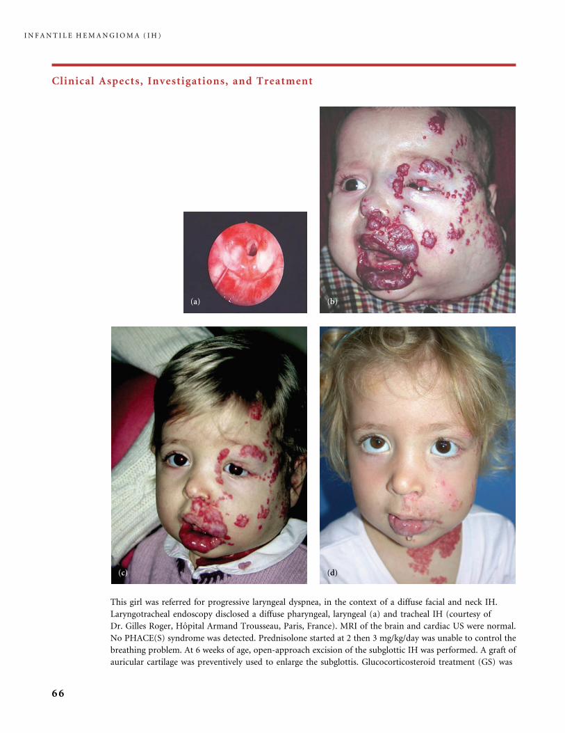
Clinical Aspects, Investigations, and Treatment
This girl was referred for progressive laryngeal dyspnea, in the context of a diffuse facial and neck IH.
Laryngotracheal endoscopy disclosed a diffuse pharyngeal, laryngeal (a) and tracheal IH (courtesy of
Dr. Gilles Roger, Hopital Armand Trousseau, Paris, France). MRI of the brain and cardiac US were normal.
No PHACE(S) syndrome was detected. Prednisolone started at 2 then 3 mg/kg/day was unable to control the
breathing problem. At 6 weeks of age, open-approach excision of the subglottic IH was performed. A graft of
auricular cartilage was preventively used to enlarge the subglottis. Glucocorticosteroid treatment (GS) was
I N F A N T I L E H E M A N G I O M A ( I H )
66

Clinical Aspects, Investigations, and Treatment
poorly effective: at 3 months a bulky superficial IH had developed with orbital involvement, ulcers, major neck and upper
thorax location, as well as extension of the tracheal IH on laryngotracheoscopy. At 4 months of age interferon alpha 2a
treatment was initiated (3 million units/m2/day subcutaneously) while GS treatment was progressively stopped over 4 weeks.
Initially, there was clear improvement but secondary rebound growth of the facial IH occurred 4 months later (b) and
obstructive dyspnea worsened. Therefore, weekly vincristine treatment was introduced (and interferon stopped) and given
over 8 months (24 injections) and stopped (c). Results were clearly good on both the superficial and airway IH. The residual
involuting IH slowly improved over the next 3 years of follow-up (d).
This large hemangioma of the forearm and hand was
seen at 1 month of age (a); glucocorticosteroid (GS) and
flashlamp pumped-pulsed dye laser (PDL) treatments
(operator: Dr. Virginie Fayard, Paris, France) were
simultaneously started; 2 months after the first laser
treatment, and while beginning tapering of GS, the area
treated with laser was clearly fading compared to the rest
of the IH (b). She had two additional laser sessions on
the whole surface of the IH. Significant regression was
obtained before 1 year of age (c) with no further
rebound growth. PDL treatment seemed to have boosted
the response of IH to GS.
I N F A N T I L E H E M A N G I O M A ( I H )
67

Clinical Aspects, Investigations, and Treatment
The IH in this girl rapidly proliferated. At 2 months she had upward displacement of the eye and her visual axis was nearly
occluded (a). MRI T2-weighted sagittal image showed deep orbital extension along the floor of the orbit (b). No PHACE
syndrome was detected. Surgical excision was the preferred therapeutic option to relieve the pressure and burden of the IH on
the globe and prevent permanent visual deficit. Both the lid and orbital part of the IH could be extracted with immediate
good results (operator: Dr. Patrick Diner, Hopital Armand Trousseau, Paris, France). Six months later no rebound growth
had occurred and the incision scar along the inferior line of eyelashes was barely detectable (c). The use of the electronic
dissector CUSA (cavitron ultra sonic aspirator) (Cavitron� and Dissectron�) facilitated the procedure. This technique
minimizes intra-operative hemorrhage and postoperative complications (25, 88). Early surgical treatment prevents the
development of amblyopia, but post-operative patching, or atropine drops (to blur the normal eye) and/or glasses, are often
required to correct the already established refractive error (astigmatism or myopia) (19, 52, 78, 89).
I N F A N T I L E H E M A N G I O M A ( I H )
68

Clinical Aspects, Investigations, and Treatment
This upper eyelid hemangioma closed the visual field and
was growing as a deep IH without superficial red
component (a). A bolus of glucocorticosteroid and then
daily oral treatment (prednisolone 3 mg/kg/day) did not
re-open the eye. MRI indicated that there was no orbital
extension (b). Thus surgical excision using an ultrasonic
device (Dissectron�) was performed at 2 months of age, to
avoid amblyopiogenisis (operator: Dr. Patrick Diner,
Hopital Armand Trousseau, Paris, France). The outcome
was excellent 1 month later (c) and perfect 3 years later
(d) with a barely visible scar. As soon as the visual axis
is re-opened ophthalmologic evaluation is essential, to
look for residual palpebral occlusion if any, strabismus
secondary to extraocular muscle infiltration, ocular
motility, objective refraction, and amblyopia; if necessary,
re-education aims at improving vision.
I N F A N T I L E H E M A N G I O M A ( I H )
69
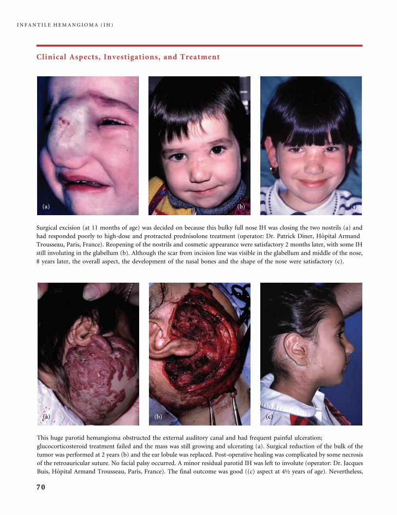
Clinical Aspects, Investigations, and Treatment
This huge parotid hemangioma obstructed the external auditory canal and had frequent painful ulceration;
glucocorticosteroid treatment failed and the mass was still growing and ulcerating (a). Surgical reduction of the bulk of the
tumor was performed at 2 years (b) and the ear lobule was replaced. Post-operative healing was complicated by some necrosis
of the retroauricular suture. No facial palsy occurred. A minor residual parotid IH was left to involute (operator: Dr. Jacques
Buis, Hopital Armand Trousseau, Paris, France). The final outcome was good ((c) aspect at 4½ years of age). Nevertheless,
Surgical excision (at 11 months of age) was decided on because this bulky full nose IH was closing the two nostrils (a) and
had responded poorly to high-dose and protracted prednisolone treatment (operator: Dr. Patrick Diner, Hopital Armand
Trousseau, Paris, France). Reopening of the nostrils and cosmetic appearance were satisfactory 2 months later, with some IH
still involuting in the glabellum (b). Although the scar from incision line was visible in the glabellum and middle of the nose,
8 years later, the overall aspect, the development of the nasal bones and the shape of the nose were satisfactory (c).
I N F A N T I L E H E M A N G I O M A ( I H )
70
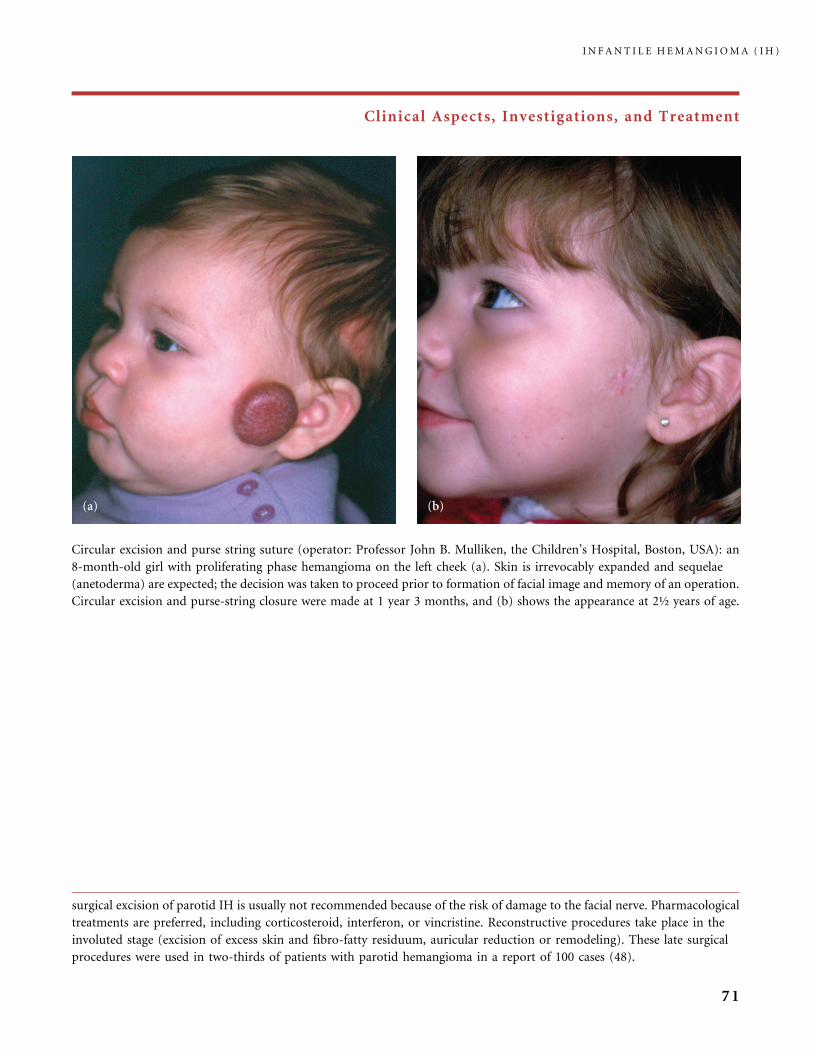
Clinical Aspects, Investigations, and Treatment
surgical excision of parotid IH is usually not recommended because of the risk of damage to the facial nerve. Pharmacological
treatments are preferred, including corticosteroid, interferon, or vincristine. Reconstructive procedures take place in the
involuted stage (excision of excess skin and fibro-fatty residuum, auricular reduction or remodeling). These late surgical
procedures were used in two-thirds of patients with parotid hemangioma in a report of 100 cases (48).
Circular excision and purse string suture (operator: Professor John B. Mulliken, the Children’s Hospital, Boston, USA): an
8-month-old girl with proliferating phase hemangioma on the left cheek (a). Skin is irrevocably expanded and sequelae
(anetoderma) are expected; the decision was taken to proceed prior to formation of facial image and memory of an operation.
Circular excision and purse-string closure were made at 1 year 3 months, and (b) shows the appearance at 2½ years of age.
I N F A N T I L E H E M A N G I O M A ( I H )
71
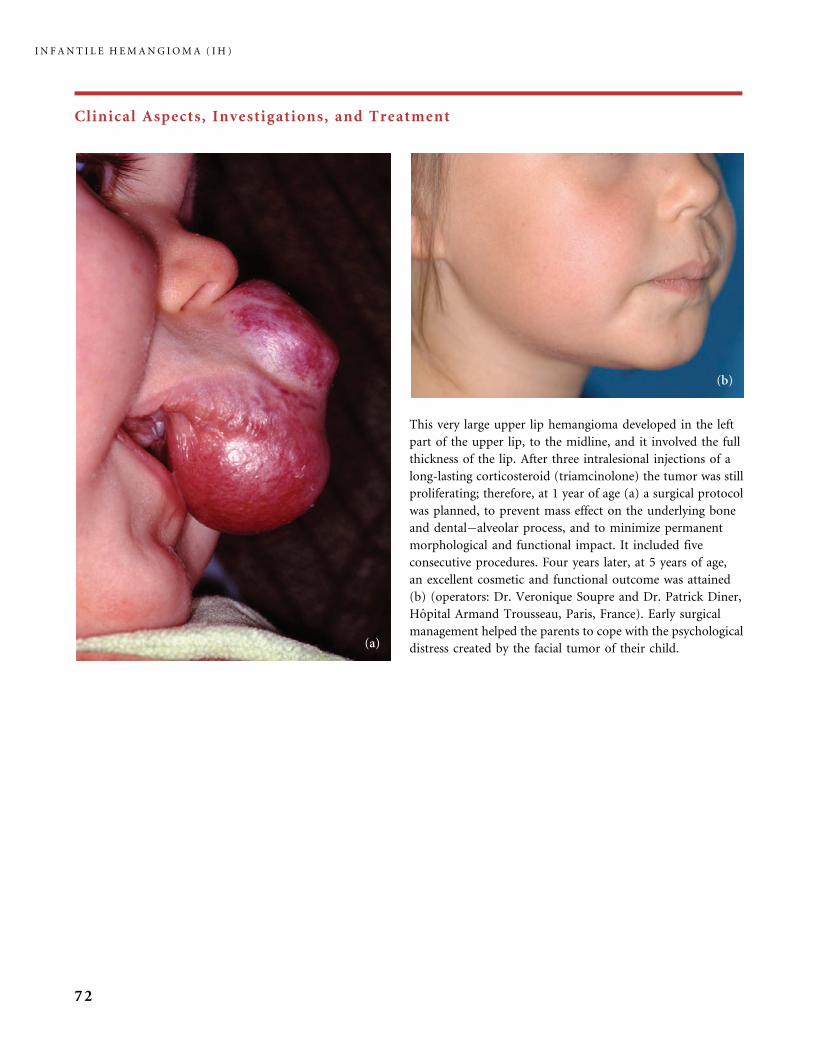
Clinical Aspects, Investigations, and Treatment
This very large upper lip hemangioma developed in the left
part of the upper lip, to the midline, and it involved the full
thickness of the lip. After three intralesional injections of a
long-lasting corticosteroid (triamcinolone) the tumor was still
proliferating; therefore, at 1 year of age (a) a surgical protocol
was planned, to prevent mass effect on the underlying bone
and dental�alveolar process, and to minimize permanent
morphological and functional impact. It included five
consecutive procedures. Four years later, at 5 years of age,
an excellent cosmetic and functional outcome was attained
(b) (operators: Dr. Veronique Soupre and Dr. Patrick Diner,
Hopital Armand Trousseau, Paris, France). Early surgical
management helped the parents to cope with the psychological
distress created by the facial tumor of their child.
I N F A N T I L E H E M A N G I O M A ( I H )
72
References
1 Adams D. in: Infantile hemangiomas: current knowledge, future directions. Proceedings
of a Research Workshop on Infantile Hemangioma. Bethesda Maryland, April 7�9,
2005. Pediatr Dermatol 2005; 22: 383�406.
2 Akyuz C, Yaris N, Kutluk MT, Buyukpamukcu M. Management of cutaneous
hemangiomas: a retrospective analysis of 1109 cases and comparison of conventional
dose prednisolone with high-dose methylprednisolone therapy. Pediatr Hematol Oncol
2001; 18: 47�55.
3 Allen RM, Sandquist MA, Piatt JH Jr, Selden NR. Ultrasonographic screening in infants
with isolated spinal strawberry nevi. J Neurosurg 2003; 98: 247�50.
4 Amir J, Metzker A, Krikler R, Reisner SH. Strawberry hemangioma in preterm infants.
Pediatr Dermatol 1986; 3: 331�2.
5 Ashinoff R, Geronemus RG. Failure of the flashlamp-pumped dye laser to prevent
progression to deep hemangioma. Pediatr Dermatol 1993; 10: 77�80.
6 Aviles R, Boyce TG, Thompson DM. Pneumocystis carinii pneumonia in a 3-month old
infant receiving high-dose corticosteroid therapy for airway hemangiomas. Mayo Clin
Proceed 2004; 79: 243�5.
7 Barlow CF, Priebe CJ, Mulliken JB, Barnes PD, MacDonald D, Folkman J. Spastic
diplegia as a complication of interferon alfa 2a treatment of hemangiomas of infancy.
J Pediatr 1998; 132: 527�30.
8 Batta K, Goodyear HM, Moss C, Williams HC, Hiller L, Waters R. Randomised
controlled study of early pulsed dye laser treatment of uncomplicated childhood
hemangiomas: results of a 1-year analysis. Lancet 2002; 360: 521�7.
9 Bauland CG, Steijlen PM, Rieu PNMA, Spauwen PHM. The pathogenesis of
hemangiomas: a review. Plast Recontr Surg 2006; 117: 29e�35e
10 Bennett ML, Fleischer AB Jr, Chamlin SN, Frieden IJ. Oral corticosteroid use is effective
for cutaneous hemangiomas: an evidence-based evaluation. Arch Dermatol 2001; 137:
1208�13.
11 Bhattacharya JJ, Luo CB, Alvarez H, Rodesch G, Pongpech S, Lasjaunias PL. PHACES
syndrome: a review of eight previously unreported cases with late arterial occlusions.
Neuroradiology 2004; 46: 227�33.
12 Bitar MA, Moukarbel RV, Zalzal GH. Management of congenital subglottic
hemangioma: trends and success over the past 17 years. Otolaryngol Head Neck Surg
2005; 132: 226�31.
13 Boon LM, MacDonald DM, Mulliken JB. Complications of systemic corticosteroid
therapy for problematic hemangiomas. Plast Reconstr Surg 1999; 104: 1616�23.
14 Bouchard S, Yazbeck S, Lallier M. Perineal hemangioma, anorectal malformation,
and genital anomaly, a new association? J Pediatr Surg 1999; 34: 1133�5.
14a Girard C, Bigorre M, Guilot B, Bessis D. PELVIS syndrome. Arch Dermatol 2006;
142: 884�8.
15 Bronzetti G, Giardini A, Patrizi A, Prandstraller D, Donti A, Formigari R, et al.
Ipsilateral hemangioma and aortic arch anomalies in posterior fossa malformations,
hemangiomas, arterial anomalies, coarctation of the aorta, and cardiac defects, and
eye abnormalities (PHACE) anomaly: report and review. Pediatrics 2004; 113: 412�15.
16 Bruckner AL, Frieden IJ. Hemangiomas of infancy. J Am Acad Dermatol 2003; 48: 477�93.
17 Burrows PE, Laor T, Paltiel H, Robertson RL. Diagnostic imaging in the evaluation
of vascular birthmarks. Dermatology Clin 1998; 16: 455�88.
18 Burrows PE, Robertson RL, Mulliken JB, Beardsley Ds, Chaloupka JC, Ezekowitz RAB,
et al. Cerebral vasculopathy and neurologic sequelae in infants with cervicofacial
hemangioma: report of eight patients. Radiology 1998; 207: 601�7.
R E F E R E N C E S
73
19 Ceisler EJ, Santos L, Blei F. Periocular hemangiomas: what every physician should know.
Pediatr Dermatol 2004; 21: 1�9.
20 Chang E, Boyd A, Nelson CC, Crowley D, Law T, Kenough KM, et al. Successful
treatment of infantile hemangiomas with interferon alfa-2b. J Pediatr Hematol Oncol
1997; 19: 237�44.
21 Chen MT, Yeong EK, Horng SY. Intralesional corticosteroid therapy in proliferating
head and neck hemangiomas: a review of 155 cases. J Pediatr Surg 2000; 35: 420�3.
22 Chiller KG, Passaro D, Frieden IJ. Hemangiomas of infancy. Clinical characteristics,
morphologic subtypes, and their relationship to race, ethnicity and gender. Arch
Dermatol 2002; 138: 1567�76.
23 David LR, Malek MM, Argenta LC. Efficacy of pulsed dye laser therapy for the treatment
of ulcerated haemangioma: a review of 78 patients. Br J Plast Surg 2003; 56: 317�27.
24 Demiri Ec, Pelissier P, Genin-Etcheberry T, Tsakoniatis N, Martin D, Baudet J.
Treatment of facial haemangiomas: the present status of surgery. Br J Plast Surg 2001; 54:
665�74.
25 Diner PA, Petit F, Soupre V, Enjolras O, Lemarchand-Venencie F, Moraillon I, et al.
Exerese precoce des hemangiomes de la face: nouvelle technique par ultrasons. Ann
Dermatol Venereol 1998; 125: 605�7.
26 Drolet BA, Dohil M, Golomb MR, Wells R, et al. Early stroke and cerebral vasculopathy in
children with facial hemangiomas and PHACE association. Pediatrics 2006; 117: 959�64.
27 Dubois J, Garel L. Imaging and therapeutic approach of hemangiomas and vascular
malformations in the pediatric age group. Pediatr Radiol 1999; 29: 879�93.
28 Dubois J, Hershon L, Carmant L, Belanger S, Leclerc JM, David M. Toxicity profile of
interferon alfa-2b in children: a prospective evaluation. J Pediatr 1999; 135: 782�5.
29 Egbert JE, Paul S, Engel WK, Summers CG. High injection pressure during intralesional
injection of corticosteroids into capillary hemangiomas. Arch Ophthalmol 2001; 119:
677�83.
30 Egbert JE, Enjolras O, Elsas FJ, Stalder JF, Lemarchand-Venencie F, Haik Bg,
Hamel-Teillac D, Magalon G, Lacour JP. Question du mois: corticotherapie intralesion-
nelle et hemangiomes orbito-palpebraux. Ann Dermatol Venereol 1998; 125: 754�8.
31 Enjolras O, Riche MC, Merland JJ, Escande JP. Management of alarming hemangiomas
in infancy: a review of 25 cases. Pediatrics 1990; 85: 491�8.
32 Enjolras O, Pouplard F, Beucher A, Ginies JL, Verret JL. Toxicite neurologique de l’inter-
feron alfa 2a au cours du traitement d’un hemangiome cephalique grave. Communication
Journees Dermatologiques de Paris 1996. Ann Dermatol Venereol 1996; 123: S1: 144�5.
33 Enjolras O, Gelbert F. Superficial hemangiomas: associations and management. Pediatr
Dermatol 1997; 14: 173�9.
34 Enjolras O. Neurotoxicity of interferonalfa in children treated for hemangiomas. J Am
Acad Dermatol 1998; 39: 1037�8.
35 Enjolras O, Breviere GM, Roger G, Tovi M, Pellegrino S, Varotti E, Soupre V, Picard A,
Leverger G. Traitement par vincristine des hemangiomes graves du nourrisson. Arch
Pediatr 2004; 11: 99�107.
36 Esterly NB. Cutaneous Hemangiomas, Vascular stains and malformations, and
associated syndromes. Curr Prob Dermatol 1995; VII: 65�108.
37 Ezekowitz RAB, Mulliken JB, Folkman J. Interferon alfa-2a therapy for life-threatening
hemangiomas of infancy. N Eng J Med 1992; 326: 1456�63.
38 Faguer K, Dompmartin A, Labbe D, Barrelier MT, Leroy D, Theron J. Early surgical
treatment of Cyrano-nose haemangiomas with Rethi incision. Br J Plast Surg 2002; 55:
498�503.
39 Fishman S. in: Infantile hemangiomas: current knowledge, future directions. Proceedings
of a Research Workshop on Infantile Hemangioma. Bethesda Maryland, April 7�9,
2005. Pediatr Dermatol 2005; 22: 383�406.
40 Frieden IJ, Reese V, Cohen D., PHACE syndrome: the association of posterior fossa brain
malformations, hemangiomas, arterial anomalies, coarctation of the aorta and cardiac
defects, and eye abnormalities. Arch Dermatol 1996; 132: 307�11.
I N F A N T I L E H E M A N G I O M A ( I H )
74
41 Frieden IJ, Haggstrom AJ, Drolet BA, Mancini AJ, Friedlander SF, Boon L, Chamlin S,
Baselga E, Garzon M, Nopper AJ, Siegel DH, Mathes EW, Goddard D, Bischoff J,
North PE, Esterly NB. Infantile hemangiomas: current knowledge, future directions.
Proceedings of a Research Workshop on Infantile Hemangiomas, April 7�9, 2005,
Bethesda, Maryland. Pediatr Dermatol 2005; 22: 383�406.
42 Frischer JS, Huang J, Serur A, Kadenhe A, Yamashiro DJ, Kandel JJ. Biomolecular
markers and involution of hemangiomas. J Pediatr Surg 2004; 39: 400�4.
43 Garden JM, Bakus AD, Paller AS. Treatment of cutaneous hemangiomas with the
flashlamp-pumped pulsed dye laser: prospective analysis. J Pediatr 1992; 120: 555�60.
44 Garmendia G, Miranda N, Borroso S, Longchong M, Martinez E, Ferrero J. Regression
of infancy hemangiomas with recombinant IFN alpha-2b. J Interferon Cytokine Res 2001;
21: 31�8.
45 Garzon MC, Lucky AW, Hawrot A, Frieden IJ. Ultrapotent corticosteroid treatment of
hemangiomas of infancy. J Am Acad Dermatol 2005; 52: 281�6.
46 George ME, Sharma V, Jacobson J, Simon S, Nopper AJ. Adverse effects of systemic gluco-
corticosteroid therapy in infants with hemangiomas. Arch Dermatol 2004; 140: 960�9.
47 Gorlin RJ, Kantaputra P, Aughton DJ, Mulliken JB. Marked female predilection in some
syndromes associated with facial hemangiomas. Am J Med Genet 1994; 52: 130�5.
48 Greene AK, Rogers GF, Mulliken JB. Management of parotid hemangioma in 100
children. Plast Reconstr Surg 2004; 113: 53�60.
49 Grosso S, De Cosmo L, Bonifazi E, Galluzzi P, Farnetani MA, Loffredo P, et al. Facial
hemangioma and malformation of the cortical development: a broadening of PHACE
spectrum or a new entity? Am J Med Genet 2004; 124A: 192�5.
50 Guggisberg D, Hadj-Rabia S, Viney C, Bodemer C, Brunelle F, Zerah M, et al. Skin
markers of occult spinal dysraphism in children. A review of 54 cases. Arch Dermatol
2004; 140: 1109�15.
51 Haggstrom A, Lammer E, Schneider R, Marcucio R, Frieden IJ. Patterns of infantile
hemangiomas: new clues to hemangioma pathogenesis and embryonic facial develop-
ment. Pediatrics 2006; 117: 698�703.
52 Hastings MM, Milot J, Barsoum-Homsy M, Hershon L, Dubois J, Leclerc JM.
Recombinant interferon alfa-2b in the treatment of vision-threatening capillary
hemangioma in childhood. J AAPOS 1997; 1: 226�30.
53 Herman TE, McAlister WH, Dehner LP. Posterior mediastinal capillary hemangioma
with extradural extension resembling neuroblastoma. Pediatr Radiol 1999; 29: 517�19.
54 Hersh JH, Waterfill D, Rutleedge J, Harrod MJ, O’Sheal SF, Verdi G, Martinez S,
Weisskopf B. Sternal malformation/vascular dysplasia association. Am J Med Genet 1985;
21: 177�86.
55 Hohenleutner S, Badur-Ganter E, Landthaler M, Hohenleutner U. Long-term results in
the treatment of childhood hemangioma with the flashlamp-pumped pulsed dye laser: an
evaluation of 617 cases. Lasers Surg Med 2001; 28: 273�7.
56 Hohenleutner U, Landthaler M. Laser treatment of childhood hemangioma: progress or
not. Lancet 2002; 360: 502�3.
57 Huang SA, Tu HM, Harney JW, Venihaki M, Butte AJ, Kozakewich HPW, Fishman SJ,
Larsen PR. Severe hypothyroidism caused by type 3 iodothyronine deiodinase in infantile
hemangioma. N Eng J Med 2000; 343: 185�9.
58 Iwata J, Sonobe H, Furihata M, Ido E, Ohtsuki Y. High frequency of apoptosis in
infantile capillary hemangioma. J Pathol 1996; 179: 403�8.
59 Jang YC, Isik FF, Bibran NS. Nerve distribution in hemangiomas depends on the
proliferative state of the microvasculature. J Surg Res 2000; 93: 144�8.
60 Kassarjian A, Zurakowsky D, Dubois J, Paltiel HJ, Fishman SJ, Burrows PE. Infantile
hepatic hemangiomas, clinical and imaging findings and their correlation with therapy.
Am J Roentgenol 2004; 182: 785�95.
61 Kniestedt C, Landau K, Brodsky MC, North PE, Waner M. Infantile orofacial
hemangioma with ipsilateral peripapillary excavation in girls: a variant of PHACE
syndrome. Arch Ophthalmol 2004; 122: 413�15.
R E F E R E N C E S
75

62 Konrad D, Ellis G, Perlman K. Spontaneous regression of severe acquired infantile
hypothyroidism associated with multiple liver hemangiomas. Pediatrics 2003; 112:
1424�6.
63 Kraling BM, Razon MJ, Boon LM, Zurakowski D, Seachord C, Darveau RP, et al.
E-selectin is present in proliferating endothelial cells in human hemangiomas. Am
J Pathol 1996; 148: 1181�91.
64 Kronenberg A, Blei F, Ceisler E, Steele M, Furlan L, Kodsi S. Ocular and systemic
manifestations of PHACES syndrome. J AAPOS 2005; 9: 169�73.
65 Kushner BJ. The treatment of periorbital infantile hemangioma with intralesional
corticosteroid. Plast Reconstr Surg 1985; 76: 517�24.
66 Lasky JB, Sandu M, Balashanmugan A. PHACE syndrome: association with persistent
fetal vasculature and coloboma-like iris defect. J AAPOS 2004; 8: 495�8.
67 Leaute-Labreze C, Labbe L, Grenier N, Berge J, Vergnes P, Taieb A. Hemangiomes graves
traites par interferon a : 7 cas. Ann Dermatol Venereol 1998; 125: 174�8.
68 Luo CM, Lasjaunias P, Teng MM, Chang FC, Lirng JF, Chang CY. Cervico-
cerebrovascular anomalies in children with PHACE syndrome. J Formos Med Assoc
2003; 102: 379�86.
69 MacArthur CJ, Senders CW, Katz J. The use of interferon alfa-2a for life-threatening
hemangiomas. Arch Otolaryngol Head Neck Surg 1995; 121: 690�3.
70 Mancini AJ, Smoller BR. Proliferation and apoptosis within juvenile capillary
hemangiomas. Am J Dermatopathol 1996; 18: 505�14.
71 Metry DW, Dowd CF, Barkovich AJ, Frieden IJ. The many faces of PHACE syndrome.
J Pediatr 2001; 139: 117�23.
72 Metry DW, Hawrot A, Altman C, Frieden IJ. Association of solitary segmental heman-
giomas of the skin with visceral hemangiomatosis. Arch Dermatol 2004; 140: 591�6.
73 Metry D. Update on hemangiomas of infancy. Curr Opin Pediatr 2004; 16: 373�7.
74 Metry D. Potential complications of segmental hemangiomas of infancy. Semin Cutan
Med Surg 2004; 23: 107�15.
75 Metz BJ, Rubinstein MC, Levy ML, Metry DW. Response of ulcerated perineal
hemangioma of infancy to beclapermin gel, a recombinant human platelet-derived
growth factor. Arch Dermatol 2004; 140: 867�70.
76 Michaud AP, Bauman NM, Burke DK, Manaligod JM, Smith RJ. Spastic diplegia and
other motor disturbances in infants receiving interferon alpha. Laryngoscope 2004; 114:
1231�6.
77 Millischer-Bellaiche AE, Enjolras O, Andre Ch, Bursztyn J, Kalifa G, Adamsbaum C. Les
hemangiomes palpebraux du nourrisson. J Radiol 2004; 85: 2019�28.
78 Momtchilova M, Pelosse B, Diner PA, Vazquez MP, Laroche L. Amblyopi et
hemangiome capillaire infantile perioculaire. J Fr Ophtalmol 2004; 27: 1135�40.
79 Mulliken JB, Rogers GF, Marler JJ. Circular excision of hemangioma and purse-string
closure: the smallest possible scar. Plast Reconstr Surg 2002; 109: 1544�54.
80 Nakayama H. Clinical and histological studies of the classification and the natural course
of the strawberry mark. J Dermatol 1981; 8: 277�91.
81 North PE, Waner M, Mizeracki A, Mihm MC Jr. GLUT1: a newly discovered immu-
nohistochemical marker for juvenile hemangiomas. Hum Pathol 2000; 31: 11�22.
82 North PE, Waner M, Mizeracki A, Mrak RE, Nicholas R, Kincannon J, Suen JY, Mihm
MC Jr. A unique microvascular phenotype shared by juvenile hemangiomas and human
placenta. Arch Dermatol 2001; 137: 559�70.
83 Orlow SJ, Isakoff MS, Blei F. Increased risk of symptomatic hemangiomas of the airway
in association with cutaneous hemangiomas in a ‘‘beard’’ distribution. J Pediatr 1997;
131: 643�6.
84 Pascual-Castroviejo I. Vascular and non-vascular intracranial malformations with
external capillary hemangiomas. Neuroradiology 1978; 16: 82�4.
85 Pascual-Castroviejo I, Viano J, Moreno F, Palencia R, Fernandez VM, Pascual-Pascual SI,
et al. Hemangiomas of the head, neck and chest with associated vascular brain anomalies:
a complex neurocutaneous syndrome. Am J Neuroradiol 1996; 17: 461�71.
I N F A N T I L E H E M A N G I O M A ( I H )
76

86 Pascual-Castroviejo I, Pascual-Castroviejo S, Viano J, Martinez V. Hemangioma
cavernoso y mega-arteriosis cerebral: involucion coetanea de ambas malformaciones
en un caso. Patologia Vascular 1999; 5: 53�60.
87 Pascual-Castroviejo I, Viano J, Pascual-Pascual S, Martinez V. Do cutaneous
hemangiomas and internal vascular anomalies follow the same evolution? Neurology
2003; 61: 140�1.
88 Picard A, Soupre V, Diner PA, Buis J, Goga D, Vazquez MP. Chirurgie precoce des
hemangiomes immatures a l’aide d’un dissecteur a ultrasons. Etude a propos de 81 cas.
Rev Stomatol Chir Maxillofac 2002; 103: 10�21.
89 Pienaar C, Graham R, Geldenhuys S, Hudson DA. Intralesional bleomycin for the
treatment of hemangiomas. Plast Reconstr Surg 2006; 117: 221�6.
90 Poetke M, Froemmeld T, Berlien HP. PHACE syndrome: new views on diagnostic
criteria. Eur J Pediatr Surg 2002; 12: 366�74.
91 Pokorny JJ, Roth F, Balfour I, Rinehart G. An unusual complication of the treatment of
a hemangioma. Ann Plast Surg 2002; 48: 83�7.
92 Rahbar R, Nicollas R, Roger G, Triglia JM, Garabedian EN, McGill TJ, Healy GB. The
biology and management of subglottic hemangioma: past, present, future. Laryngoscope
2004; 114: 1880�91.
93 Razon MJ, Kraling BM, Mulliken JB, Bischoff J. Increased apoptosis coincides with onset
of involution in infantile hemangioma. Microcirculation 1998; 5: 189�95.
94 Reischle S, Schuller-Petrovic S. Treatment of capillary hemangiomas of early childhood
with a new method of cryosurgery. J Am Acad Dermatology 2000; 42: 809�13.
95 Ritter MR, Dorrell MI, Edmonds J, Friedlander SF, Friedlander M. Insulin-like growth
factor 2 and potential regulators of hemangioma growth and involution identified by
large-scale expression analysis. Proc Natl Acad Sci USA 2002; 99: 7455�60.
96 Rothe MJ, Rowse D, Grant-Kels JM. Benign neonatal hemangiomatosis with aggressive
growth of cutaneous lesions. Pediatr Dermatol 1991; 8: 140�6.
97 Scheepers JH, Quaba AA. Does the pulsed tunable dye laser have a role in the
mangement of infantile hemangioma? Observations based on a 3 years experience. Plast
Reconstr Surg 1995; 95: 305�12.
98 Shorr N, Seiff SR. Central retinal artery occlusion associated with periocular
corticosteroid injection for juvenile hemangioma. Ophthalmic Surg 1986; 17: 229�231.
99 Takahashi K, Mulliken JB, Kozakewich HPW, Rogers RA, Folkman J, Ezekowitz RA.
Cellular markers that distinguish the phases of hemangioma during infancy and
childhood. J Clin Invest 1994; 93: 2357�64.
100 The Denat B, Leaute-Labreze C, Boralevi F, Roul S, Labbe L, Marliere V, Taieb A.
Surveillance tensionnelle des nourrissons traits par corticotherapie generale pour un
hemangiome. Ann Dermatol Venereol 2002; 129: 183�5.
101 Vazquez MP, Diner PA, Picard A, Soupre V, Enjolras O. Les levres angiomateuses. Ann
Chir Plast Esthet 2002; 47: 561�79.
102 Vazquez-Botet R, Reyes BA, Vazquez-Botet M. Sclerodermiform linear atrophy after the
use of intralesional steroids for periorbital hemangiomas: a review of complications.
J Pediatr Ophthalmol Strabismus 1989; 26: 124�7.
103 Van Den Abbelee T, Triglia JM, Lescanne E, Roger G, Nocollas R, Ployet MJ, Garabedian
EN. Surgical removal of subglottic hemangiomas in children. Laryngoscope 1999; 109:
1281�6.
104 Waner M, North PE, Scherer KA, Frieden IJ, Mihm MC, Suen JY. The non-random
distribution of facial hemangiomas. Arch Dermatol 2003; 139: 869�75.
105 Yeh TF, Lin YJ, Huang CC, Chen YJ, Lin CH, Lin HC. Early dexamethasone therapy in
preterm infants: a follow-up study. Pediatrics 1998; 101: E7.
106 Yu Y, Fuhr J, Boye E, Gyorffy S, Soker S, Atala A, Mulliken JB, Bischoff J. Mesenchymal
stem cells and adipogenesis in hemangioma evolution. Stem Cells 2006, Feb epub ahead
of print.
R E F E R E N C E S
77
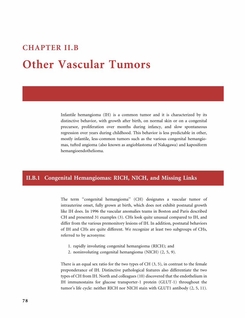
CHAPTER II.B
Other Vascular Tumors
Infantile hemangioma (IH) is a common tumor and it is characterized by its
distinctive behavior, with growth after birth, on normal skin or on a congenital
precursor, proliferation over months during infancy, and slow spontaneous
regression over years during childhood. This behavior is less predictable in other,
mostly infantile, less-common tumors such as the various congenital hemangio-
mas, tufted angioma (also known as angioblastoma of Nakagawa) and kaposiform
hemangioendothelioma.
II.B.1 Congenital Hemangiomas: RICH, NICH, and Missing Links
The term ‘‘congenital hemangioma’’ (CH) designates a vascular tumor of
intrauterine onset, fully grown at birth, which does not exhibit postnatal growth
like IH does. In 1996 the vascular anomalies teams in Boston and Paris described
CH and presented 31 examples (3). CHs look quite unusual compared to IH, and
differ from the various premonitory lesions of IH. In addition, postnatal behaviors
of IH and CHs are quite different. We recognize at least two subgroups of CHs,
referred to by acronyms:
1. rapidly involuting congenital hemangioma (RICH); and
2. noninvoluting congenital hemangioma (NICH) (2, 5, 9).
There is an equal sex ratio for the two types of CH (3, 5), in contrast to the female
preponderance of IH. Distinctive pathological features also differentiate the two
types of CH from IH. North and colleagues (10) discovered that the endothelium in
IH immunostains for glucose transporter-1 protein (GLUT-1) throughout the
tumor’s life cycle: neither RICH nor NICH stain with GLUT1 antibody (2, 5, 11).
78

Table
11
Clinical
andhistological
comparisonbetweeninfantile
hem
angioma,
RIC
H,an
dNIC
H.
Com
mon
infantilehem
angiom
aRICH
NICH
Sex
F:M
3�
5:1
1:1
1:1
Lo
cati
on
Hea
dan
dn
eck
in2
/3,
1/4
on
tru
nk
Hea
dan
dn
eck¼
extr
emit
ies,
rare
on
tru
nk
-(b
ased
on
bio
psi
ed/r
esec
ted
spec
imen
s)
Hea
dan
dn
eck¼
extr
emit
ies,
tru
nk
-un
com
mo
n
Ave
rage
size
Var
iab
le6
cm5
cm
Cu
tan
eou
s
app
eara
nce
Pro
life
rati
vep
has
eS
up
erfi
cial
:b
righ
tre
d,
bo
ssed
Dee
p:
no
rmal
or
blu
ish
Rai
sed
and
do
me-
shap
ed,
rou
nd
or
ovo
id,
tela
ngi
ecta
ses,
oft
enp
ale
rim
,ce
ntr
alu
lcer
,
scar
,o
rd
epre
ssio
n
Sli
ghtl
yra
ised
,ro
un
do
ro
void
,p
urp
le,
wel
l-d
elin
eate
d,
tela
ngi
ecta
ses,
cen
tral
/per
iph
eral
pal
lor
Invo
luti
vep
has
eA
tro
ph
y,te
lan
giec
tase
s,
dys
chro
mia
,an
eto
der
ma
Dep
ress
ed,
pal
e,fe
wre
sid
ual
tela
ngi
ecta
ses,
pro
min
ent
vein
s
Gen
eral
arch
itec
ture
Pro
life
rati
vep
has
eL
ob
ule
scl
ose
lysp
aced
or
con
flu
ent
Un
kn
ow
nL
ob
ule
sse
par
ated
by
den
sefi
bro
us
tiss
ue,
pro
min
ent
inte
rlo
bu
lar
vasc
ula
ture
,
usu
ally
no
zon
atio
n
Invo
luti
vep
has
eIn
tral
ob
ula
ran
d
inte
rlo
bu
lar
fib
rou
sti
ssu
e
Inte
rlo
bu
lar
fib
rou
sti
ssu
e,o
ften
zon
atio
n
wit
had
van
ced
invo
luti
on
inle
sio
nal
cen
ter
Lo
bu
les
Pro
life
rati
vep
has
eU
sual
lyla
rge
Un
kn
ow
nU
sual
lyla
rge
wit
hcu
rved
chan
nel
san
d
pro
min
ent
cen
tril
ob
ula
rd
rain
ing
vess
els(
s)
Invo
luti
vep
has
eS
mal
l-to
-lar
geS
mal
l,m
ediu
m,
or
larg
ew
ith
pro
min
ent
cen
tril
ob
ula
rd
rain
ing
chan
nel
(s)
En
do
thel
ium
Pro
life
rati
vep
has
eM
ark
edly
pro
min
ent,
GL
UT
-1p
osi
tive
Un
kn
ow
nH
ob
nai
led
,cy
top
lasm
icin
clu
sio
n,
GL
UT
-1n
egat
ive
Invo
luti
vep
has
eF
lat,
GL
UT
-1p
osi
tive
Mo
der
atel
yp
rom
inen
t,ra
rely
ho
bn
aile
d,
GL
UT
-1u
sual
lyn
egat
ive
Cap
illa
ryb
asem
ent
mem
bra
ne
Pro
life
rati
vep
has
eT
hin
Un
kn
ow
nT
hin
,fo
call
yth
ick
ened
Invo
luti
vep
has
eT
hic
kT
hin
,th
ick
ened
inla
test
age
Ext
ralo
bu
lar
vasc
ula
ture
Pro
life
rati
vep
has
eM
inim
ally
or
mo
der
atel
y
pro
min
ent
arte
ries
and
vein
s
Un
kn
ow
nP
rom
inen
tar
teri
esan
dab
no
rmal
vein
s,
arte
rio
lob
ula
ran
dar
teri
ove
no
us
fist
ula
e
Invo
luti
vep
has
eF
eed
ing
arte
ries
and
vein
s
may
no
tre
gres
sco
mp
lete
ly
Lar
gean
dab
no
rmal
dra
inin
gch
ann
els,
par
ticu
larl
yin
lesi
on
alce
nte
r
Ad
dit
ion
alfe
atu
res
Pro
life
rati
vep
has
eU
nk
no
wn
Th
rom
bi
Invo
luti
vep
has
eF
atT
hro
mb
i,in
farc
tio
n,
hem
osi
der
in,
calc
ific
atio
n,
cyst
s,an
eury
sms,
extr
amed
ull
ary
hem
ato
po
iesi
s
RIC
H¼
rap
idly
invo
luti
ng
con
gen
tial
hem
angi
om
a;N
ICH¼
no
nin
volu
tin
gco
nge
nti
alh
eman
gio
ma;
GL
UT
-1¼
glu
cose
tran
spo
rter
-1p
rote
in.
Reproducedwithpermission
from
:B
eren
guer
B,
Mu
llik
enJB
,E
njo
lras
O,
Bo
on
LM
,W
asse
fM
,Jo
sset
P,
Bu
rro
ws
PE
,P
erez
-Ata
yde
AR
,K
oza
kew
ich
HP
W.
Rap
idly
invo
luti
ng
con
gen
ital
hem
angi
om
a:cl
inic
alan
dh
isto
pat
ho
logi
cfe
atu
res.Ped
Dev
Biol
20
03
;6
:4
95�
61
0(T
able
1).
C O N G E N I T A L H E M A N G I O M A S : R I C H , N I C H , A N D M I S S I N G L I N K S
79
NICH and RICH have distinct clinical and pathological features (Table 11).
However, in a number of infants, a RICH may to some extent regress but it results
in a lesion similar to NICH: we call these cases ‘‘missing links’’ as they probably
indicate some relationship between both tumors, or at least a subset of these
congenital hemangiomas.
I I .B .1 . 1 RICH
RICH are fully grown in utero and full-blown at birth, no postnatal proliferation
happens, and regression quickly follows over 6 to 14 months. This clinical pattern
and course are unique and differ from those of IH. Because of their remarkable
postnatal accelerated regression we called these tumors Rapidly Involuting Con-
genital Hemangioma (RICH) to stress this distinctive behavior, quite different
from the protracted regression of a large IH (2, 3, 6). Six analogous tumors were
called ‘‘congenital nonprogressive hemangioma’’ by North and colleagues in
a study that focused on histological findings; none of the six congenital tumors had
vascular immunoreactivity for GLUT1 and Lewis Y antigen; conversely the 25 IH
tested in parallel demonstrated strong lesional endothelial immunoreactivity for
these two antigens (11).
Due to their fast-flow nature RICH are more and more commonly detected
during antenatal ultrasonographic evaluations (6). Most of the lesions discovered
during the fetal life were tumors of the scalp or neck, and less frequently of
an extremity. In the literature there are various examples of RICH detected during
the second or third trimester by antenatal ultrasonography and/or prenatal MRI
(2, 3, 8). Tumors discovered during fetal life either exhibited rapid postnatal
regression (14) or were excised in infancy (4). In rare instances, the neonate died
of complications of the tumor (13).
All tumors initially exhibit fast-flow on ultrasonography and magnetic reso-
nance imaging (3, 12). The ultrasonic characteristics of RICH were reported by
Rogers et al. in a group of 10 patients: all lesions were uniformly hypoechoic,
confined to the subcutaneous fat and traversed by multiple vascular channels (12).
MRI and angiographic characteristics of RICH have both similarities and differen-
ces from those of infantile hemangioma. MRI shows a dense tumor with, or
without, tortuous large flow voids, usually located near the surface of the tumor,
and sometimes areas of inhomogeneity. Angiographic features indicate a fast-flow
lesion, which, unlike IH, may include inhomogeneous parenchymal staining,
direct AV fistulae, large and irregularly organized feeding arteries, multiple arterial
aneurysms, cyst, intralesional bleeding and thrombi; because of significant intra-
tumoral arteriovenous shunting some newborns with RICH may present with
high-output cardiac failure (7).
Although RICH have a slightly variable morphology depending on the
patients, common features include: a protuberant round lump, pink or purple
O T H E R V A S C U L A R T U M O R S
80

in color, with coarse often radiating telangiectasia, and habitually a thin surround-
ing whitish halo. Some have central ulceration or a linear scar or depression.
Others exhibit a few central red nodules. Rarely, a nearly normal slightly blanched
overlying skin is observed. There is increased local warmth and in some patients
dilated veins and pulsations. After regression the involved area is left with either an
area of lipoatrophy, or an area of dermal atrophy with persisting blanching-bluish
hue. Some lesions leave a rather prominent telangiectatic round macule or plaque.
The most typical locations are the extremities, close to a joint (by frequency:
mainly the knee, then ankle, shoulder, hip, and wrist) and the head around the
ear (forehead, cheek, or scalp). So far, centrofacial lesions of RICH have not been
observed.
Management
Most lesions are left to shrink spontaneously. Some are excised early in life,
specifically if large arterial vessels are detected close to the surface by ultrasonic and
MRI investigation, or if they have a thick central crust with a risk of ulceration: in
both cases the aim is to avoid profuse hemorrhage (1). In our experience, none of
the excised tumors recurred. Also, as some RICH are now known to result in a
pink plaque of telangiectasia with increasing veins over the years, the achievability
of excision early in life must be discussed with the pediatric plastic surgeon.
I I .B .1 . 2 NICH
Not all congenital hemangiomas spontaneously shrink in the first year of life.
Some do not regress at all. We call these Non-Involuting Congenital Hemangioma
(NICH), as opposed to the RICH behavior (5, 6).
NICH has an almost equal sex distribution. It is solitary, and like RICH it has
a predilection for the head or a limb close to a joint. Lesions on the trunk are
uncommon. In a group of 53 patients followed from 2 to 30 years of age � mean
age at last consultation ¼ 10 years � 43% of the tumors were in the cephalic area,
38% in the limbs and only 19% on the trunk (5). NICH are round or oval,
pink-to-purple, plaque-type or slightly raised, and always warm on palpation.
There is a rim of peripheral white or bluish pallor, or a bluish hue of the whole
surface, punctuated by more or less conspicuous telangiectasia.
NICH persists indefinitely, with a slight tendency to worsening. NICH remains
a fast-flow lesion, as documented by duplex Doppler examination often exhibiting
arteriovenous fistulas. MRI and angiographic findings in NICH are reminiscent
of those of a common IH in the proliferating phase. On MRI a NICH is iso-
intense on T1, hyperintense on T2, and it is enhanced with gadolinium injection.
C O N G E N I T A L H E M A N G I O M A S : R I C H , N I C H , A N D M I S S I N G L I N K S
81
In the past, NICH was misdiagnosed arteriovenous malformation (AVM) or
called ‘‘arteriolo-capillary malformation’’ because angiography demonstrates fast-
flow and rapid arterial filling, but no early venous return could be documented,
as in AVM.
Management
NICH can usually be excised without recurrence, and, unlike AVM, they do not
re-expand in cases of partial or serial excision (5, 6). For large lesions we prefer to
have pre-operative angiography, and, if arterial feeders are numerous, arterial
embolization with particles is performed the day before surgery to limit intra-
operative bleeding.
I I .B .1 . 3 MISSING LINKS
RICH, NICH and IH have some overlapping clinical and/or pathological features.
This has led us to consider that they may be variations of a single tumoral entity
(2, 6, 9). A number of RICH or NICH, present at birth, are later associated with
a growing IH. Rare cases show the superposition of an IH growing on top of
a RICH. In some of our patients, the postnatal course of some RICH suggests the
possibility that NICH could be a late in utero-stage of RICH. The typical story is as
follows: an infant is born with a large congenital hemispherical lump typical of a
RICH. The lesion involutes over 6 to 12 months, then it ceases to regress. Lastly,
a pink telangiectatic plaque, with a white peripheral rim, resembling a NICH will
persist indefinitely. In addition, in some NICH excised early in life the histology
may be indistinguishable from RICH or there are combined features of NICH and
RICH. Based on these postnatal observations we hypothesized that the same
phenomenon may arise in utero and that NICH may be the end result of a RICH
that has regressed during the end of the intrauterine life. Better prenatal ultrasonic
detection and prenatal US follow-up should confirm or refute this hypothesis that
RICH can transform to NICH before birth, and that at least some subtypes of
RICH are a precursor of NICH.
O T H E R V A S C U L A R T U M O R S
82
Figures
CONGENITAL HEMANGIOMAS (RICH AND NICH)
Pathology of RICH
RICH is made up of large-,
medium- or small-sized channels
grouped in lobules. Extralobular
large abnormal vessels are also seen.
Several large nodules are present
in the dermis of a resected RICH,
each of them being made of small
capillary lobules embedded in
fibrosis.
O T H E R V A S C U L A R T U M O R S
83
Pathology of RICH
At a higher magnification some
endothelial cells appear plump and a
mild anisocaryosis is evident.
RICH contains areas of small lobules
with fibrosis. Vessels have a moderately
plump endothelium.
O T H E R V A S C U L A R T U M O R S
84
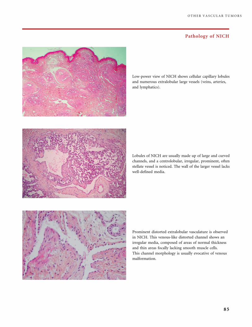
Pathology of NICH
Prominent distorted extralobular vasculature is observed
in NICH. This venous-like distorted channel shows an
irregular media, composed of areas of normal thickness
and thin areas focally lacking smooth muscle cells.
This channel morphology is usually evocative of venous
malformation.
Lobules of NICH are usually made up of large and curved
channels, and a centrolobular, irregular, prominent, often
stellate vessel is noticed. The wall of the larger vessel lacks
well-defined media.
Low-power view of NICH shows cellular capillary lobules
and numerous extralobular large vessels (veins, arteries,
and lymphatics).
O T H E R V A S C U L A R T U M O R S
85
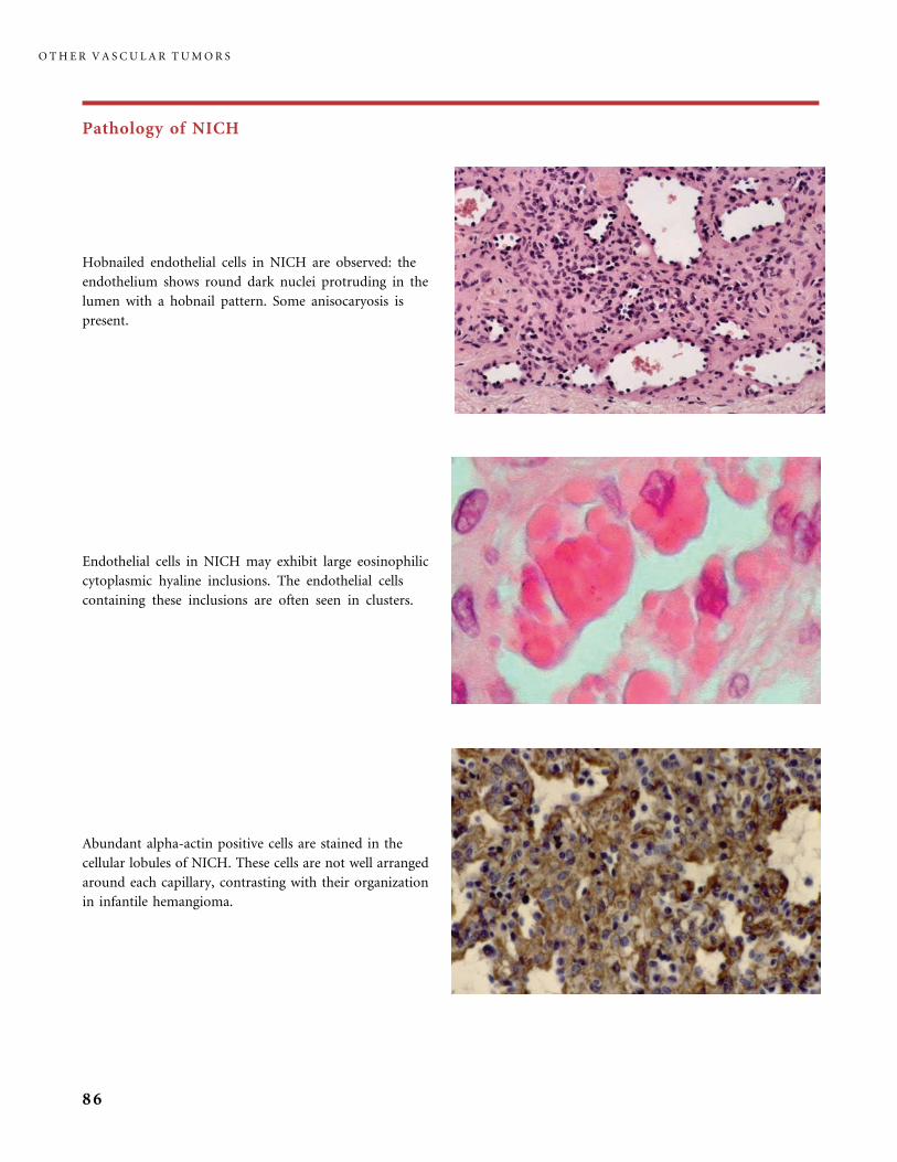
Pathology of NICH
Endothelial cells in NICH may exhibit large eosinophilic
cytoplasmic hyaline inclusions. The endothelial cells
containing these inclusions are often seen in clusters.
Hobnailed endothelial cells in NICH are observed: the
endothelium shows round dark nuclei protruding in the
lumen with a hobnail pattern. Some anisocaryosis is
present.
Abundant alpha-actin positive cells are stained in the
cellular lobules of NICH. These cells are not well arranged
around each capillary, contrasting with their organization
in infantile hemangioma.
O T H E R V A S C U L A R T U M O R S
86
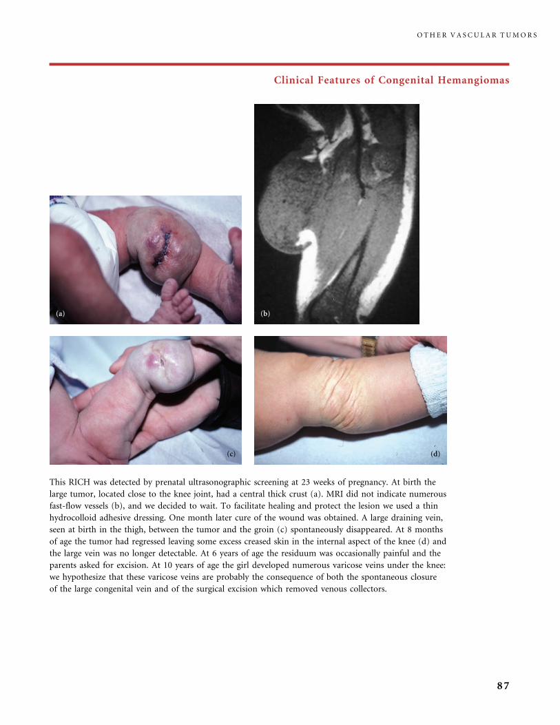
Clinical Features of Congenital Hemangiomas
This RICH was detected by prenatal ultrasonographic screening at 23 weeks of pregnancy. At birth the
large tumor, located close to the knee joint, had a central thick crust (a). MRI did not indicate numerous
fast-flow vessels (b), and we decided to wait. To facilitate healing and protect the lesion we used a thin
hydrocolloid adhesive dressing. One month later cure of the wound was obtained. A large draining vein,
seen at birth in the thigh, between the tumor and the groin (c) spontaneously disappeared. At 8 months
of age the tumor had regressed leaving some excess creased skin in the internal aspect of the knee (d) and
the large vein was no longer detectable. At 6 years of age the residuum was occasionally painful and the
parents asked for excision. At 10 years of age the girl developed numerous varicose veins under the knee:
we hypothesize that these varicose veins are probably the consequence of both the spontaneous closure
of the large congenital vein and of the surgical excision which removed venous collectors.
O T H E R V A S C U L A R T U M O R S
87
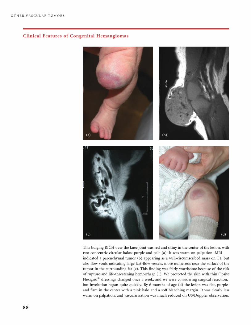
Clinical Features of Congenital Hemangiomas
This bulging RICH over the knee joint was red and shiny in the center of the lesion, with
two concentric circular halos: purple and pale (a). It was warm on palpation. MRI
indicated a parenchymal tumor (b) appearing as a well-circumscribed mass on T1, but
also flow voids indicating large fast-flow vessels, more numerous near the surface of the
tumor in the surrounding fat (c). This finding was fairly worrisome because of the risk
of rupture and life-threatening hemorrhage (1). We protected the skin with thin Opsite
Flexigrid� dressings changed once a week, and we were considering surgical resection,
but involution began quite quickly. By 6 months of age (d) the lesion was flat, purple
and firm in the center with a pink halo and a soft blanching margin. It was clearly less
warm on palpation, and vascularization was much reduced on US/Doppler observation.
O T H E R V A S C U L A R T U M O R S
88
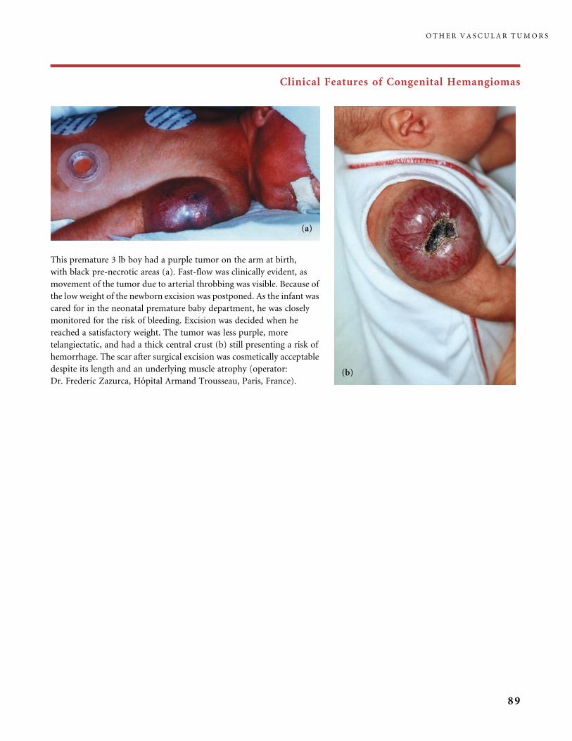
Clinical Features of Congenital Hemangiomas
This premature 3 lb boy had a purple tumor on the arm at birth,
with black pre-necrotic areas (a). Fast-flow was clinically evident, as
movement of the tumor due to arterial throbbing was visible. Because of
the low weight of the newborn excision was postponed. As the infant was
cared for in the neonatal premature baby department, he was closely
monitored for the risk of bleeding. Excision was decided when he
reached a satisfactory weight. The tumor was less purple, more
telangiectatic, and had a thick central crust (b) still presenting a risk of
hemorrhage. The scar after surgical excision was cosmetically acceptable
despite its length and an underlying muscle atrophy (operator:
Dr. Frederic Zazurca, Hopital Armand Trousseau, Paris, France).
O T H E R V A S C U L A R T U M O R S
89

Clinical Features of Congenital Hemangiomas
A typical example of partial regression of a
telangiectatic-type RICH located close to the knee
joint: at birth the large round mauve telangiectatic
bump had a spontaneous thin linear crusted crease in
the center and a faint blanched margin (a). The center
healed in 1 month using hydrocolloid dressing.
Regression progressed over a year. In the meantime
radiated telangiectasia and a white margin became
prominent, and normal skin appeared at the basis of
the lesion (b1, b2). Then involution stopped and the
residual lesion, reminiscent of a thick NICH, was
excised at 18 months. It presented aspects of both
RICH and NICH on pathology.
O T H E R V A S C U L A R T U M O R S
90

Clinical Features of Congenital Hemangiomas
These RICH presenting as large bulging tumors, purple or
bluish, always with coarse radiated telangiectasia, a linear
central scar or a flattened center, and often a thin pale
peripheral rim, tend to only partially involute and result in
a plaque telangiectatic NICH-like residuum. This residuum
is often cosmetically problematic. Therefore, if the tumor
can be excised in infancy, with an anticipated satisfactory
surgical scar, surgical treatment should be considered in
infancy or early childhood.
O T H E R V A S C U L A R T U M O R S
91
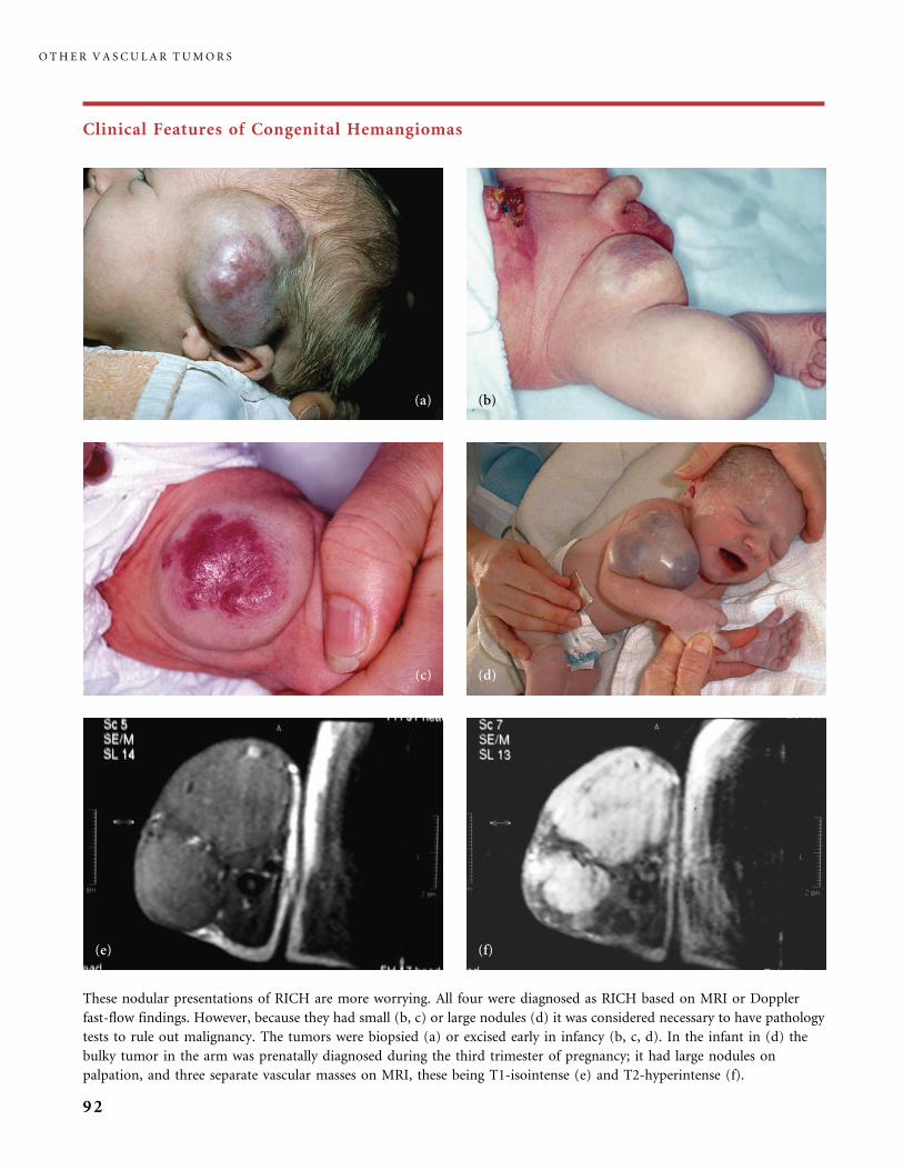
Clinical Features of Congenital Hemangiomas
These nodular presentations of RICH are more worrying. All four were diagnosed as RICH based on MRI or Doppler
fast-flow findings. However, because they had small (b, c) or large nodules (d) it was considered necessary to have pathology
tests to rule out malignancy. The tumors were biopsied (a) or excised early in infancy (b, c, d). In the infant in (d) the
bulky tumor in the arm was prenatally diagnosed during the third trimester of pregnancy; it had large nodules on
palpation, and three separate vascular masses on MRI, these being T1-isointense (e) and T2-hyperintense (f).
O T H E R V A S C U L A R T U M O R S
92
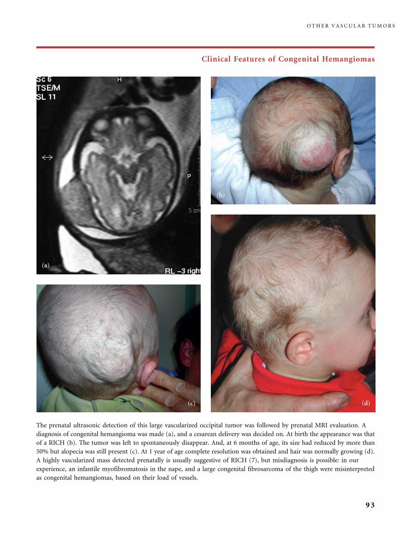
Clinical Features of Congenital Hemangiomas
The prenatal ultrasonic detection of this large vascularized occipital tumor was followed by prenatal MRI evaluation. A
diagnosis of congenital hemangioma was made (a), and a cesarean delivery was decided on. At birth the appearance was that
of a RICH (b). The tumor was left to spontaneously disappear. And, at 6 months of age, its size had reduced by more than
50% but alopecia was still present (c). At 1 year of age complete resolution was obtained and hair was normally growing (d).
A highly vascularized mass detected prenatally is usually suggestive of RICH (7), but misdiagnosis is possible: in our
experience, an infantile myofibromatosis in the nape, and a large congenital fibrosarcoma of the thigh were misinterpreted
as congenital hemangiomas, based on their load of vessels.
O T H E R V A S C U L A R T U M O R S
93
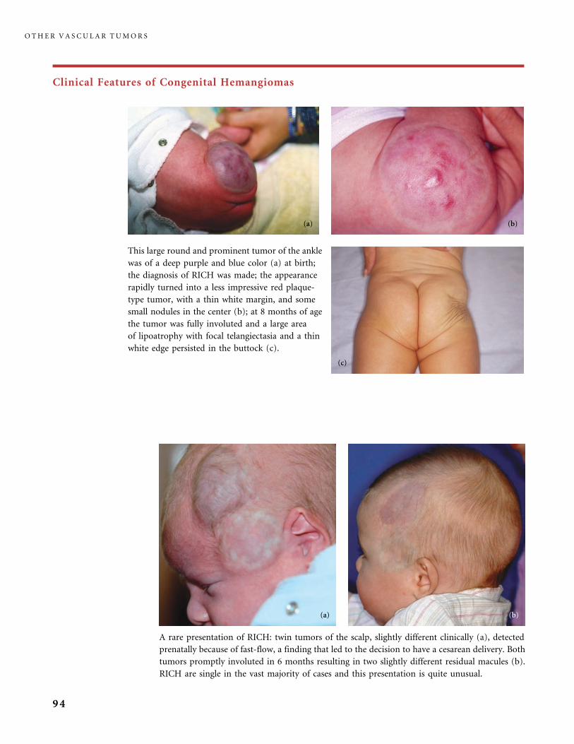
Clinical Features of Congenital Hemangiomas
This large round and prominent tumor of the ankle
was of a deep purple and blue color (a) at birth;
the diagnosis of RICH was made; the appearance
rapidly turned into a less impressive red plaque-
type tumor, with a thin white margin, and some
small nodules in the center (b); at 8 months of age
the tumor was fully involuted and a large area
of lipoatrophy with focal telangiectasia and a thin
white edge persisted in the buttock (c).
A rare presentation of RICH: twin tumors of the scalp, slightly different clinically (a), detected
prenatally because of fast-flow, a finding that led to the decision to have a cesarean delivery. Both
tumors promptly involuted in 6 months resulting in two slightly different residual macules (b).
RICH are single in the vast majority of cases and this presentation is quite unusual.
O T H E R V A S C U L A R T U M O R S
94
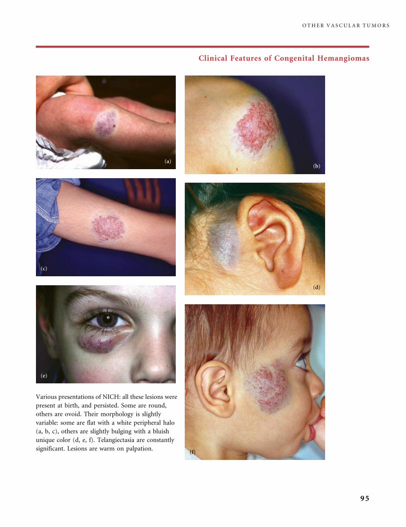
Clinical Features of Congenital Hemangiomas
Various presentations of NICH: all these lesions were
present at birth, and persisted. Some are round,
others are ovoid. Their morphology is slightly
variable: some are flat with a white peripheral halo
(a, b, c), others are slightly bulging with a bluish
unique color (d, e, f). Telangiectasia are constantly
significant. Lesions are warm on palpation.
O T H E R V A S C U L A R T U M O R S
95

Clinical Features of Congenital Hemangiomas
NICH in this boy had been present from birth on the arm. It was reminiscent of an involuting IH.
NICH are round or oval, and fairly well delineated. There is often a rim of peripheral pallor or, as in
this case, a bluish hue punctuated by more or less coarse telangiectasia. The angiogram performed for
embolization, before excision, revealed a large arterial feeder, a tumor-like capillary blush, dilated
draining veins but there was no early venous opacification.
An example of what we call the ‘‘missing links’’
(9), cases showing the possible transformation
after birth of a RICH (a) into a NICH (b)
suggesting that NICH could be a late,
intrauterine stage of a RICH involuted before
birth. The large telangiectatic bluish lump
present at birth (RICH) involuted over
a few months and left a slightly atrophic
telangiectatic residuum with a thin peripheral
pale halo reminiscent of NICH, which
persisted and was excised.
O T H E R V A S C U L A R T U M O R S
96
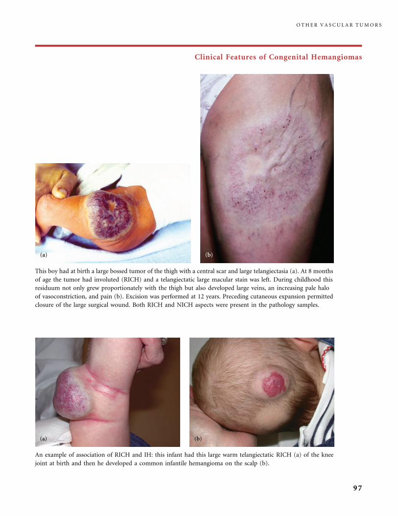
Clinical Features of Congenital Hemangiomas
This boy had at birth a large bossed tumor of the thigh with a central scar and large telangiectasia (a). At 8 months
of age the tumor had involuted (RICH) and a telangiectatic large macular stain was left. During childhood this
residuum not only grew proportionately with the thigh but also developed large veins, an increasing pale halo
of vasoconstriction, and pain (b). Excision was performed at 12 years. Preceding cutaneous expansion permitted
closure of the large surgical wound. Both RICH and NICH aspects were present in the pathology samples.
An example of association of RICH and IH: this infant had this large warm telangiectatic RICH (a) of the knee
joint at birth and then he developed a common infantile hemangioma on the scalp (b).
O T H E R V A S C U L A R T U M O R S
97
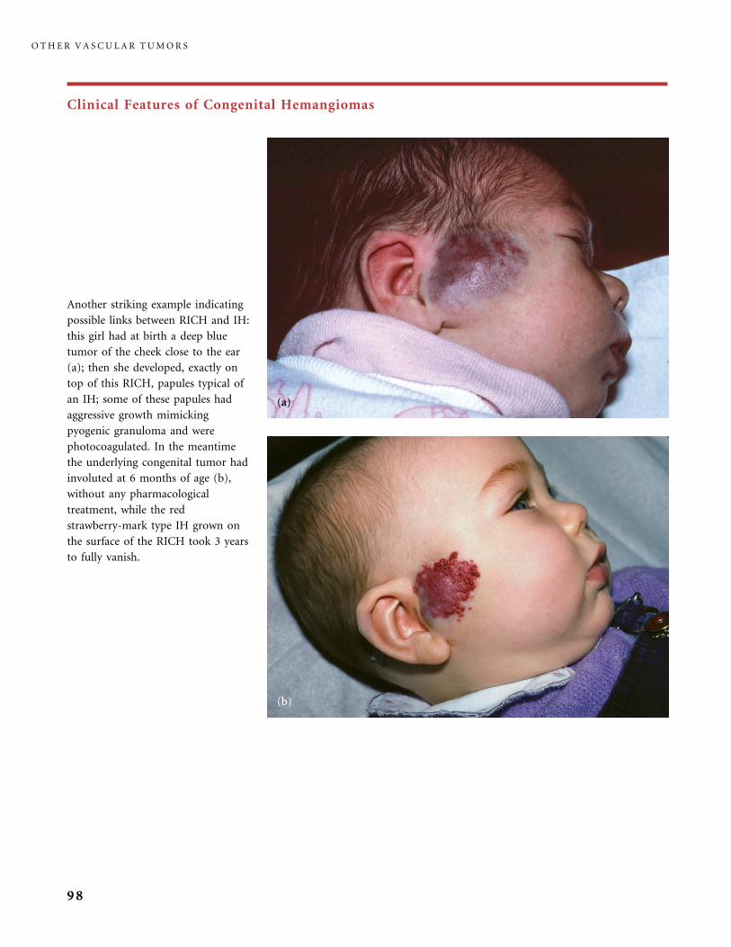
Clinical Features of Congenital Hemangiomas
Another striking example indicating
possible links between RICH and IH:
this girl had at birth a deep blue
tumor of the cheek close to the ear
(a); then she developed, exactly on
top of this RICH, papules typical of
an IH; some of these papules had
aggressive growth mimicking
pyogenic granuloma and were
photocoagulated. In the meantime
the underlying congenital tumor had
involuted at 6 months of age (b),
without any pharmacological
treatment, while the red
strawberry-mark type IH grown on
the surface of the RICH took 3 years
to fully vanish.
O T H E R V A S C U L A R T U M O R S
98
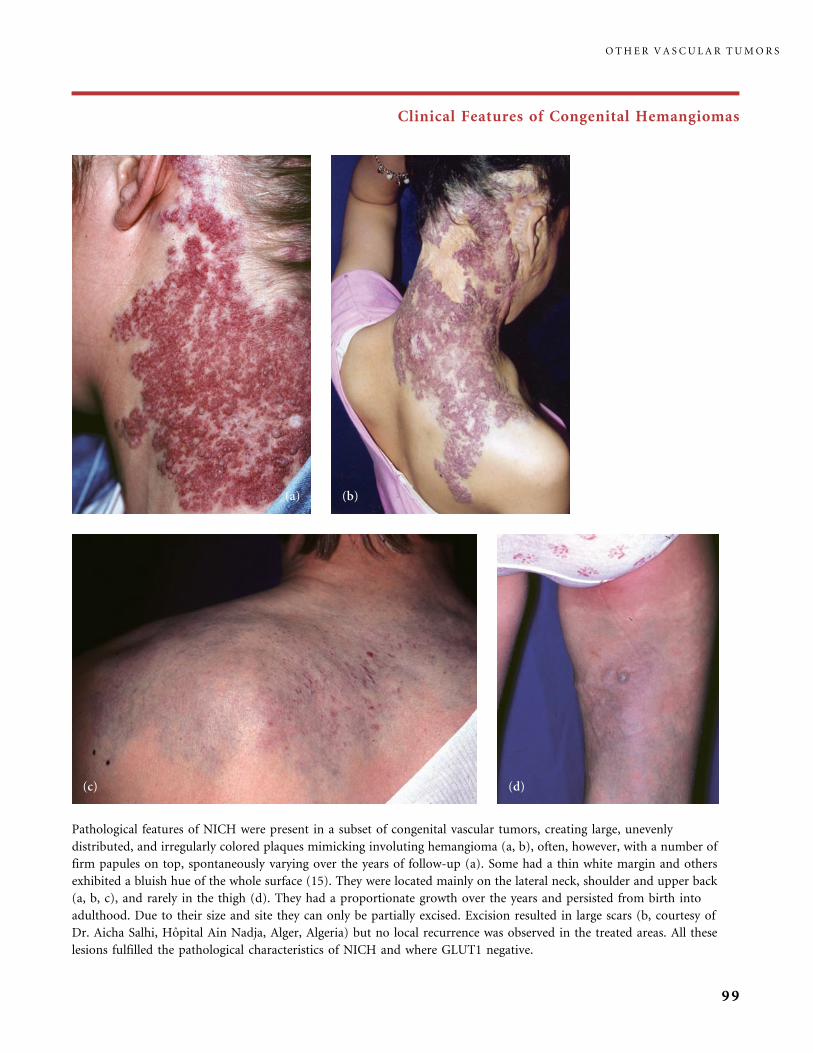
Clinical Features of Congenital Hemangiomas
Pathological features of NICH were present in a subset of congenital vascular tumors, creating large, unevenly
distributed, and irregularly colored plaques mimicking involuting hemangioma (a, b), often, however, with a number of
firm papules on top, spontaneously varying over the years of follow-up (a). Some had a thin white margin and others
exhibited a bluish hue of the whole surface (15). They were located mainly on the lateral neck, shoulder and upper back
(a, b, c), and rarely in the thigh (d). They had a proportionate growth over the years and persisted from birth into
adulthood. Due to their size and site they can only be partially excised. Excision resulted in large scars (b, courtesy of
Dr. Aicha Salhi, Hopital Ain Nadja, Alger, Algeria) but no local recurrence was observed in the treated areas. All these
lesions fulfilled the pathological characteristics of NICH and where GLUT1 negative.
O T H E R V A S C U L A R T U M O R S
99
References
1 Agesta N, Boralevi F, Sarlangue J, Vergnes P, Grenier N, Leaute-Labreze C.
Life-threatening hemorrhage of a congenital haemangioma. Acta Paediatr 2003; 92:
1216�18.
2 Berenguer B, Mulliken JB, Enjolras O, Boon LM, Wassef M, Josset P, Burrows PE, Perez-
Atayde AR, Kozakewich HPW. Rapidly involuting congenital hemangioma: clinical and
histopathologic features. Pediatr Dev Biol 2003; 6: 495�610.
3 Boon LM, Enjolras O, Mulliken JB. Congenital hemangioma: evidence for accelerated
regression. J Pediatr 1996; 128: 329�35.
4 Bulas DI, Johnson D, Allen JF, Kapur S. Fetal hemangioma. Sonographic and color flow
Doppler findings. J Ultrasound Med 1992; 11: 499�501.
5 Enjolras O, Mulliken JB, Boon LM, Wassef M, Kozakewich HP, Burrows PE.
Noninvoluting congenital hemangioma: a rare cutaneous vascular anomaly; Plast
Reconstr Surg 2001; 107: 1647�54.
6 Enjolras O. Hemangiomes congenitaux. Ann Dermatol Venereol 2003; 130: 367�71.
7 Konez O, Burrows PE, Mulliken JB, Fishman SJ, Kozakewich HP. Angiographic features of
rapidly involuting congenital hemangioma RICH. Pediatr Radiol 2003; 33: 15�19.
8 Marler JJ, Fishman SJ, Upton J, Burrows PE, Paltiel HJ, Jennings RW, Mulliken JB.
Prenatal diagnosis of vascular anomalies. J Pediatr Surg 2002; 37: 318�26.
9 Mulliken JB, Enjolras O. Congenital hemangiomas and infantile hemangioma: missing
links. J Am Acad Dermatol 2004; 50: 875�82.
10 North PE, Waner M, Mizeracki A, Mihm MC Jr., GLUT1: a newly discovered immuno-
histochemical marker for juvenile hemangioma. Hum Pathol 2000; 31: 11�22.
11 North PE, Waner M, James CA, Mizeracki A, Frieden IJ, Mihm MC Jr., Congenital
nonprogressive hemangioma. A distinct clinicopathologic entity unlike infantile
hemangioma. Arch Dermatol 2001; 137: 1607�20.
12 Rogers M, Lam A, Fischer G. Sonographic findings in a series of Rapidly Involuting
Congenital Hemangiomas (RICH). Pediatric Dermatol 2002; 19: 5�11.
13 Shiraishi H, Nakamura M, Ichihashi K. MRI in a fetus with a giant neck hemangioma:
a case report. Prenat Diagn 2000; 20: 1004�7.
14 Viora E, Grassi PP, Comoglio F, Bastonero S, Campogrande M. Ultrasonic detection of
fetal cranio-facial hemangioma: case report and review of the literature. Ultrasound Obstet
Gynecol 2000; 15: 431�4.
15 Wassef M, Salhi A, Kozakewich HPW, Breviere GM, Mulliken JB, Enjolras O. Atypical
cervical NICH: report of 11 cases. Communication 15th Workshop ISSVA, Wellington
(NZ), 22�25 Feb 2005.
O T H E R V A S C U L A R T U M O R S
100
Tufted Angioma, Kaposiform Hemangioendothelioma,Kasabach�Merritt Phenomenon (KMP)
TUFTED ANGIOMA
Wilson-Jones and Orkin in 1989 described a distinctive cutaneous lesion as ‘‘tufted
angioma’’ (TA), because of the ‘‘tufts of hypertrophied endothelial cells’’ scattered
through the whole dermis ‘‘in a cannonball distribution’’ (38). These aggregates of
endothelial cells are more prominent in the middle and lower part of the dermis,
going down to the fat (28). It is clear today that TA is identical to ‘‘angioblastoma
of Nakagawa’’ in the Japanese literature (4, 17, 26).
TA is congenital or acquired usually before 5 years of age. The same
pathological features characterize congenital, infantile or ‘‘acquired’’ TA, as well as
TA associated with Kasabach�Merritt phenomenon (KMP) (8�10).
TA seems more common in the limbs (16, 39). Herron and colleagues (16)
stressed the various clinical patterns and course in infancy. TA is often tender to
touch to a variable degree, and it may change its size and shape over time. Pink,
reddish or brownish, single or multiple, macules or variably infiltrated plaques,
some with hyperhidrosis or hypertrichosis, or tiny red papules on the surface, are
observed. In some infants TA appears as a congenital lump with a blanching halo;
this uncommon aspect is difficult to differentiate from RICH, thus requiring
a biopsy.
Although some of these vascular tumors spontaneously regress in about 6 to
24 months, a majority persists indefinitely, becoming more or less indurated
and slightly painful (30). TA may worsen during pregnancy in our experience.
The platelet-trapping syndrome, KMP, sometimes engrafts on TA, exactly like on
kaposiform hemangioendothelioma (see below).
Treatment
No treatment is really satisfactory. Topical corticosteroid, systemic corticosteroid,
intralesional interferon alpha 2 or systemic interferon alpha and laser treatments
gave both encouraging results and failure (6, 22, 27, 31, 37).
KAPOSIFORM HEMANGIOENDOTHELIOMA
Zukerberg and coworkers (40) used the term ‘‘kaposiform hemangioendothelioma
of infancy and childhood’’ for a tumor that could be mistaken for Kaposi sarcoma.
This lesion was also called ‘‘hemangioma with Kaposi sarcoma features’’ (24)
T U F T E D A N G I O M A , K A P O S I F O R M H E M A N G I O E N D O T H E L I O M A , K A S A B A C H M E R R I T T P H E N O M E N O N ( K M P )
101
and ‘‘Kaposi-like infantile hemangioendothelioma’’ of the retroperitoneum (32).
The lymphatic nature was long discussed based on pathological features (the
presence of lymphatic channels). In 2005, Debelenko et al. demonstrated that
the lymphatic vessels present in kaposiform hemangioendothelioma (KHE) and
the spindled neoplastic cells exhibit distinct staining for the lymphatic marker
D2 40, suggesting lymphothelial differentiation of the proliferation (5).
KHE is aggressive: it permeates soft tissues, muscles, and even bones. There
is no report of distant metastatic disease but perinodal infiltration or involvement
of the subcapsular sinus in a regional lymph node was observed (18).
Skin location of KHE is uncommon: it develops in skin as a reddish tender
plaque or a conglomerate of nodules and macules, with a chronic course
(19). Involution rarely occurs. Visceral KHE affects the neck, mediastinum
and thymus, or retroperitoneum. These visceral forms are massively infiltrative
and usually platelet trapping, and thrombocytopenia engraft on these KHE: the
KMP, the identical hematological disorder occurring with TA (see above and
below). TA without KMP is not rare, and conversely cutaneous KHE without
KMP is exceptional. Based on pathology overlap is commonly observed in
superficial cases of KMP, with tumors exhibiting gradation between TA and KHE.
These two tumors are now considered to be of the same pathological spectrum
and closely related if not identical (8, 18, 29, 36). Considering the visceral KMP,
including the retroperitoneal location, all reported cases were linked to KHE,
except one case demonstrating overlapping pathological features (KHE þ TA) in
the retroperitoneal tumor (2).
Treatment
There are no clear guidelines for the treatment of KHE without KMP. Gluco-
corticosteroids, vincristine, interferon alpha 2 a or 2b or excision may be indicated.
KASABACH�MERRITT PHENOMENON
Since its description by Kasabach and Merritt in 1940, the association of a large
vascular tumor and thrombocytopenia has been called ‘‘Kasabach�Merritt
syndrome’’ (KMS) or ‘‘Kasabach�Merritt phenomenon’’ (KMP). It was long
believed to be a complication of infantile hemangioma. However, the original
microscopic report specified that there were: ‘‘spindle-shaped cells supported by
a delicate fibrillar stroma.’’ It is now established that KMP occurs with distinct
vascular tumors, KHE, and TA (8�10, 14, 29, 34, 40).
KHE and TA can be congenital or appear after birth, and KMP develops
at birth or in infancy mainly before 5 months of age. Thrombocytopenia is
O T H E R V A S C U L A R T U M O R S
102
profound (platelets rapidly fall to less than 10000/mm3). There is anemia,
low fibrinogen, and elevated D-dimers.
KMP with KHE and TA has identical clinical appearance. KMP developed
on a previously biopsy-proven TA is not less severe in our experience: the tender
tumor becomes larger, purple or purplish-blue, often with a shiny smooth fragile
surface, and purpura comes out. The sudden development of KMP or the sudden
worsening of a minor pre-existing skin lesion is striking. After cure of the
hematologic anomalies, three types of residual lesions have been documented (10).
Type 1 is a ‘‘pseudo port-wine stain.’’ Type II is ‘‘red telangiectatic streaks
and swelling.’’ Type III is a ‘‘firm irregular subcutaneous lesion’’ or a firm
‘‘sclerodermiform infiltration’’ appraised by palpation, or ‘‘a deep infiltration’’ as
demonstrated by CT or MRI. All these lesions are not fixed: they slightly modify
their presentation over the years and they occasionally ache, requiring low doses
of aspirin to soothe the pain.
Nosology
There is a tendency in the literature to use the term Kasabach�Merritt syndrome
(KMS) or KMP for various diseases with associated hematological and coagula-
tion disorder, e.g. coagulation disorder associated with malignancies (infantile
fibrosarcoma, angiosarcoma), and the chronic coagulopathy associated with an
extensive venous or lymphatic�venous malformation (see page 170 and Table 15
p. 171). This is confusing: the pathological background and prognosis are
very different, and there is a more problematic issue: their treatments are also
completely different. Such an extension of the use of the label KMS or KMP
is inappropriate and dangerous for the management of these patients.
Treatment
Excision, when possible, is curative (7, 33). Heparin is contraindicated. Platelet
infusions may boost both the hematologic phenomenon and growth of the tumor
(21). Platelet infusions must not be prescribed because of a very low platelet count;
they are only indicated in case of life-threatening hemorrhage, or immediately
before the resection of the tumor, when surgical treatment is feasible. Platelets are
rapidly entrapped within the tumor and they are destroyed. Pharmacological
therapy should be given promptly. However, large variations in the platelet counts
often occur during the first weeks or months of treatment: this is not an indication
for more aggressive treatment with combinations of drugs (1) if no life-threatening
symptom emerges. KMP remains a life-threatening disease, although the prognosis
has improved. From a review of cases it appeared to us that more deaths resulted
T U F T E D A N G I O M A , K A P O S I F O R M H E M A N G I O E N D O T H E L I O M A , K A S A B A C H M E R R I T T P H E N O M E N O N ( K M P )
103
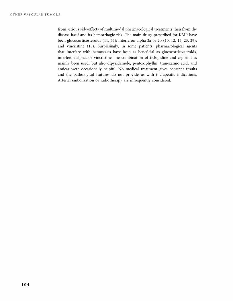
from serious side-effects of multimodal pharmacological treatments than from the
disease itself and its hemorrhagic risk. The main drugs prescribed for KMP have
been glucocorticosteroids (11, 35); interferon alpha 2a or 2b (10, 12, 13, 23, 29);
and vincristine (15). Surprisingly, in some patients, pharmacological agents
that interfere with hemostasis have been as beneficial as glucocorticosteroids,
interferon alpha, or vincristine; the combination of ticlopidine and aspirin has
mainly been used, but also dipyridamole, pentoxiphyllin, tranexamic acid, and
amicar were occasionally helpful. No medical treatment gives constant results
and the pathological features do not provide us with therapeutic indications.
Arterial embolization or radiotherapy are infrequently considered.
O T H E R V A S C U L A R T U M O R S
104
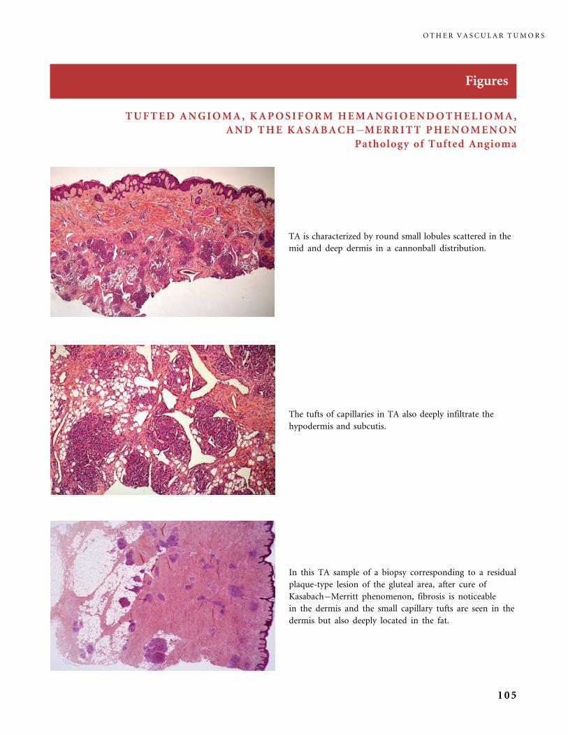
Figures
TUFTED ANGIOMA, KAPOSIFORM HEMANGIOENDOTHELIOMA,
AND THE KASABACH�MERRITT PHENOMENON
Pathology of Tufted Angioma
In this TA sample of a biopsy corresponding to a residual
plaque-type lesion of the gluteal area, after cure of
Kasabach�Merritt phenomenon, fibrosis is noticeable
in the dermis and the small capillary tufts are seen in the
dermis but also deeply located in the fat.
TA is characterized by round small lobules scattered in the
mid and deep dermis in a cannonball distribution.
The tufts of capillaries in TA also deeply infiltrate the
hypodermis and subcutis.
O T H E R V A S C U L A R T U M O R S
105
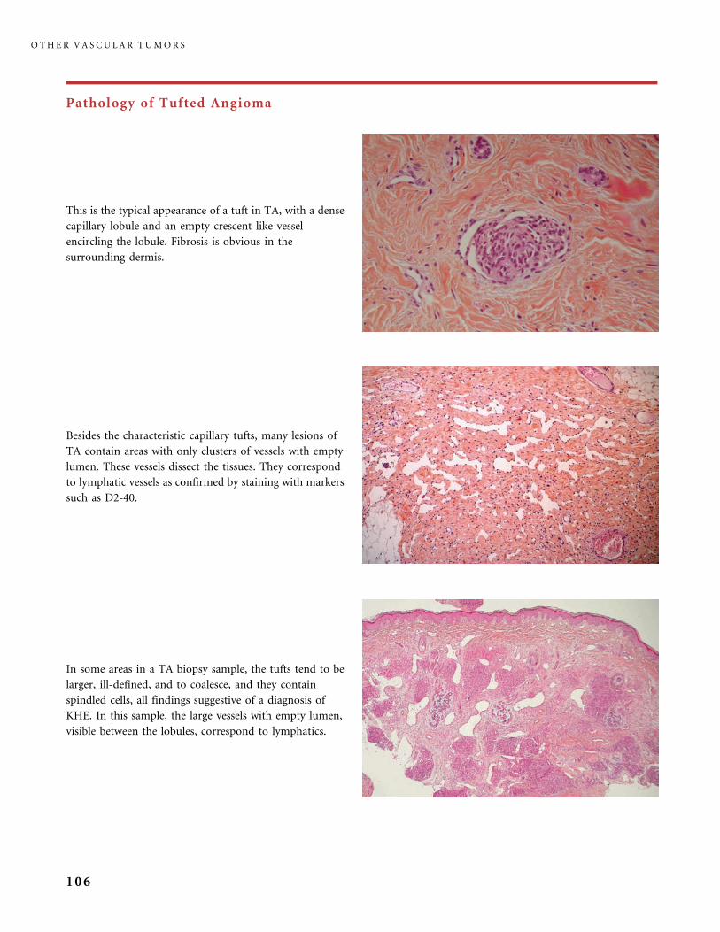
Pathology of Tufted Angioma
In some areas in a TA biopsy sample, the tufts tend to be
larger, ill-defined, and to coalesce, and they contain
spindled cells, all findings suggestive of a diagnosis of
KHE. In this sample, the large vessels with empty lumen,
visible between the lobules, correspond to lymphatics.
This is the typical appearance of a tuft in TA, with a dense
capillary lobule and an empty crescent-like vessel
encircling the lobule. Fibrosis is obvious in the
surrounding dermis.
Besides the characteristic capillary tufts, many lesions of
TA contain areas with only clusters of vessels with empty
lumen. These vessels dissect the tissues. They correspond
to lymphatic vessels as confirmed by staining with markers
such as D2-40.
O T H E R V A S C U L A R T U M O R S
106
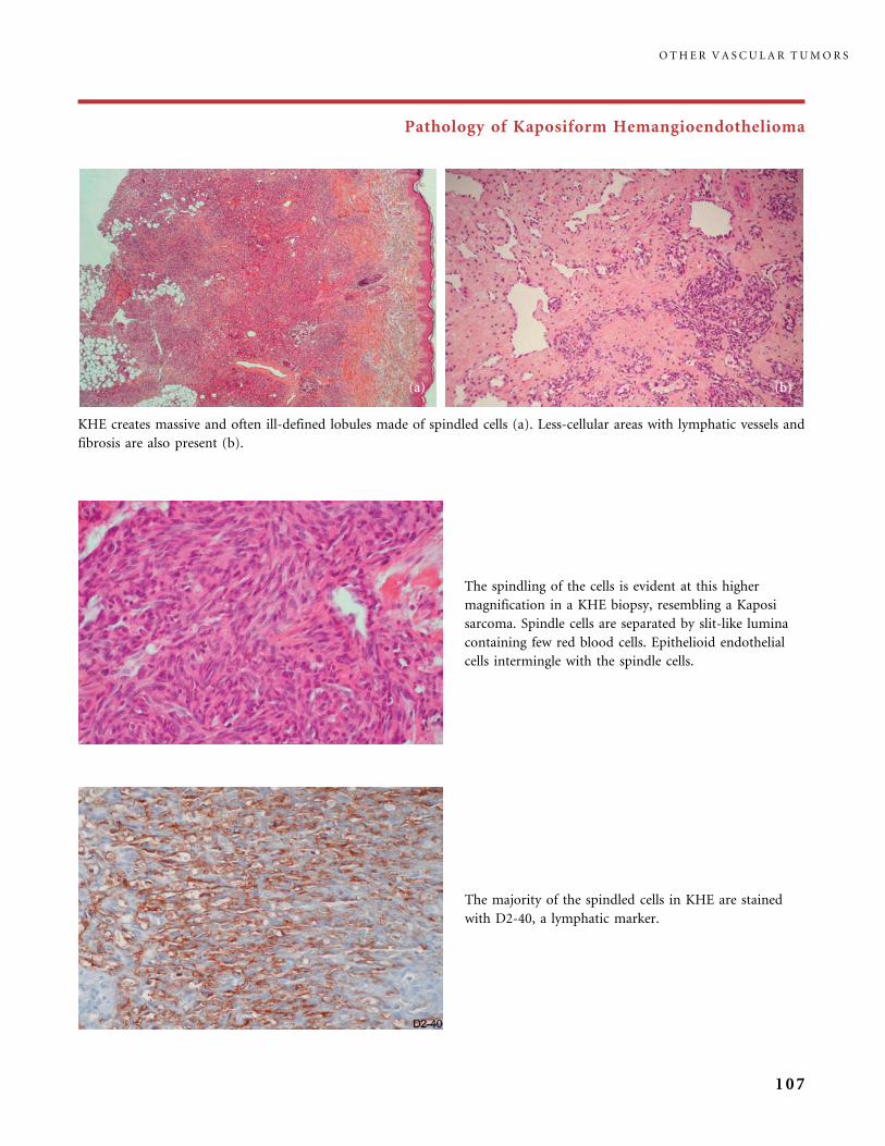
Pathology of Kaposiform Hemangioendothelioma
The spindling of the cells is evident at this higher
magnification in a KHE biopsy, resembling a Kaposi
sarcoma. Spindle cells are separated by slit-like lumina
containing few red blood cells. Epithelioid endothelial
cells intermingle with the spindle cells.
KHE creates massive and often ill-defined lobules made of spindled cells (a). Less-cellular areas with lymphatic vessels and
fibrosis are also present (b).
The majority of the spindled cells in KHE are stained
with D2-40, a lymphatic marker.
O T H E R V A S C U L A R T U M O R S
107

Pathology of Kaposiform Hemangioendothelioma
Microthrombi are detected in cellular areas.
One can observe better-defined lobules of spindled cells in
KHE, with surrounding fibrosis, a pattern reminiscent
of 2TA.
The infiltrating cells may permeate and invade a large
lymphatic channel.
O T H E R V A S C U L A R T U M O R S
108
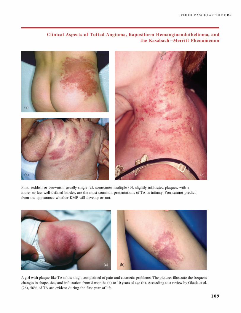
Clinical Aspects of Tufted Angioma, Kaposiform Hemangioendothelioma, and
the Kasabach�Merritt Phenomenon
Pink, reddish or brownish, usually single (a), sometimes multiple (b), slightly infiltrated plaques, with a
more- or less-well-defined border, are the most common presentations of TA in infancy. You cannot predict
from the appearance whether KMP will develop or not.
A girl with plaque-like TA of the thigh complained of pain and cosmetic problems. The pictures illustrate the frequent
changes in shape, size, and infiltration from 8 months (a) to 10 years of age (b). According to a review by Okada et al.
(26), 56% of TA are evident during the first year of life.
O T H E R V A S C U L A R T U M O R S
109
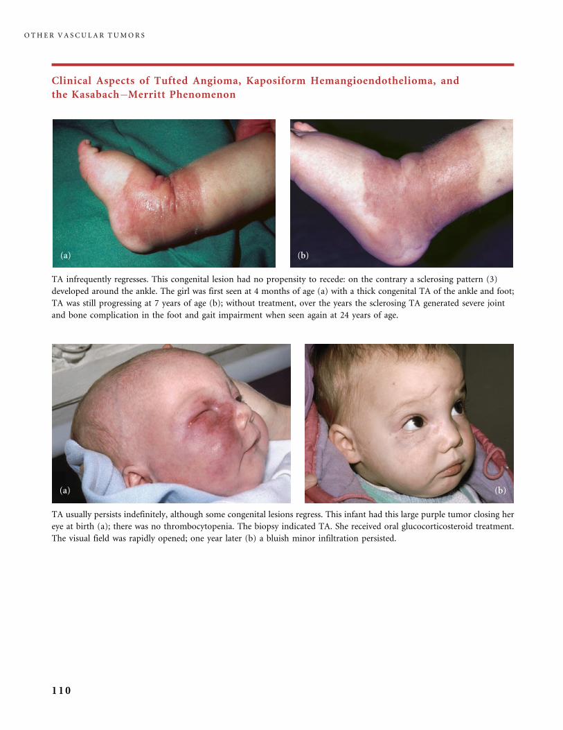
Clinical Aspects of Tufted Angioma, Kaposiform Hemangioendothelioma, and
the Kasabach�Merritt Phenomenon
TA usually persists indefinitely, although some congenital lesions regress. This infant had this large purple tumor closing her
eye at birth (a); there was no thrombocytopenia. The biopsy indicated TA. She received oral glucocorticosteroid treatment.
The visual field was rapidly opened; one year later (b) a bluish minor infiltration persisted.
TA infrequently regresses. This congenital lesion had no propensity to recede: on the contrary a sclerosing pattern (3)
developed around the ankle. The girl was first seen at 4 months of age (a) with a thick congenital TA of the ankle and foot;
TA was still progressing at 7 years of age (b); without treatment, over the years the sclerosing TA generated severe joint
and bone complication in the foot and gait impairment when seen again at 24 years of age.
O T H E R V A S C U L A R T U M O R S
110

Clinical Aspects of Tufted Angioma, Kaposiform Hemangioendothelioma, and
the Kasabach�Merritt Phenomenon
KHE has long been known possibly to grow on a large
lymphatic malformation, as in this infant who never
developed KMP (40).
This congenital, biopsy-proven KHE of the leg was slowly
enlarging and obviously painful on palpation in this infant.
KHE usually presents as a reddish or purplish-blue tender
plaque with an ill-defined border, and is clinically difficult
to differentiate from TA (20).
A large congenital plaque-type, biopsy-proven KHE
infiltrates the dorsum and lateral aspect of the hand. It first
slightly progressed to a more important infiltration in the
center. Then spontaneous involution occurred, leaving a
minor pink residuum.
O T H E R V A S C U L A R T U M O R S
111

Clinical Aspects of Tufted Angioma, Kaposiform Hemangioendothelioma, and
the Kasabach�Merritt Phenomenon
KMP, the severe platelet trapping phenomenon within the
pre-existing tumor, suddenly modified the clinical aspects
of a lesion described as a minor red infiltrated plaque,
present at birth, in a twin boy hospitalized in the neonatal
unit because of severe congenital cardiac ventricular septal
malformation. Purpura appeared on the arm, distant to
the tumor. The KMP was cured with vincristine treatment
in association with pentoxyfillin, and the infant could have
the cardiac surgery.
This large infiltrating plaque and lump, covering the full
right buttock, was of a light purple color with a single blue
nodule, and some purpura in the periphery; it had
enlarged from birth; at this stage the biopsy evidenced
KHE and the platelet count was subnormal; but a few days
later a sudden drop of the platelet count confirmed the
diagnosis of KMP on KHE.
This massive congenital tumor with KMP (courtesy of
Dr. Paul Rieu, St. Radboud Hospital, Nijmegen,
The Netherlands) had pathological features of TA on
the first biopsy and then aspects of both TA and KHE
depending on the biopsy sections. Various
pharmacological treatments were tried over 4 years,
with poor response and local severe deterioration, and
finally the boy had amputation at the hip level.
O T H E R V A S C U L A R T U M O R S
112
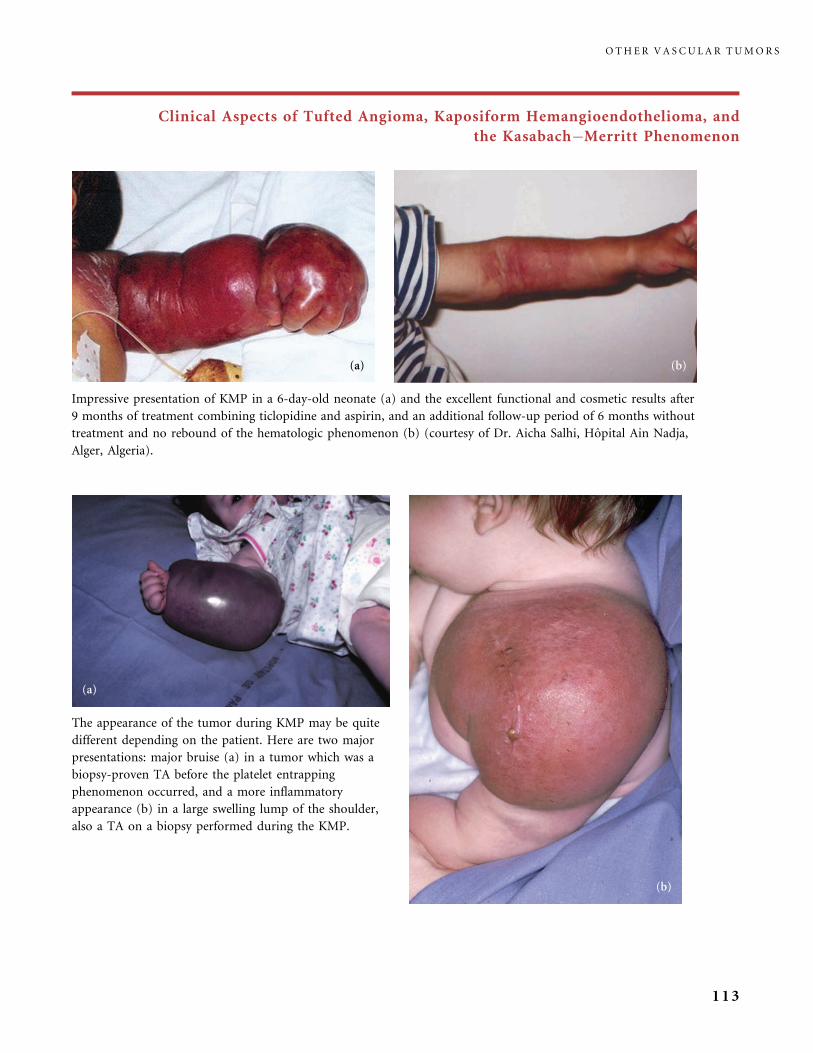
Clinical Aspects of Tufted Angioma, Kaposiform Hemangioendothelioma, and
the Kasabach�Merritt Phenomenon
The appearance of the tumor during KMP may be quite
different depending on the patient. Here are two major
presentations: major bruise (a) in a tumor which was a
biopsy-proven TA before the platelet entrapping
phenomenon occurred, and a more inflammatory
appearance (b) in a large swelling lump of the shoulder,
also a TA on a biopsy performed during the KMP.
Impressive presentation of KMP in a 6-day-old neonate (a) and the excellent functional and cosmetic results after
9 months of treatment combining ticlopidine and aspirin, and an additional follow-up period of 6 months without
treatment and no rebound of the hematologic phenomenon (b) (courtesy of Dr. Aicha Salhi, Hopital Ain Nadja,
Alger, Algeria).
O T H E R V A S C U L A R T U M O R S
113

Clinical Aspects of Tufted Angioma, Kaposiform Hemangioendothelioma, and
the Kasabach�Merritt Phenomenon
The thrombocytopenia and bulk of the tumor in this boy
(a) affected with KMP was controlled after multiple
therapeutic approaches including glucocorticosteroids and
ticlopidine plus aspirin, and he also received radiotherapy.
Years later, a slight ‘‘fibrosis’’ persisted and made the ankle
thicker (b): this was considered to be a sequela of radiation
therapy until the biopsy indicated residual TA (type III
residuum of the KMP tumor).
O T H E R V A S C U L A R T U M O R S
114

Clinical Aspects of Tufted Angioma, Kaposiform Hemangioendothelioma, and
the Kasabach�Merritt Phenomenon
This massive congenital tumor of the nape and upper back with thrombocytopenia was present at birth (a);
on MRI it permeated the muscles. Prednisone (5 mg/kg/day) did not improve the platelet count. The KMP was
controlled using vincristine treatment. At 7 years of age, the residual lesion was a pseudocapillary malformation
(type I residuum of KMP) and MRI showed only minor signal modification of the muscles of the nape.
However, comparing the appearance of the nape at 5 years (b) and 7 years (c) it appeared that a fibrotic
muscular band had developed. Although the child complained of intermittent pain and weakness of the neck,
neurological examination remained normal during the 7 years of follow-up.
O T H E R V A S C U L A R T U M O R S
115
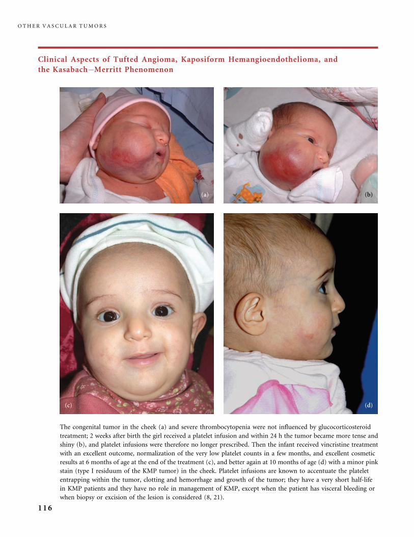
Clinical Aspects of Tufted Angioma, Kaposiform Hemangioendothelioma, and
the Kasabach�Merritt Phenomenon
The congenital tumor in the cheek (a) and severe thrombocytopenia were not influenced by glucocorticosteroid
treatment; 2 weeks after birth the girl received a platelet infusion and within 24 h the tumor became more tense and
shiny (b), and platelet infusions were therefore no longer prescribed. Then the infant received vincristine treatment
with an excellent outcome, normalization of the very low platelet counts in a few months, and excellent cosmetic
results at 6 months of age at the end of the treatment (c), and better again at 10 months of age (d) with a minor pink
stain (type I residuum of the KMP tumor) in the cheek. Platelet infusions are known to accentuate the platelet
entrapping within the tumor, clotting and hemorrhage and growth of the tumor; they have a very short half-life
in KMP patients and they have no role in management of KMP, except when the patient has visceral bleeding or
when biopsy or excision of the lesion is considered (8, 21).
O T H E R V A S C U L A R T U M O R S
116

Clinical Aspects of Tufted Angioma, Kaposiform Hemangioendothelioma, and
the Kasabach�Merritt Phenomenon
A congenital purple tumor with a double contour was
located along the nose in the right cheek at birth. It then
slowly enlarged to create a large mauve mass displacing the
lips commissure, which was present when the infant was
brought to us for diagnostic advice; at that stage (a) the
platelet count was very low and KMP was diagnosed.
Vincristine treatment was very effective. Three years after
the end of treatment, a residual pink infiltration persisted
in the lower part of the cheek and nasolabial fold (type III
residuum of KMP), but it was still slowly improving (b).
O T H E R V A S C U L A R T U M O R S
117

Clinical Aspects of Tufted Angioma, Kaposiform Hemangioendothelioma, and
the Kasabach�Merritt Phenomenon
This massive telangiectatic tumor of the right arm and back (a) had not responded to the pharmacological treatments used
in infancy. When the girl was seen she had been without treatment for years; she was in a poor general condition, had a
severe thrombocytopenia, was bleeding easily, also had lymphatic fluid oozing from vesicles on the lateral aspect of the
chest. Among complications, she endured pain, cardiac failure, and scoliosis, and was unable to go to school. Combined
ticlopidine and aspirin treatment was initiated (she had never received this before) and she had a dramatic clinical and
hematological improvement within a few months (b). The good results were maintained with the same treatment sustained
over the next 3 years. Residual lesions in the lateral neck and chest were slightly infiltrated red stains (type I residuum (10))
clinically reminiscent of TA (c); on the trunk telangiectasia were observed as type II residuum of KMP.
O T H E R V A S C U L A R T U M O R S
118
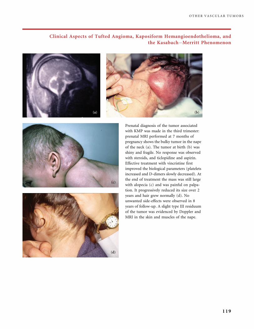
Clinical Aspects of Tufted Angioma, Kaposiform Hemangioendothelioma, and
the Kasabach�Merritt Phenomenon
Prenatal diagnosis of the tumor associated
with KMP was made in the third trimester:
prenatal MRI performed at 7 months of
pregnancy shows the bulky tumor in the nape
of the neck (a). The tumor at birth (b) was
shiny and fragile. No response was observed
with steroids, and ticlopidine and aspirin.
Effective treatment with vincristine first
improved the biological parameters (platelets
increased and D-dimers slowly decreased). At
the end of treatment the mass was still large
with alopecia (c) and was painful on palpa-
tion. It progressively reduced its size over 2
years and hair grew normally (d). No
unwanted side-effects were observed in 8
years of follow-up. A slight type III residuum
of the tumor was evidenced by Doppler and
MRI in the skin and muscles of the nape.
O T H E R V A S C U L A R T U M O R S
119

Clinical Aspects of Tufted Angioma, Kaposiform Hemangioendothelioma, and
the Kasabach�Merritt Phenomenon
This adolescent was treated soon after birth for a congenital
KMP with a large bumped thoracic tumor. She received
glucocorticosteroids, ticlopidine and aspirin, radiotherapy, and
finally remained under ticlopidine and aspirin treatment for
years. We saw her when she was 13 years old because of
recurrence of thrombocytopenia after cessation of her treatment.
Her lesion was a deeply infiltrated plaque with well-defined
margins on the lateral thorax. Long telangiectasia were running
over her torso. There was also a newly developed bump under
normal skin in her back and bony lytic lesions were discovered in
her ribs. A biopsy was performed to rule out radiation-induced
angiosarcoma, and it indicated KHE. Platelet counts were
between 5000 and 15000. She was treated consecutively with
vincristine, interferon alpha 2b, thalidomide, and again
ticlopidine and aspirin, and topical imiquimod, without any
further clinical and hematological improvement over the next
3 years. Relapse of KMP after a long interval is rare (25).
O T H E R V A S C U L A R T U M O R S
120
References
1 Blei F, Karp N, Rofsky N, Rosen R, Greco MA. Successful multimodal therapy for
kaposiform hemangioendothelioma complicated by Kasabach�Merritt phenomenon,
case report and review of the literature. Pediatr Hematol Oncol 1998; 15: 295�305.
2 Brasansac D, Janic D, Boricic I, Jovanovic N, Dokamanovic L. Retroperitoneal kaposiform
hemangioendothelioma with tufted angioma like features in an infant with
Kasabach�Merritt syndrome. Pathol Int 2003; 53: 627�31.
3 Catteau B, Enjolras O, Delaporte E, Friedel J, Breviere G, Wassef M, Lecomte-Houcke M,
Piette F, Bergoend H. Angiome en touffes sclerosant. Ann Dermatol Venereol 1998; 125:
682�7.
4 Cho KH, Kim SH, Park KC, Lee AY, Song KY, Chi JG, Lee YS, Kim KJ. Angioblastoma
(Nakagawa) � is it the same as tufted angioma? Clin Exp Dermatol 1991; 16: 110�13.
5 Debelenko, Perz-Atayde AR, Mulliken JB, Liang MG, Archibald TH, Kozakewich HP.
D2-40 immunohistochemical analysis of pediatric vascular tumors reveals positivity in
kaposiform hemangioendothelioma. Mod Pathol 2005; 18: 1454�60.
6 Dewerdt S, Callens A, Machet L, Grangeponte MC, Vaillant L, Lorette G. Acquired tufted
angioma in an adult: failure of pulsed dye laser therapy. Ann Dermatol Venereol 1998; 125:
47�9.
7 Drolet BA, Scott LA, Esterly NB, Gosain AK. Early surgical intervention in a patient with
Kasabach�Merritt phenomenon. J Pediatr 2001; 138: 756�8.
8 Enjolras O, Wassef M, Mazoyer E, Frieden IJ, Rieu PN, Drouet L, Taieb A, Stalder JF,
Escande JP. Infants with Kasabach�Merritt syndrome do not have ‘true‘ hemangioma.
J Pediatr 1997; 130: 631�40.
9 Enjolras O, Wassef M, Dosquet Ch, Drouet L, Fortier G, Josset P, Merland JJ, Escande JP.
Syndrome de Kasabach�Merritt sur angiome en touffes congenital. Ann Dermatol
Venereol 1998; 125: 257�60.
10 Enjolras O, Mulliken JB, Wassef M, Frieden IJ, Rieu PN, Burrows PE, Salhi A, Leaute-
Labreze C, Kozakewich HP. Residual lesions after Kasabach�Merritt phenomenon in 41
patients. J Am Acad Dermatol 2000; 42: 275�9.
11 Esterly N. Kasabach�Merritt syndrome in infants. J Am Acad Dermatol 1983; 8: 504�13.
12 Ettlinger JJ, Fleming PJ, Joffe HS, Kennedy CTC. Cavernous haemangioma with
Kasabach�Merritt syndrome: treatment with alpha interferon. J R Soc Med 1996; 89:
55P�56P.
13 Ezekowitz RA, Mulliken JB, Folkman J. Interferon alfa-2a therapy for life-threatening
hemangiomas of infancy. N Engl J Med 1992; 326: 1456�63.
14 Fukunaga M, Ushigome S, Ishikawa E. Kaposiform hemangioendothelioma associated
with Kasabach�Merritt syndrome. Histopathology 1996; 28: 281�4.
15 Haisley-Royster C, Enjolras O, Frieden IJ, Garzon MD, Oranje A, Gonzalez F, Frangoul H,
LeMoine P, Prose NS, Adams D. Kasabach�Merritt phenomenon: a retrospective study
of treatment with vincristine. J Pediatr Hematol Oncol 2002; 24: 459�62.
16 Herron MD, Coffin CM, Vanderhooft SL. Tufted angioma: variability in clinical
morphology. Pediatr Dermatol 2002; 19: 394�401.
17 Igarashi M, Oh-I T, Koga M. The relationship between angioblastoma (Nakagawa) and
tufted angioma: report of four cases with angioblastoma and a literature-based
comparison of the two conditions. J Dermatol 2000; 27: 537�42.
18 Lyons LL, North PE, Mac-Moune Lai F, Stoler MH, Folpe AL, Weiss SW. Kaposiform
hemangioendothelioma. A study of 33 cases emphasizing its pathologic, immunopheno-
typic, and biologic uniqueness from juvenile hemangioma. Am J Surg Pathol 2004;
28: 559�68.
R E F E R E N C E S
121
19 Mac-Moune Lai F, To KF, Choi PC, Leung PC, Kumta SM, Yuen PP, Lam WY, Cheung
AN, Allen PW. Kaposiform hemangioendothelioma: five patients with cutaneous lesions
and long follow-up. Mod Pathol 2001; 14: 1087�92.
20 Mentzel T, Mazzoleni G, Dei Tos A, Fletcher CDM. Kaposiform hemangioendothelioma
in adults. Clinicopathologic and immunohistochemical study of three cases. Am J Clin
Pathol 1997; 108: 450�5.
21 Mulliken JB, Anupindi S, Ezekowitz RAB, Mihm MC Jr. Case 13-2004: a newborn girl
with a large cutaneous lesion, thrombocytopenia, and anemia. N Engl J Med 2004; 350:
1764�75.
22 Munn SE, Jackson JE, Russel Jones R. Tufted angioma responding to high dose systemic
steroids. Clin Exp Dermatol 1994; 19: 511�14.
23 Nako Y, Fukushima N, Igarashi T, Hoshino M, Sugiyama M, Tomomasa T, Morikawa A.
Successful interferon therapy in a neonate with life-threatening Kasabach�Merritt
syndrome. J Perinatol 1997; 17: 244�7.
24 Niedt GW, Greco MA, Wieczorek R, Blanc WA, Knowles DM. Hemangioma with
Kaposi’s sarcoma-like features: report of two cases. Pediatr Pathol 1989; 9: 567�75.
25 Ohtsuka T, Saegusa M, Yamakage A, Yamazaki S. Angioblastoma (Nakagawa) with
hyperhidrosis, and relapse after a 10-year interval. Br J Dermatol 2000; 143: 223�4.
26 Okada E, Tamura A, Ishikawa O, Miyachi Y. Tufted angioma (angioblastoma) case report
and review of 41 cases in the Japanese literature. Clin Exp Dermatol 2000; 25: 627�30.
27 Park KC, Ahn PS, Lee YS, Kim KH, Cho KH. Treatment of angioblastoma with
recombinant interferon-a2. Pediatr Dermatol 1995; 12: 184�6.
28 Requena L, Sangueza OP. Cutaneous vascular proliferation. Part II. J Am Acad Dermatol
1997; 37: 887�919.
29 Sarkar M, Mulliken JB, Kozakewich HPW, Robertson RL, Burrows PE.
Thrombocytopenic coagulopathy (Kasabach�Merritt phenomenon) is associated with
kaposiform hemangioendothelioma and not with common infantile hemangioma. Plast
Reconstr Surg 1997; 100: 1377�86.
30 Satter EK, Graham BS, Gibbs NF. Congenital tufted angioma. Pediatr Dermatol 2002; 19:
445�7.
31 Suarez SM, Pensler JM, Paller AS. Response of deep tufted angioma to interferon alfa.
J Am Acad Dermatol 1995; 33: 124�6.
32 Tsang WYW, Chan JKC. Kaposi-like hemangioendothelioma. A distinctive vascular
neoplasm of the retroperineum. Am J Surg Pathol 1991; 15: 982�9.
33 Velin P, Dupont D, Golkar A, Valla JS. Syndrome de Kasabach�Merritt neonatal gueri
par exerese chirurgicale complete de l’angiome. Arch Pediatr 1998; 5: 295�7.
34 Vin-Christian K, McCalmont TH, Frieden IJ. Kaposiform hemangioendothelioma,
an aggressive locally invasive vascular tumor that can minic hemangioma of infancy.
Arch Dermatol 1997; 133: 1573�8.
35 Wananukul S, Nuchprayoon I, Seksarun P. Treatment of Kasabach�Merritt syndrome:
a stepwise regimen of prednisolone, dipyridamole and interferon. Int J Dermatol 2003;
42: 741�8.
36 Weiss SW, Goldblum JR (eds). Enzinger and Weiss’s Soft Tissue Tumors. 4th Edn, St Louis:
Mosby; ch. 23: pp. 837�890; ch. 24: pp. 891�915, 2001.
37 Wilmer A, Katz M, Bocker T, Wollina U. Tufted angioma. Eur J Dermatol 1999; 9: 51�3.
38 Wilson-Jones E, Orkin M. Tufted angioma (angioblastoma): a benign progressive
angioma not to be confused with Kaposi’s sarcoma or low-grade angiosarcoma. J Am Acad
Dermatol 1989; 20: 214�25.
39 Wong SN, Tay YK. Tufted angioma: a report of five cases. Pediatr Dermatol 2002; 19:
388�93.
40 Zukerberg LR, Nickoloff BJ, Weiss SW. Kaposiform hemangioendothelioma of infancy
and childhood. An aggressive neoplasm associated with Kasabach�Merritt syndrome
and lymphangiomatosis. Am J Surg Pathol 1993; 17: 321�8.
O T H E R V A S C U L A R T U M O R S
122
PART III
Vascular Malformations

CHAPTER III.A
Capillary Malformations (CM)
Introduction
Capillary malformations (CM) are hemodynamically inactive, slow-flow vascular
malformations affecting the capillary network of skin and mucosa, sometimes
invading deeper underlying structures specifically in the facial area. They include
common CMs also known as ‘‘port-wine stains’’ and ‘‘telangiectasia.’’ Both can
occur as a single isolated anomaly or in association with other abnormalities. Some
are included in complex syndromes, the majority being sporadic and some being
familial. We can also include the various ‘‘angiokeratomas’’, localized, system-
atized or diffuse (angiokeratoma corporis diffusum), familial or not.
III.A.1 Common Capillary Malformations: Port-wine Stains (PWS)
Clinical Aspects
This is the most common type of vascular malformation. Typically a port wine stain
(PWS) is present at birth and persists lifelong growing proportionately. However,
rare acquired PWSs develop and progress in adolescents or adults; the possible role
of trauma has been stressed (1), and reported as Fegeler syndrome (51).
A PWS is a more- or less-extensive well-demarcated red macular stain. Local-
ized segmental PWSs are common on the face. On the face, CMs are commonly
sub-classified as lateral CM (PWS) and medial CM (also known as salmon patch).
Lateral PWSs of the face always persist whereas medial lesions usually become
lighter and some disappear, particularly those of the mid-face. Metameric
distribution occurs on the trunk and limbs; on the other hand it seems that PWSs
never spread along Blaschko lines. A stain with geographical contour on the lateral
aspect of the thigh and knee at birth usually predicts the further development of
a complex syndrome (Klippel�Trenaunay syndrome) including capillary, venous,
and lymphatic abnormalities (19, 44). Diffusely scattered PWSs all over the body
are less common, except in Proteus syndrome.
125
A PWS often has a bright red, scarlet color at birth, because of the high
neonatal hemoglobin content of skin capillaries. Then it fades over 1 or 2 months
to reach a pink or red hue. The color is the result of ectatic capillaries in the dermis
carrying more blood than small normal channels. Deficiency in perivascular
innervation has been reported (61, 62). Some PWSs in adults take on a darker
color, tissular hyperplasia gives them a cobblestone appearance: this is obvious in
some facial CMs undergoing progressive dilatation of the capillaries, an increasing
number of ectatic capillaries, and often sebaceous hyperplasia and fibrosis. A
striking nodular hyperplasia may develop in some patients, creating disfigurement
(36). According to Sanchez-Carpintero et al. (58), thickening and nodularity can,
at least in some cases, be explained by hamartomatous changes in the connective
tissue surrounding the dilated engorged capillaries: in addition to the prominent
vascular ectasia they found pilo-sebaceous abnormalities, arrector pili-type
smooth-muscle bundles, and neural and mesenchymal hamartomatous changes.
PWSs of the face, particularly those involving the cheek and lips, are sometimes
associated with hyperplasia of underlying soft tissues and bones resulting in
macrocheilia, gum hypertrophy, epulis, and bony maxilla hypertrophy in the three
planes with dental malocclusion.
Diagnosis
Diagnosis of CM/PWS is made clinically without the need for complementary
investigation. However, two vascular lesions may generate confusion: the red,
sometimes telangiectatic, congenital stain precursor of hemangioma (see the
section on Hemangioma page 53) and an AVM in a quiescent stage (stage 1 AVM)
(see the section on AVM, page 261) which requires US/Doppler duplex scan
confirmation.
Treatment
Since the 1980s the flashlamp pumped-pulsed dye laser (PDL) has been considered
the best laser system for the treatment of PWS. Modified devices enabling
longer pulse widths, longer wavelengths, and bigger spot sizes have improved PDL
efficacy (43). The use of dynamic surface cooling, reducing the risk of epidermal
damage and minimizing the pain of treatment, has permitted the application of
more-effective, higher energy fluence. Nonetheless, only one-fifth of PWSs clear
completely, although the color of the majority of treated CMs improves signif-
icantly (28, 68). Various methods have been tested to try to predict and improve
the response to laser treatment, depending on the color of the stain, depth
of vessels and size of ectatic capillaries. Spectrophotometry first gave a better
correlation between color, clinical appearance, and hemoglobin content.
Videomicroscopy permits localization of the ectatic capillary network and allowed
C A P I L L A R Y M A L F O R M A T I O N S ( C M )
126

Eubanks and McBurney (21) to correlate areas of poorer response to laser
treatment (for example the V2 facial PWS) with the type of ectasia: type 2 pattern
(rings) corresponded to ectasia of deeper horizontal plexus. High-frequency
ultrasound evaluation confirmed that thicker areas of skin with PWS respond
worse to PDL (48). Under confocal microscopy of biopsies of PWSs, decreased
nerve density, increased capillary density, and increased mean blood vessel
diameter correlated with poor response to PDL (61).
III.A.2 Capillary Malformations and Associations
We will illustrate quite common associations occurring with CM/PWS, such as
CM and nevus anemicus, CM and pigmentary disorders with the various types
of phakomatosis pigmentovascularis, the problem of CM as a marker of spinal
dysraphism, CM with hyperhidrosis due to increased eccrine glands in eccrine
angiomatous hamartoma or sudoriparous angioma.
I I I .A .2 . 1 PHAKOMATOSIS PIGMENTOVASCULARIS
This association of cutaneous vascular anomalies (CM and nevus anemicus)
and epidermal or pigmentary changes (29, 30, 57) is considered to be the result
of the ‘‘twin-spotting’’ phenomenon (30). Five types are currently described:
type I is CM and epidermal nevus; type II is CM with dermal melanocytosis
(aberrant Mongolian spots) a very common finding in skin of color; type III
is the association of CM and nevus spilus (54); type IV may combine CM and
aberrant Mongolian spots and nevus spilus; type V is the association of cutis
marmorata telangiectatica congenita and Mongolian spots (65). Nevus anemicus
can be also present in types II, III and IV. Each group is classified as A (without
another anomaly) or B (with additional abnormalities, including nevus of Ota,
KTS, and SWS (27)). Extracutaneous anomalies have also been reported.
I I I .A .2 . 2 ECCRINE ANGIOMATOUS HAMARTOMA (EAH)
This clinically heterogeneous lesion, often congenital or appearing in children (46)
is more common on the distal extremities, notably a toe or ankle. Some patients
have only a purple or bluish nodule, others have a plaque-type lesion and
some have a very large infiltrated blue-purple plaque with increased sweating,
C A P I L L A R Y M A L F O R M A T I O N S A N D A S S O C I A T I O N S
127
occasionally dampening the clothes of the trunk. The lesion is usually solitary and
often not well-defined at its edges; it hurts on palpation or contact, and in our
experience sweating is obvious if the patient feels anxious during examination.
Infiltration is variable. Color varies from yellowish to brownish or pink to bluish-
purple. Hypertrichosis can be present. The lesion grows slowly and recurs after
incomplete excision. Pain increases in adulthood and it is often because of the pain
that patients seek treatment. If surgical excision is reasonably possible, we advise
doing it during childhood, when the lesion has a reasonable size. Histopathologic
features include closely associated thin-walled irregular capillaries and abundant
eccrine glands and ducts; larger channels may be present (veins) as well as fatty
tissue, hair follicles, and a hyperplastic overlying epidermis (55).
III.A.3 Syndromic Capillary Malformations
I I I .A .3 . 1 STURGE�WEBER SYNDROME (SWS)
This systematized vascular syndrome is likely caused by a somatic mutation in the
anterior neural primordium. The ‘‘whole’’ disease includes a facial PWS always
staining the so-called V1 area (forehead and upper eyelid) and sometimes
extending further, ipsilateral vascular anomalies of the leptomeninges, and
ipsilateral ocular abnormalities. The dermis of the face is made of cells originating
from the cephalic neural crests (except endothelial cells, of mesodermal origin).
SWS is hypothesized to be the result of a very early mutational event arising in the
prosencephalic neural crest, a region providing cells to the dermis of the supra-
ocular area and nasal bud. This may arise at a time when precursors of the V1
dermis, ocular choroid and piamater are still in the anterior neural primordium,
before they start their migration to their final destination. The dermis of the V2
(maxillary) and V3 (mandibular) areas is made of cells from the mesencephalic
neural crests, cells not forming leptomeninges: this is why SWS is not linked to
a PWS occupying the V2 and V3 skin, without V1 location. However, the limits are
not strict at the boundaries of these three regions (20). At these boundaries, cells
of various origin may mix together: this explains slight variation in the limits of
the so-called V1, V2, and V3 PWSs. It is noteworthy that, although we still retain
this terminology of V1, V2, and V3 distribution of facial PWS, the sensitive
trigeminal nerve (Vth nerve) has nothing to do with the pathogenesis of facial
PWS and SWS, as was thought in the past.
Epilepsy in SWS may be devastating and it usually starts very early in
life between birth and one year. About 10% of infants with V1 PWS actually
have leptomeningeal vascular anomalies. Cognitive deficit and mental retarda-
tion, loss of developmental milestones, and motor deficit contralateral to the
C A P I L L A R Y M A L F O R M A T I O N S ( C M )
128

meningeal lesions follow the onset of epilepsy, with a correlation between
prolonged seizure and further severe developments, and motor and intellectual
deficits (Table 12).
Prevention of the first seizure by prophylactic antiepileptic treatment aims at
avoiding these severe developments (67). The seizures decrease strongly after
puberty. Ocular follow-up is also mandatory because of the risk of choroidal
vascular anomaly and of glaucoma. The PWS can be treated with the PDL after
pharmacological control of epilepsy is obtained. Surgical treatment (callosotomy,
hemispherotomy, hemispherectomy) is performed in patients with seizures that
are intractable to medication, first to end seizures, and hopefully to improve
function secondarily (37).
I I I .A .3 . 2 KLIPPEL�TRENAUNAY SYNDROME AND
RELATED SYNDROMES
At the limb and trunk level CM/PWSs occur in various clinical situations
including:
1. diffuse CMs scattered over an extremity and adjacent trunk with apparently
unsystematic spreading and no associated abnormality;
Table 12 Neurological risks associated with vascular anomalies involving the cephalic region.
Diagnosis Neurological associations Neurological consequences
Infantile hemangioma PHACE(S) syndrome: posterior
fossa malformations, arterial
intracranial anomalies
Symptom-free or seizures, ischemic
attacks, stroke
Sturge�Weber syndrome Leptomeningeal vascular malformation,
cerebral atrophy and calcifications
Epilepsy, hemiplegia, developmental delay,
mental retardation, headaches
Proteus syndrome Hemimegalencephaly, hydrocephalus,
abnormal cerebral cortex, tumors
Seizures, developmental delay, and mental
retardation
Cutis marmorata�macrocephaly
syndrome
Hemimegalencephaly, hydrocephalus Mental retardation
Ataxia telangiectasia Ataxia
HHT (Rendu�Osler�
Weber disease)
Cerebral AVM, risk of cerebral abscess
if pulmonary AVF
Headaches, bacterial emboli, and stroke
Cephalic VM Developmental venous anomalies (DVA) Symptom-free or headaches
Bonnet�Dechaume�Blanc
or Wyburn-Mason syndrome
Brain AVM Headaches, seizures, cerebral hemorrhage
Orbital LM Dural AVF, DVA Exophthalmos, conjunctival chemosis,
intracranial hypertension
HHT¼hereditary hemorrhagic telangiectasia; AVM¼arteriovenous malformation; AVF¼arteriovenous fistula; VM¼venous
malformation; LM¼lymphatic malformation.
S Y N D R O M I C C A P I L L A R Y M A L F O R M A T I O N S
129

2. PWS fully covering a limb with congenital hypertrophy of the same
extremity, with proportionate growth over the years;
3. a slow-flow combined and complex vascular malformation with CM,
varicose veins, very often LM manifesting as either lymphedema or
lymphatic vesicles or both, and progressive overgrowth in length and
girth of the affected limb: this is Klippel�Trenaunay syndrome (KTS) (7)
(Table 13).
KTS is associated with SWS in some patients. It may occur in Proteus syndrome.
In infants a geographic skin stain on the external aspect of the thigh and knee,
rapidly colonized by clear and purple lymphatic vesicles, is predictive of com-
plicated KTS, with lymphatic anomalies increasing over the years and a usually
severe progressive discrepancy in limb growth (44).
The work-up for young patients with KTS should not be invasive: we rarely
need angiographic or lymphoscintigraphic data for their management, and
an US/Doppler duplex scan, sometimes coupled with MRI, gives adequate data
on the vascular anomalies, extent of lesions, and possible associated intestinal and
urinary vascular anomalies. Patients with KTS and limb hypertrophy are not
at higher risk of Wilms tumor than the general population and thus they do
not need routine Wilms tumor screening (24). Another study based on a literature
Table 13 Clinical characteristics of two different limb complex-combined vascular malformations
with progressive overgrowth of the affected extremity.
Characteristics Klippel�Trenaunay syndrome Parkes Weber syndrome
Hemodynamics Slow-flow Fast-flow
Type of vascular anomaly CM (geographic or not),
VM (varicose veins),
LM (lymphedema,
lymphatic vesicles)
Multiple AVFs/AVM,
pseudo-CM on skin
(stage I dormant AVM),
lymphedema
Skin temperature Normal Increased
Progressive overgrowth in
length and girth
þ þ
Visceral involvement Possibly: GI tract, urinary, genitalia Rare: AVM (pelvic),
Cobb syndrome
Chronic coagulopathy with high
D-dimers and low fibrinogen
þ �
Increased cardiac output and
possibly congestive cardiac failure
� þ
Deep vein thrombosis, risk of
pulmonary embolism
þ �
Venous stasis skin alteration, ulcers þ �
Pseudo Kaposi, skin ischemic
changes, ulcers
� þ
CM¼capillary malformation; VM¼ venous malformation; LM¼ lymphatic malformation; AVM¼arteriovenous malformation;
AVF¼arteriovenous fistula.
C A P I L L A R Y M A L F O R M A T I O N S ( C M )
130
review found only one report of hemihypertrophy, Wilms tumor, and KTS
(a picture confirms the diagnosis) in 58 cases, and it concludes that routine
screening for Wilms tumor in patients with KTS is unnecessary, unless the patient
has generalized hemihypertrophy (39).
The mainstay of management of KTS in pediatric patients with KTS is
orthopedic assessment once a year, and wearing elastic garments on a lifelong
basis. Laser (FPDL or other) treatments are rarely very useful and adverse
effects are more frequent than at other sites. Epiphyseodesis is considered at
around 10 to 13 years of age, depending on the growth curve of the child, when leg
discrepancy is important (if more than 2 or 3 cm). Surgical treatment of varicose
veins is not considered before puberty, except for very enlarged marginal veins in
the thigh.
I I I .A .3 . 3 PROTEUS SYNDROME AND PROTEIFORM SYNDROMES
Proteus syndrome is a complex syndrome (34, 69). It combines asymmetric
growth of bones in a haphazard distribution, mainly in the distal extremities
(macrodactyly in digits of the hands or feet), spinal cord (creating scoliosis),
and skull (with exostoses). Subcutaneous benign tumors such as lipoma, fibroma,
and plantar collagenoma (this connective tissue nevus gives to the sole of foot, and
sometimes the palm of hand, a cerebriform aspect, and is considered as an absolute
criterion for a diagnosis of Proteus syndrome (Table 14) (8, 9)). Epidermal nevus
and vascular anomalies are also present. Hyperplasia is the most obvious
presentation of Proteus lesions, but hypoplasia is also observed: either global
hypoplasia, e.g. of an extremity, or patchy dermal hypoplasia (31). The vascular
anomalies in Proteus syndrome have been underappraised until recently:
Hoeger et al. (33) stressed the fact that they are present in 100% of patients
with Proteus syndrome and that more than one type is often identified in a single
patient. Extensive PWSs of a crimson red color at birth, macrocystic LM that
may develop suddenly and quickly, varicose veins, and complex combined
slow-flow vascular malformations of the KTS type, are all very common in Proteus
syndrome. SWS is rarely present: it may occur in a patient whose PWS is in the V1
area, in the context of Proteus syndrome (unpublished personal data). Also,
in our experience, no fast-flow anomaly with arteriovenous shunting (either
localized or of the Parkes Weber syndrome type) is observed in Proteus syndrome,
all the associated vascular anomalies being of the slow-flow type. Other reported
findings are visceral anomalies. Hemimegalencephaly and migrational disorders
seem to be the most common demonstrated brain anomaly, among a range of
other situations, but seizures and intellectual impairment have rarely been
reported (14). Benign and malignant neoplasias may affect the ovaries, testes, and
central nervous system. In the most severe cases abnormalities tend to expand
earlier in life.
S Y N D R O M I C C A P I L L A R Y M A L F O R M A T I O N S
131
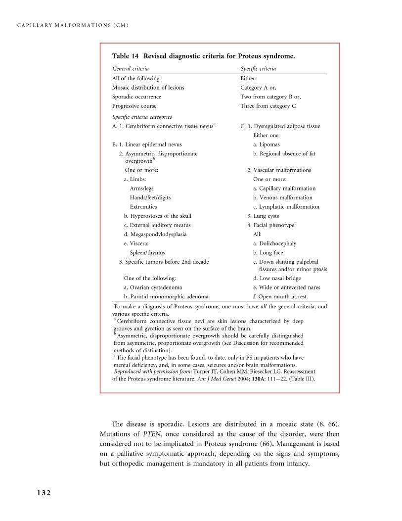
The disease is sporadic. Lesions are distributed in a mosaic state (8, 66).
Mutations of PTEN, once considered as the cause of the disorder, were then
considered not to be implicated in Proteus syndrome (66). Management is based
on a palliative symptomatic approach, depending on the signs and symptoms,
but orthopedic management is mandatory in all patients from infancy.
Table 14 Revised diagnostic criteria for Proteus syndrome.
General criteria Specific criteria
All of the following: Either:
Mosaic distribution of lesions Category A or,
Sporadic occurrence Two from category B or,
Progressive course Three from category C
Specific criteria categories
A. 1. Cerebriform connective tissue nevusa C. 1. Dysregulated adipose tissue
Either one:
B. 1. Linear epidermal nevus a. Lipomas
2. Asymmetric, disproportionate
overgrowthb
b. Regional absence of fat
One or more: 2. Vascular malformations
a. Limbs: One or more:
Arms/legs a. Capillary malformation
Hands/feet/digits b. Venous malformation
Extremities c. Lymphatic malformation
b. Hyperostoses of the skull 3. Lung cysts
c. External auditory meatus 4. Facial phenotypec
d. Megaspondylodysplasia All:
e. Viscera: a. Dolichocephaly
Spleen/thymus b. Long face
3. Specific tumors before 2nd decade c. Down slanting palpebral
fissures and/or minor ptosis
One of the following: d. Low nasal bridge
a. Ovarian cystadenoma e. Wide or anteverted nares
b. Parotid monomorphic adenoma f. Open mouth at rest
To make a diagnosis of Proteus syndrome, one must have all the general criteria, and
various specific criteria.a Cerebriform connective tissue nevi are skin lesions characterized by deep
grooves and gyration as seen on the surface of the brain.b Asymmetric, disproportionate overgrowth should be carefully distinguished
from asymmetric, proportionate overgrowth (see Discussion for recommended
methods of distinction).c The facial phenotype has been found, to date, only in PS in patients who have
mental deficiency, and, in some cases, seizures and/or brain malformations.Reproduced with permission from: Turner JT, Cohen MM, Biesecker LG. Reassessment
of the Proteus syndrome literature. Am J Med Genet 2004; 130A: 111�22. (Table III).
C A P I L L A R Y M A L F O R M A T I O N S ( C M )
132

Criteria for diagnosing Proteus syndrome have been established (see Table 14).
‘‘Proteiform’’ syndromes exist with similar, predominant vascular anomalies, and
a milder degree of orthopedic abnormalities and complications, often a moderately
progressive hemi-hypertrophy and macrodactyly.
III.A.4 Telangiectasia and Syndromes with Telangiectasia
I I I .A .4 . 1 CUTIS MARMORATA TELANGIECTATICA CONGENITA
Also known as Van Lohuizen syndrome (19, 22) cutis marmorata telangiectatica
congenita (CMTC) is mainly a sporadic idiopathic disease. Rare familial cases have
been reported but these diagnoses are not fully convincing. A predilection for
females is mentioned in some series (15, 52). The main vascular feature is
telangiectasia. Skin is marbled: a more or less purple vascular reticulated network
differs from common livedo in that there are associated telangiectases and
focal linear atrophy in the center of some purple bands and meshes. Some thin
branching telangiectatic edges of the strips are reminiscent of livedo racemosa.
Atrophy can be prominent over the joints: these scarlet, atrophic, sometimes
slightly hyperkeratotic lesions tend to ulcerate and end in bad scars. Some patients
have associated blotchy, often pale PWSs. With time, lesions of CMTC tend to
fade. However, if some CMTC may disappear, many patients, in our experience,
remain with a residual violet telangiectatic more or less conspicuous vascular
network throughout life (17). Ulcerations leave scars, usually over the knee or
elbow. Some phlebectasia may be visible in late lesions. CMTC is either widespread
or more frequently localized to one or more limbs and the trunk, or only part of
an extremity. De Villers et al. (15) beautifully illustrated the various patterns of
extent and distribution of CMTC in their 35 patients. CMTC is a benign vascular
disease in a majority of infants. Many cases are limited to the skin (52). The most
common associated feature is hypotrophy of the involved limb, or hemiatrophy if
two limbs are affected. This discrepancy is obvious in infants with partial
segmental CMTC, the most common situation: 65% of the 85 cases reported by
Ben Amitai (6) had a localized CMTC. Hypotrophy mainly affects the circum-
ference of the involved arm or leg, and predominantly concerns the subcutaneous
fat; it does not get worse over the years of growth of the child. Diffuse CMTC
is rare but it tends to have more severe associated abnormalities: beside orthopedic
anomalies, as already described, ocular anomalies (glaucoma) and various
neurological abnormalities are the most frequently reported associations. For the
treatment of residual CMTC, laser treatment with PDL should be used with
caution because these lesions tend to ulcerate and make crusts, even with low
fluence, and scars are easily left (17).
T E L A N G I E C T A S I A A N D S Y N D R O M E S W I T H T E L A N G I E C T A S I A
133

I I I .A .4 . 2 ADAMS�OLIVER SYNDROME
This syndrome is described as the association of CMTC, scalp aplasia cutis,
and transverse limb defects (16).
I I I .A .4 . 3 MACROCEPHALY � CUTIS MARMORATA SYNDROME
This syndrome combines macrocephaly, with or without intellectual impairment
and brain anomalies, ocular anomalies, a midfacial CM, staining the glabellum,
nose and lip philtrum, a diffusely marbled skin (in fact a marbled skin � cutis
marmorata � but not a classic CMTC) and orthopedic anomalies (56).
I I I .A .4 . 4 RETICULATE DIFFUSE CM
A diffuse reticulate capillary malformation, giving the skin a marbled livedoid
pattern, without the telangiectasia and depressed lines seen in typical CMTC,
is rarely present in an infant. This apparance, fixed throughout life, carries
a high risk of associated internal vascular abnormalities, including transient
brain ischemic attacks and visceral vascular anomalies (pulmonary, renal, and
ocular) (17).
I I I .A .4 . 5 RENDU�OSLER�WEBER DISEASE OR HEREDITARY
HEMORRHAGIC TELANGIECTASIA
This autosomal-dominant disorder is inherited with varying penetrance and
expressivity, and there are a wide variety of phenotypes (5). It is characterized
by skin and mucosal telangiectasia, particularly nasal telangiectasia accountable
for recurrent prolonged epistaxis. Nasal telangiectasia are mainly located in
the anterior nasal cavity and on the middle turbinates (22). Telangiectasia can
also affect the stomach and GI tract and can create severe visceral hemorrhages.
Anemia requires iron supplementation. Arteriovenous malformations (AVM) and
arteriovenous fistulas (AVF) are also observed in some hereditary hemorrhagic
telangiectasia (HHT) patients: pulmonary, cerebral, spinal and liver fast-flow
lesions can develop.
Two genotypes of HHT are currently known: HHT1 is linked to endoglin
(ENG) mutations (chromosome 9q33) and HHT2 is linked to ALK1 mutations
(chromosome 12q13). A third locus has been localized: HHT3 maps to 5q31.5�32
C A P I L L A R Y M A L F O R M A T I O N S ( C M )
134

(13), but the mutated gene is unknown. According to Kuehl et al. (38) AVMs
occur more frequently in patients with ALK1 mutations than in patients with ENG
mutations. DNA testing for HHT1 and HHT2 is now available and it will enable
us to detect asymptomatic patients in a given family, even during infancy, and
screen them for their risk of AVM, as AVM of lungs and brain may be controlled
by endovascular embolization (13). Pulmonary AVMs place the patient at risk of
brain abscess due to the loss of the pulmonary filter, and prophylactic antibiotic
treatment is advised. Molecular diagnosis also permits avoidance of unnecessary
radiological imaging in nonaffected relatives (4).
I I I .A .4 . 6 ATAXIA TELANGIECTASIA
Ataxia telangiectasia (AT) is a rare autosomal recessive disease that combines
ocular and cutaneous telangiectasia, progressive cerebellar ataxia and humoral
immune deficiency (mainly IgA deficiency, but also IgG deficiency, and B and
T lymphocytes altered response) responsible for recurrent sinopulmonary
infections. Gait abnormalities tend to appear before telangiectasia (10). These
patients also have a predisposition to cancer and a sensitivity to ionizing radiation.
Alpha-fetoprotein levels are elevated (26). The disease is linked to mutations of
ATM gene mapping to chromosome 11q22�23. Heterozygous carriers of germline
ATM mutations are also at higher risk of malignancy (breast cancer) (32).
III.A.5 Angiokeratomas
Angiokeratoma defines the combination of ectatic capillaries of the dermis and
hyperkeratotic epidermis giving the lesion both a dark-red hue and a scaly or warty
central surface (55).
Various presentations exist, from solitary lesions to diffusely scattered plaques
with always a distinctive dark-red color and a more or less obvious hyperkeratosis
(60). ‘‘Fordyce angiokeratomas’’ occur as red to dark, 2 to 4 mm papules on the
genitalia of males (mainly on the scrotum) and females (on the vulva) in late
adulthood (55). ‘‘Angiokeratomas of Mibelli’’ appear on the fingers and toes and
they may ulcerate and bleed easily. These acral tiny angiokeratomas sometimes
involve other sites than hands and feet (63) and familial forms have been reported.
‘‘Solitary angiokeratoma’’ may mimic melanoma. ‘‘Angiokeratoma circumscrip-
tum’’ develops as one or few large warty purple plaques or in a metameric
linear distribution over an entire limb. ‘‘Hutchinson angioma serpiginosum’’
is made up of tiny papules of a bright dark red color. This metameric sporadic
A N G I O K E R A T O M A S
135
and idiopathic lesion is also known as Fabry II disease. It occurs on the limbs and
corresponding trunk, and should be differentiated from ‘‘unilateral nevoid
telangiectasia’’ developed around puberty and in young adults on the trunk, neck,
and upper extremities. ‘‘Angiokeratoma corporis diffusum’’ is often indicative of
an hereditary enzymatic disease.
Angiokeratoma corporis diffusum and Anderson�Fabry disease
This is an X-linked hereditary lysosomal storage disorder (OMIM # 301 500)
caused by an absence or a deficit in the enzyme alpha galactosidase A (a-gal A),
leading to lysosomal neutral glycosphingolipids (globotriaosylceramides) accu-
mulation in various cellular types (in nearly all tissues). The mutated gene maps
to Xq22. A large number of mutations have been reported but no genotype-
phenotype correlation has been established. Hemizygous men are predominantly
affected and in the past the disease was lethal at around 40 years of age. Carrier
females may develop a milder form of the disease and 30% of them have
angiokeratomas (40). In men, symptoms developing in childhood include
angiokeratoma corporis diffusum (a majority of the tiny dark-red palpable lesions
being in the buttocks and thigh, but they may also cover the elbows, feet, hands
and navel (40); hyperkeratosis may be clinically inconspicuous), acroparesthesia (a
burning and tingling pain of hands and feet that spreads to more proximal sites),
hypohidrosis or anhidrosis, cornea verticillata. Visceral lesions appear in young
adults: cardiac and pulmonary symptoms, transient ischemic brain attacks, stroke,
and renal failure. The biopsy of an angiokeratoma shows lysosomal fine lamellar
inclusions on electron microscopy. The diagnosis relies on the detection of a-gal A
deficit in leucocytes and plasma. To avoid misinterpretation of inclusion bodies in
cells in a biopsy of a skin lesion, the diagnosis requires biochemical confirmation
(enzymatic analysis) and DNA mutation analysis (23, 42). Now-available enzyme
replacement therapy with recombinant human alpha-galactosidase A (rh-alpha
GalA ¼ agalsidase alfa�) is a major step in the management of Fabry disease and
the safety of the treatment has been reported in a multicenter phase 3 trial (70). It
stabilizes, limits or prevents visceral lesions and it should improve not only the
quality of life but also the prognosis, particularly the kidney and myocardial
function in patients diagnosed early before the development of visceral
complications (23).
Angiokeratoma corporis diffusum may also be the vascular skin marker of
other lysosomal enzyme deficit and lysosomal storage disorders (b mannosidase
deficiency, a fucosidase deficiency, a NAGA deficiency, sialidase deficiency, etc.),
or may occur without any currently detectable inborn error of metabolism.
C A P I L L A R Y M A L F O R M A T I O N S ( C M )
136
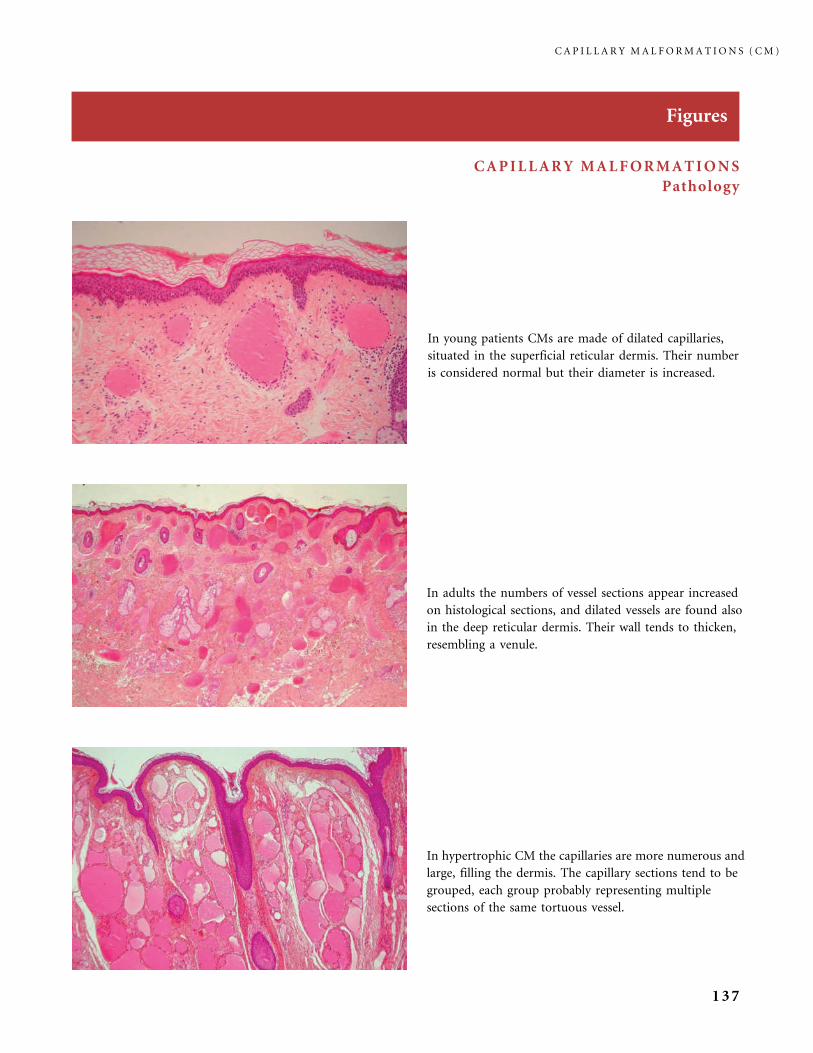
Figures
CAPILLARY MALFORMATIONS
Pathology
In hypertrophic CM the capillaries are more numerous and
large, filling the dermis. The capillary sections tend to be
grouped, each group probably representing multiple
sections of the same tortuous vessel.
In young patients CMs are made of dilated capillaries,
situated in the superficial reticular dermis. Their number
is considered normal but their diameter is increased.
In adults the numbers of vessel sections appear increased
on histological sections, and dilated vessels are found also
in the deep reticular dermis. Their wall tends to thicken,
resembling a venule.
C A P I L L A R Y M A L F O R M A T I O N S ( C M )
137
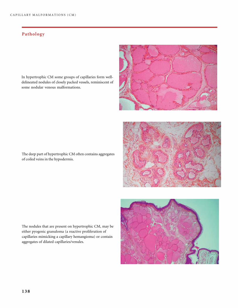
Pathology
The nodules that are present on hypertrophic CM, may be
either pyogenic granuloma (a reactive proliferation of
capillaries mimicking a capillary hemangioma) or contain
aggregates of dilated capillaries/venules.
The deep part of hypertrophic CM often contains aggregates
of coiled veins in the hypodermis.
In hypertrophic CM some groups of capillaries form well-
delineated nodules of closely packed vessels, reminiscent of
some nodular venous malformations.
C A P I L L A R Y M A L F O R M A T I O N S ( C M )
138
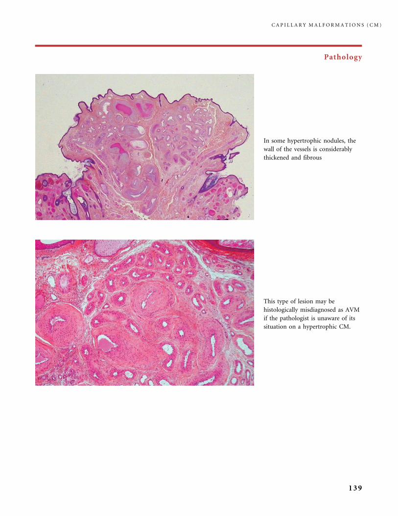
Pathology
This type of lesion may be
histologically misdiagnosed as AVM
if the pathologist is unaware of its
situation on a hypertrophic CM.
In some hypertrophic nodules, the
wall of the vessels is considerably
thickened and fibrous
C A P I L L A R Y M A L F O R M A T I O N S ( C M )
139
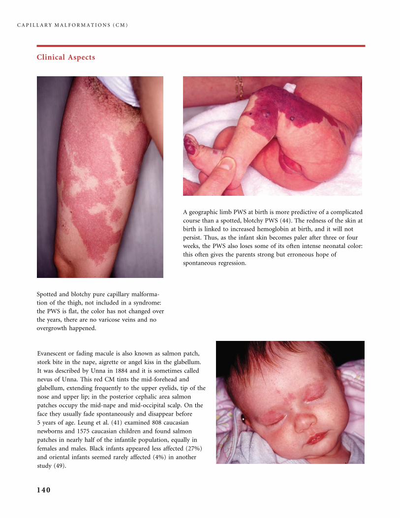
Clinical Aspects
Spotted and blotchy pure capillary malforma-
tion of the thigh, not included in a syndrome:
the PWS is flat, the color has not changed over
the years, there are no varicose veins and no
overgrowth happened.
A geographic limb PWS at birth is more predictive of a complicated
course than a spotted, blotchy PWS (44). The redness of the skin at
birth is linked to increased hemoglobin at birth, and it will not
persist. Thus, as the infant skin becomes paler after three or four
weeks, the PWS also loses some of its often intense neonatal color:
this often gives the parents strong but erroneous hope of
spontaneous regression.
Evanescent or fading macule is also known as salmon patch,
stork bite in the nape, aigrette or angel kiss in the glabellum.
It was described by Unna in 1884 and it is sometimes called
nevus of Unna. This red CM tints the mid-forehead and
glabellum, extending frequently to the upper eyelids, tip of the
nose and upper lip; in the posterior cephalic area salmon
patches occupy the mid-nape and mid-occipital scalp. On the
face they usually fade spontaneously and disappear before
5 years of age. Leung et al. (41) examined 808 caucasian
newborns and 1575 caucasian children and found salmon
patches in nearly half of the infantile population, equally in
females and males. Black infants appeared less affected (27%)
and oriental infants seemed rarely affected (4%) in another
study (49).
C A P I L L A R Y M A L F O R M A T I O N S ( C M )
140
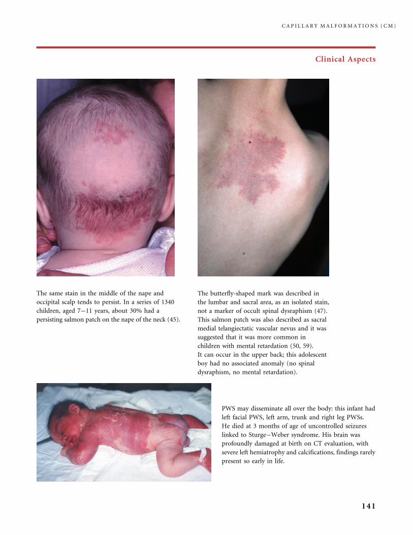
Clinical Aspects
The same stain in the middle of the nape and
occipital scalp tends to persist. In a series of 1340
children, aged 7�11 years, about 30% had a
persisting salmon patch on the nape of the neck (45).
The butterfly-shaped mark was described in
the lumbar and sacral area, as an isolated stain,
not a marker of occult spinal dysraphism (47).
This salmon patch was also described as sacral
medial telangiectatic vascular nevus and it was
suggested that it was more common in
children with mental retardation (50, 59).
It can occur in the upper back; this adolescent
boy had no associated anomaly (no spinal
dysraphism, no mental retardation).
PWS may disseminate all over the body: this infant had
left facial PWS, left arm, trunk and right leg PWSs.
He died at 3 months of age of uncontrolled seizures
linked to Sturge�Weber syndrome. His brain was
profoundly damaged at birth on CT evaluation, with
severe left hemiatrophy and calcifications, findings rarely
present so early in life.
C A P I L L A R Y M A L F O R M A T I O N S ( C M )
141
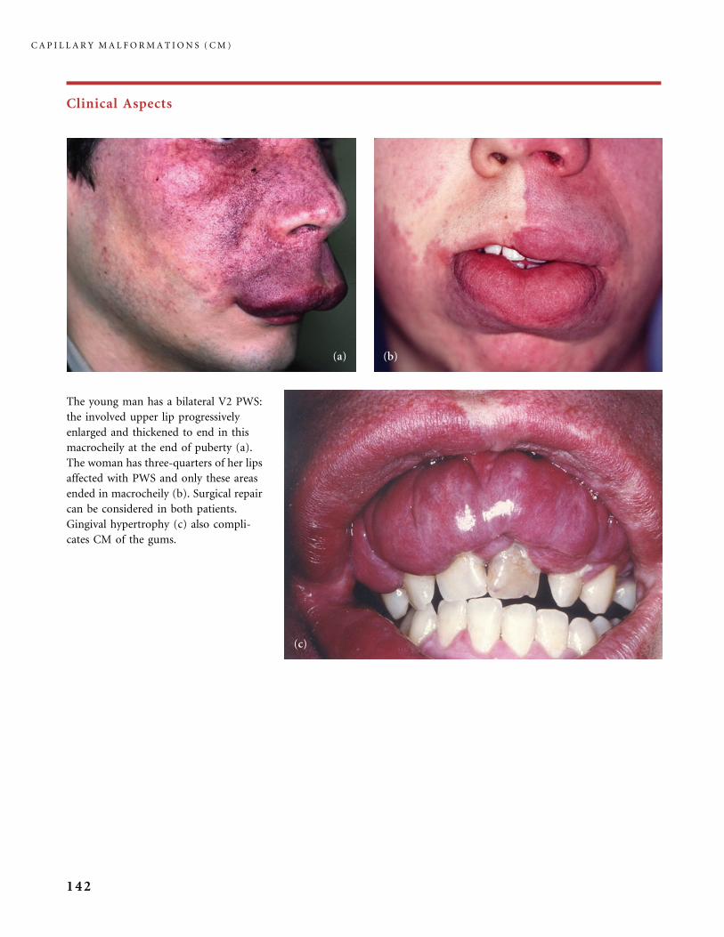
Clinical Aspects
The young man has a bilateral V2 PWS:
the involved upper lip progressively
enlarged and thickened to end in this
macrocheily at the end of puberty (a).
The woman has three-quarters of her lips
affected with PWS and only these areas
ended in macrocheily (b). Surgical repair
can be considered in both patients.
Gingival hypertrophy (c) also compli-
cates CM of the gums.
C A P I L L A R Y M A L F O R M A T I O N S ( C M )
142
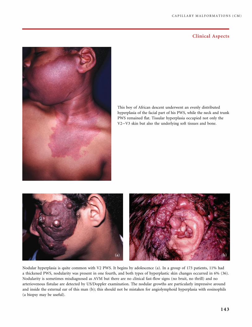
Clinical Aspects
This boy of African descent underwent an evenly distributed
hyperplasia of the facial part of his PWS, while the neck and trunk
PWS remained flat. Tissular hyperplasia occupied not only the
V2�V3 skin but also the underlying soft tissues and bone.
Nodular hyperplasia is quite common with V2 PWS. It begins by adolescence (a). In a group of 173 patients, 11% had
a thickened PWS, nodularity was present in one fourth, and both types of hyperplastic skin changes occurred in 6% (36).
Nodularity is sometimes misdiagnosed as AVM but there are no clinical fast-flow signs (no bruit, no thrill) and no
arteriovenous fistulae are detected by US/Doppler examination. The nodular growths are particularly impressive around
and inside the external ear of this man (b); this should not be mistaken for angiolymphoid hyperplasia with eosinophils
(a biopsy may be useful).
C A P I L L A R Y M A L F O R M A T I O N S ( C M )
143
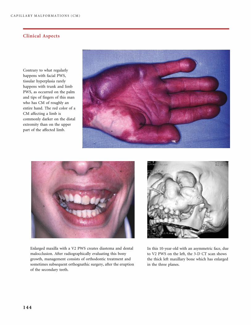
Clinical Aspects
Contrary to what regularly
happens with facial PWS,
tissular hyperplasia rarely
happens with trunk and limb
PWS, as occurred on the palm
and tips of fingers of this man
who has CM of roughly an
entire hand. The red color of a
CM affecting a limb is
commonly darker on the distal
extremity than on the upper
part of the affected limb.
Enlarged maxilla with a V2 PWS creates diastema and dental
malocclusion. After radiographically evaluating this bony
growth, management consists of orthodontic treatment and
sometimes subsequent orthognathic surgery, after the eruption
of the secondary teeth.
In this 10-year-old with an asymmetric face, due
to V2 PWS on the left, the 3-D CT scan shows
the thick left maxillary bone which has enlarged
in the three planes.
C A P I L L A R Y M A L F O R M A T I O N S ( C M )
144
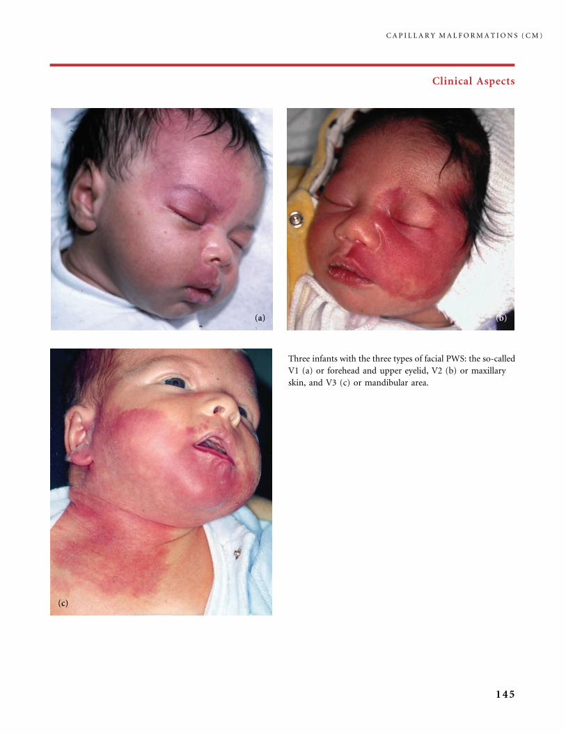
Clinical Aspects
Three infants with the three types of facial PWS: the so-called
V1 (a) or forehead and upper eyelid, V2 (b) or maxillary
skin, and V3 (c) or mandibular area.
C A P I L L A R Y M A L F O R M A T I O N S ( C M )
145

Clinical Aspects
The picture (a) and the drawing (b, c) show
the controversial watersheds of V1�V2. In
most cases this type of involvement in (a) is
not an association of V2 and V1 CM, but
a pure V2 PWS reaching the external and/or
internal part of the upper eyelid, with no risk
of SWS (a). The dermis of these territories is
made of mesoderm-derived endothelial cells
ensheathed with pericytes and smooth muscle
cells originating from the cephalic neural crest
(CNC) (20). The boundaries, between these
inflows of cells originating from the CNC, are
not strictly defined. This explains the overlap
of V1 and V2. In a large facial PWS, the risk of
SWS is connected with the V1 location of
PWS (10% of infants with such skin
involvement are at risk). The variable water-
sheds between V1 and V2 may be puzzling for
those taking the decision to screen the infant
neuroradiologically or not.
C A P I L L A R Y M A L F O R M A T I O N S ( C M )
146
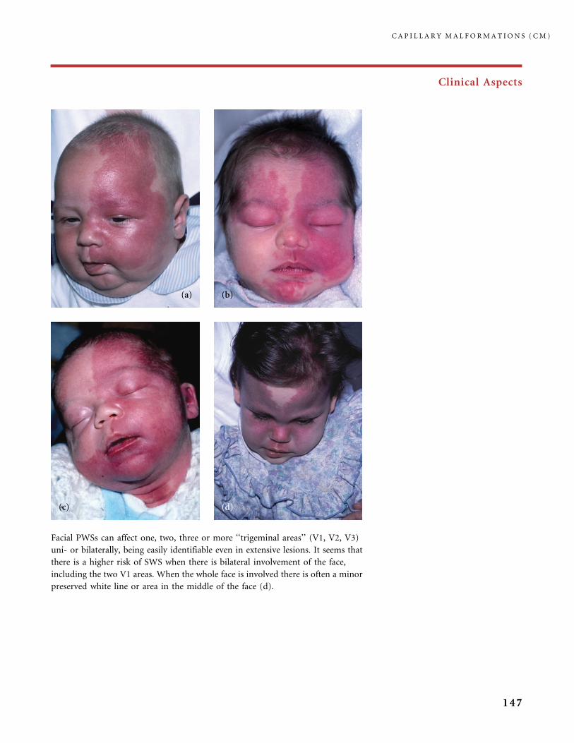
Clinical Aspects
Facial PWSs can affect one, two, three or more ‘‘trigeminal areas’’ (V1, V2, V3)
uni- or bilaterally, being easily identifiable even in extensive lesions. It seems that
there is a higher risk of SWS when there is bilateral involvement of the face,
including the two V1 areas. When the whole face is involved there is often a minor
preserved white line or area in the middle of the face (d).
C A P I L L A R Y M A L F O R M A T I O N S ( C M )
147
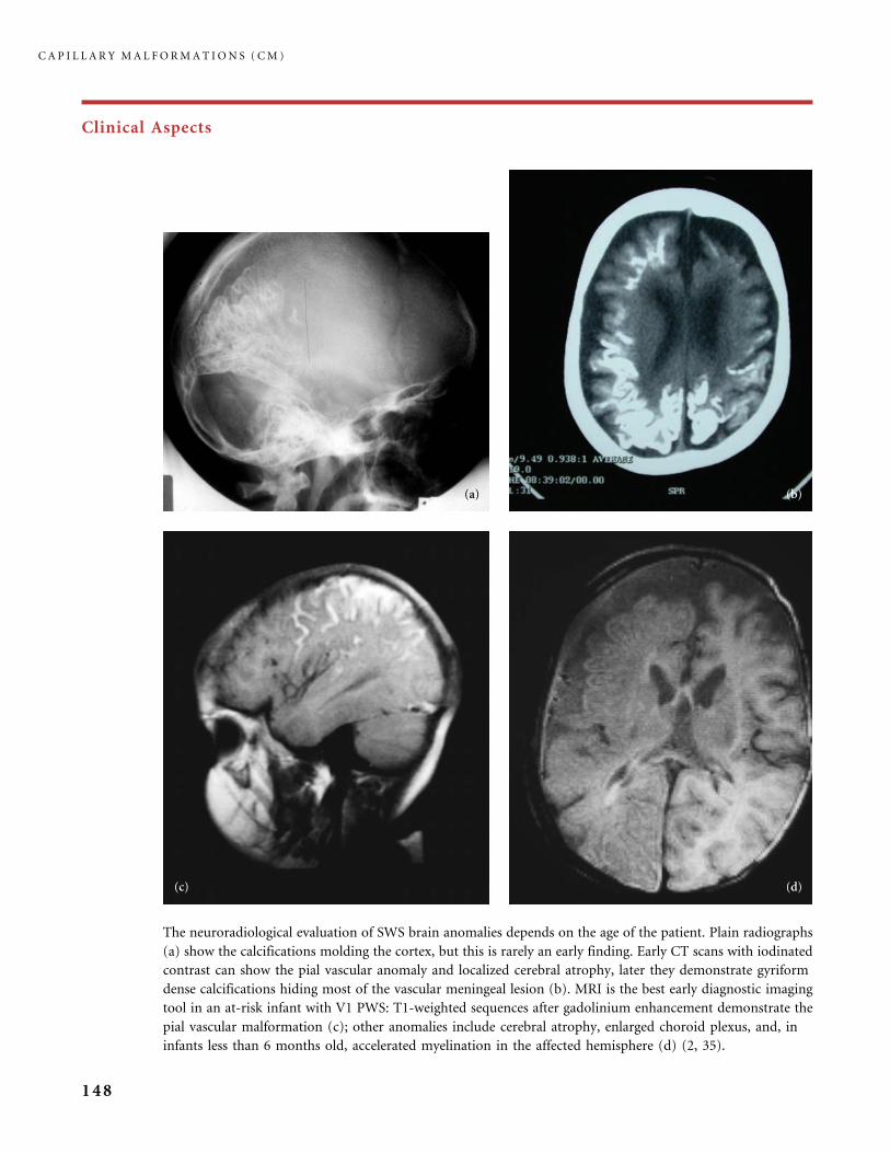
Clinical Aspects
The neuroradiological evaluation of SWS brain anomalies depends on the age of the patient. Plain radiographs
(a) show the calcifications molding the cortex, but this is rarely an early finding. Early CT scans with iodinated
contrast can show the pial vascular anomaly and localized cerebral atrophy, later they demonstrate gyriform
dense calcifications hiding most of the vascular meningeal lesion (b). MRI is the best early diagnostic imaging
tool in an at-risk infant with V1 PWS: T1-weighted sequences after gadolinium enhancement demonstrate the
pial vascular malformation (c); other anomalies include cerebral atrophy, enlarged choroid plexus, and, in
infants less than 6 months old, accelerated myelination in the affected hemisphere (d) (2, 35).
C A P I L L A R Y M A L F O R M A T I O N S ( C M )
148

Clinical Aspects
SPECT scans (Single Photon Emission Computed Tomography) with
Xe-133 (courtesy of Dr. C. Chiron, Hopital Necker-Enfants Malades, Paris,
France) permit the study of the regional cerebral blood flow (rCBF). In
infants it gives a good indication of the extent of the involvement: the
picture shows that the decrease of rCBF is usually wider than the lesion
evidenced by CT scan. This functional cerebral imaging shows a decreased
rCBF after the first seizures in the area of the pial vascular anomaly, whereas
rCBF is temporarily increased, before the first seizure (11, 53). The more
extensive the pial vascular lesion is, the more severe the course of the
neurological symptoms will be. According to Ville et al. (67) motor
functions are affected by ischemic events whereas the cognitive function
alteration is likely linked to epilepsy. PET scans (Positron Emission
Tomography) study the cerebral glucose metabolism; results are compar-
able to data obtained with SPECT scans in SWS (12).
Above: Nevus anemicus is often intermingled with CM, in both
facial or trunk and limb lesions, as a twin-spotting
phenomenon (29). These pale blanching macules do not
redden when the skin is rubbed, in contrast to the surrounding
CM and normal skin.
Right: Phakomatosis pigmentovascularis (PPV) type II a in a girl at
1 year. Spontaneous fading of both the Mongolian spots and PWS
is likely to occur after a few years. Type II is the most common type
of PPV, and it seems particularly frequent in infants of Asian or
African ancestry.
C A P I L L A R Y M A L F O R M A T I O N S ( C M )
149
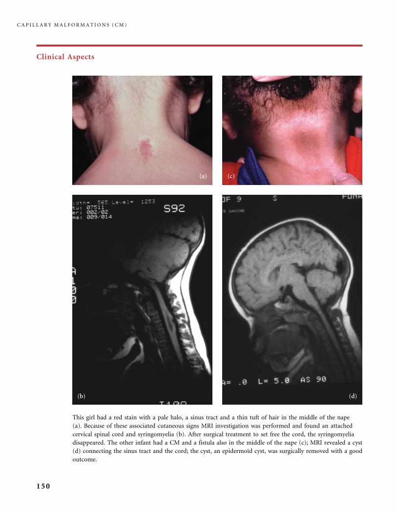
Clinical Aspects
This girl had a red stain with a pale halo, a sinus tract and a thin tuft of hair in the middle of the nape
(a). Because of these associated cutaneous signs MRI investigation was performed and found an attached
cervical spinal cord and syringomyelia (b). After surgical treatment to set free the cord, the syringomyelia
disappeared. The other infant had a CM and a fistula also in the middle of the nape (c); MRI revealed a cyst
(d) connecting the sinus tract and the cord; the cyst, an epidermoıd cyst, was surgically removed with a good
outcome.
C A P I L L A R Y M A L F O R M A T I O N S ( C M )
150
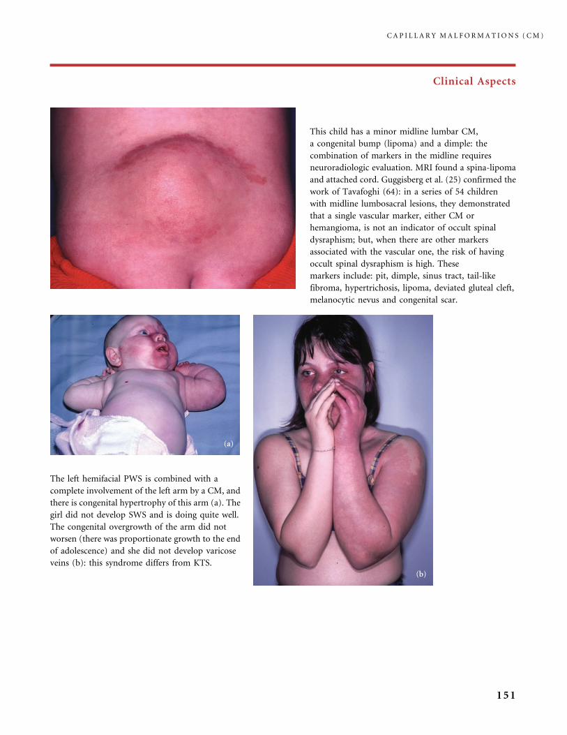
Clinical Aspects
This child has a minor midline lumbar CM,
a congenital bump (lipoma) and a dimple: the
combination of markers in the midline requires
neuroradiologic evaluation. MRI found a spina-lipoma
and attached cord. Guggisberg et al. (25) confirmed the
work of Tavafoghi (64): in a series of 54 children
with midline lumbosacral lesions, they demonstrated
that a single vascular marker, either CM or
hemangioma, is not an indicator of occult spinal
dysraphism; but, when there are other markers
associated with the vascular one, the risk of having
occult spinal dysraphism is high. These
markers include: pit, dimple, sinus tract, tail-like
fibroma, hypertrichosis, lipoma, deviated gluteal cleft,
melanocytic nevus and congenital scar.
The left hemifacial PWS is combined with a
complete involvement of the left arm by a CM, and
there is congenital hypertrophy of this arm (a). The
girl did not develop SWS and is doing quite well.
The congenital overgrowth of the arm did not
worsen (there was proportionate growth to the end
of adolescence) and she did not develop varicose
veins (b): this syndrome differs from KTS.
C A P I L L A R Y M A L F O R M A T I O N S ( C M )
151
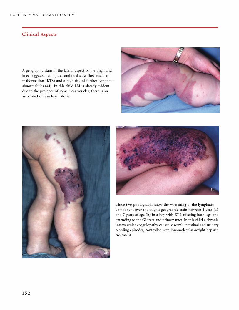
Clinical Aspects
A geographic stain in the lateral aspect of the thigh and
knee suggests a complex combined slow-flow vascular
malformation (KTS) and a high risk of further lymphatic
abnormalities (44). In this child LM is already evident
due to the presence of some clear vesicles; there is an
associated diffuse lipomatosis.
These two photographs show the worsening of the lymphatic
component over the thigh’s geographic stain between 1 year (a)
and 7 years of age (b) in a boy with KTS affecting both legs and
extending to the GI tract and urinary tract. In this child a chronic
intravascular coagulopathy caused visceral, intestinal and urinary
bleeding episodes, controlled with low-molecular-weight heparin
treatment.
C A P I L L A R Y M A L F O R M A T I O N S ( C M )
152
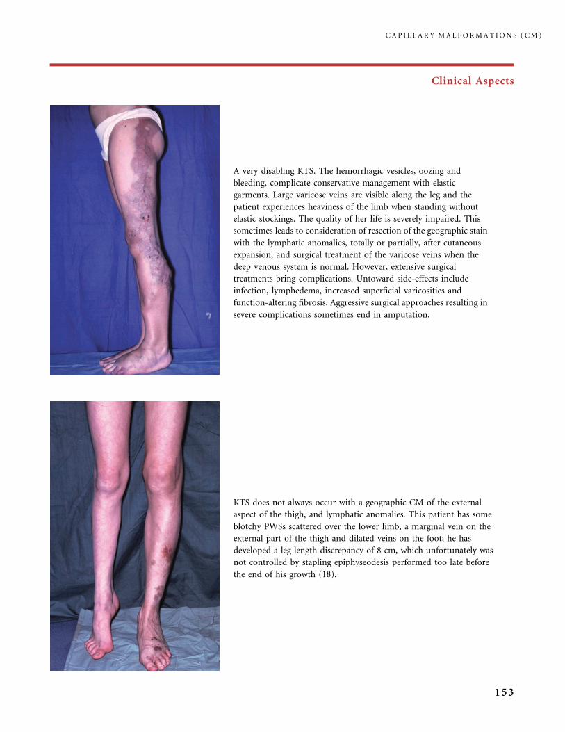
Clinical Aspects
A very disabling KTS. The hemorrhagic vesicles, oozing and
bleeding, complicate conservative management with elastic
garments. Large varicose veins are visible along the leg and the
patient experiences heaviness of the limb when standing without
elastic stockings. The quality of her life is severely impaired. This
sometimes leads to consideration of resection of the geographic stain
with the lymphatic anomalies, totally or partially, after cutaneous
expansion, and surgical treatment of the varicose veins when the
deep venous system is normal. However, extensive surgical
treatments bring complications. Untoward side-effects include
infection, lymphedema, increased superficial varicosities and
function-altering fibrosis. Aggressive surgical approaches resulting in
severe complications sometimes end in amputation.
KTS does not always occur with a geographic CM of the external
aspect of the thigh, and lymphatic anomalies. This patient has some
blotchy PWSs scattered over the lower limb, a marginal vein on the
external part of the thigh and dilated veins on the foot; he has
developed a leg length discrepancy of 8 cm, which unfortunately was
not controlled by stapling epiphyseodesis performed too late before
the end of his growth (18).
C A P I L L A R Y M A L F O R M A T I O N S ( C M )
153

Clinical Aspects
Diffusely scattered PWSs, over the trunk and limbs, are a common
finding in infants with Proteus or Proteiform syndromes. Their bright
red tint, at birth, as a rule significantly lightens after a few years; thus
it is not worth undertaking extensive pulsed dye laser treatment: laser
will be used only for CMs of visible areas (the face). This infant has
right hemihypertrophy affecting one vascular extremity (leg) and one
nonvascular one (arm).
Patients with Proteus syndrome develop large lipomas (particularly on the
back and abdomen), recurring after excision (see the large scars on the back,
evidence of the previous resection). Some lipomas have intra-abdominal
extension. This young man also has a slow-flow vascular malformation
combining large CM of the trunk and underlying dilated veins.
C A P I L L A R Y M A L F O R M A T I O N S ( C M )
154
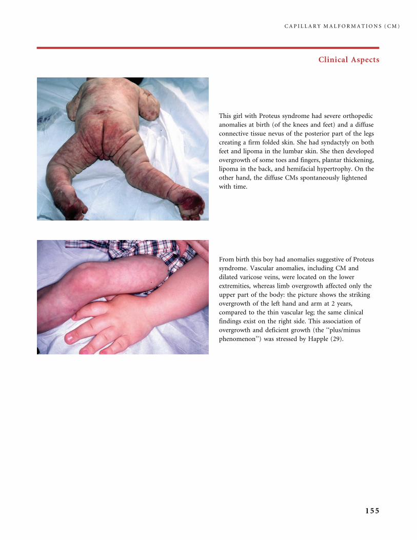
Clinical Aspects
This girl with Proteus syndrome had severe orthopedic
anomalies at birth (of the knees and feet) and a diffuse
connective tissue nevus of the posterior part of the legs
creating a firm folded skin. She had syndactyly on both
feet and lipoma in the lumbar skin. She then developed
overgrowth of some toes and fingers, plantar thickening,
lipoma in the back, and hemifacial hypertrophy. On the
other hand, the diffuse CMs spontaneously lightened
with time.
From birth this boy had anomalies suggestive of Proteus
syndrome. Vascular anomalies, including CM and
dilated varicose veins, were located on the lower
extremities, whereas limb overgrowth affected only the
upper part of the body: the picture shows the striking
overgrowth of the left hand and arm at 2 years,
compared to the thin vascular leg; the same clinical
findings exist on the right side. This association of
overgrowth and deficient growth (the ‘‘plus/minus
phenomenon’’) was stressed by Happle (29).
C A P I L L A R Y M A L F O R M A T I O N S ( C M )
155
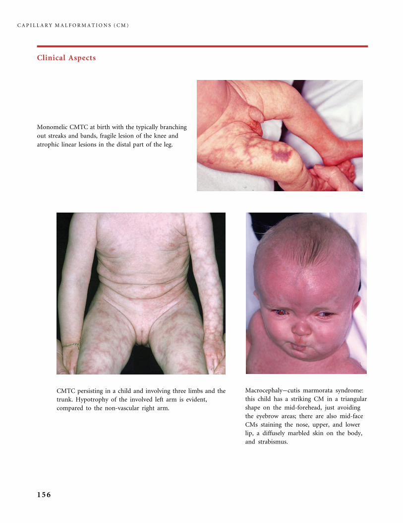
Clinical Aspects
Monomelic CMTC at birth with the typically branching
out streaks and bands, fragile lesion of the knee and
atrophic linear lesions in the distal part of the leg.
CMTC persisting in a child and involving three limbs and the
trunk. Hypotrophy of the involved left arm is evident,
compared to the non-vascular right arm.
Macrocephaly�cutis marmorata syndrome:
this child has a striking CM in a triangular
shape on the mid-forehead, just avoiding
the eyebrow areas; there are also mid-face
CMs staining the nose, upper, and lower
lip, a diffusely marbled skin on the body,
and strabismus.
C A P I L L A R Y M A L F O R M A T I O N S ( C M )
156
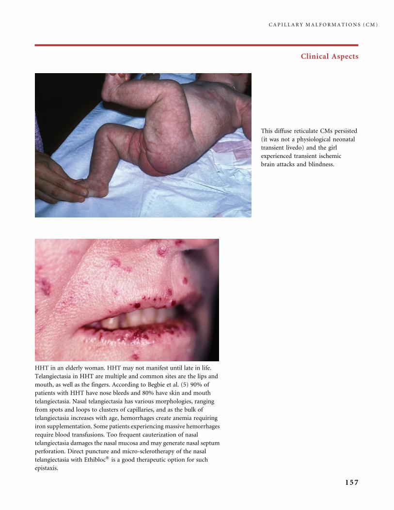
Clinical Aspects
This diffuse reticulate CMs persisted
(it was not a physiological neonatal
transient livedo) and the girl
experienced transient ischemic
brain attacks and blindness.
HHT in an elderly woman. HHT may not manifest until late in life.
Telangiectasia in HHT are multiple and common sites are the lips and
mouth, as well as the fingers. According to Begbie et al. (5) 90% of
patients with HHT have nose bleeds and 80% have skin and mouth
telangiectasia. Nasal telangiectasia has various morphologies, ranging
from spots and loops to clusters of capillaries, and as the bulk of
telangiectasia increases with age, hemorrhages create anemia requiring
iron supplementation. Some patients experiencing massive hemorrhages
require blood transfusions. Too frequent cauterization of nasal
telangiectasia damages the nasal mucosa and may generate nasal septum
perforation. Direct puncture and micro-sclerotherapy of the nasal
telangiectasia with Ethibloc� is a good therapeutic option for such
epistaxis.
C A P I L L A R Y M A L F O R M A T I O N S ( C M )
157

Clinical Aspects
Angiokeratoma circumscriptum (AKC): the plaque-type lesion is obviously hyperkeratotic and in this
knee area (a), barely protected from trauma in an active child, bleeding is problematic. Epidermal
hyperkeratosis increases over the years and this allows the lesion to bleed with minor trauma. Often an
underlying capillary-venous malformation gives the skin around the AKC a bluish color: in this newborn
(b) this allows us to predict a further worsening of the AKC. AKC of toes or fingers are usually called AK of
Mibelli (c). Excision is the only effective treatment for AKC. However, in very large lesions surgical
treatment is not realistic, even after cutaneous expansion. Then, CO2 laser or 1064 nm Nd-YAG laser
resurfacing treatment, sometimes combined with pulsed dye laser, can be considered to achieve a smoother
surface and decrease the color, but it is seldom possible to eradicate the angiokeratoma.
C A P I L L A R Y M A L F O R M A T I O N S ( C M )
158

Clinical Aspects
This large solitary angiokeratoma circumscriptum of the thigh of
a 15-year-old girl, which had recently thickened and darkened,
was misdiagnosed as melanoma before she had a biopsy.
Fordyce angiokeratomas speckled on the
scrotum often have a smooth surface and
are associated with symptoms of local
venous hypertension (varicose veins,
varicocele). In this patient lesions were
improved with 3% polidocanol
intralesional injections.
C A P I L L A R Y M A L F O R M A T I O N S ( C M )
159
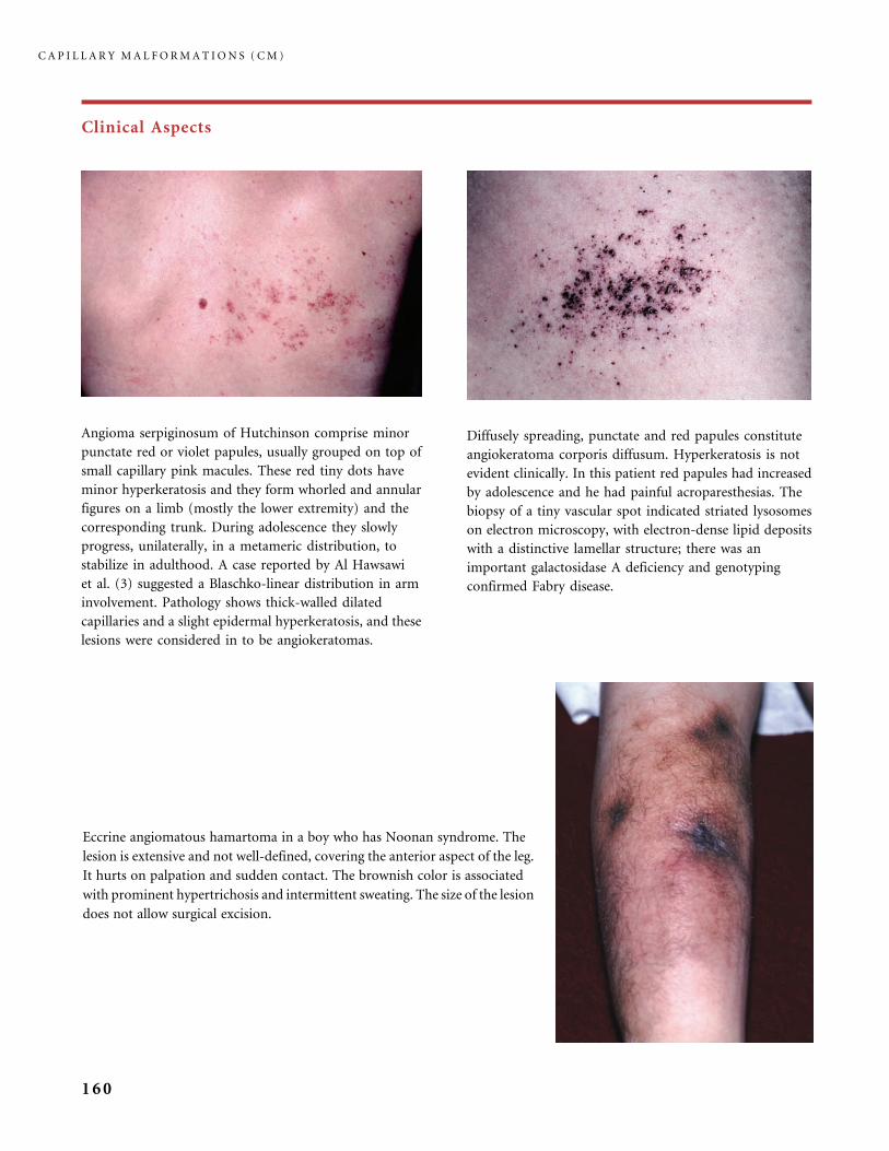
Clinical Aspects
Angioma serpiginosum of Hutchinson comprise minor
punctate red or violet papules, usually grouped on top of
small capillary pink macules. These red tiny dots have
minor hyperkeratosis and they form whorled and annular
figures on a limb (mostly the lower extremity) and the
corresponding trunk. During adolescence they slowly
progress, unilaterally, in a metameric distribution, to
stabilize in adulthood. A case reported by Al Hawsawi
et al. (3) suggested a Blaschko-linear distribution in arm
involvement. Pathology shows thick-walled dilated
capillaries and a slight epidermal hyperkeratosis, and these
lesions were considered in to be angiokeratomas.
Diffusely spreading, punctate and red papules constitute
angiokeratoma corporis diffusum. Hyperkeratosis is not
evident clinically. In this patient red papules had increased
by adolescence and he had painful acroparesthesias. The
biopsy of a tiny vascular spot indicated striated lysosomes
on electron microscopy, with electron-dense lipid deposits
with a distinctive lamellar structure; there was an
important galactosidase A deficiency and genotyping
confirmed Fabry disease.
Eccrine angiomatous hamartoma in a boy who has Noonan syndrome. The
lesion is extensive and not well-defined, covering the anterior aspect of the leg.
It hurts on palpation and sudden contact. The brownish color is associated
with prominent hypertrichosis and intermittent sweating. The size of the lesion
does not allow surgical excision.
C A P I L L A R Y M A L F O R M A T I O N S ( C M )
160

Clinical Aspects
This eccrine angiomatous hamartoma creates ill-defined
infiltration of the leg and blotchy pink areas. Some
dilated venous-like channels are visible as bluish streaks.
Sweating became obvious in this anxious patient during
examination, and droplets of sweat emerged only in this
area of the skin. With his pain increasing, this patient is
looking for a treatment but excision is difficult due to
the site and size.
During pregnancy diffuse telangiectasia sometimes appear, as on the arms and trunk of this young woman
(a); 6 months after the birth of her baby the telangiectasia had completely disappeared (b). This patient also
had an arteriovenous malformation of one hand, known from childhood, which did not change during and
after her pregnancy.
C A P I L L A R Y M A L F O R M A T I O N S ( C M )
161
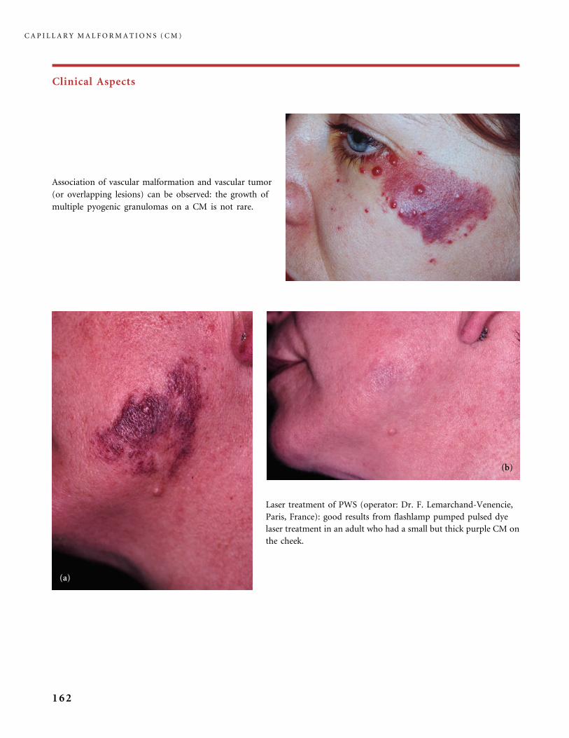
Clinical Aspects
Association of vascular malformation and vascular tumor
(or overlapping lesions) can be observed: the growth of
multiple pyogenic granulomas on a CM is not rare.
Laser treatment of PWS (operator: Dr. F. Lemarchand-Venencie,
Paris, France): good results from flashlamp pumped pulsed dye
laser treatment in an adult who had a small but thick purple CM on
the cheek.
C A P I L L A R Y M A L F O R M A T I O N S ( C M )
162
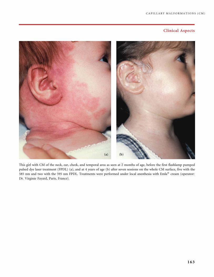
Clinical Aspects
This girl with CM of the neck, ear, cheek, and temporal area as seen at 2 months of age, before the first flashlamp pumped
pulsed dye laser treatment (FPDL) (a), and at 4 years of age (b) after seven sessions on the whole CM surface, five with the
585 nm and two with the 595 nm FPDL. Treatments were performed under local anesthesia with Emla� cream (operator:
Dr. Virginie Fayard, Paris, France).
C A P I L L A R Y M A L F O R M A T I O N S ( C M )
163
References
1 Adams BB, Lucky AW. Acquired port-wine stains and antecedent trauma: case report
and review of the literature. Arch Dermatol 2000; 136: 897�9.
2 Adamsbaum C, Pinton F, Rolland Y, Chiron C, Dulac O, Kalifa G. Accelerated
myelination in early Sturge�Weber syndrome: MRI-SPECT correlations. Pediatr Radiol
1996; 26: 759�62.
3 Al Hawsawi K, Al Aboud K, Al Aboud D, Al Githami A. Linear angioma serpiginosum.
Pediatr Dermatol 2003; 20: 167�8.
4 Bayrak-Toydemir P, Mao R, Lewin S, McDonald J. Hereditary hemorrhagic telangiectasia:
an overview of diagnosis and management in the molecular era for clinicians. Genet Med
2004; 6: 175�91.
5 Begbie ME, Wallace GM, and Shovlin CL. Hereditary haemorrhagic telangiectasia
(Osler�Weber�Rendu syndrome): a view from the 21st century. Postgrad Med J 2003;
79: 18�24.
6 Ben-Amitai D, Davidson S, Schwartz M, Prais D, Shamir R, Metzker A et al. Sacral nevus
flammeus simplex: the role of imaging. Pediatr Dermatol 2000; 17: 469�71.
7 Berry SA, Peterson C, Mize W, Bloom K, Zachary C, Blasco P et al. Klippel�Trenaunay
syndrome. Am J Med Genet 1998; 79: 319�26.
8 Biesecker LG. The multifaceted challenges of Proteus syndrome. JAMA 2001; 285:
2240�3.
9 Biesecker LG, Peters KF, Darling TN, Choyke P, Hill S, Schimke N et al. Clinical
differentiation between Proteus syndrome and hemihyperplasia: description of a distinct
form of hemihyperplasia. Am J Med Genet 1998; 79: 311�18.
10 Cabana MD, Crawford TO, Winkelstein JA, Christensen JR, Lederman HM.
Consequences of the delayed diagnosis of ataxia�telangiectasia. Pediatrics 1998; 102:
98�100.
11 Chiron C, Raynaud C, Tzourio N, Diebler C, Dulac O, Zilbovicius M et al. Regional
cerebral blood flow by SPECT imaging in Sturge�Weber disease: an aid for diagnosis.
J Neurol Neurosurg Psychiatry 1989; 52: 1402�9.
12 Chugani HT. The role of PET in childhood epilepsy. J Child Neurol 1994; 9 Suppl 1:
S82�8.
13 Cole SG, Begbie ME, Wallace GM, Shovlin CL. A new locus for Hereditary Haemorrhagic
Telangiectasia (HHT3) maps to chromosome 5. J Med Genet 2005; 42: 577�82.
14 DeLone DR, Brown WD, Gentry LR. Proteus syndrome: craniofacial and cerebral MRI.
Neuroradiology 1999; 41: 840�3.
15 Devillers AC, de Waard-van der Spek FB, Oranje A P. Cutis marmorata telangiectatica
congenita: clinical features in 35 cases. Arch Dermatol 1999; 135: 34�8.
16 Dyall-Smith D, Ramsden A, Laurie S. Adams�Oliver syndrome: aplasia cutis congenita,
terminal transverse limb defects and cutis marmorata telangiectatica congenita. Australas
J Dermatol. 1994; 35: 19�22.
17 Enjolras O. Cutis marmorata telangiectatica congenita. Ann Dermatol Venereol 2001; 128:
161�6.
18 Enjolras O, Chapot R, Merland JJ. Vascular anomalies and the growth of limbs: a review.
J Pediatr Orthop B 2004; 13: 349�57.
19 Enjolras O, Riche MC, Mulliken JB, Merland JJ, Hemangiomes et Malformations
Vasculaires. Atlas. Paris: Medsi/McGraw Hill, 1990,.
20 Etchevers HC, Vincent C, Le Douarin NM, Couly GF. The cephalic neural crest provides
pericytes and smooth muscle cells to all blood vessels of the face and forebrain.
Development 2001; 128: 1059�68.
C A P I L L A R Y M A L F O R M A T I O N S ( C M )
164

21 Eubanks LE, McBurney EI. Videomicroscopy of port-wine stains: correlation of location
and depth of lesion. J Am Acad Dermatol 2001; 44: 948�51
22 Folz BJ, Wollstein AC, Lippert BM, Werner JA. Morphology and distribution of nasal
telangiectasia in HHT-patients with epistaxis. Am J Rhinol 2005; 19: 65�70.
23 Germain DP. Fabry disease in 2004. Rev Prat 2003; 53: 2215�20.
24 Greene AK, Kieran M, Burrows PE, Mulliken JB, Kasser J, Fishman SJ. Wilms
tumor screening is unnecessary in Klippel�Trenaunay syndrome. Pediatrics 2004; 113:
e326�9.
25 Guggisberg D, Hadj-Rabia S, Viney C, Bodemer C, Brunelle F, Zerah M et al. Skin markers
of occult spinal dysraphism in children: a review of 54 cases. Arch Dermatol 2004; 140:
1109�15.
26 Gungor T, Buhring I, Cremer R, Gartenschlager M, Zielen S. Pathogenesis, diagnosis,
clinical and therapeutic aspects of ataxia telangiectasia. Klin Padiatr 1997; 209: 328�35.
27 Hagiwara K, Uezato H, Nonaka S. Phacomatosis pigmentovascularis type IIb associated
with Sturge�Weber syndrome and pyogenic granuloma. J Dermatol 1998; 25: 721�9.
28 Hansen K, Kreiter CD, Rosenbaum M, Whitaker DC, Arpey CJ. Long-term psychological
impact and perceived efficacy of pulsed-dye laser therapy for patients with port-wine
stains. Dermatol Surg 2003; 29: 49�55.
29 Happle R. Loss of heterozygosity in human skin. J Am Acad Dermatol 1999; 41: 143�64.
30 Happle R, Steijlen PM. Phacomatosis pigmentovascularis interpreted as a phenomenon
of twin spots. Hautarzt 1989; 40: 721�4.
31 Happle R, Steijlen PM, Theile U, Karitzky D, Tinschert S, Albrecht-Nebe H et al. Patchy
dermal hypoplasia as a characteristic feature of Proteus syndrome. Arch Dermatol 1997;
133: 77�80.
32 Heikkinen K, Rapakko K, Karppinen SM, Erkko H, Nieminen P, Winqvist R. Association
of common ATM polymorphism with bilateral breast cancer. Int J Cancer 2005; 116:
69�72.
33 Hoeger PH, Martinez A, Maerker J, Harper JI. Vascular anomalies in Proteus syndrome.
Clin Exp Dermatol 2004; 29: 222�30.
34 Hotamisligil GS, Ertogan F. The Proteus syndrome: association with nephrogenic diabetes
insipidus. Clin Genet 1990; 38: 139�44.
35 Jacoby CG, Yuh WT, Afifi AK, Bell WE, Schelper RL, Sato Y. Accelerated myelination in
early Sturge�Weber syndrome demonstrated by MR imaging. J Comput Assist Tomogr
1987; 11: 226�31.
36 Klapman MH, Yao JF. Thickening and nodules in port-wine stains. J Am Acad Dermatol
2001; 44: 300�2.
37 Kossoff EH, Buck C, Freeman JM. Outcomes of 32 hemispherectomies for Sturge�Weber
syndrome worldwide. Neurology 2002; 59: 1735�8.
38 Kuehl HK, Caselitz M, Hasenkamp S, Wagner S, El-Harith el HA, Manns MP et al.
Hepatic manifestation is associated with ALK1 in hereditary hemorrhagic telangiectasia:
identification of five novel ALK1 and one novel ENG mutations. Hum Mutat 2005;
25: 320.
39 Kundu RV, Frieden IJ. Presence of vascular anomalies with congenital hemihypertrophy
and Wilms tumor: an evidence-based evaluation. Pediatr Dermatol 2003; 20: 199�206.
40 Larralde M, Boggio P, Amartino H, Chamoles N. Fabry disease: a study of 6 hemizygous
men and 5 heterozygous females with emphasis on dermatologic manifestations. Arch
Dermatol 2004; 140: 1440�6.
41 Leung AK, Telmesani AM. Salmon patches in Caucasian children. Pediatr Dermatol 1989;
6: 185�7.
42 Linthorst GE, De Rie MA, Tjiam KH, Aerts JM, Dingemans KP, Hollak CE. Misdiagnosis
of Fabry disease: importance of biochemical confirmation of clinical or pathological
suspicion. Br J Dermatol 2004; 150: 575�7.
43 Loo WJ, Lanigan SW. Recent advances in laser therapy for the treatment of cutaneous
vascular disorders. Lasers Med Sci 2002; 17: 9�12.
R E F E R E N C E S
165
44 Maari C, Frieden IJ. Klippel�Trenaunay syndrome: the importance of ‘‘geographic stains’’
in identifying lymphatic disease and risk of complications. J Am Acad Dermatol 2004; 51:
391�8.
45 Maniscalco M, Guareschi E, Noto G, Patrizi A. Midline telangiectatic nevus (salmon
patch) of the nape of the neck. Eur J Pediatr Dermatol 2003; 13: 81�4.
46 Martinelli PT, Tschen JA. Eccrine angiomatous hamartoma: a case report and review
of the literature. Cutis 2003; 71: 449�55.
47 Metzker A, Shamir R. Butterfly-shaped mark: a variant form of nevus flammeus simplex.
Pediatrics 1990; 85: 1069�71.
48 Nagore E, Requena C, Sevila A, Coll J, Costa D, Botella-Estrada R et al. Thickness
of healthy and affected skin of children with port wine stains: potential repercussions on
response to pulsed dye laser treatment. Dermatol Surg 2004; 30: 1457�61.
49 Osburn K, Schosser RH, Everett MA. Congenital pigmented and vascular lesions in
newborn infants. J Am Acad Dermatol 1987; 16: 788�92.
50 Patrizi A, Neri I, Orlandi C, Marini R. Sacral medial telangiectatic vascular nevus: a study
of 43 children. Dermatology 1996; 192: 301�6.
51 Piaserico S, Belloni Fortina A. Posttraumatic port-wine stain in a 4-year-old girl: Fegeler
syndrome. Pediatr Dermatol 2004; 21: 131�3.
52 Picascia DD, Esterly NB. Cutis marmorata telangiectatica congenita: report of 22 cases.
J Am Acad Dermatol 1989; 20: 1098�104.
53 Pinton F, Chiron C, Enjolras O, Motte J, Syrota A, Dulac O. Early single photon emission
computed tomography in Sturge�Weber syndrome. J Neurol Neurosurg Psychiatry 1997;
63: 616�21.
54 Prigent F, Kahn A, Martinet C, Saigot T. Pigmented-vascular phakomatosis type IIIa. Ann
Dermatol Venereol 1991; 118: 531�3.
55 Requena L, Sangueza OP. Cutaneous vascular anomalies. Part I. Hamartomas,
malformations, and dilation of preexisting vessels. J Am Acad Dermatol 1997; 37:
523�49; quiz 549�52.
56 Robertson SP, Gattas M, Rogers M, Ades LC. Macrocephaly�cutis marmorata
telangiectatica congenita: report of five patients and a review of the literature. Clin
Dysmorphol 2000; 9: 1�9.
57 Ruiz-Maldonado R, Tamayo L, Laterza AM, Brawn G, Lopez A. Phacomatosis
pigmentovascularis: a new syndrome? Report of four cases. Pediatr Dermatol 1987;
4: 189�96.
58 Sanchez-Carpintero I, Mihm MC, Mizeracki A, Waner M, North PE. Epithelial and
mesenchymal hamartomatous changes in a mature port-wine stain: Morphologic evidence
for a multiple germ layer field defect. J Am Acad Dermatol 2004; 50: 608�12.
59 Schepis C, Greco D, Failla P, Siragusa M, Romano C, Scaffidi M et al. Medial telangiectatic
sacral nevi and MCA/MR syndromes. Pediatr Dermatol 2003; 20: 370�1.
60 Schiller PI, Itin PH, Angiokeratomas: an update. Dermatology 1996; 193: 275�82.
61 Selim MM, Kelly KM, Nelson JS, Wendelschafer-Crabb G, Kennedy WR, Zelickson BD.
Confocal microscopy study of nerves and blood vessels in untreated and treated port wine
stains: preliminary observations. Dermatol Surg 2004; 30: 892�7.
62 Smoller BR, Rosen S. Port-wine stains. A disease of altered neural modulation of blood
vessels? Arch Dermatol 1986; 122: 177�9.
63 Sommer S, Merchant WJ, Sheehan-Dare R. Severe predominantly acral variant of
angiokeratoma of Mibelli: response to long-pulse Nd:YAG (1064 nm) laser treatment.
J Am Acad Dermatol 2001; 45: 764�6.
64 Tavafoghi V, Ghandchi A, Hambrick GW Jr, Udverhelyi GB. Cutaneous signs of spinal
dysraphism. Report of a patient with a tail-like lipoma and review of 200 cases in the
literature. Arch Dermatol 1978; 114: 573�7.
65 Torrelo A, Zambrano A, Happle R. Cutis marmorata telangiectatica congenita and
extensive mongolian spots: type 5 phacomatosis pigmentovascularis. Br J Dermatol 2003;
148: 342�5.
C A P I L L A R Y M A L F O R M A T I O N S ( C M )
166
66 Turner JT, Cohen MM Jr, Biesecker LG. Reassessment of the Proteus syndrome literature:
application of diagnostic criteria to published cases. Am J Med Genet A 2004; 130: 111�22.
67 Ville D, Enjolras O, Chiron C, Dulac O. Prophylactic antiepileptic treatment in
Sturge�Weber disease. Seizure 2002; 11: 145�50.
68 Waner M. Recent developments in lasers and the treatment of birthmarks. Arch Dis Child
2003; 88: 372�4.
69 Wiedemann HR, Burgio GR, Aldenhoff P, Kunze J, Kaufmann HJ, Schirg E. The proteus
syndrome. Partial gigantism of the hands and/or feet, nevi, hemihypertrophy,
subcutaneous tumors, macrocephaly or other skull anomalies and possible accelerated
growth and visceral affections. Eur J Pediatr 1983; 140: 5�12.
70 Wilcox WR, Banikazemi M, Guffon N, Waldek S, Lee P, Linthorst GE et al. Long-term
safety and efficacy of enzyme replacement therapy for Fabry disease. Am J Hum Genet
2004; 75: 65�74.
R E F E R E N C E S
167

CHAPTER III.B
Venous Malformations (VM)
III.B.1 Common Venous Malformations
Introduction and Clinical Patterns
Venous malformations (VM) are hemodynamically inactive, slow-flow vascular
malformations involving the collecting side of the vascular network. They are the
most common vascular malformation seen in clinics for vascular anomalies. This
localized defect in vascular morphogenesis is characterized by enlarged and
distorted venous channels, with irregular lack of smooth muscle cells around the
flat continuous layer of endothelial cells in their walls (5).
Most VMs occur sporadically. Rare familial cases exist: these account for
about 1% of all VMs (3). A VM is usually visible at birth, as a minor bluish
patch or plaque, or as a network of dilated veins. It slowly worsens over the life
of the patient to varying degrees. The overload of distorted venous channels
within the dermal layers give the skin or mucous membranes the characteristic
blue hue. The blue color becomes particularly significant when the dilated
venous-like channels invade the superficial dermis. This blue color is obvious
on skin as well as on the mucous membranes of the mouth, conjunctiva and
genitalia. A VM never regresses, unlike hemangioma and various vascular
tumors present at birth or developing in infancy. It affects males and females
equally.
VMs are localized or extensive, minor or distorting, flat or spongy, single or
multiple. They involve skin, mucous membranes, soft tissues and particularly
muscles, joints and bones, and even viscera. Depending on the size and site of
their lesion, patients complain of swelling and pain sensation. Stiffness and pain
upon awakening are common symptoms in patients with extremity VM. VMs
swell with physical exertion and when the region is dependent, in fact in every
situation when blood pressure increases. Deformation of anatomical structures
happens slowly over the years. With a VM in the cheek, facial features are altered
by the soft-tissue filling and bony deformity results from a mass-compressing
effect. Skin temperature is usually normal or mildly increased. There is no thrill,
no bruit like in AVM. Head-and-neck VM and trunk-and-limbs VM have
168

different clinical consequences due to the unique environment they occur in
(20, 21, 31).
Worsening over the years is due to the progressive ‘‘opening’’ of the distorted,
poorly defined and interconnecting anomalous venous network that dissects the
normal tissues.
Pathology
Whatever their location, the pathological features of VM are similar (40).
Macroscopic evaluation of surgical specimens may be deceptive, as the blood
content of the lesion is lost during surgery. The lesion may be elusive or appear as
soft sponge-like fibrous tissue, unless modified by thrombosis or embolization or
containing spherical phleboliths. Histologically, VMs are badly delimited, made of
interconnecting slit-like or open lumen, dissecting the host tissues and surround-
ing some normal elements (arteries, nerves, muscle bundles, etc.). The frequent
intralesional thrombosis may be organized as concentric laminated collagen
deposits that are prone to calcification (phleboliths), or as thin fibrous papillary
fronds (Masson intravascular papillary hyperplasia).
In rare cases the vascular channels are more grouped together, packed
against each other without intervening normal constituents of the host tissue,
forming fairly delineated nodules. This type of nodular VM, often called
‘‘cavernoma’’ when localized in the central nervous system, is rarely diagnosed
preoperatively as a VM in the skin and soft tissues and may be operated on as
a cyst or a tumor.
Investigations
First of all, one should stress the point that the best imaging tool for VM
is MRI.
ULTR A SONOGRA PHY AND CO LOR DO P P L E R give information on the angio-
architecture, vessel density, peak flow velocities, and resistive indexes (32). It easily
distinguishes between hemangioma and vascular malformations, and between VM
and fast-flow anomalies. Heterogeneous echogenicity and ill-defined hypoechoic
lacunar or tubular pattern are noted. Arterial structures are not identified.
Calcifications are detected.
C O M M O N V E N O U S M A L F O R M A T I O N S
169

CT SC AN S with iodinated contrast detect soft-tissues and skeletal alterations,
but the images are not specific for a VM.
MRI with spin�echo T1-weighted images allowing anatomical evaluation, and fat
saturation T2-weighted sequences characteristic of slow-flow lesions, visualizes the
VM in axial, sagittal, and coronal sections. In a majority of patients MRI is the only
indispensable radiological investigation to ensure the diagnosis and delineate the
VM. On T1-sequences the VM gives an intermediate signal, hypo- or isointense to
muscles. On T2-sequences with fat saturation, a VM is composed of well-
delineated venous pouches with high signal intensity (17). Round areas of absent
signal (black dots) correspond to phleboliths (round calcification secondary to
thrombi). Thrombus and hemosiderin deposits modify the signal. MRI
demonstrates the involved structures and the extent of the vascular anomaly.
Muscle involvement by the VM may be localized or diffusely distributed to a
muscle or a group of muscles, and the abnormal venous network creates either
large pouches or multiple small hyperintense areas outlined by fibrous septa.
Gadolinium contrast infusion enhances the VM lesions in T1-weighted sequences,
a finding that distinguishes between VM and lymphatic malformations (LM),
or in the craniofacial area between VM and various cystic lesions (thyroglosal duct,
cysts of pharyngeal arch, etc.).
DIG I T A L COMPUT ED AR T E R I OG RAM is not recommended as it usually
poorly fills a VM; in large spongy VM the late venous phase of the femoral
arteriogram shows the contrast collecting or pudding in the anomalous
network (9).
PHL E BOGRA PH Y reveals the abnormal veins of VMs located in the extremities,
but it does not precisely demonstrate their anatomical location and the size of the
lesion, and therefore has limited usefulness in comparison to Doppler plus MRI
evaluation. ‘‘Direct percutaneous puncture of the malformation with contrast
injection’’ gives a phlebogram that best shows the VM and its drainage prior to
sclerotherapy (10, 17, 34).
VM can be associated with ‘‘modifications of the blood coagulation profile,’’
e.g. an activation of coagulation in the distorted and enlarged stagnant venous
channels, with consumption of coagulation factors (29). This localized intra-
vascular coagulation (LIC) due to local conditions has to be distinguished from
disseminated intravascular coagulation (DIC) linked with general conditions
such as septicemia, and from KMP (Table 15). The slow flow and increased
blood volume within the VM are responsible for blood stagnation and activation
of the intravascular coagulation. Consequences are: local thrombotic episodes
with pain, activation of coagulation factors and consumption of inhibitors,
and consumption of coagulation factors potentiating blood loss from the VM (29).
V E N O U S M A L F O R M A T I O N S ( V M )
170

D-dimers increase and fibrinogen level is low, while platelet count is variably
lowered. Improving these parameters counteracts the clinical problems: patients
are moderately anticoagulated with low-molecular-weight heparin in a preventive
dosage (for example enoxaparine 100 units/kg once a day, and we monitor
anti Xa activity to reach a target value of 0.5 u/ml) (29). This LIC is common
in extensive limb and trunk VMs, and, for unknown reasons, seems unusual in
cephalic ones.
Table 15 Differences between the Kasabach�Merritt syndrome (KMS)
or phenomenon (KMP) and the VM/LM-associated localized chronic
intravascular coagulopathy (LIC).
KMP VM/LM LIC
Age of occurrence Infancy Flares from birth to old age
Pathologic subset A tumor: tufted
angioma or kaposiform
hemangioendothelioma
A vascular malformation:
VM or LM, usually an extensive
one in the limbs or trunk
Platelet counts Very low
(5000�10000/mm3)
Moderately low (usually around
80000�100000/mm3
Other
coagulopathic
anomalies
High D-dimers, low
fibrinogen
Very high D-dimers, very low
fibrinogen
Pathogenesis Platelet trapping inside the
tumor
Abnormal channel wall function
Clinical patterns
and risks
The tumor (usually
congenital) becomes
suddenly raised, shiny,
bruising, and occasionally
inflammatory purpura
Visceral bleeding
Intralesional bruise, local pain,
phlebolith formation secondary
to thrombosis may progress
to DIC with hemorrhages,
in case of trauma, surgical
treatment, embolization/
sclerotherapy
Therapeutic
management
Tumor resection when
feasible
Pharmacological treatment
(GS, IFN, VCR, etc.)
LMWH
Pharmacological treatments
totally ineffective
Course Residual tumor (usually
minor) after cure of the
KMP
VM and LM persist lifelong
Remark This phenomenon matches
the original description by
Kasabach and Merritt
in 1940
This phenomenon should not
be called KMP/KMS, as is often
done in the literature: this
brings a risk of therapeutic
mismanagement
VM¼ venous malformation; LM¼ lymphatic malformation; DIC¼ disseminated
intravascular coagulopathy; GS ¼glucocorticosteroids; IFN¼ interferon alpha 2a or 2b;
VCR¼ vincristine; LMWH¼ low-molecular-weight heparin.
C O M M O N V E N O U S M A L F O R M A T I O N S
171
Treatment
Treatment depends on the location and likely consequences of the lesion. It
relies on sclerotherapy and surgery. Selective catheterization of slow-flow lesions is
difficult, and, therefore, arterial embolization is now very seldom used. The best
strategy for treatment is intralesional treatment after direct puncture of the lesions
(1, 18, 39, 41). Various sclerosant injections have been used, mainly Ethibloc�
(a mixture of the corn protein zein, ethanol, contrast medium, and additives
(19, 35)) and absolute ethanol.
Acrylic polymers are not suitable for VM sclerotherapy; however, just before
resection of some VMs they are sometimes preferred, as they help limit bleeding
and better delineate the lesion, they also cause less immediate post-treatment
inflammation and edema than Ethibloc or ethanol (10). The adjunctive technique
consists of introducing platinum coils in very large VM before injecting the
sclerosant, and sometimes inflating a balloon in a large collecting vein, during
the intralesional injection of ethanol, to reduce the washout of the sclerosant (28).
Ethanol is the best sclerosant but it carries a serious risk of side-effects,
locally (swelling, necrosis and scarring, nerve injury) and systemically (pulmonary
vasospasm, pulmonary embolism, myocardial toxicity, CNS depression, etc.) (10).
In the literature the currently admitted ‘‘safe’’ limit dose was 1 ml/kg of body-
weight of ethanol for sclerotherapy (27, 28). This dose is in fact probably unsafe
and should be redefined. Rare lethal complications have happened with lower
dosages (15). The dose of 1 ml/kg also results in an alcohol-intoxicated state,
which can pose risks during and after the procedure (28). Procedures are performed
under fluoroscopic control with real-time digital subtraction. For safety,
a procedure with ethanol requires the use of an adjuvant to visualize this X-ray-
undetectable sclerosant (37). The treatment is given under general anesthesia
with careful monitoring. Some small lesions are treated utilizing ‘‘detergent
sclerosants’’ that are milder than ethanol (12, 36) � either sodium tetradecyl sulfate
(Sotradecol or Trombovar�) or polidocanol (Lauromacrogol or Aetoxisclerol�) �
also aiming at destroying the endothelium. These sclerosants are given every 2
or 3 weeks, with either local (contact) anesthesia and/or neuroleptanalgesia,
or without anesthesia, and they are effective enough for bluish cutaneous VM,
with the advantage of carrying a more minor risk of necrosis of the skin than
Ethibloc or ethanol. Microfoam prepared with polidocanol seems to have few
local side-effects, as this mixture allows reduction of the amount of injected
sclerosant (13, 14). US-guided sclerotherapy avoids misdirected injections and
provides the best results.
For cephalic VMs, treatment begins as soon as deformity or functional
problems (mainly misalignment of teeth) develop. Multiple sessions are required,
combining sclerotherapy and surgical procedures, with varying timing. In
addition, open-bite deformity requires orthodontic management and some-
times orthognathic surgery is also performed after the secondary teeth erupt.
V E N O U S M A L F O R M A T I O N S ( V M )
172

The final aim of this long-lasting treatment is to maintain or restore facial
symmetry and improve the patient’s quality of life, reducing the painful episodes
of swelling of the VM. Good resolution of small and medium-sized VMs in the
trunk or limbs, involving skin and/or muscles, may be obtained with percutaneous
sclerotherapy (41). However, large VMs in the extremities or trunk, affecting skin,
muscles, and joints, cannot be eradicated without functional risk and marked
scarring; in this case treatments are palliative, sclerotherapy limiting areas
of pain, or excision of some bulges improving shape. Sclerotherapy and resection
help alleviate volume and symptoms in some parts of the lesion, but can never
suppress it. Elastic stockings are indispensable: they provide comfort by reducing
the venous pressure, and they may reduce the risk of intralesional thrombosis and
ensuing pain.
III.B.2 Syndromic Venous Malformations, Nosology
A number of syndromes have long been associated or confused with common VM.
Some are familial, others are sporadic.
I I I .B .2 . 1 BEAN SYNDROME OR BLUE RUBBER
BLEB NAEVUS SYNDROME
Blue rubber bleb naevus (BRBN) syndrome consists principally of multiple
circumscribed venous lesions disseminating on skin, of three types: dark blue
often keratotic spots, blebs of normal skin color, and large venous or venous
and lymphatic masses resembling common VM. The second important aspect is
bleeding from lesions of the gastrointestinal tract. Many other sites of involvement
have been reported but they are all uncommon. BRBN has long been described in
the literature as familial, probably because of a confusion with VMCM. In the
literature several cases reported as BRBN are probably different diseases (mainly
VMCM, glomuvenous malformation or Maffucci syndrome (see pages 174�6).
Bean syndrome occurs sporadically in our experience with a large number of
patients. Both sexes are equally affected. Vascular skin abnormalities are usually
visible from birth, and they rapidly increase in number. This proliferation may
continue lifelong. Among the gastrointestinal (GI) lesions, the small bowel ones
are the most frequent (22, 23), but GI distribution may display a widespread
involvement in many patients. Easy bleeding causes anemia and severe iron
deficiency (melena, rectal bleeding, gastric blood vomiting). Other reported
S Y N D R O M I C V E N O U S M A L F O R M A T I O N S , N O S O L O G Y
173
visceral locations include: the brain, bladder, liver, spleen, lung, heart, etc.
All these lesions are often reported as ‘‘hemangiomas,’’ which is misleading: they
encompass venous channels and they are VMs. Imaging is characteristic of a
VM: in addition to the small blue ‘‘nipples’’ and ‘‘buttons’’ (26) the lesions may
also create a large soft bluish mass and affect the muscles. MRI clearly shows
them as well-circumscribed, septated, strongly hyperintense masses on T2.
Gastrointestinal lesions are evaluated using endoscopy, and capsular videoendo-
scopy, barium studies, nuclear imaging, CT, MRI, or selective mesenteric artery
angiograms (26). Intestinal lesions are treated when anemia is severe. Bleeding
requires blood transfusions when conservative treatment (iron supplementation)
is insufficient. Other complications include intussusception, infarction, and
volvulus. Endoscopic sclerotherapy and laser photocoagulation or intestinal
resection are applied, but new lesions often develop with time (16). A chronic
intravascular coagulopathy, identical to what happens with large VM in an
extremity, often manifests early in childhood (and even at birth) with high
D-dimers, very low fibrinogen levels and a moderate thrombocytopenia; it
enhances intestinal bleeding and can be improved using low-molecular-weight
heparin treatment.
I I I .B .2 . 2 FAMILIAL CUTANEOUS AND MUCOUS VENOUS
MALFORMATIONS
In familial cutaneous and mucous venous malformations (VMCM) patients dis-
play multiple skin, mucosa, and muscle venous malformations, in an apparently
haphazard distribution. They do not usually have a visceral location. Histologi-
cally, the lesions are identical to common sporadic VMs. Vikkula et al. (38)
identified a single aminoacid change in the endothelial-specific angiopoietin
receptor TIE2/TEK, leading to a gain-of-function genomic mutation. The mutated
gene, located on 9p21, is the cause of this rare familial autosomal dominant
vascular malformation (OMIM 600195), the occurrence being about 1% of
all VM (3).
I I I .B .2 . 3 GLOMUVENOUS MALFORMATIONS (GVM)
A venous malformation (VM) is composed of malformed venous channels lined
by a media which is focally deficient in smooth muscle cells. In contrast,
glomuvenous malformations (GVM) have a variable increased number of layers
of rounded or cuboidal cells in their walls, long known as glomus cells. These are
V E N O U S M A L F O R M A T I O N S ( V M )
174
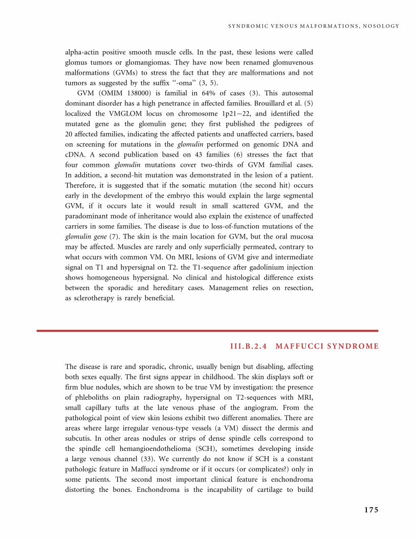
alpha-actin positive smooth muscle cells. In the past, these lesions were called
glomus tumors or glomangiomas. They have now been renamed glomuvenous
malformations (GVMs) to stress the fact that they are malformations and not
tumors as suggested by the suffix ‘‘-oma’’ (3, 5).
GVM (OMIM 138000) is familial in 64% of cases (3). This autosomal
dominant disorder has a high penetrance in affected families. Brouillard et al. (5)
localized the VMGLOM locus on chromosome 1p21�22, and identified the
mutated gene as the glomulin gene; they first published the pedigrees of
20 affected families, indicating the affected patients and unaffected carriers, based
on screening for mutations in the glomulin performed on genomic DNA and
cDNA. A second publication based on 43 families (6) stresses the fact that
four common glomulin mutations cover two-thirds of GVM familial cases.
In addition, a second-hit mutation was demonstrated in the lesion of a patient.
Therefore, it is suggested that if the somatic mutation (the second hit) occurs
early in the development of the embryo this would explain the large segmental
GVM, if it occurs late it would result in small scattered GVM, and the
paradominant mode of inheritance would also explain the existence of unaffected
carriers in some families. The disease is due to loss-of-function mutations of the
glomulin gene (7). The skin is the main location for GVM, but the oral mucosa
may be affected. Muscles are rarely and only superficially permeated, contrary to
what occurs with common VM. On MRI, lesions of GVM give and intermediate
signal on T1 and hypersignal on T2. the T1-sequence after gadolinium injection
shows homogeneous hypersignal. No clinical and histological difference exists
between the sporadic and hereditary cases. Management relies on resection,
as sclerotherapy is rarely beneficial.
I I I .B .2 . 4 MAFFUCCI SYNDROME
The disease is rare and sporadic, chronic, usually benign but disabling, affecting
both sexes equally. The first signs appear in childhood. The skin displays soft or
firm blue nodules, which are shown to be true VM by investigation: the presence
of phleboliths on plain radiography, hypersignal on T2-sequences with MRI,
small capillary tufts at the late venous phase of the angiogram. From the
pathological point of view skin lesions exhibit two different anomalies. There are
areas where large irregular venous-type vessels (a VM) dissect the dermis and
subcutis. In other areas nodules or strips of dense spindle cells correspond to
the spindle cell hemangioendothelioma (SCH), sometimes developing inside
a large venous channel (33). We currently do not know if SCH is a constant
pathologic feature in Maffucci syndrome or if it occurs (or complicates?) only in
some patients. The second most important clinical feature is enchondroma
distorting the bones. Enchondroma is the incapability of cartilage to build
S Y N D R O M I C V E N O U S M A L F O R M A T I O N S , N O S O L O G Y
175

normal bone. Radiographically there are translucent metaphyseal and diaphyseal
masses. The hands and feet are affected in nearly 90% of patients, while one third
of them have long-bone involvement creating distortion and dwarfism.
Neurological complications may occur with cranial involvement. Enchondrosar-
coma occurs; however, this malignant change may have been overestimated
(30% of cases) in the literature.
V E N O U S M A L F O R M A T I O N S ( V M )
176
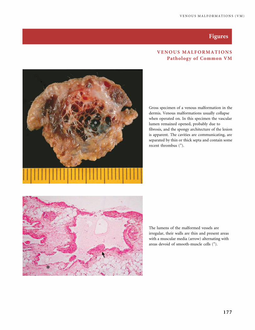
Figures
VENOUS MALFORMATIONS
Pathology of Common VM
The lumens of the malformed vessels are
irregular, their walls are thin and present areas
with a muscular media (arrow) alternating with
areas devoid of smooth-muscle cells (�).
Gross specimen of a venous malformation in the
dermis. Venous malformations usually collapse
when operated on. In this specimen the vascular
lumen remained opened, probably due to
fibrosis, and the spongy architecture of the lesion
is apparent. The cavities are communicating, are
separated by thin or thick septa and contain some
recent thrombus (�).
V E N O U S M A L F O R M A T I O N S ( V M )
177

Pathology of Common VM
Venous malformation of the skin. The
lesion seems to replace the dermis
collagen. Hair follicles (arrows) and
a small nerve (arrowhead) are floating
in the cavities.
Immunohistochemistry with an
anti-alpha smooth-muscle cell actin
antibody (decorating the smooth-
muscle cells in brown) highlights the
defective character of the media,
with large areas devoid of smooth-
muscle cells.
V E N O U S M A L F O R M A T I O N S ( V M )
178
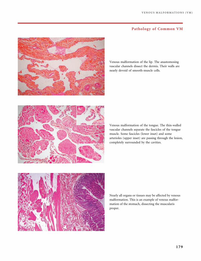
Pathology of Common VM
Nearly all organs or tissues may be affected by venous
malformation. This is an example of venous malfor-
mation of the stomach, dissecting the muscularis
proper.
Venous malformation of the lip. The anastomosing
vascular channels dissect the dermis. Their walls are
nearly devoid of smooth-muscle cells.
Venous malformation of the tongue. The thin-walled
vascular channels separate the fascicles of the tongue
muscle. Some fascicles (lower inset) and some
arterioles (upper inset) are passing through the lesion,
completely surrounded by the cavities.
V E N O U S M A L F O R M A T I O N S ( V M )
179
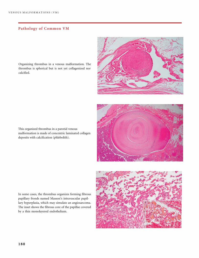
Pathology of Common VM
In some cases, the thrombus organizes forming fibrous
papillary fronds named Masson’s intravascular papil-
lary hyperplasia, which may simulate an angiosarcoma.
The inset shows the fibrous core of the papillae covered
by a thin monolayered endothelium.
Organizing thrombus in a venous malformation. The
thrombus is spherical but is not yet collagenized nor
calcified.
This organized thrombus in a parotid venous
malformation is made of concentric laminated collagen
deposits with calcification (phlebolith).
V E N O U S M A L F O R M A T I O N S ( V M )
180

Clinical Patterns, Investigations, and Treatment
In the cephalic area VMs are usually unilateral, but some are bilateral, and may also be associated with another trunk or limb
location of VM. The blue color of a facial VM is easily observed in skin. The full lesion may appear blue (a). In patient (b)
there is some swelling of the cheek, which is fully involved by VM, but the blue hue mainly affects the lower lid and upper lip,
a very common presentation.
The blue hue is also clearly seen on the affected mucosa: in the mouth area the surface of
the tongue is of a deep blue.
V E N O U S M A L F O R M A T I O N S ( V M )
181
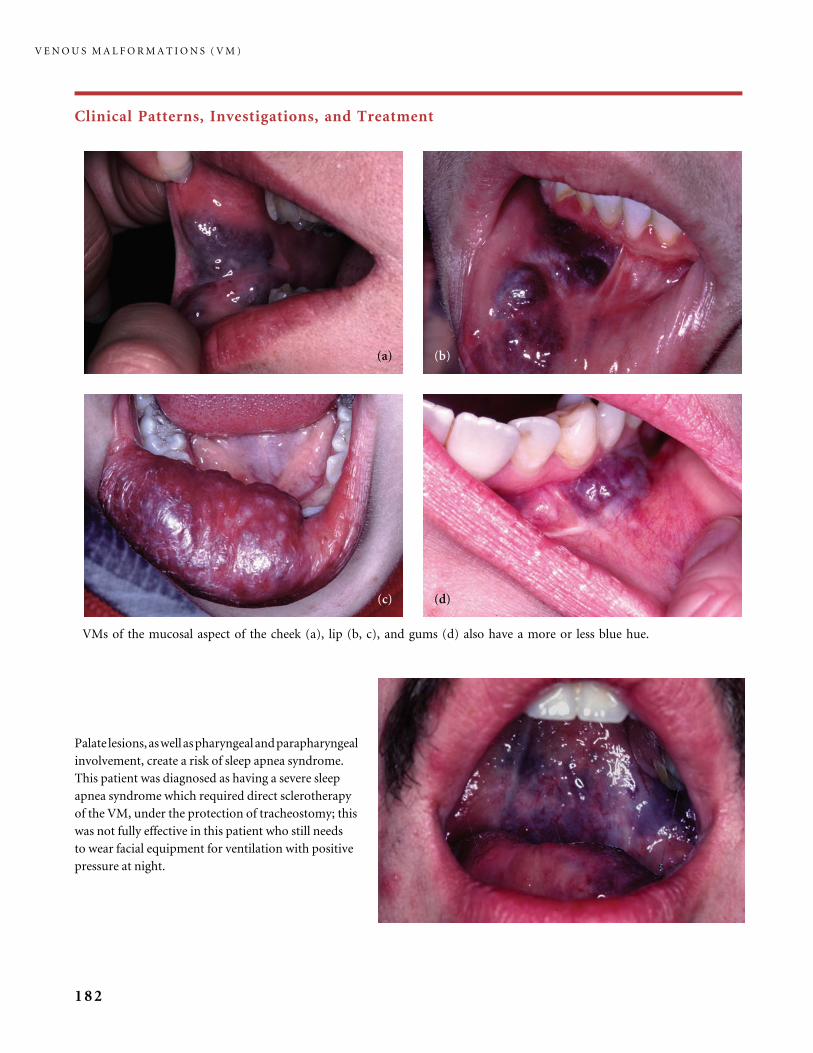
Clinical Patterns, Investigations, and Treatment
VMs of the mucosal aspect of the cheek (a), lip (b, c), and gums (d) also have a more or less blue hue.
Palate lesions,aswellaspharyngealandparapharyngeal
involvement, create a risk of sleep apnea syndrome.
This patient was diagnosed as having a severe sleep
apnea syndrome which required direct sclerotherapy
of the VM, under the protection of tracheostomy; this
was not fully effective in this patient who still needs
to wear facial equipment for ventilation with positive
pressure at night.
V E N O U S M A L F O R M A T I O N S ( V M )
182
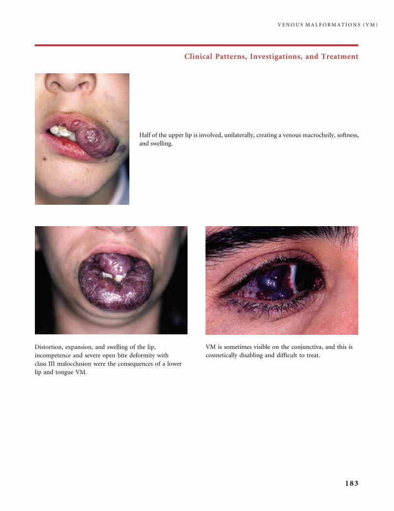
Clinical Patterns, Investigations, and Treatment
Half of the upper lip is involved, unilaterally, creating a venous macrocheily, softness,
and swelling.
Distortion, expansion, and swelling of the lip,
incompetence and severe open bite deformity with
class III malocclusion were the consequences of a lower
lip and tongue VM.
VM is sometimes visible on the conjunctiva, and this is
cosmetically disabling and difficult to treat.
V E N O U S M A L F O R M A T I O N S ( V M )
183
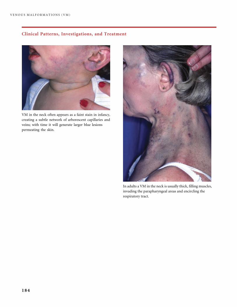
Clinical Patterns, Investigations, and Treatment
VM in the neck often appears as a faint stain in infancy,
creating a subtle network of arborescent capillaries and
veins; with time it will generate larger blue lesions
permeating the skin.
In adults a VM in the neck is usually thick, filling muscles,
invading the parapharyngeal areas and encircling the
respiratory tract.
V E N O U S M A L F O R M A T I O N S ( V M )
184
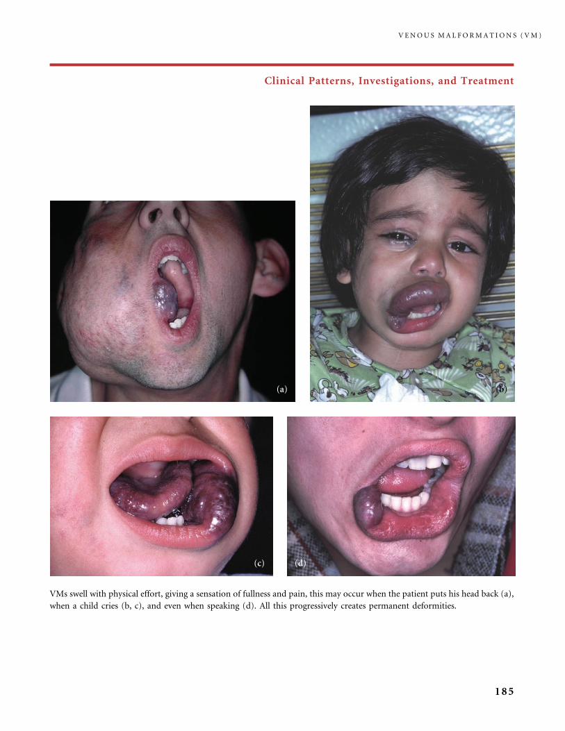
Clinical Patterns, Investigations, and Treatment
VMs swell with physical effort, giving a sensation of fullness and pain, this may occur when the patient puts his head back (a),
when a child cries (b, c), and even when speaking (d). All this progressively creates permanent deformities.
V E N O U S M A L F O R M A T I O N S ( V M )
185
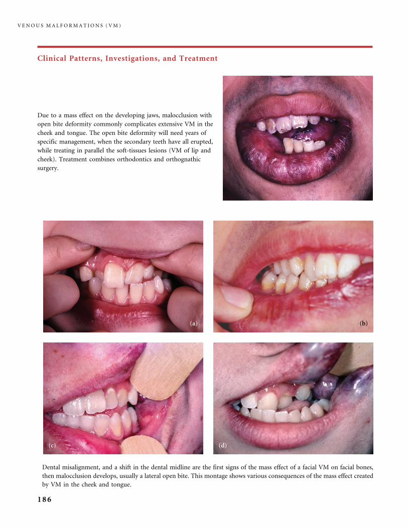
Clinical Patterns, Investigations, and Treatment
Dental misalignment, and a shift in the dental midline are the first signs of the mass effect of a facial VM on facial bones,
then malocclusion develops, usually a lateral open bite. This montage shows various consequences of the mass effect created
by VM in the cheek and tongue.
Due to a mass effect on the developing jaws, malocclusion with
open bite deformity commonly complicates extensive VM in the
cheek and tongue. The open bite deformity will need years of
specific management, when the secondary teeth have all erupted,
while treating in parallel the soft-tissues lesions (VM of lip and
cheek). Treatment combines orthodontics and orthognathic
surgery.
V E N O U S M A L F O R M A T I O N S ( V M )
186
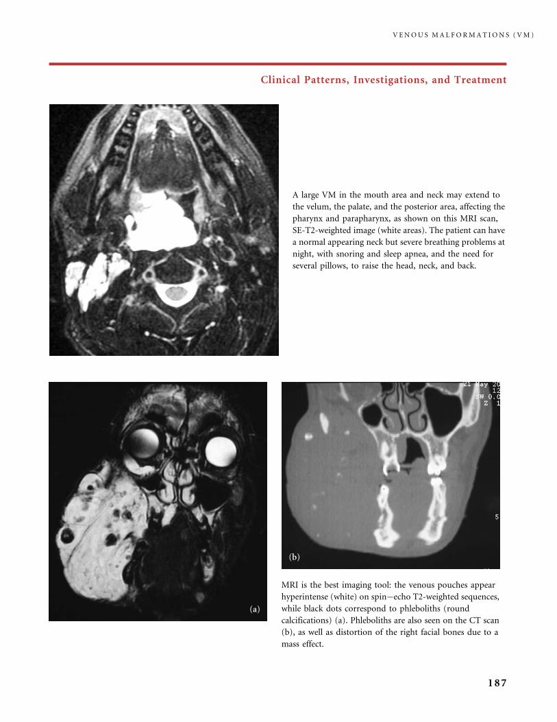
Clinical Patterns, Investigations, and Treatment
A large VM in the mouth area and neck may extend to
the velum, the palate, and the posterior area, affecting the
pharynx and parapharynx, as shown on this MRI scan,
SE-T2-weighted image (white areas). The patient can have
a normal appearing neck but severe breathing problems at
night, with snoring and sleep apnea, and the need for
several pillows, to raise the head, neck, and back.
MRI is the best imaging tool: the venous pouches appear
hyperintense (white) on spin�echo T2-weighted sequences,
while black dots correspond to phleboliths (round
calcifications) (a). Phleboliths are also seen on the CT scan
(b), as well as distortion of the right facial bones due to a
mass effect.
V E N O U S M A L F O R M A T I O N S ( V M )
187
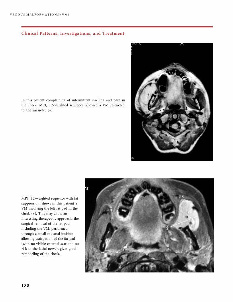
Clinical Patterns, Investigations, and Treatment
MRI, T2-weighted sequence with fat
suppression, shows in this patient a
VM involving the left fat pad in the
cheek (�). This may allow an
interesting therapeutic approach: the
surgical removal of the fat pad,
including the VM, performed
through a small mucosal incision
allowing extirpation of the fat pad
(with no visible external scar and no
risk to the facial nerve), gives good
remodeling of the cheek.
In this patient complaining of intermittent swelling and pain in
the cheek; MRI, T2-weighted sequence, showed a VM restricted
to the masseter (�).
V E N O U S M A L F O R M A T I O N S ( V M )
188

Clinical Patterns, Investigations, and Treatment
MRI in this patient with VM in the entire neck
revealed not only deep lesions around the
respiratory tract, but also VM close to the
parotid gland, a finding suggesting a high risk
of facial palsy if ethanol sclerosing treatment
of this part of the VM is planned.
VM on the mobile part of the tongue creates
dental malposition and malocclusion due to
the mass effect and pressure, but it rarely
impairs speech, while VM of the base of the
tongue and pharynx can cause difficulties in
swallowing, airway compromise, and sleep
apnea syndrome.
V E N O U S M A L F O R M A T I O N S ( V M )
189
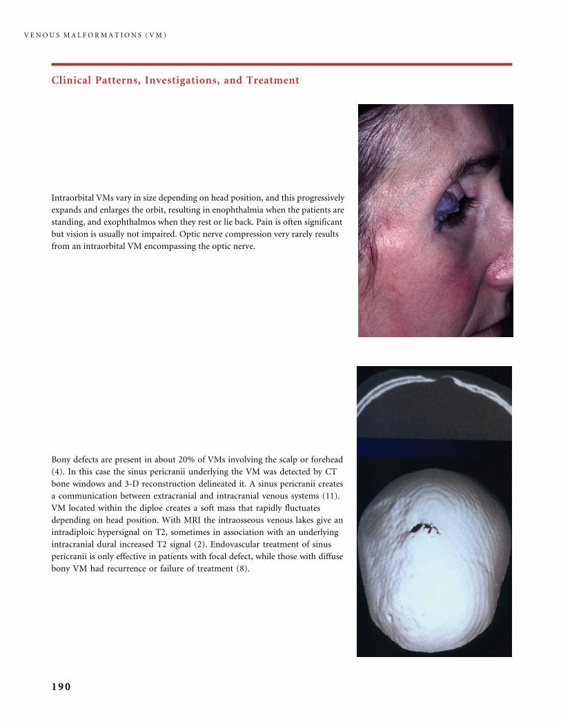
Clinical Patterns, Investigations, and Treatment
Intraorbital VMs vary in size depending on head position, and this progressively
expands and enlarges the orbit, resulting in enophthalmia when the patients are
standing, and exophthalmos when they rest or lie back. Pain is often significant
but vision is usually not impaired. Optic nerve compression very rarely results
from an intraorbital VM encompassing the optic nerve.
Bony defects are present in about 20% of VMs involving the scalp or forehead
(4). In this case the sinus pericranii underlying the VM was detected by CT
bone windows and 3-D reconstruction delineated it. A sinus pericranii creates
a communication between extracranial and intracranial venous systems (11).
VM located within the diploe creates a soft mass that rapidly fluctuates
depending on head position. With MRI the intraosseous venous lakes give an
intradiploic hypersignal on T2, sometimes in association with an underlying
intracranial dural increased T2 signal (2). Endovascular treatment of sinus
pericranii is only effective in patients with focal defect, while those with diffuse
bony VM had recurrence or failure of treatment (8).
V E N O U S M A L F O R M A T I O N S ( V M )
190

Clinical Patterns, Investigations, and Treatment
Cerebral developmental venous anomaly (DVA) consists of dilated intramedullary veins converging into a large draining
vein. This uncommon trajectory of the brain venous drainage occurs in less than 0.5% of the general population, while in
our experience it is observed in 20% patients with head and neck VMs. In patients with cephalic VMs, DVAs usually
consist of ectatic and dilated veins converging into the drainage system of the deep brain (4). In contrast to cerebral
‘‘cavernoma’’ (previously known as ‘‘angiographically occult’’ vascular malformation), opacification of DVA appears in
the angiographic venous phase, as do normal veins. DVA is imaged using CT and MRI with MRA. Patients with DVA
usually complain of headaches, but they are not at risk of cerebral hemorrhage, seizures or neurological deficit.
These very limited VMs in a finger were both present at birth; the blue color is distinctive: it indicates that the malformed
venous channels permeate the skin reaching the very superficial dermis. Treatment of such a lesion in a child is nearly
impossible.
V E N O U S M A L F O R M A T I O N S ( V M )
191

Clinical Patterns, Investigations, and Treatment
VM can affect an entire upper or lower
extremity and the adjacent trunk. It brings
about swelling of the soft tissues, deformity,
some increase in limb girth, but no limb
length discrepancy. In time function is
affected, with pain; symptoms are increased
after exercising and upon waking in the
morning and tend to abate with rest. Elastic
garments for both the hand and the arm, or
leg and foot, are absolutely indispensable, as
they bring comfort, limit swelling and pain,
and reduce the consequence of the localized
intravascular coagulopathy (LIC).
V E N O U S M A L F O R M A T I O N S ( V M )
192
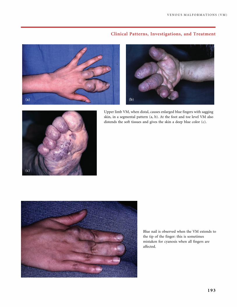
Clinical Patterns, Investigations, and Treatment
Upper limb VM, when distal, causes enlarged blue fingers with sagging
skin, in a segmental pattern (a, b). At the foot and toe level VM also
distends the soft tissues and gives the skin a deep blue color (c).
Blue nail is observed when the VM extends to
the tip of the finger: this is sometimes
mistaken for cyanosis when all fingers are
affected.
V E N O U S M A L F O R M A T I O N S ( V M )
193
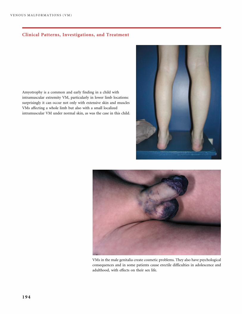
Clinical Patterns, Investigations, and Treatment
VMs in the male genitalia create cosmetic problems. They also have psychological
consequences and in some patients cause erectile difficulties in adolescence and
adulthood, with effects on their sex life.
Amyotrophy is a common and early finding in a child with
intramuscular extremity VM, particularly in lower limb locations:
surprisingly it can occur not only with extensive skin and muscles
VMs affecting a whole limb but also with a small localized
intramuscular VM under normal skin, as was the case in this child.
V E N O U S M A L F O R M A T I O N S ( V M )
194
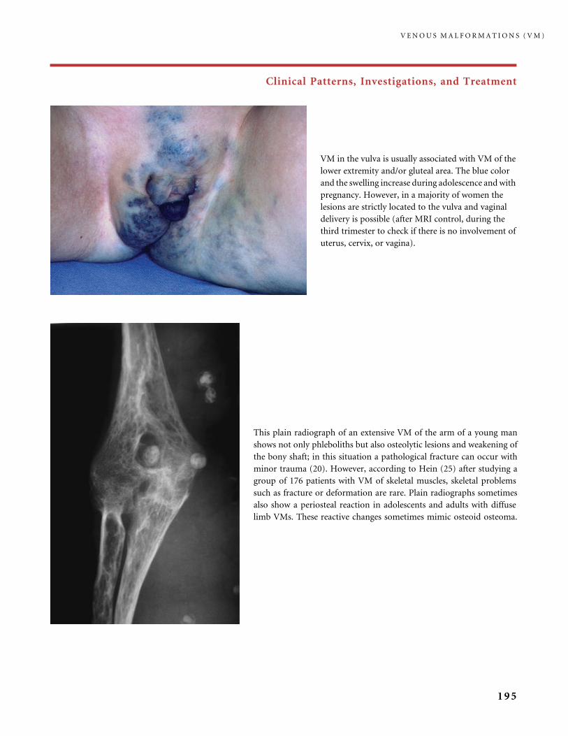
Clinical Patterns, Investigations, and Treatment
This plain radiograph of an extensive VM of the arm of a young man
shows not only phleboliths but also osteolytic lesions and weakening of
the bony shaft; in this situation a pathological fracture can occur with
minor trauma (20). However, according to Hein (25) after studying a
group of 176 patients with VM of skeletal muscles, skeletal problems
such as fracture or deformation are rare. Plain radiographs sometimes
also show a periosteal reaction in adolescents and adults with diffuse
limb VMs. These reactive changes sometimes mimic osteoid osteoma.
VM in the vulva is usually associated with VM of the
lower extremity and/or gluteal area. The blue color
and the swelling increase during adolescence and with
pregnancy. However, in a majority of women the
lesions are strictly located to the vulva and vaginal
delivery is possible (after MRI control, during the
third trimester to check if there is no involvement of
uterus, cervix, or vagina).
V E N O U S M A L F O R M A T I O N S ( V M )
195

Clinical Patterns, Investigations, and Treatment
Such a diffuse involvement of the arm and trunk in this young man (a) precludes surgical treatment: muscles
are all extensively filled and excision will be too disabling. The extent of the VM also does not allow us to
offer efficient percutaneous sclerotherapy: this challenging treatment will end in fairly deceptive results, and
there is also a high risk of adverse effects, particularly migration of the embolic material with possible nerve
damage or pulmonary embolism. In this case, elastic garments are indispensable, as well as medical treatment
(low-molecular-weight heparin injections) because of flares of the associated localized intravascular
coagulopathy (LIC): the heparin treatment minimizes pain, a common complaint linked to episodes of
venous thrombosis. In most cases thrombosis affects superficial veins, but occasionally deep vein thrombosis
happens, carrying, rarely, a risk of pulmonary embolism. Thrombosis precedes phlebolith formation (b, c).
T2-sequence of MRI (d) shows the VM filling the muscles.
V E N O U S M A L F O R M A T I O N S ( V M )
196
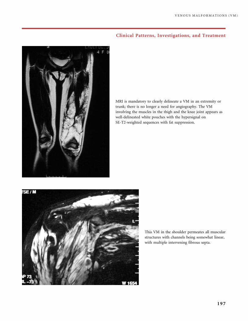
Clinical Patterns, Investigations, and Treatment
This VM in the shoulder permeates all muscular
structures with channels being somewhat linear,
with multiple intervening fibrous septa.
MRI is mandatory to clearly delineate a VM in an extremity or
trunk; there is no longer a need for angiography. The VM
involving the muscles in the thigh and the knee joint appears as
well-delineated white pouches with the hypersignal on
SE-T2-weighted sequences with fat suppression.
V E N O U S M A L F O R M A T I O N S ( V M )
197

Clinical Patterns, Investigations, and Treatment
This is an example of the full work-up required for a VM located in the thigh; the lesion is
detected by US (a) and Doppler (b), a venous flow is barely visible on the MRI T1 sequence (c),
but clearly shown on the SE T1-sequence after gadolinium injection (d). Figure continues at top
of page 201.
V E N O U S M A L F O R M A T I O N S ( V M )
198
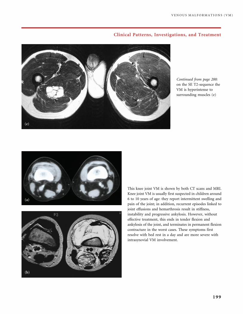
Clinical Patterns, Investigations, and Treatment
This knee joint VM is shown by both CT scans and MRI.
Knee joint VM is usually first suspected in children around
6 to 10 years of age: they report intermittent swelling and
pain of the joint; in addition, recurrent episodes linked to
joint effusions and hemarthrosis result in stiffness,
instability and progressive ankylosis. However, without
effective treatment, this ends in tender flexion and
ankylosis of the joint, and terminates in permanent flexion
contracture in the worst cases. These symptoms first
resolve with bed rest in a day and are more severe with
intrasynovial VM involvement.
Continued from page 200:
on the SE T2-sequence the
VM is hyperintense to
surrounding muscles (e)
V E N O U S M A L F O R M A T I O N S ( V M )
199
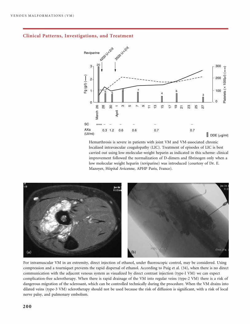
Clinical Patterns, Investigations, and Treatment
Hemarthrosis is severe in patients with joint VM and VM-associated chronic
localized intravascular coagulopathy (LIC). Treatment of episodes of LIC is best
carried out using low-molecular-weight heparin as indicated in this scheme: clinical
improvement followed the normalization of D-dimers and fibrinogen only when a
low molecular weight heparin (reviparine) was introduced (courtesy of Dr. E.
Mazoyer, Hopital Avicenne, APHP Paris, France).
For intramuscular VM in an extremity, direct injection of ethanol, under fluoroscopic control, may be considered. Using
compression and a tourniquet prevents the rapid dispersal of ethanol. According to Puig et al. (34), when there is no direct
communication with the adjacent venous system as visualized by direct contrast injection (type-I VM) we can expect
complication-free sclerotherapy. When there is rapid drainage of the VM into regular veins (type-2 VM) there is a risk of
dangerous migration of the sclerosant, which can be controlled technically during the procedure. When the VM drains into
dilated veins (type-3 VM) sclerotherapy should not be used because the risk of diffusion is significant, with a risk of local
nerve palsy, and pulmonary embolism.
V E N O U S M A L F O R M A T I O N S ( V M )
200

Clinical Patterns, Investigations, and Treatment
A soft-palate VM, shown on MRI scan (a), induced severe dyspnea. As surgical removal of the VM-enlarged soft palate
could cause important functional impairment, it was contraindicated, and thus direct sclerotherapy was decided on.
The soft palate was accessed by direct puncture (b) and a mixture of ethanol, Ethibloc� and lipiodol was injected, after
a tracheostomy because of the expected serious inflammatory reaction with risk of breathing impairment. Tracheostomy
is also recommended when there is bulking of the posterior tongue and pharyngeal VM, creating a risk of local wound
and hemorrhage during intubation for general anesthesia. CT immediately after sclerosis showed filling of the VM with
the sclerosing (�) agent (c). Nearly complete resolution of the VM was shown on MRI scan in (d) 3 months later, while the
functional symptoms had disappeared.
V E N O U S M A L F O R M A T I O N S ( V M )
201
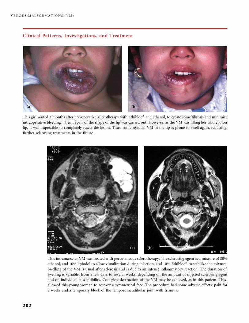
Clinical Patterns, Investigations, and Treatment
This intramasseter VM was treated with percutaneous sclerotherapy. The sclerosing agent is a mixture of 80%
ethanol, and 10% lipiodol to allow visualization during injection, and 10% Ethibloc� to stabilize the mixture.
Swelling of the VM is usual after sclerosis and is due to an intense inflammatory reaction. The duration of
swelling is variable, from a few days to several weeks, depending on the amount of injected sclerosing agent
and on individual susceptibility. Complete destruction of the VM may be achieved, as in this patient. This
allowed this young woman to recover a symmetrical face. The procedure had some adverse effects: pain for
2 weeks and a temporary block of the temporomandibular joint with trismus.
This girl waited 3 months after pre-operative sclerotherapy with Ethibloc� and ethanol, to create some fibrosis and minimize
intraoperative bleeding. Then, repair of the shape of the lip was carried out. However, as the VM was filling her whole lower
lip, it was impossible to completely resect the lesion. Thus, some residual VM in the lip is prone to swell again, requiring
further sclerosing treatments in the future.
V E N O U S M A L F O R M A T I O N S ( V M )
202
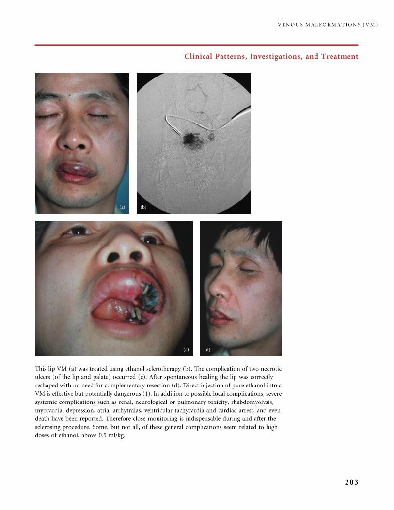
Clinical Patterns, Investigations, and Treatment
This lip VM (a) was treated using ethanol sclerotherapy (b). The complication of two necrotic
ulcers (of the lip and palate) occurred (c). After spontaneous healing the lip was correctly
reshaped with no need for complementary resection (d). Direct injection of pure ethanol into a
VM is effective but potentially dangerous (1). In addition to possible local complications, severe
systemic complications such as renal, neurological or pulmonary toxicity, rhabdomyolysis,
myocardial depression, atrial arrhytmias, ventricular tachycardia and cardiac arrest, and even
death have been reported. Therefore close monitoring is indispensable during and after the
sclerosing procedure. Some, but not all, of these general complications seem related to high
doses of ethanol, above 0.5 ml/kg.
V E N O U S M A L F O R M A T I O N S ( V M )
203
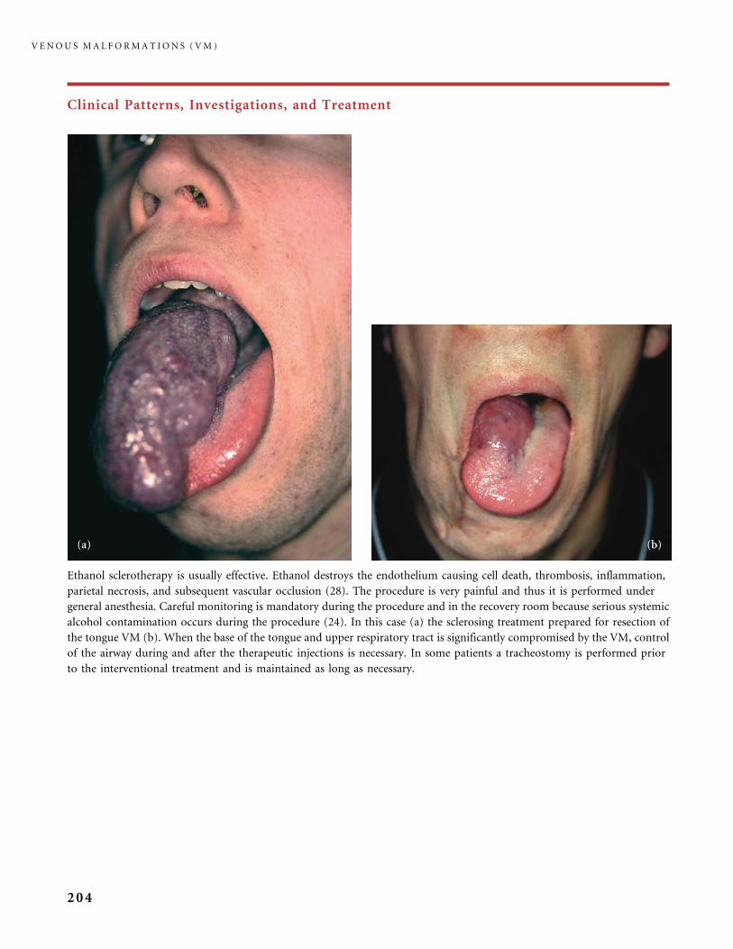
Clinical Patterns, Investigations, and Treatment
Ethanol sclerotherapy is usually effective. Ethanol destroys the endothelium causing cell death, thrombosis, inflammation,
parietal necrosis, and subsequent vascular occlusion (28). The procedure is very painful and thus it is performed under
general anesthesia. Careful monitoring is mandatory during the procedure and in the recovery room because serious systemic
alcohol contamination occurs during the procedure (24). In this case (a) the sclerosing treatment prepared for resection of
the tongue VM (b). When the base of the tongue and upper respiratory tract is significantly compromised by the VM, control
of the airway during and after the therapeutic injections is necessary. In some patients a tracheostomy is performed prior
to the interventional treatment and is maintained as long as necessary.
V E N O U S M A L F O R M A T I O N S ( V M )
204

Clinical Patterns, Investigations, and Treatment
Results of 15 years of therapeutic management including multiple use of direct puncture sclerotherapy with Ethibloc�,
various surgical procedures, with progressive excision of areas of VM bumping in the mouth, and blue skin locations on the
cheek. In addition orthodontic management was followed by bi-maxillary surgical treatment. With the use of Ethibloc�,
inflammation, swelling, and bruising are immediate and last from weeks to a few months. Usually surgery takes place a few
weeks later. Local complications of this sclerosing agent occur in about 10% of procedures and they include skin necrosis,
aseptic chronic drainage, and finally some degree of scarring. Peripheral nerve damage is extremely rare with Ethibloc�,
by contrast with absolute ethanol sclerotherapy. Fever commonly occurs after the procedure, but no severe systemic
complications have been reported with Ethibloc�.
V E N O U S M A L F O R M A T I O N S ( V M )
205
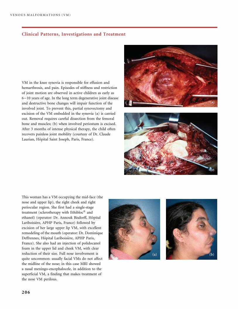
Clinical Patterns, Investigations and Treatment
VM in the knee synovia is responsible for effusion and
hemarthrosis, and pain. Episodes of stiffness and restriction
of joint motion are observed in active children as early as
6�10 years of age. In the long term degenerative joint disease
and destructive bone changes will impair function of the
involved joint. To prevent this, partial synovectomy and
excision of the VM embedded in the synovia (a) is carried
out. Removal requires careful dissection from the femoral
bone and muscles; (b) when involved periostum is excised.
After 3 months of intense physical therapy, the child often
recovers painless joint mobility (courtesy of Dr. Claude
Laurian, Hopital Saint Joseph, Paris, France).
This woman has a VM occupying the mid-face (the
nose and upper lip), the right cheek and right
periocular region. She first had a single-stage
treatment (sclerotherapy with Ethibloc� and
ethanol) (operator: Dr. Annouk Bisdorff, Hopital
Lariboisiere, APHP Paris, France) followed by
excision of her large upper lip VM, with excellent
remodeling of the mouth (operator: Dr. Dominique
Deffrennes, Hopital Lariboisiere, APHP Paris,
France). She also had an injection of polidocanol
foam in the upper lid and cheek VM, with clear
reduction of their size. Full nose involvement is
quite uncommon: usually facial VMs do not affect
the midline of the nose; in this case MRI showed
a nasal meningo-encephalocele, in addition to the
superficial VM, a finding that makes treatment of
the nose VM perilous.
V E N O U S M A L F O R M A T I O N S ( V M )
206
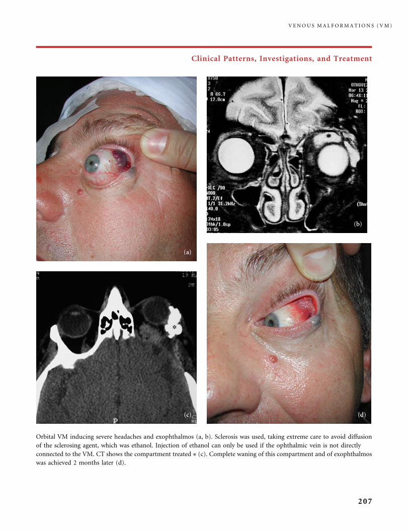
Clinical Patterns, Investigations, and Treatment
Orbital VM inducing severe headaches and exophthalmos (a, b). Sclerosis was used, taking extreme care to avoid diffusion
of the sclerosing agent, which was ethanol. Injection of ethanol can only be used if the ophthalmic vein is not directly
connected to the VM. CT shows the compartment treated � (c). Complete waning of this compartment and of exophthalmos
was achieved 2 months later (d).
V E N O U S M A L F O R M A T I O N S ( V M )
207
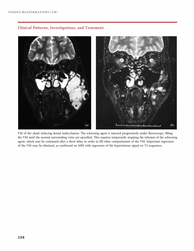
Clinical Patterns, Investigations, and Treatment
VM of the cheek inducing dental malocclusion. The sclerosing agent is injected progressively under fluoroscopy, filling
the VM until the normal surrounding veins are opacified. This requires temporarily stopping the infusion of the sclerosing
agent, which may be continued after a short delay in order to fill other compartments of the VM. Important regression
of the VM may be obtained, as confirmed on MRI with regression of the hyperintense signal on T2-sequences.
V E N O U S M A L F O R M A T I O N S ( V M )
208
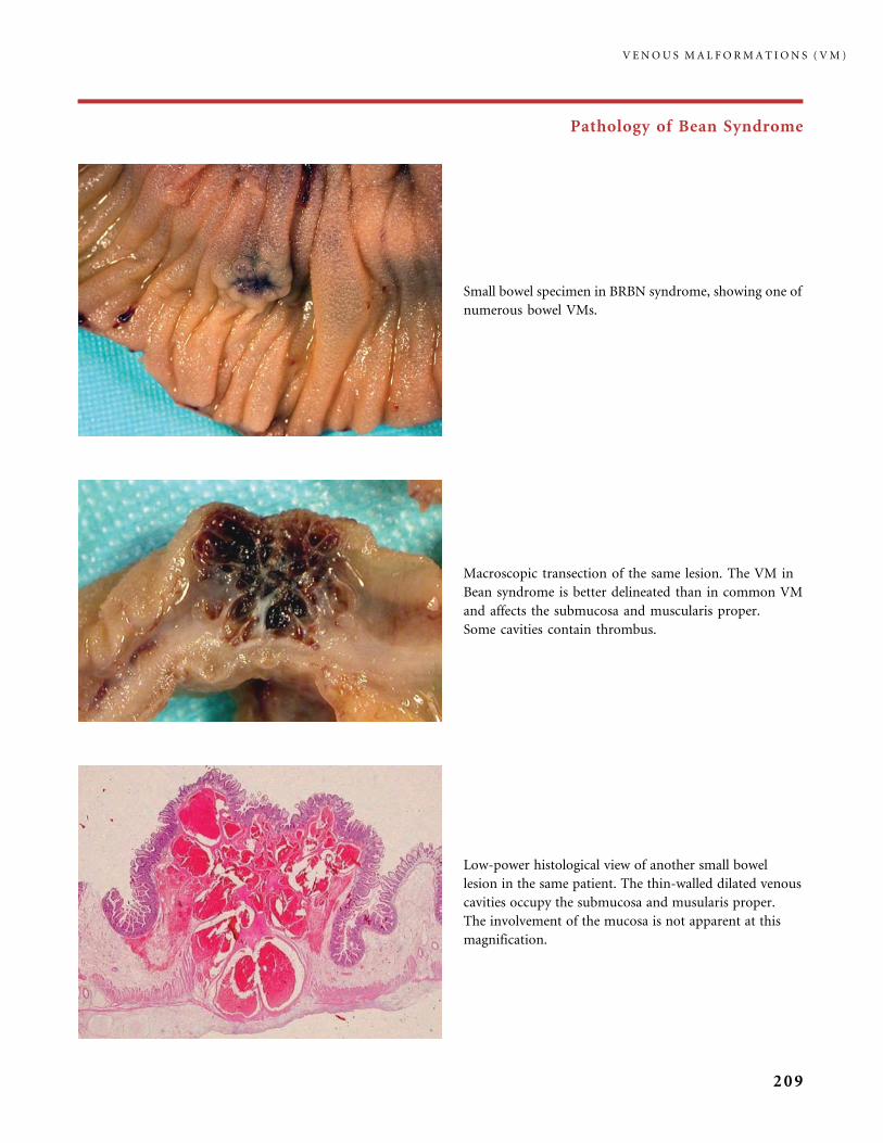
Pathology of Bean Syndrome
Low-power histological view of another small bowel
lesion in the same patient. The thin-walled dilated venous
cavities occupy the submucosa and musularis proper.
The involvement of the mucosa is not apparent at this
magnification.
Small bowel specimen in BRBN syndrome, showing one of
numerous bowel VMs.
Macroscopic transection of the same lesion. The VM in
Bean syndrome is better delineated than in common VM
and affects the submucosa and muscularis proper.
Some cavities contain thrombus.
V E N O U S M A L F O R M A T I O N S ( V M )
209
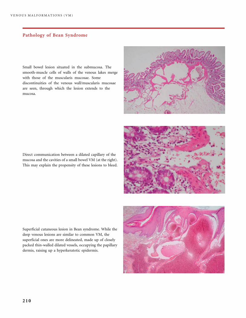
Pathology of Bean Syndrome
Superficial cutaneous lesion in Bean syndrome. While the
deep venous lesions are similar to common VM, the
superficial ones are more delineated, made up of closely
packed thin-walled dilated vessels, occupying the papillary
dermis, raising up a hyperkeratotic epidermis.
Small bowel lesion situated in the submucosa. The
smooth-muscle cells of walls of the venous lakes merge
with those of the muscularis mucosae. Some
discontinuities of the venous wall/muscularis mucosae
are seen, through which the lesion extends to the
mucosa.
Direct communication between a dilated capillary of the
mucosa and the cavities of a small bowel VM (at the right).
This may explain the propensity of these lesions to bleed.
V E N O U S M A L F O R M A T I O N S ( V M )
210
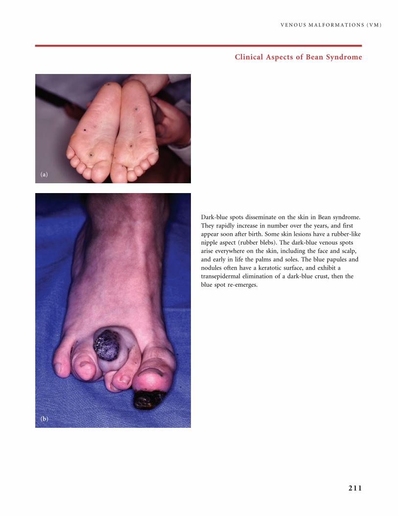
Clinical Aspects of Bean Syndrome
Dark-blue spots disseminate on the skin in Bean syndrome.
They rapidly increase in number over the years, and first
appear soon after birth. Some skin lesions have a rubber-like
nipple aspect (rubber blebs). The dark-blue venous spots
arise everywhere on the skin, including the face and scalp,
and early in life the palms and soles. The blue papules and
nodules often have a keratotic surface, and exhibit a
transepidermal elimination of a dark-blue crust, then the
blue spot re-emerges.
V E N O U S M A L F O R M A T I O N S ( V M )
211
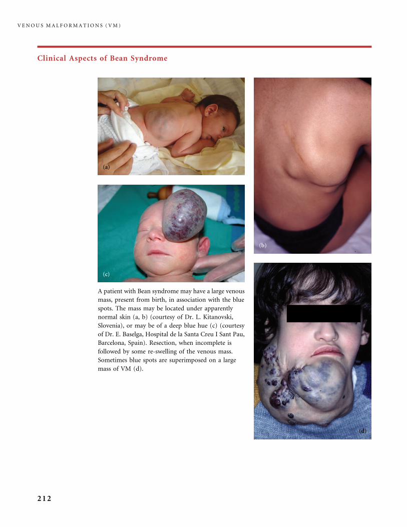
Clinical Aspects of Bean Syndrome
A patient with Bean syndrome may have a large venous
mass, present from birth, in association with the blue
spots. The mass may be located under apparently
normal skin (a, b) (courtesy of Dr. L. Kitanovski,
Slovenia), or may be of a deep blue hue (c) (courtesy
of Dr. E. Baselga, Hospital de la Santa Creu I Sant Pau,
Barcelona, Spain). Resection, when incomplete is
followed by some re-swelling of the venous mass.
Sometimes blue spots are superimposed on a large
mass of VM (d).
V E N O U S M A L F O R M A T I O N S ( V M )
212
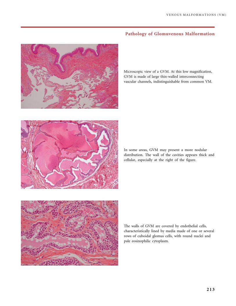
Pathology of Glomuvenous Malformation
The walls of GVM are covered by endothelial cells,
characteristically lined by media made of one or several
rows of cuboidal glomus cells, with round nuclei and
pale eosinophilic cytoplasm.
Microscopic view of a GVM. At this low magnification,
GVM is made of large thin-walled interconnecting
vascular channels, indistinguishable from common VM.
In some areas, GVM may present a more nodular
distribution. The wall of the cavities appears thick and
cellular, especially at the right of the figure.
V E N O U S M A L F O R M A T I O N S ( V M )
213
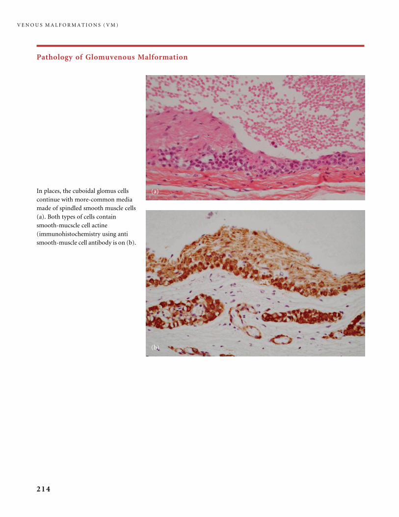
Pathology of Glomuvenous Malformation
In places, the cuboidal glomus cells
continue with more-common media
made of spindled smooth muscle cells
(a). Both types of cells contain
smooth-mucscle cell actine
(immunohistochemistry using anti
smooth-muscle cell antibody is on (b).
V E N O U S M A L F O R M A T I O N S ( V M )
214
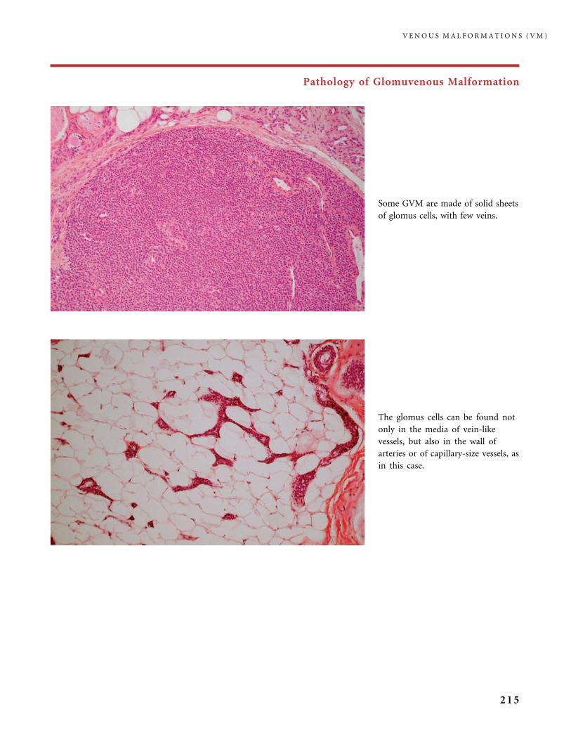
Pathology of Glomuvenous Malformation
The glomus cells can be found not
only in the media of vein-like
vessels, but also in the wall of
arteries or of capillary-size vessels, as
in this case.
Some GVM are made of solid sheets
of glomus cells, with few veins.
V E N O U S M A L F O R M A T I O N S ( V M )
215
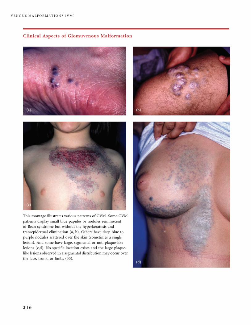
Clinical Aspects of Glomuvenous Malformation
This montage illustrates various patterns of GVM. Some GVM
patients display small blue papules or nodules reminiscent
of Bean syndrome but without the hyperkeratosis and
transepidermal elimination (a, b). Others have deep blue to
purple nodules scattered over the skin (sometimes a single
lesion). And some have large, segmental or not, plaque-like
lesions (c,d). No specific location exists and the large plaque-
like lesions observed in a segmental distribution may occur over
the face, trunk, or limbs (30).
V E N O U S M A L F O R M A T I O N S ( V M )
216
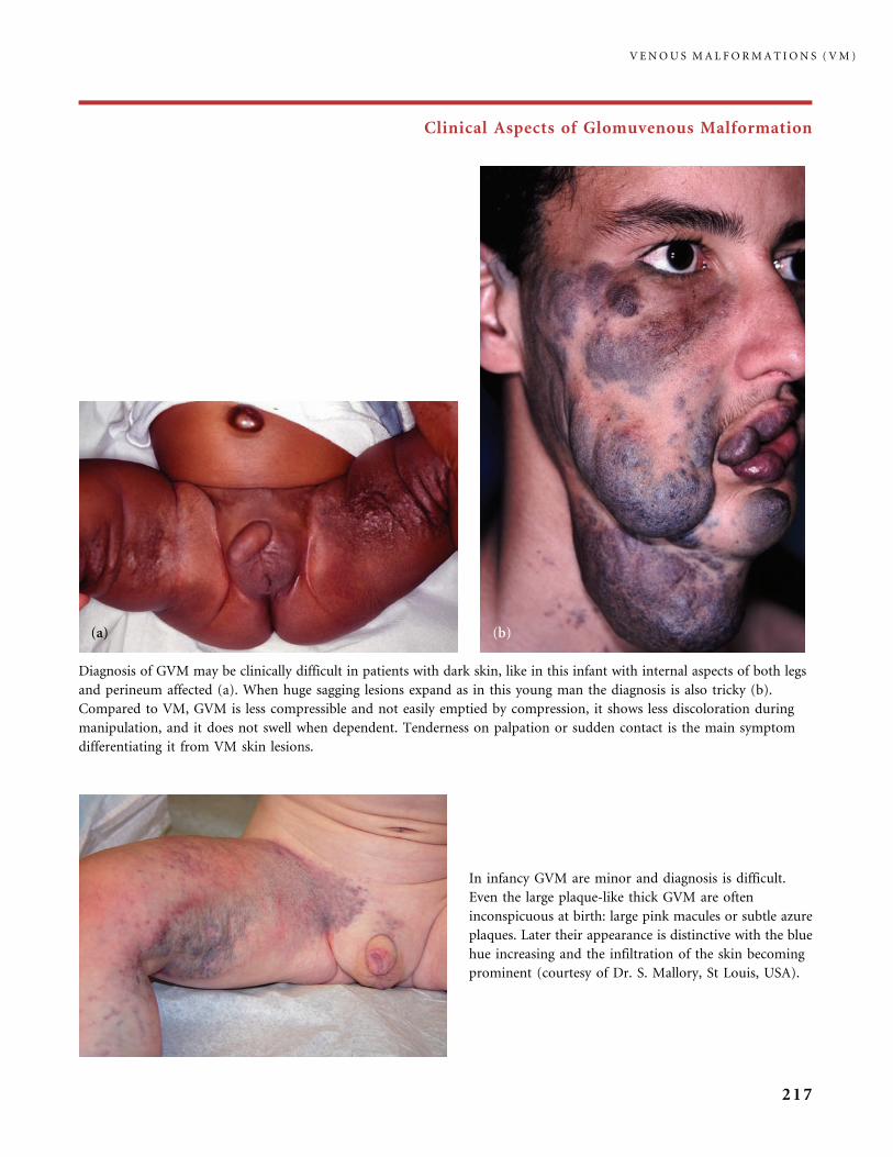
Clinical Aspects of Glomuvenous Malformation
Diagnosis of GVM may be clinically difficult in patients with dark skin, like in this infant with internal aspects of both legs
and perineum affected (a). When huge sagging lesions expand as in this young man the diagnosis is also tricky (b).
Compared to VM, GVM is less compressible and not easily emptied by compression, it shows less discoloration during
manipulation, and it does not swell when dependent. Tenderness on palpation or sudden contact is the main symptom
differentiating it from VM skin lesions.
In infancy GVM are minor and diagnosis is difficult.
Even the large plaque-like thick GVM are often
inconspicuous at birth: large pink macules or subtle azure
plaques. Later their appearance is distinctive with the blue
hue increasing and the infiltration of the skin becoming
prominent (courtesy of Dr. S. Mallory, St Louis, USA).
V E N O U S M A L F O R M A T I O N S ( V M )
217
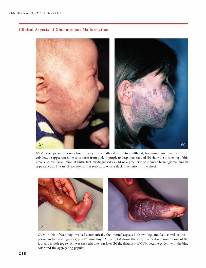
Clinical Aspects of Glomuvenous Malformation
GVM develops and thickens from infancy into childhood and into adulthood, becoming raised with a
cobblestone appearance; the color turns from pink or purple to deep blue: (a) and (b) show the thickening of this
inconspicuous facial lesion at birth, first misdiagnosed as CM or a precursor of infantile hemangioma, and its
appearance at 7 years of age after a first resection, with a thick blue lesion in the cheek.
GVM in this African boy involved symmetrically the internal aspects both two legs and feet, as well as the
perineum (see also figure (a) p. 217, same boy). At birth, (a) shows the shiny plaque-like lesion on one of the
foot and a sixth toe (which was excised); one year later (b) the diagnosis of GVM became evident with the blue
color and the aggregating papules.
V E N O U S M A L F O R M A T I O N S ( V M )
218
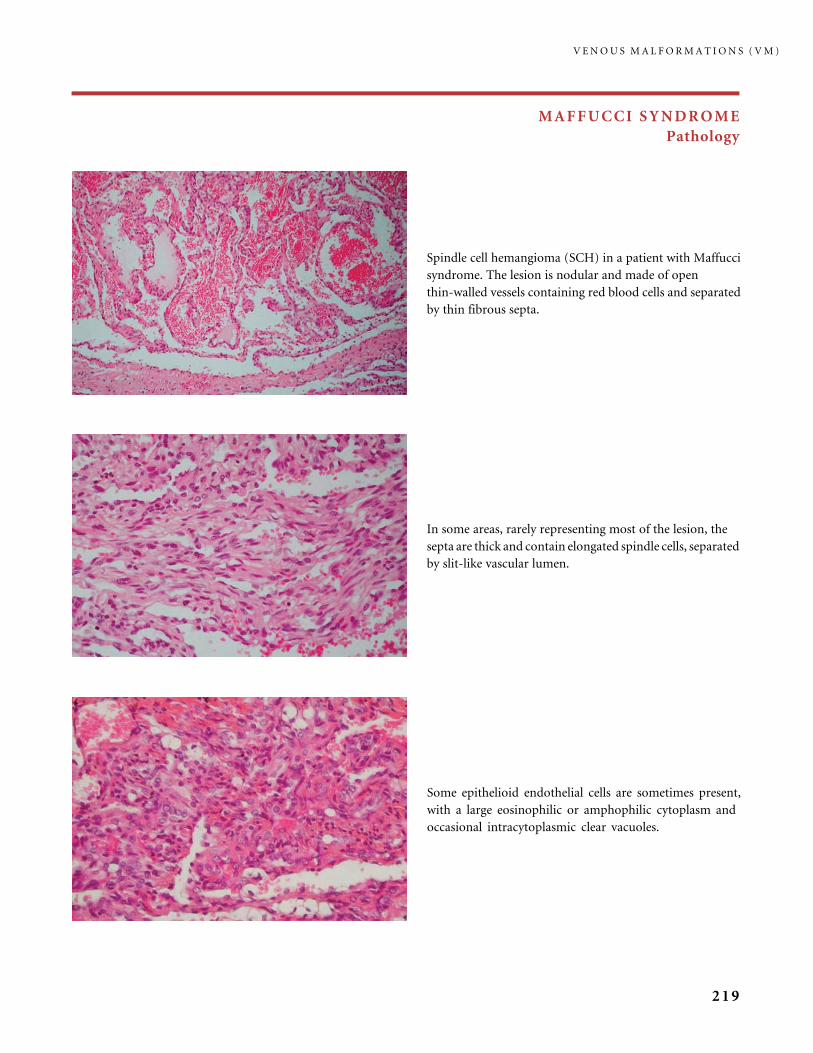
MAFFUCCI SYNDROME
Pathology
Some epithelioid endothelial cells are sometimes present,
with a large eosinophilic or amphophilic cytoplasm and
occasional intracytoplasmic clear vacuoles.
Spindle cell hemangioma (SCH) in a patient with Maffucci
syndrome. The lesion is nodular and made of open
thin-walled vessels containing red blood cells and separated
by thin fibrous septa.
In some areas, rarely representing most of the lesion, the
septa are thick and contain elongated spindle cells, separated
by slit-like vascular lumen.
V E N O U S M A L F O R M A T I O N S ( V M )
219
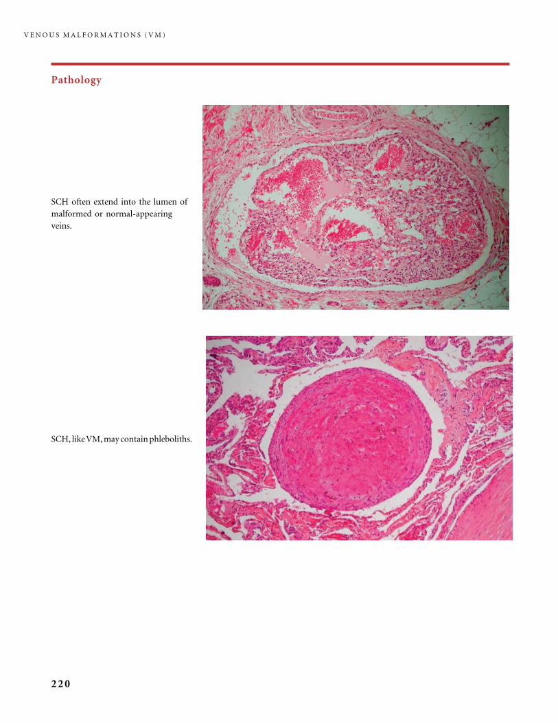
Pathology
SCH, like VM, may contain phleboliths.
SCH often extend into the lumen of
malformed or normal-appearing
veins.
V E N O U S M A L F O R M A T I O N S ( V M )
220

Clinical aspects
Maffucci syndrome. The hand lesions in patient
(a) comprised blue venous nodules (with phle-
boliths) while the bumps in the fingers in patient
(b) are mainly enchondromas; enchondromas give
translucent areas of the bones on the radiograph
in patient (c). Both distort the fingers.
V E N O U S M A L F O R M A T I O N S ( V M )
221

References
1 Berenguer B, Burrows PE, Zurakowski D, Mulliken JB. Sclerotherapy of craniofacial
venous malformations: complications and results. Plast Reconstr Surg 1999; 104: 1�11;
discussion 12�5.
2 Bigot JL, Iacona C, Lepreux A, Dhellemmes P, Motte J, Gomes H. Sinus pericranii:
advantages of MR imaging. Pediatr Radiol 2000; 30: 710�12.
3 Boon LM, Mulliken JB, Enjolras O, Vikkula M. Glomuvenous malformation (gloman-
gioma) and venous malformation: distinct clinicopathologic and genetic entities. Arch
Dermatol 2004; 140: 971�6.
4 Boukobza M, Enjolras O, Guichard JP, Gelbert F, Herbreteau D, Reizine D et al. Cerebral
developmental venous anomalies associated with head and neck venous malformations.
AJNR Am J Neuroradiol 1996; 17: 987�94.
5 Brouillard P, Boon LM, Mulliken JB, Enjolras O, Ghassibe M, Warman ML et al.
Mutations in a novel factor, glomulin, are responsible for glomuvenous malformations
(‘‘glomangiomas’’). Am J Hum Genet 2002; 70: 866�74.
6 Brouillard P, Ghassibe M, Penington A, Boon LM, Dompmartin A, Temple IK et al. Four
common glomulin mutations cause two thirds of glomuvenous malformations (‘‘familial
glomangiomas’’): evidence for a founder effect. J Med Genet 2005; 42: e13
7 Brouillard P, Vikkula M. Vascular malformations: localized defects in vascular
morphogenesis. Clin Genet 2003; 63: 340�51.
8 Burrows PE. Treatment of sinus pericranii. Communication in 15th ISSVA Workshop.
Wellington, NZ. 22�25 February 2004.
9 Burrows PE, Laor T, Paltiel H, Robertson RL. Diagnostic imaging in the evaluation of
vascular birthmarks. Dermatol Clin 1998; 16: 455�88.
10 Burrows PE, Mason KP. Percutaneous treatment of low flow vascular malformations.
J Vasc Interv Radiol 2004; 15: 431�45.
11 Buxton N, Vloeberghs M. Sinus pericranii. Report of a case and review of the literature.
Pediatr Neurosurg 1999; 30: 96�9.
12 Cabrera J, Cabrera J Jr, Garcia-Olmedo MA. Sclerosants in microfoam. A new approach in
angiology. Int Angiol 2001; 20: 322�9.
13 Cabrera J, Cabrera J Jr, Garcia-Olmedo MA, Redondo P. Treatment of venous
malformations with sclerosant in microfoam form. Arch Dermatol 2003; 139: 1409�16.
14 Cabrera J, Redondo P, Becerra A, Garrido C, Cabrera J Jr, Garcia-Olmedo MA et al.
Ultrasound-guided injection of polidocanol microfoam in the management of venous leg
ulcers. Arch Dermatol 2004; 140: 667�73.
15 Chapot R, Laurent A, Enjolras O, Payen D, Houdart E. Fatal cardiovascular collapse
during ethanol sclerotherapy of a venous malformation. Intervent Neuroradiol 2002; 8:
321�4.
16 Domini M, Aquino A, Fakhro A, Tursini S, Marino N, Di Matteo S et al. Blue rubber bleb
nevus syndrome and gastrointestinal haemorrhage: which treatment? Eur J Pediatr Surg
2002; 12: 129�33.
17 Dubois J, Garel L, Grignon A, David M, Laberge L, Filiatrault D et al. Imaging of
hemangiomas and vascular malformations in children. Acad Radiol 1998; 5: 390�400.
18 Dubois J, Soulez G, Oliva VL, Berthiaume MJ, Lapierre C, Therasse E. Soft-tissue venous
malformations in adult patients: imaging and therapeutic issues. Radiographics 2001; 21:
1519�31.
V E N O U S M A L F O R M A T I O N S ( V M )
222
19 Dubois JM, Sebag GH, De Prost Y, Teillac D, Chretien B, Brunelle FO. Soft-tissue venous
malformations in children: percutaneous sclerotherapy with Ethibloc. Radiology 1991;
180: 195�8.
20 Enjolras O, Ciabrini D, Mazoyer E, Laurian C, Herbreteau D. Extensive pure venous
malformations in the upper or lower limb: a review of 27 cases. J Am Acad Dermatol 1997;
36: 219�25.
21 Enjolras O, Mulliken JB. Vascular tumors and vascular malformations (new issues).
Adv Dermatol 1997; 13: 375�423.
22 Ertem D, Acar Y, Kotiloglu E, Yucelten D, Pehlivanoglu E. Blue rubber bleb nevus
syndrome. Pediatrics 2001; 107: 418�20.
23 Gallo SH, McClave S. A Blue rubber bleb nevus syndrome: gastrointestinal involvement
and its endoscopic presentation. Gastrointest Endosc 1992; 38: 72�6.
24 Hammer FD, Boon LM, Mathurin P, Vanwijck RR. Ethanol sclerotherapy of venous
malformations: evaluation of systemic ethanol contamination. J Vasc Interv Radiol 2001;
12: 595�600.
25 Hein KD, Mulliken JB, Kozakewich HP, Upton J, Burrows PE. Venous malformations of
skeletal muscle. Plast Reconstr Surg 2002; 110: 1625�35.
26 Kassarjian A, Fishman SJ, Fox VL, Burrows PE. Imaging characteristics of blue rubber bleb
nevus syndrome. AJR Am J Roentgenol 2003; 181: 1041�8.
27 Lasjaunias P, Berenstein A. Endovascular treatment of craniofacial lesions. Surgical
Neuroangiography. Vol. 2. Berlin: Springer Verlag, 1987.
28 Mason KP, Michna E, Zurakowski D, Koka BV, Burrows PE. Serum ethanol levels in
children and adults after ethanol embolization or sclerotherapy for vascular anomalies.
Radiology 2000; 217: 127�32.
29 Mazoyer E, Enjolras O, Laurian C, Houdart E, Drouet L. Coagulation abnormalities
associated with extensive venous malformations of the limbs: differentiation from
Kasabach�Merritt syndrome. Clin Lab Haematol 2002; 24: 243�51.
30 Mounayer C, Wassef M, Enjolras O, Boukobza M, Mulliken JB. Facial ‘‘glomangiomas’’:
large facial venous malformations with glomus cells. J Am Acad Dermatol 2001; 45:
239�45.
31 Mulliken JB, Fishman SJ, Burrows PE. Vascular anomalies. Curr Probl Surg 2000; 37:
517�84.
32 Paltiel HJ, Burrows PE, Kozakewich HP, Zurakowski D, Mulliken JB. Soft-tissue vascular
anomalies: utility of US for diagnosis. Radiology 2000; 214: 747�54.
33 Perkins P, Weiss SW. Spindle cell hemangioendothelioma. An analysis of 78 cases with
reassessment of its pathogenesis and biologic behavior. Am J Surg Pathol 1996; 20:
1196�204.
34 Puig S, Aref H, Chigot V, Bonin B, Brunelle F. Classification of venous malformations in
children and implications for sclerotherapy. Pediatr Radiol 2003; 33: 99�103.
35 Riche MC, Hadjean E, Tran-Ba-Huy P, Merland JJ. The treatment of capillary-venous
malformations using a new fibrosing agent. Plast Reconstr Surg 1983; 71: 607�14.
36 Siniluoto TM, Svendsen PA, Wikholm GM, Fogdestam I, Edstrom S. Percutaneous
sclerotherapy of venous malformations of the head and neck using sodium tetradecyl
sulphate (sotradecol). Scand J Plast Reconstr Surg Hand Surg 1997; 31: 145�50.
37 Suh JS, Shin KH, Na JB, Won JY, Hahn SB. Venous malformations: sclerotherapy with
a mixture of ethanol and lipiodol. Cardiovasc Intervent Radiol 1997; 20: 268�73.
38 Vikkula M, Boon LM, Carraway KL 3rd, Calvert JT, Diamonti AJ, Goumnerov B et al.
Vascular dysmorphogenesis caused by an activating mutation in the receptor tyrosine
kinase TIE2. Cell 1996; 87: 1181�90.
39 Waner M. Recent developments in lasers and the treatment of birthmarks. Arch Dis Child
2003; 88: 372�4.
40 Wassef M, Enjolras O. Les malformations vasculaires superficielles, classification
et histopathologie. Ann Pathol 1999; 19: 253�64.
41 Yakes WF. Extremity venous malformations. Semin Intervent Radiol 1994; 11: 332�9.
R E F E R E N C E S
223
CHAPTER III.C
Lymphatic Malformations (LM)
III.C.1 Common Lymphatic Malformations
Introduction
Lymphatic malformation (LM) is a malformation of the lymphatic system, and it
consists of small vesicles or large pouches filled with lymphatic fluid. The lymphatic
system is an open-ended unidirectional system returning interstitial fluid, macro-
molecules and immune cells from the tissues to the blood circulatory system (34).
Microcystic LMs (also known as tissular LMs) infiltrate soft tissues including
the skin and mucosa where they cause the emergence of clear or hemorrhagic
vesicles, and they also can occupy visceral territories in the thorax or abdomen,
and even bones.
Macrocystic LMs create large translucent lumps under normal skin, or are
deeply located.
Combined micro- and macrocystic forms are relatively common, in all
locations, superficially and internally.
LMs suddenly expand in the presence of regional inflammation or intralesional
bleeding. Three quarters of LMs are clinically evident before 5 years of age (14).
In the same series of 145 patients the most common location was the head and
neck (36.5%) followed by the extremities and axilla (31%) and the trunk (24.1%),
while visceral intrathoracic or abdominal lesions accounted for 8.2% of cases. In the
group of 186 patients studied by Alqahtani et al. (2) 48% of lesions were in the head
and neck and 42% were in the trunk or limbs, while 10% were internally located.
Ultrasonography detects intrauterine macrocystic LM (‘‘cystic hygroma’’) as
early as the late first trimester of pregnancy. Fetal hygroma colli cysticum are
septated or nonseptated on sonography for morphological features, and they carry
a risk of associated chromosomal abnormalities, particularly Turner syndrome and
Trisomy 18 (36).
Clinical Aspects
Depending on their location both the microcystic and macrocystic LMs have
distinctive effects and complications (11).
224
Microcystic LM
Long known as ‘‘lymphangioma circumscriptum,’’ microcystic LM is a plaque-like
ill-defined lesion involving the skin or visible on the mucous membranes. Clear,
yellowish, or blood-filled and dark-red vesicles spread over the surface of a smooth
area of swelling. Recurrent inflammation, bulging and bruises, or lymph and blood
oozing, are regularly observed during the chronic course of this malformation.
In childhood the surface occupied by vesicles is often minor compared to the
true future visible extent of the lesion. This is why very often an early resection
gives poor results, with vesicles later developing all around the surgical scar.
Macrocystic LM
In this condition (classically known as ‘‘cystic hygroma’’) the large cyst creates
a soft lump under a normal or slightly bluish skin. A macrocystic LM in the mouth
area may suddenly expand, usually during a nose or throat, viral or bacterial
infection, or because there is dental caries or gingivitis. Whatever their location
this sometimes sudden expansion results from intracystic spontaneous bleeding.
As a consequence, the mass becomes inflammatory, red, firm on palpation,
and tender.
Associated micro- and macrocystic LM is a common feature in the orbital
area. Intraorbital and periorbital LM produces exophthalmos, and it has visual
consequences (functional amblyopia, refraction abnormalities, mainly astigmatism
and strabismus). Sudden orbital proptosis was observed in 45% of cases in a group
of 42 children (16). In the same series one-half of the patients had both intraconal
and extraconal involvement, and it was extraconal or intraconal in one-quarter.
Visual loss may result from bleeding into orbital cysts encircling the optic nerve. In
the group of 42 patients reported by Greene et al. (16) 40% of children had
permanently diminished vision of multifactorial causes (amblyopia, exposure
keratitis and corneal ulcer, congenital cataract, retinal detachment, and glaucoma),
and three patients were blind in the affected eye.
Visceral LMs usually combine micro- and macrocystic cysts; however,
microcystic infiltrating lesions preponderate in most patients. They occur in the
thorax, abdomen, and buttocks and also involve bones. A number of the patients
with visceral lesions endure a life-threatening disease, which begins usually by
childhood or adolescence. It is frequently complicated by intralesional bleeding
episodes because of chronic coagulopathy with elevated D-dimers and low
fibrinogen.
Bony LMs have different presentations. In the trunk and limbs bone involve-
ment is either benign with no symptoms and no progression (15) or it carries
a risk of fracture in the affected bones, with possible neurological consequences
C O M M O N L Y M P H A T I C M A L F O R M A T I O N S
225

when the spine is involved. In the cervicofacial region LM creates various patterns
of osseous hypertrophy ending in widened interdental spaces, progressive maxil-
lary deformity or mandibular distortion, increased mandibular height, class III
malocclusion, anterior open bite, and prognathism (31).
Visceral LM with intestinal and pulmonary involvement is often called
‘‘lymphangiectasia.’’ It results in pleural effusions, abdominal pain, protein-losing
enteropathy, hypoalbuminemia and hypogammaglobulinemia, and lymphopenia.
It often manifests during childhood or adolescence and has a bad prognosis and
outcome. Intestinal lesions are managed by bowel rest and then a low-fat and
medium-chain triglyceride diet. Various pharmacological agents, including inter-
feron alpha, corticosteroids and vincristine, have usually been of no help. When
there is a coexisting coagulopathy, protracted treatment with a low-molecular-
weight heparin brings some improvement. There is one report of improved
gastrointestinal symptoms with tranexamic acid (25), and another with the
combination of corticosteroid and octreotide (a somatostatine analog).
Investigations
When clinical diagnosis for a macrocystic LM is unclear, the following techniques
are all valuable:
ULTRA SONOGRA PH Y (US ) AND US DOP P L E R of a macrocystic LM show
multiloculated anechoic cysts with no flow, while microcystic LM are hetero-
geneously hypoechoic with no flow.
CT SCAN S With these cysts are hypodense.
MRI shows a septated mass, cystic spaces being hypointense on SE T1-sequences
and usually there is no gadolinium contrast enhancement; cysts are of high signal
intensity on SE T2-sequences with fat suppression.
DIR E C T PUNC TUR E of a macrocystic lesion permits liquid analysis, to rule
out any other cystic lesion, in particular cystic malignancies. Direct iodinated
contrast injection, under fluoroscopic control, better delineates the lesion before
sclerotherapy.
Treatment
Sudden enlargement of a macrocystic LM is usually the result of intralesional
bleeding or infection. Pain relief, antibiotics, and anti-inflammatory drugs
(corticosteroids or nonsteroidal anti-inflammatory drugs) are prescribed.
Rarely, incision and drainage are necessary. Some lesions may spontaneously
L Y M P H A T I C M A L F O R M A T I O N S ( L M )
226

regress after such an episode. Large cysts are treated with aspiration of the
lymphatic fluid, followed by percutaneous intralesional injection of a sclerosing
agent, under fluoroscopic guidance. Puig et al. (33) recommend the use of a
double-needle to avoid elevation of pressure inside the lesion and to allow outflow
of excess sclerosant and contrast. After the procedure erythema and swelling with
variable pain are commonly observed: they require pain relief medication, and
a few days of corticosteroid treatment. Complications of sclerotherapy include
fistula with leakage of a mixture of the sclerosing agent, fibrin, and inflammatory
cells, and consequently a scar. Partial regression of the cyst requires re-injection or
excision. New cysts may develop months or years after apparently satisfactory
sclerosing treatments.
Surgical resection of the macrocystic LM is proposed either as a second step
after failure or incomplete results of sclerotherapy, or as first procedure (2, 16, 38).
Recurrence after incomplete excision is not surprising and adequate management
may be either a new surgical treatment or sclerotherapy. Post-operative compli-
cations include fistula with leakage of lymph fluid requiring drainage for weeks or
months, infection, burst of vesicles on a previously apparently undamaged skin
or mucosa, varying cosmetic damage with unaesthetic scarring, depending on the
LM location, and lymphedema (32).
Nd-YAG and diode laser photocoagulation have been employed to reduce
microcystic LM in two modalities: superficial, with or without continuous ice-cube
surface cooling, or interstitial after puncture or the lesions, but large lesions tend
to recur. Radiofrequency treatment is under evaluation for microcystic LM.
In extensive LM with lymphedema in the limbs, treatment will include
compression with a pneumatic device, compressive bandaging, and adapted elastic
support garments.
Macrocystic LM enlarges progressively during fetal life, but no prenatal
intervention is required in the majority of cases (10). Prenatal MRI identifies the
need for cesarean section. Massive lesions in the neck, tongue, and mouth areas
may require tracheotomy after birth because of significant airway compromise,
and a gastric tube may be necessary for feeding. Then various combined and
multiple therapeutic procedures and operations will aim at restoring the airway
and oropharyngeal function, and improving cosmesis (4).
III.C.2 Syndromic Lymphatic Malformations and Lymphedemas
II I .C .2 . 1 LYMPHEDEMAS
The primary lymphedemas are usually divided into two categories, the rare
congenital Milroy disease (OMIM 153100) and the more frequent late-onset Meige
lymphedema (OMIM 153200).
S Y N D R O M I C L Y M P H A T I C M A L F O R M A T I O N S A N D L Y M P H E D E M A S
227

Lymphedema can be associated with Turner and Noonan syndromes. In
Turner syndrome acral congenital lymphedema of the extremities is a presenting
manifestation as are the pterygium colli and redundant neck folds. They are
sequels of altered lymphatic pathogenesis; when detected during pregnancy, rarely
hygroma cysticum colli persists at birth; it usually resolves leaving the pterygium
colli (24).
This subcutaneous swelling, lymphedema, is more common in the lower
extremities, although it also occurs in the upper limbs, trunk, and even face.
Aplasia or hypoplasia of lymph vessels results in clinical edema either at birth
(early-onset forms) or later in life, until adulthood (late-onset forms).
Autosomal-dominant forms exist isolated or as part of a syndrome: primary
congenital lymphedema (locus on 5q34�35, the mutated gene FLT4 (VEGFR3) is
a tyrosine kinase receptor for vascular endothelial growth factor) (21), early-onset
familial lymphedema of Milroy, late-onset familial lymphedema of Meige,
lymphedema�distichiasis syndrome (with locus on 16q24.3, the mutated gene
being FOXC2), and hypotrichosis�lymphedema�telangiectasia syndrome (locus
on 20q, mutated gene SOX18)(7).
I I I .C .2 . 2 AAGENAES SYNDROME OR
CHOLESTASIS�LYMPHEDEMA SYNDROME
This disease was identified in 1968 by Aagenaes (1) and half of the reported cases are
of Norwegian origin. It combines a neonatal intrahepatic cholestasis that improves,
and chronic severe lymphedema mainly in the lower extremities that worsens.
Other features include: peripheral pulmonary stenosis, vertebral anomalies,
and a distinctive facies. The gene has been located to chromosome 15q (8).
I I I .C .2 . 3 HENNEKAM SYNDROME
Hennekam syndrome was described in 1989 as an autosomal recessive disease
comprising intestinal lymphangiectasia, lymphedema, facies anomalies, including
downslanting palpebral fissures and epicanthal folds, and mental retardation (18).
The phenotype was later expanded to other visceral anomalies (3).
I I I .C .2 . 4 GORHAM SYNDROME
Vanishing bone disease, the Gorham�Stout syndrome, or phantom bone,
or vanishing bone syndrome is a sporadic progressive and spontaneous
L Y M P H A T I C M A L F O R M A T I O N S ( L M )
228

bone-destructive process. Numerous dilated blood or lymph vascular channels are
present. They occupy the site of the vanished bones. The disease demineralizes and
destroys the affected bones. On plain radiographs, radiolucency of the involved
bones gives them a ‘‘licked stick of candy’’ appearance. Gorham syndrome causes
death in 16% of cases. Phantom bone disorder can be associated with chronic
coagulopathy and bleeding. An increase in osteoclast formation (from mono-
nuclear precursors) and differentiation, promotes osteolysis (20). When the
disease affects the spine and ribs it can cause chylothorax and paraplegia (40).
S Y N D R O M I C L Y M P H A T I C M A L F O R M A T I O N S A N D L Y M P H E D E M A S
229

Figures
LYMPHATIC MALFORMATIONS
Pathology
The dilated lymph vessels can be superficial and occupy
and expand the papillae of the dermis or mucosa, covered
with a thin epithelium. This is the pathological substratum
to the vesicles that are present in some LM. Thin
endothelial cell-covered fibrous projections, reminiscent of
valves, are seen in the cavities.
The histological structure of LM is similar to VM.
The malformed vessels can however be somewhat more
round or open with less anastomosis. The lumen contain
a clear fluid with rare macrophages.
Some vessels have very thin walls and contain a clear fluid,
sometimes with numerous intravascular lymphocytes.
Aggregates of lymphocytes are also present in the tissue,
closely associated to the malformed vessels. Inset: a vessel
lumen containing lymphocytes, a macrophage,
and red blood cells.
L Y M P H A T I C M A L F O R M A T I O N S ( L M )
230
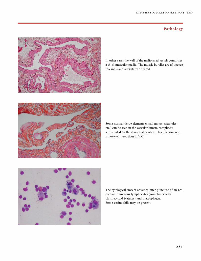
Pathology
Some normal tissue elements (small nerves, arterioles,
etc.) can be seen in the vascular lumen, completely
surrounded by the abnormal cavities. This phenomenon
is however rarer than in VM.
In other cases the wall of the malformed vessels comprises
a thick muscular media. The muscle bundles are of uneven
thickness and irregularly oriented.
The cytological smears obtained after puncture of an LM
contain numerous lymphocytes (sometimes with
plasmacytoid features) and macrophages.
Some eosinophils may be present.
L Y M P H A T I C M A L F O R M A T I O N S ( L M )
231
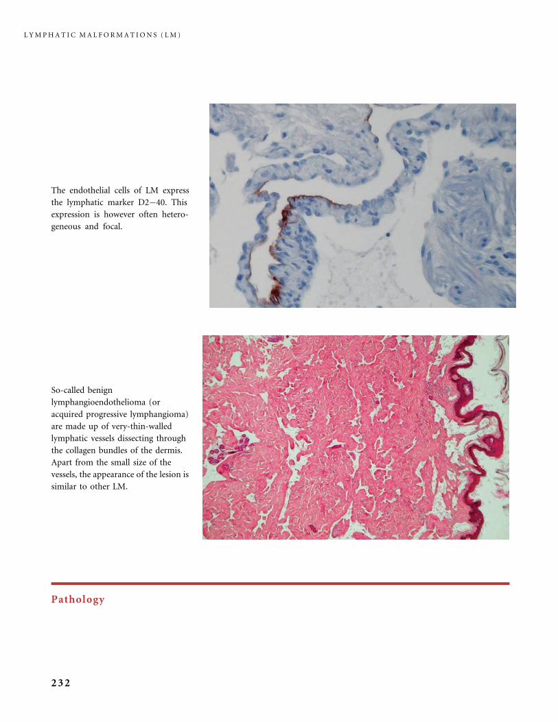
Pathology
So-called benign
lymphangioendothelioma (or
acquired progressive lymphangioma)
are made up of very-thin-walled
lymphatic vessels dissecting through
the collagen bundles of the dermis.
Apart from the small size of the
vessels, the appearance of the lesion is
similar to other LM.
The endothelial cells of LM express
the lymphatic marker D2�40. This
expression is however often hetero-
geneous and focal.
L Y M P H A T I C M A L F O R M A T I O N S ( L M )
232
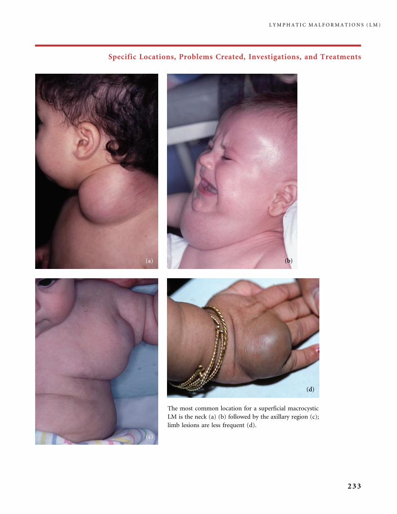
Specific Locations, Problems Created, Investigations, and Treatments
The most common location for a superficial macrocystic
LM is the neck (a) (b) followed by the axillary region (c);
limb lesions are less frequent (d).
L Y M P H A T I C M A L F O R M A T I O N S ( L M )
233
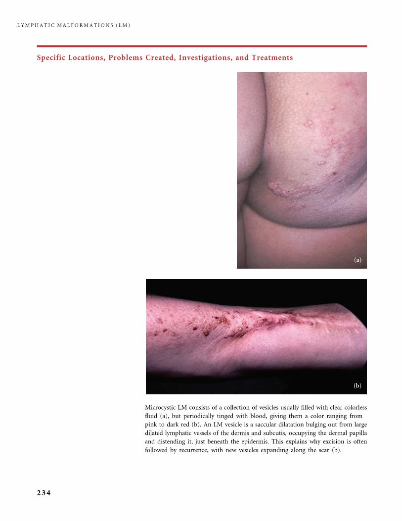
Specific Locations, Problems Created, Investigations, and Treatments
Microcystic LM consists of a collection of vesicles usually filled with clear colorless
fluid (a), but periodically tinged with blood, giving them a color ranging from
pink to dark red (b). An LM vesicle is a saccular dilatation bulging out from large
dilated lymphatic vessels of the dermis and subcutis, occupying the dermal papilla
and distending it, just beneath the epidermis. This explains why excision is often
followed by recurrence, with new vesicles expanding along the scar (b).
L Y M P H A T I C M A L F O R M A T I O N S ( L M )
234
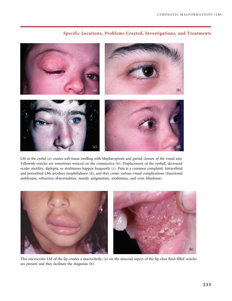
Specific Locations, Problems Created, Investigations, and Treatments
This microcystic LM of the lip creates a macrocheily; (a) on the mucosal aspect of the lip clear fluid-filled vesicles
are present and they facilitate the diagnosis (b).
LM in the eyelid (a) creates soft tissue swelling with blepharoptosis and partial closure of the visual axis.
Yellowish vesicles are sometimes noticed on the conjunctiva (b). Displacement of the eyeball, decreased
ocular motility, diplopia, or strabismus happen frequently (c). Pain is a common complaint. Intraorbital
and periorbital LMs produce exophthalmos (d), and they create various visual complications (functional
amblyopia, refraction abnormalities, mainly astigmatism, strabismus, and even blindness).
L Y M P H A T I C M A L F O R M A T I O N S ( L M )
235
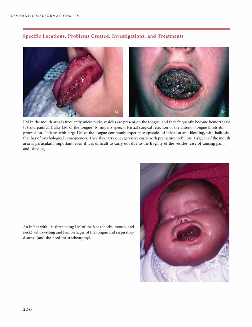
Specific Locations, Problems Created, Investigations, and Treatments
LM in the mouth area is frequently microcystic; vesicles are present on the tongue, and they frequently become hemorrhagic
(a) and painful. Bulky LM of the tongue (b) impairs speech. Partial surgical resection of the anterior tongue limits its
protraction. Patients with large LM of the tongue commonly experience episodes of infection and bleeding, with halitosis
that has of psychological consequences. They also carry out aggressive caries with premature teeth loss. Hygiene of the mouth
area is particularly important, even if it is difficult to carry out due to the fragility of the vesicles, ease of causing pain,
and bleeding.
An infant with life-threatening LM of the face (cheeks, mouth, and
neck) with swelling and hemorrhages of the tongue and respiratory
distress (and the need for tracheotomy).
L Y M P H A T I C M A L F O R M A T I O N S ( L M )
236
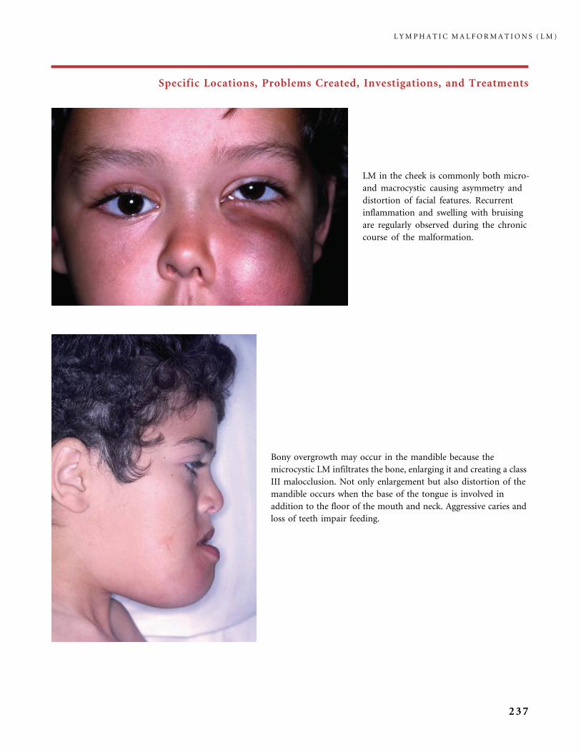
Specific Locations, Problems Created, Investigations, and Treatments
Bony overgrowth may occur in the mandible because the
microcystic LM infiltrates the bone, enlarging it and creating a class
III malocclusion. Not only enlargement but also distortion of the
mandible occurs when the base of the tongue is involved in
addition to the floor of the mouth and neck. Aggressive caries and
loss of teeth impair feeding.
LM in the cheek is commonly both micro-
and macrocystic causing asymmetry and
distortion of facial features. Recurrent
inflammation and swelling with bruising
are regularly observed during the chronic
course of the malformation.
L Y M P H A T I C M A L F O R M A T I O N S ( L M )
237

Specific Locations, Problems Created, Investigations, and Treatments
In the neck area, a diffuse LM creates airway obstruction, the
most extensive cases necessitating tracheotomy on a lifelong basis
(a, MRI showing the diffuse extent of a cervical and facial LM).
LM in the trunk and axilla can invade the thorax (b), some of
them encompassing recurrent pleural and pericardial effusion.
L Y M P H A T I C M A L F O R M A T I O N S ( L M )
238
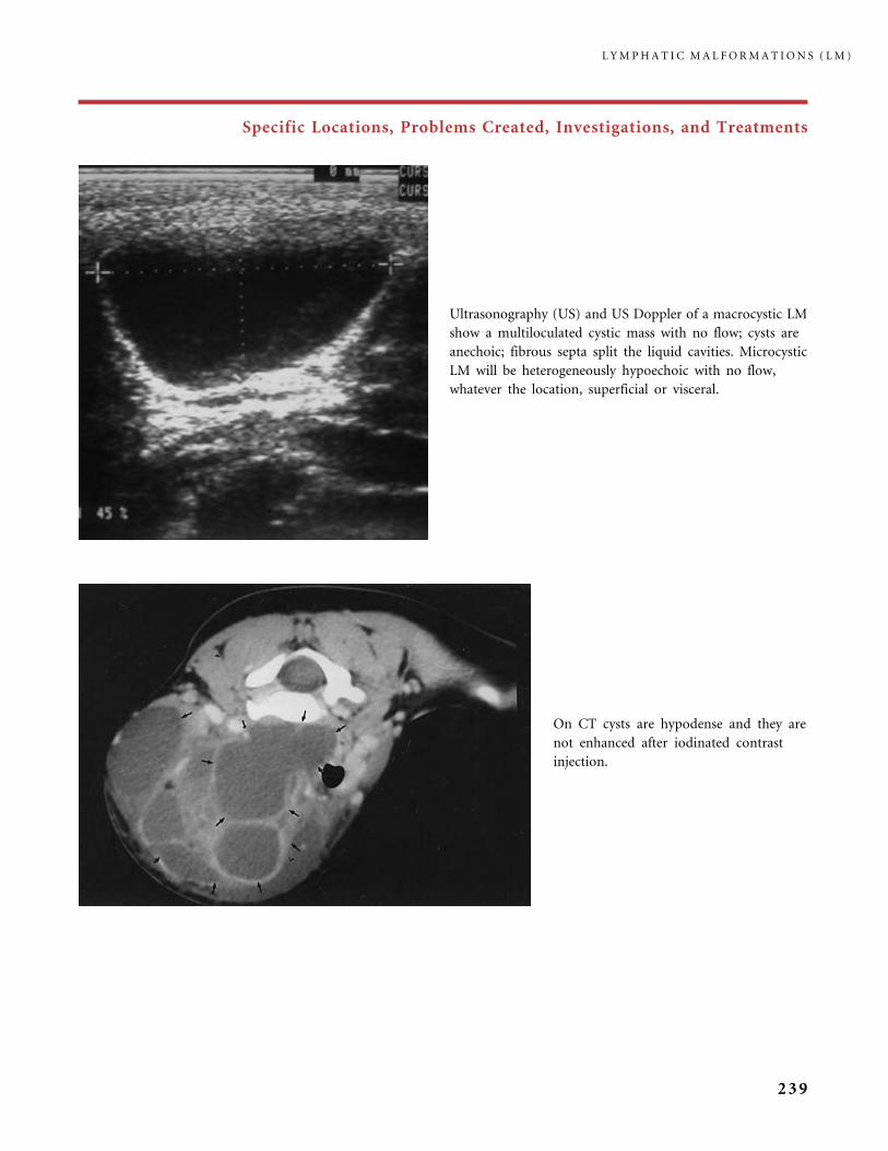
Specific Locations, Problems Created, Investigations, and Treatments
Ultrasonography (US) and US Doppler of a macrocystic LM
show a multiloculated cystic mass with no flow; cysts are
anechoic; fibrous septa split the liquid cavities. Microcystic
LM will be heterogeneously hypoechoic with no flow,
whatever the location, superficial or visceral.
On CT cysts are hypodense and they are
not enhanced after iodinated contrast
injection.
L Y M P H A T I C M A L F O R M A T I O N S ( L M )
239
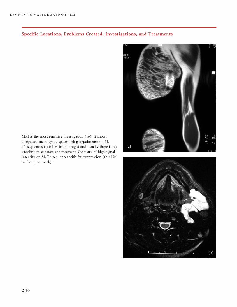
Specific Locations, Problems Created, Investigations, and Treatments
MRI is the most sensitive investigation (16). It shows
a septated mass, cystic spaces being hypointense on SE
T1-sequences ((a): LM in the thigh) and usually there is no
gadolinium contrast enhancement. Cysts are of high signal
intensity on SE T2-sequences with fat suppression ((b): LM
in the upper neck).
L Y M P H A T I C M A L F O R M A T I O N S ( L M )
240
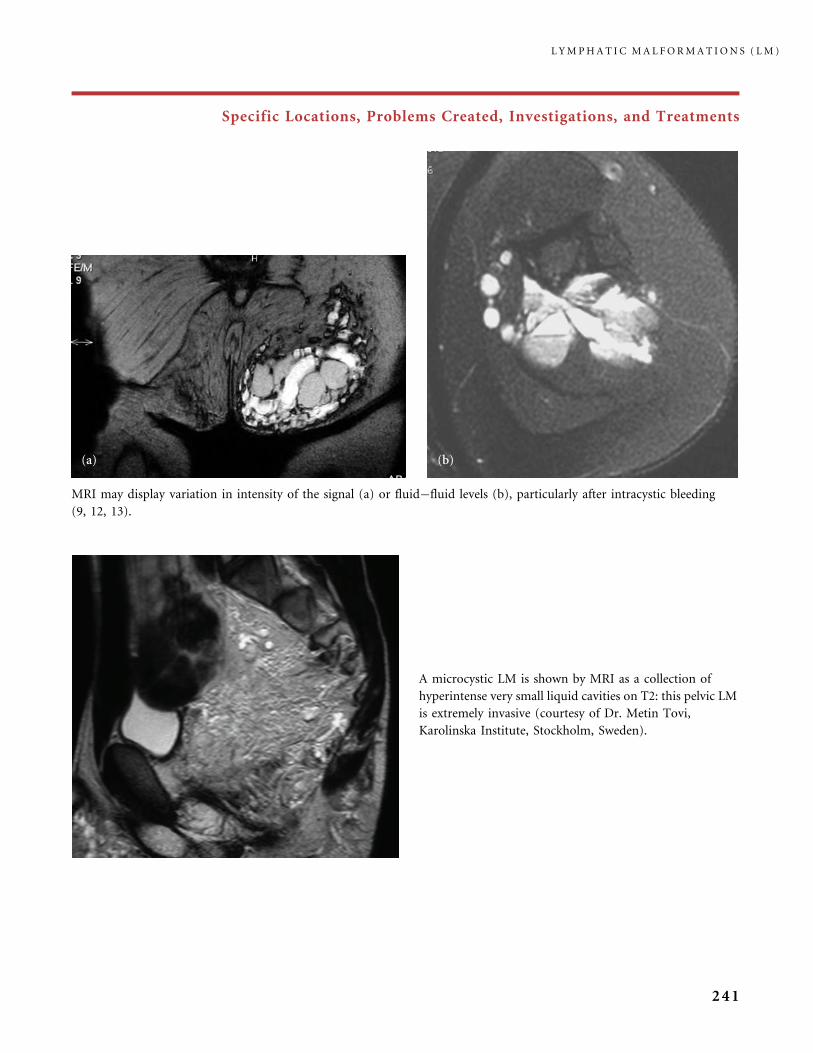
Specific Locations, Problems Created, Investigations, and Treatments
A microcystic LM is shown by MRI as a collection of
hyperintense very small liquid cavities on T2: this pelvic LM
is extremely invasive (courtesy of Dr. Metin Tovi,
Karolinska Institute, Stockholm, Sweden).
MRI may display variation in intensity of the signal (a) or fluid�fluid levels (b), particularly after intracystic bleeding
(9, 12, 13).
L Y M P H A T I C M A L F O R M A T I O N S ( L M )
241

Specific Locations, Problems Created, Investigations, and Treatments
Chronic LIC, with hemorrhages, as seen in VM, also occurs in a number of patients with diffuse LM, particularly LM
involving an entire extremity and internal organs. Oozing of skin vesicles and chronic lymphorrhea (a), or swelling
and bruises (b) are observed. The patient with diffuse LM in limbs is at high risk of sepsis.
Pelvic LM sometimes manifests in females as vulvar lymph-
angiectasia and vaginal chylous discharge, even before puberty.
L Y M P H A T I C M A L F O R M A T I O N S ( L M )
242

Specific Locations, Problems Created, Investigations, and Treatments
Lymphatic vesicles developing after acquired lymphatic obstruction secondary to radiotherapy for genital
cancer (a), or lymphangiectasia occurring in the perineum of a female patient with Crohn disease (b) are
indiscernible from microcystic vulvar LM (courtesy of Dr. M.Pelisse, Tarnier-Cochin Hospital, APHP Paris,
France). And lymphatic vesicles resembling a congenital LM also develop on the lateral aspect of the thorax
after mastectomy and radiotherapy for breast cancer (22).
In males pelvic LM is revealed by scrotal and penis lymphedema
with vesicles on top, or is discovered when an MR evaluation is
performed because of abdominal pain.
L Y M P H A T I C M A L F O R M A T I O N S ( L M )
243
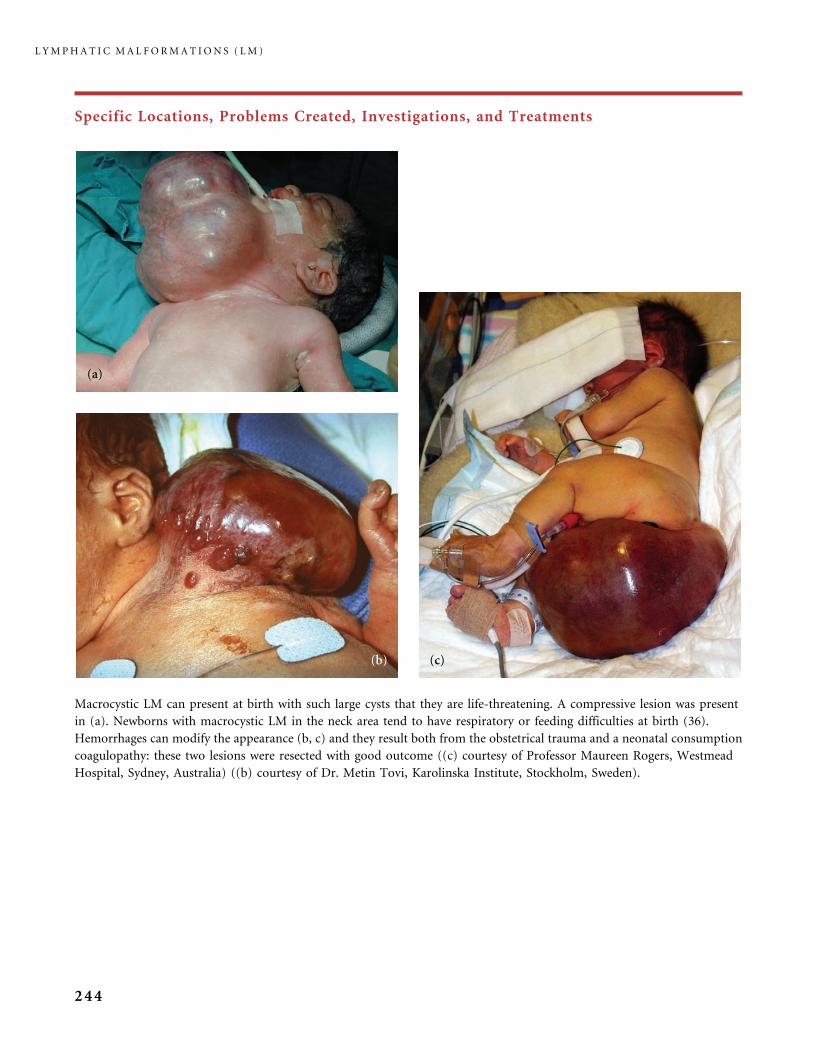
Specific Locations, Problems Created, Investigations, and Treatments
Macrocystic LM can present at birth with such large cysts that they are life-threatening. A compressive lesion was present
in (a). Newborns with macrocystic LM in the neck area tend to have respiratory or feeding difficulties at birth (36).
Hemorrhages can modify the appearance (b, c) and they result both from the obstetrical trauma and a neonatal consumption
coagulopathy: these two lesions were resected with good outcome ((c) courtesy of Professor Maureen Rogers, Westmead
Hospital, Sydney, Australia) ((b) courtesy of Dr. Metin Tovi, Karolinska Institute, Stockholm, Sweden).
L Y M P H A T I C M A L F O R M A T I O N S ( L M )
244
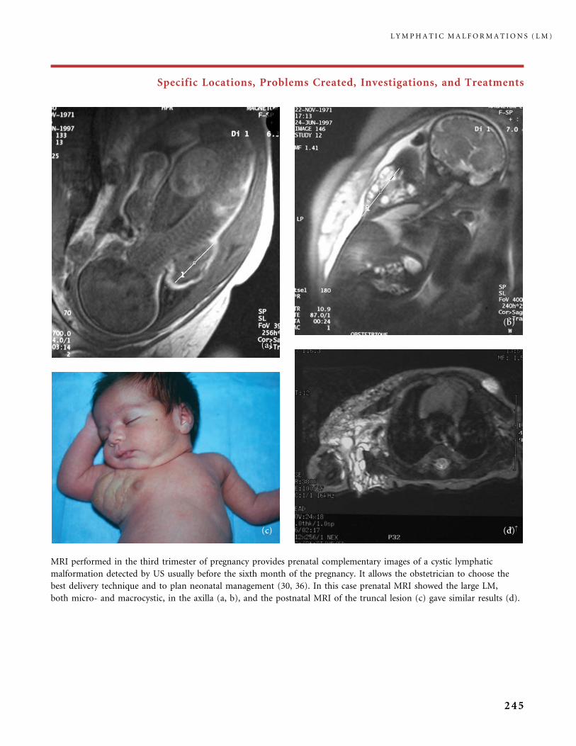
Specific Locations, Problems Created, Investigations, and Treatments
MRI performed in the third trimester of pregnancy provides prenatal complementary images of a cystic lymphatic
malformation detected by US usually before the sixth month of the pregnancy. It allows the obstetrician to choose the
best delivery technique and to plan neonatal management (30, 36). In this case prenatal MRI showed the large LM,
both micro- and macrocystic, in the axilla (a, b), and the postnatal MRI of the truncal lesion (c) gave similar results (d).
L Y M P H A T I C M A L F O R M A T I O N S ( L M )
245
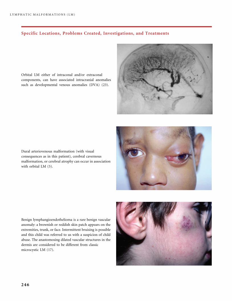
Specific Locations, Problems Created, Investigations, and Treatments
Benign lymphangioendothelioma is a rare benign vascular
anomaly: a brownish or reddish skin patch appears on the
extremities, trunk, or face. Intermittent bruising is possible
and this child was referred to us with a suspicion of child
abuse. The anastomosing dilated vascular structures in the
dermis are considered to be different from classic
microcystic LM (17).
Orbital LM either of intraconal and/or extraconal
components, can have associated intracranial anomalies
such as developmental venous anomalies (DVA) (23).
Dural arteriovenous malformation (with visual
consequences as in this patient), cerebral cavernous
malformation, or cerebral atrophy can occur in association
with orbital LM (5).
L Y M P H A T I C M A L F O R M A T I O N S ( L M )
246
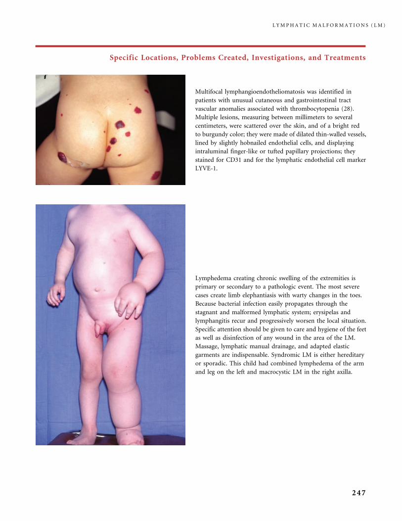
Specific Locations, Problems Created, Investigations, and Treatments
Lymphedema creating chronic swelling of the extremities is
primary or secondary to a pathologic event. The most severe
cases create limb elephantiasis with warty changes in the toes.
Because bacterial infection easily propagates through the
stagnant and malformed lymphatic system; erysipelas and
lymphangitis recur and progressively worsen the local situation.
Specific attention should be given to care and hygiene of the feet
as well as disinfection of any wound in the area of the LM.
Massage, lymphatic manual drainage, and adapted elastic
garments are indispensable. Syndromic LM is either hereditary
or sporadic. This child had combined lymphedema of the arm
and leg on the left and macrocystic LM in the right axilla.
Multifocal lymphangioendotheliomatosis was identified in
patients with unusual cutaneous and gastrointestinal tract
vascular anomalies associated with thrombocytopenia (28).
Multiple lesions, measuring between millimeters to several
centimeters, were scattered over the skin, and of a bright red
to burgundy color; they were made of dilated thin-walled vessels,
lined by slightly hobnailed endothelial cells, and displaying
intraluminal finger-like or tufted papillary projections; they
stained for CD31 and for the lymphatic endothelial cell marker
LYVE-1.
L Y M P H A T I C M A L F O R M A T I O N S ( L M )
247
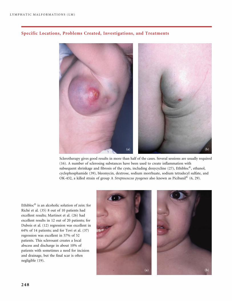
Specific Locations, Problems Created, Investigations, and Treatments
Ethibloc� is an alcoholic solution of zein: for
Riche et al. (35) 8 out of 10 patients had
excellent results; Martinot et al. (26) had
excellent results in 12 out of 20 patients; for
Dubois et al. (12) regression was excellent in
64% of 14 patients; and for Tovi et al. (37)
regression was excellent in 57% of 52
patients. This sclerosant creates a local
abscess and discharge in about 10% of
patients with sometimes a need for incision
and drainage, but the final scar is often
negligible (19).
Sclerotherapy gives good results in more than half of the cases. Several sessions are usually required
(16). A number of sclerosing substances have been used to create inflammation with
subsequent shrinkage and fibrosis of the cysts, including doxycycline (27), Ethibloc�, ethanol,
cyclophosphamide (39), bleomycin, dextrose, sodium morrhuate, sodium tetradecyl sulfate, and
OK-432, a killed strain of group A Streptococcus pyogenes also known as Picibanil� (6, 29).
L Y M P H A T I C M A L F O R M A T I O N S ( L M )
248

Specific Locations, Problems Created, Investigations, and Treatments
This huge cervical macrocystic LM (a) was diagnosed by prenatal US. After birth the first treatment option was
sclerotherapy using Ethibloc�. The first procedure was performed at 1 month and five injections were carried out
during the first year of life; she then had five additional procedures between 1 and 6 years. She had very good,
long-standing improvement, as shown in picture (b) at 15 years of age (courtesy of Dr. GM Breviere, Hopital
Cardiologique, Lille, France).
Per-operative view of a bulky macrocystic LM of the neck
during surgical dissection (operator: Professor MP
Vazquez, Hopital Armand Trousseau, APHP, Paris,
France).
L Y M P H A T I C M A L F O R M A T I O N S ( L M )
249
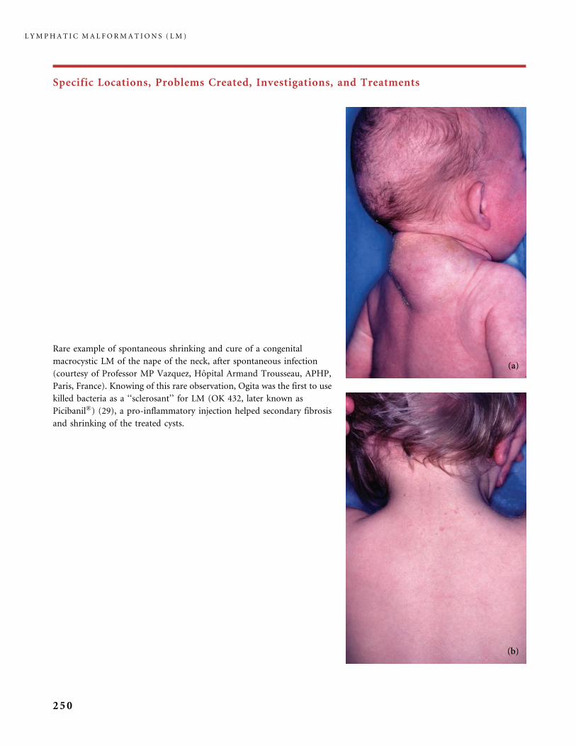
Specific Locations, Problems Created, Investigations, and Treatments
Rare example of spontaneous shrinking and cure of a congenital
macrocystic LM of the nape of the neck, after spontaneous infection
(courtesy of Professor MP Vazquez, Hopital Armand Trousseau, APHP,
Paris, France). Knowing of this rare observation, Ogita was the first to use
killed bacteria as a ‘‘sclerosant’’ for LM (OK 432, later known as
Picibanil�) (29), a pro-inflammatory injection helped secondary fibrosis
and shrinking of the treated cysts.
L Y M P H A T I C M A L F O R M A T I O N S ( L M )
250

Specific Locations, Problems Created, Investigations, and Treatments
This newborn (a) had a large hemorrhagic mass at birth; MRI, T2-weighted sequence demonstrated large lymphatic
cysts (b) and direct-puncture sclerotherapy was performed (c) with improvement (d) after two sessions over a year
(courtesy: Dr. Metin Tovi, Karolinska Institute, Stockholm, Sweden).
L Y M P H A T I C M A L F O R M A T I O N S ( L M )
251
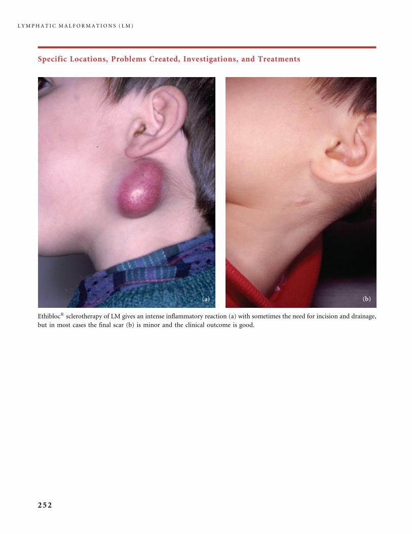
Specific Locations, Problems Created, Investigations, and Treatments
Ethibloc� sclerotherapy of LM gives an intense inflammatory reaction (a) with sometimes the need for incision and drainage,
but in most cases the final scar (b) is minor and the clinical outcome is good.
L Y M P H A T I C M A L F O R M A T I O N S ( L M )
252
References
1 Aagenaes O. Hereditary cholestasis with lymphoedema (Aagenaes syndrome,
cholestasis�lymphoedema syndrome). New cases and follow-up from infancy to adult
age. Scand J Gastroenterol 1998; 33: 335�45.
2 Alqahtani A, Nguyen LT, Flageole H, Shaw K, Laberge JM. 25 years’ experience with
lymphangiomas in children. J Pediatr Surg 1999; 34: 1164�8.
3 Angel B, Hersh JH. Expansion of the phenotype in Hennekam syndrome: a case with
new manifestations. Am J Med Genet 1997; 71: 211�14.
4 Azizkhan RG, Rutter MJ, Cohen A, Mason J, Lim L, Cotton RT. Lymphatic malformation
of the tongue base: management and long-term outcome. Communication W21 in 15th
ISSVA Workshop. Wellington, NZ. 22�25 February 2004.
5 Bisdorff A, Burrows P, Carrico J, Robertson R, Mulliken JB. Intracranial vascular
anomalies in patients with fronto-orbital lymphatic malformation. Communiaction in
15th ISSVA Workshop. Wellington, NZ. 22�25 February 2004.
6 Brewis C, Pracy JP, Albert DM. Treatment of lymphangiomas of the head and neck
in children by intralesional injection of OK-432 (Picibanil). Clin Otolaryngol Allied Sci
2000; 25: 130�4.
7 Brouillard P, Vikkula M. Vascular malformations: localized defects in vascular
morphogenesis. Clin Genet 2003; 63: 340�51.
8 Bull LN, Roche E, Song EJ, Pedersen J, Knisely AS, van Der Hagen CB et al. Mapping
of the locus for cholestasis-lymphedema syndrome (Aagenaes syndrome) to a 6.6-cM
interval on chromosome 15q. Am J Hum Genet 2000; 67: 994�9.
9 Burrows PE, Laor T, Paltiel H, Robertson RL. Diagnostic imaging in the evaluation
of vascular birthmarks. Dermatol Clin 1998; 16: 455�88.
10 Chen CP, Chen HC, Liu FF, Jan SW, Lin SP, Sheu JC et al. Progressive fetal axillary cystic
lymphangioma with coexistent naevus flammeus. Br J Dermatol 1997; 136: 102�4.
11 Davies D, Rogers M. Morphology of lymphatic malformations: a pictorial review.
Australas J Dermatol 2000; 41: 1�5.; quiz 6�7
12 Dubois J, Garel L, Abela A, Laberge L, Yazbeck S. Lymphangiomas in children:
percutaneous sclerotherapy with an alcoholic solution of zein. Radiology 1997; 204:
651�4.
13 Dubois J, Garel L, Grignon A, David M, Laberge L, Filiatrault D et al. Imaging
of hemangiomas and vascular malformations in children. Acad Radiol 1998; 5: 390�400.
14 Gimeno Aranguez M, Colomar Palmer P, Gonzalez Mediero I, Ollero Caprani JM.
The clinical and morphological aspects of childhood lymphangiomas: a review of 145
cases. An Esp Pediatr 1996; 45: 25�8.
15 Gomez CS, Calonje E, Ferrar DW, Browse NL, Fletcher CD. Lymphangiomatosis of the
limbs. Clinicopathologic analysis of a series with a good prognosis. Am J Surg Pathol 1995;
19: 125�33.
16 Greene AK, Burrows PE, Smith L, Mulliken JB. Periorbital lymphatic malformation:
clinical course and management in 42 patients. Plast Reconstr Surg 2005; 115: 22�30.
17 Guillou L, Fletcher CD. Benign lymphangioendothelioma (acquired progressive
lymphangioma): a lesion not to be confused with well-differentiated angiosarcoma and
patch stage Kaposi’s sarcoma: clinicopathologic analysis of a series. Am J Surg Pathol 2000;
24: 1047�57.
18 Hennekam RC, Geerdink RA, Hamel BC, Hennekam FA, Kraus P, Rammeloo JA et al.
Autosomal recessive intestinal lymphangiectasia and lymphedema, with facial anomalies
and mental retardation. Am J Med Genet 1989; 34: 593�600.
19 Herbreteau D, Riche MC, Enjolras O, Lemarchand F, Brette MD, Laurian C et al. Cystic
lymphatic malformations and their treatment. J Mal Vasc 1992; 17: 54�6.
R E F E R E N C E S
253
20 Hirayama T, Sabokbar A, Itonaga I, Watt-Smith S, Athanasou NA. Cellular and humoral
mechanisms of osteoclast formation and bone resorption in Gorham�Stout disease.
J Pathol 2001; 195: 624�30.
21 Irrthum A, Karkkainen MJ, Devriendt K, Alitalo K, Vikkula M. Congenital hereditary
lymphedema caused by a mutation that inactivates VEGFR3 tyrosine kinase.
Am J Hum Genet 2000; 67: 295�301.
22 Jappe U, Zimmermann T, Kahle B, Petzoldt D. Lymphangioma circumscriptum of the
vulva following surgical and radiological therapy of cervical cancer. Sex Transm Dis 2002;
29: 533�5.
23 Katz SE, Rootman J, Vangveeravong S, Graeb D. Combined venous lymphatic
malformations of the orbit (so-called lymphangiomas). Association with noncontiguous
intracranial vascular anomalies. Ophthalmology 1998; 105: 176�84.
24 Lowenstein EJ, Kim KH, Glick SA. Turner’s syndrome in dermatology. J Am Acad
Dermatol 2004; 50: 767�76.
25 MacLean JE, Cohen E, Weinstein M. Primary intestinal and thoracic lymphangiectasia:
a response to antiplasmin therapy. Pediatrics 2002; 109: 1177�80.
26 Martinot V, Descamps S, Fevrier P, Patenotre P, Breviere JM, Piette F et al. Evaluation
of the treatment of cystic lymphangioma by percutaneous injection of Ethibloc in
20 patients. Arch Pediatr 1997; 4: 8�14.
27 Molitch HI, Unger EC, Witte CL, vanSonnenberg E. Percutaneous sclerotherapy
of lymphangiomas. Radiology 1995; 194: 343�7.
28 North PE, Kahn T, Cordisco MR, Dadras SS, Detmar M, Frieden IJ. Multifocal
lymphangioendotheliomatosis with thrombocytopenia: a newly recognized clinicopatho-
logical entity. Arch Dermatol 2004; 140: 599�606.
29 Ogita S, Tsuto T, Deguchi E, Tokiwa K, Nagashima M, Iwai N. OK-432 therapy for
unresectable lymphangiomas in children. J Pediatr Surg 1991; 26: 263�8; discussion
268�70
30 Ogura T, Hamada H, Obata-Yasuoka M, Watanabe H, Okuno S, Fujiki Y et al.
Antepartum assessment of fetal cystic lymphangioma by magnetic resonance imaging.
Gynecol Obstet Invest 2002; 53: 237�9.
31 Padwa BL, Hayward PG, Ferraro NF, Mulliken JB. Cervicofacial lymphatic malformation:
clinical course, surgical intervention, and pathogenesis of skeletal hypertrophy. Plast
Reconstr Surg 1995; 95: 951�60.
32 Paradies G, Leggio S, Leggio A. The treatment of lymphangioma. Surgery versus
sclerotherapy. Eur J Pediat Dermatol 2001; 11: 23�8.
33 Puig S, Aref H, Brunelle F. Double-needle sclerotherapy of lymphangiomas and venous
angiomas in children: a simple technique to prevent complications. Am J Roentgenol 2003;
180: 1399�401.
34 Rafii S, Skobe M. Splitting vessels: keeping lymph apart from blood. Nat Med 2003;
9: 166�8.
35 Riche MC, Lemarchand-Venencie F, Enjolras O, Hadjean E, Merland JJ, Laurian C.
Nonsurgical treatment of cystic lymphangioma. Ann Otolaryngol Chir Cervicofac 1986;
103: 67�70.
36 Tanriverdi HA, Hendrik HJ, Ertan AK, Axt R, Schmidt W. Hygroma colli cysticum:
prenatal diagnosis and prognosis. Am J Perinatol 2001; 18: 415�20.
37 Tovi M, Herbreteau D, Enjolras O, Merland JJ. 52 patients with cystic lymphatic vascular
malformations. Percutaneous sclerotherapy � simple, fast and repeatable. Lakartidningen
1998; 95: 643�7.
38 Tunc M, Sadri E, Char DH. Orbital lymphangioma: an analysis of 26 patients.
Br J Ophthalmol 1999; 83: 76�80.
39 Turner C, Gross S. Treatment of recurrent suprahyoid cervicofacial lymphangioma with
intravenous cyclophosphamide. Am J Pediatr Hematol Oncol 1994; 16: 325�8.
40 Van der Horst CMAM. Gorham Stout syndrome. A rare complication of lymphatic
malformation. Communication DC1 in 15th ISSVA Workshop. Wellington, NZ. 22�25
February 2004.
L Y M P H A T I C M A L F O R M A T I O N S ( L M )
254
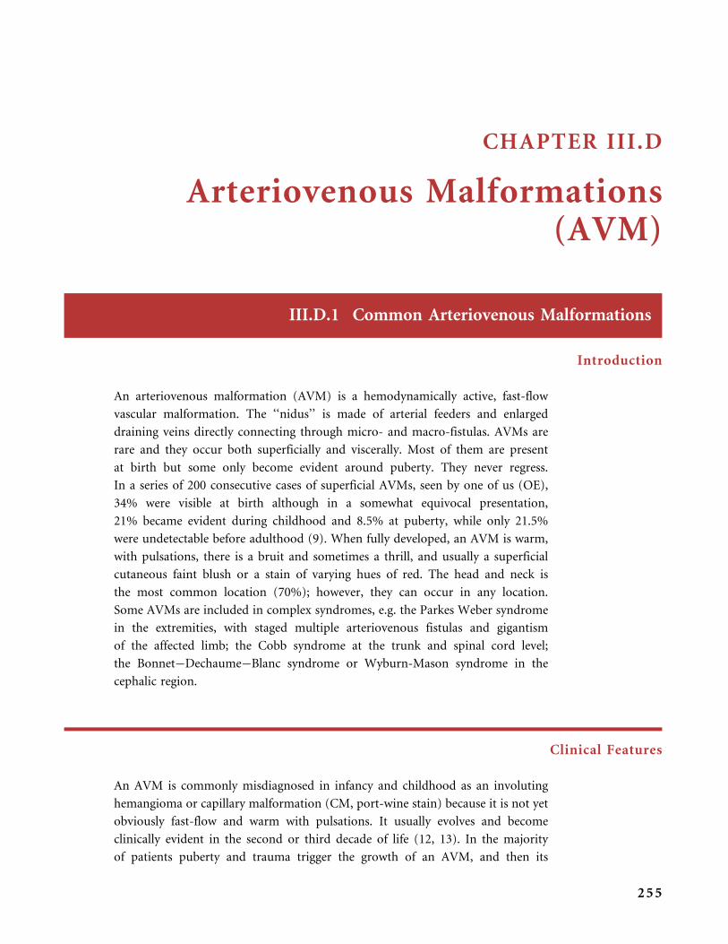
CHAPTER III.D
Arteriovenous Malformations(AVM)
III.D.1 Common Arteriovenous Malformations
Introduction
An arteriovenous malformation (AVM) is a hemodynamically active, fast-flow
vascular malformation. The ‘‘nidus’’ is made of arterial feeders and enlarged
draining veins directly connecting through micro- and macro-fistulas. AVMs are
rare and they occur both superficially and viscerally. Most of them are present
at birth but some only become evident around puberty. They never regress.
In a series of 200 consecutive cases of superficial AVMs, seen by one of us (OE),
34% were visible at birth although in a somewhat equivocal presentation,
21% became evident during childhood and 8.5% at puberty, while only 21.5%
were undetectable before adulthood (9). When fully developed, an AVM is warm,
with pulsations, there is a bruit and sometimes a thrill, and usually a superficial
cutaneous faint blush or a stain of varying hues of red. The head and neck is
the most common location (70%); however, they can occur in any location.
Some AVMs are included in complex syndromes, e.g. the Parkes Weber syndrome
in the extremities, with staged multiple arteriovenous fistulas and gigantism
of the affected limb; the Cobb syndrome at the trunk and spinal cord level;
the Bonnet�Dechaume�Blanc syndrome or Wyburn-Mason syndrome in the
cephalic region.
Clinical Features
An AVM is commonly misdiagnosed in infancy and childhood as an involuting
hemangioma or capillary malformation (CM, port-wine stain) because it is not yet
obviously fast-flow and warm with pulsations. It usually evolves and become
clinically evident in the second or third decade of life (12, 13). In the majority
of patients puberty and trauma trigger the growth of an AVM, and then its
255

fast-flow nature manifests: redness increases as well as local warmth, a thrill and
a bruit point to a fast-flow anomaly. As an AVM gets worse, draining veins become
obvious and then tortuous, tense, and large.
AVM worsens at any time of the life but particularly at puberty and with
trauma. Skin alterations, secondary to a capillary steal syndrome, develop, includ-
ing modification of skin color, pigmentary changes, skin atrophy, ulcers with
intractable pain, sudden life-threatening hemorrhage, or recurrent, intermittent
bleeding. In the limbs skin changes resembling purple plaques of Kaposi sarcoma
may expand. All of these are rare in childhood, and they arise by adolescence
or later (14).
A facial AVM localized to the skin and/or facial bones leads to facial asym-
metry, gingival hypertrophy, unstable teeth and periodontal bleeding, and skin or
mucosal ulcers with secondary infection. The ear is a quite common location in the
head and neck. A nasal AVM causes epistaxis. Ischemia of the tips of the fingers or
toes complicates an extremity distal AVM; it is linked to arterial steal and venous
hypertension, with a lack of normal blood supply to the skin. AVM in a finger or
a toe gradually narrows the distal phalanx, causing purple necrotic skin changes;
chronic adhesive crusts arise, and there is a progressive miniaturization of the
nail. Bony AVM creates osteolysis, sometimes due to the venous drainage through
the bones of an adjacent AVM. Shunting through the fistulas in large AVMs
can cause congestive heart failure, but this occurs in less than 2% of cases, and
in two situations: soon after birth in infants with massive AVM, and later in life,
often in young adults, in patients with a large rapidly worsening AVM of an
extremity or trunk (9).
SCHOB I NG E R S T AG I NG is a severity scoring system for AVMs: stage I is the
quiescent stage when the AVM mimics a capillary malformation or an involuting
hemangioma; stage II is expansion: the lesion becomes warmer, bigger, throbbing,
with a thrill and a bruit; stage III is destruction, with all the symptoms above plus
ulcers, hemorrhages, and bony lytic lesions; stage IV is rare and is comprised of all
of above plus congestive cardiac failure with increased cardiac output and left
ventricle hypertrophy (9, 13).
Radiological Investigations
ULTRA SONOGRA PHY COMB I N ED W I TH CO LOR DO P P L E R documents the AV
shunting. An AVM exhibits low-resistance high-velocity arterial flow, above the
baseline, with high diastolic flux, and pulsatile venous flow below the baseline.
Vessels are tortuous.
PU L S ED DO P P L E R measures the arterial output (of carotid, humeral or femoral
arteries) on the affected side as compared with the normal side. This is an excellent
A R T E R I O V E N O U S M A L F O R M A T I O N S ( A V M )
256

and reliable noninvasive technique to follow the course of an AVM or to monitor
the stability of the results of a treatment.
COMPUT ED TOMOGRA PHY (CT) W I TH I OD I N A T ED CONT RA S T demonstrates
soft tissue involvement, a highly enhancing lesion, and dilated feeding
and draining vessels, but it cannot definitely differentiate between hemangioma,
VM and AVM.
CT ANG I OGRA PHY gives interesting representation and 3-D reconstruction of the
vascular network in AVMs.
MRI of an AVM shows a collection of vascular flow voids (black tubular
structures), corresponding to fast-flow vessels, in all sequences (spin�echo T1-
and T2-weighted sequences) and there is no contrast parenchymal enhancement
(no tumor aspect). Some signal abnormalities may exist in relation to a fibrofatty
matrix (4, 17).
MAGNET I C R E SONANC E ANG I OGRA PHY (MRA) shows the anomalous
vascular network. MRA can replace an arteriogram in the work-up when a
treatment is not considered (for example in the case of a quiescent AVM in a child
or an elderly patient) and for follow-up of AVM, but is not yet discussed for
therapy.
DIG I T A L A R T E R I O G RA PH Y remains an indispensable tool to depict the angio-
architecture of the AVM, prior to the discussion about therapy, and prior
to therapeutic embolization; it demonstrates the dilated, lengthened and often
tortuous feeding arteries, localizes the nidus, and detects the early venous drainage
through enlarged veins (6).
Treatment
An AVM is usually not treated in its quiescent stage I, except for example when
complete resection is possible without conspicuous cosmetic damage; however,
early embolic and/or surgical treatment of a quiescent AVM remains controversial.
Partial excision leads to transient improvement, then the AVM inevitably
re-expands over time. Ligature or proximal closure by embolization of arterial
feeding vessels is contraindicated, as incomplete surgical treatment is not
recommended. After a period of apparent benefit, a vascular recruitment phenom-
enon occurs, new collateral arteries supply the nidus, intense capillarogenesis
spreads out, while the residual lesion regrows and progresses. Treatment of
an AVM is always challenging, and is either palliative (arterial embolization)
C O M M O N A R T E R I O V E N O U S M A L F O R M A T I O N S
257

to control a complication (ulcer, bleeding, and bone lytic lesion with a risk of
fracture) or aims at being curative (embolization followed by wide surgical
resection and reconstruction) (5, 15, 25). Therapeutic intervention becomes
necessary whenever local (Schobinger stage III) and/or cardiac complications
(Schobinger stage IV) develop. Long-term post-treatment follow-up is indis-
pensable to ensure the permanent results.
III.D.2 Syndromic Arteriovenous Malformations
I I I .D .2 . 1 BONNET�DECHAUME�BLANC SYNDROME
OR WYBURN-MASON SYNDROME
These are the two names for a sporadic syndrome with AVM involving the face,
retina, and brain (2, 23). The most extensive cases result in distortion of facial
features, recurrent epistaxis, gingival bleeding, blindness and cerebral hemorrhage.
The facial AVM occupies the mid-face (the nose, forehead, and lip) or is hemi-
facial. In a retrospective study of 15 patients the most common presenting sign
was reduced acuity or visual field, and a cutaneous lesion was present in only four
patients; 14 had orbital involvement, and the neurologic involvement includes
the optic nerve (in 13/15), the retina (in 11/15), the thalamus (in 9/15), and the
chiasm/hypothalamus (in 9/15); two patients also had a maxillofacial AVM (1).
In a series of 10 patients from our department (personal communication,
Dr. Monique Boukobza, Department of Neuroradiodiagnostics, Hopital
Lariboisiere, Paris, France) facial and cerebral involvement was present in all.
The syndrome is hypothesized to be the result of a somatic mutation in the region
of the neural crest, taking place before the cell migrations occur (thus, before the
fourth week of embryo development) to produce this cerebrofacial arteriovenous
metameric syndrome (CAMS) (1).
I I I .D .2 . 2 COBB SYNDROME
This is another sporadic arteriovenous metameric syndrome at the trunk level,
linking the spinal cord to a skin AVM in the same metamere. Symptoms related
to the spinal cord AVM usually appear in late childhood and they occur suddenly
or progressively. They include pain, sensory and motor deficit, and loss of control
of sphincters. Depending on the level of the spinal lesion, the patient may also have
AVM in the arm or leg. In a series of 155 patients with spinal cord arteriovenous
malformations or fistulae, 10 had Cobb syndrome (18).
A R T E R I O V E N O U S M A L F O R M A T I O N S ( A V M )
258
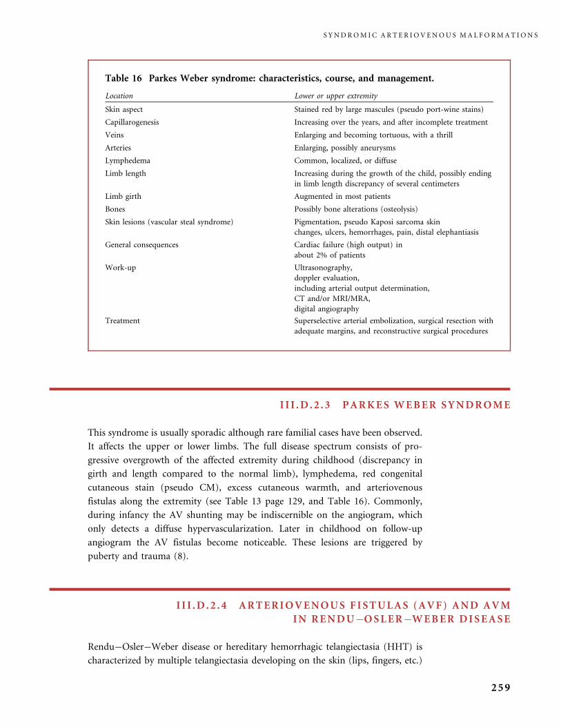
I I I .D .2 . 3 PARKES WEBER SYNDROME
This syndrome is usually sporadic although rare familial cases have been observed.
It affects the upper or lower limbs. The full disease spectrum consists of pro-
gressive overgrowth of the affected extremity during childhood (discrepancy in
girth and length compared to the normal limb), lymphedema, red congenital
cutaneous stain (pseudo CM), excess cutaneous warmth, and arteriovenous
fistulas along the extremity (see Table 13 page 129, and Table 16). Commonly,
during infancy the AV shunting may be indiscernible on the angiogram, which
only detects a diffuse hypervascularization. Later in childhood on follow-up
angiogram the AV fistulas become noticeable. These lesions are triggered by
puberty and trauma (8).
I I I .D .2 . 4 ARTERIOVENOUS FISTULAS (AVF) AND AVM
IN RENDU�OSLER�WEBER DISEASE
Rendu�Osler�Weber disease or hereditary hemorrhagic telangiectasia (HHT) is
characterized by multiple telangiectasia developing on the skin (lips, fingers, etc.)
Table 16 Parkes Weber syndrome: characteristics, course, and management.
Location Lower or upper extremity
Skin aspect Stained red by large mascules (pseudo port-wine stains)
Capillarogenesis Increasing over the years, and after incomplete treatment
Veins Enlarging and becoming tortuous, with a thrill
Arteries Enlarging, possibly aneurysms
Lymphedema Common, localized, or diffuse
Limb length Increasing during the growth of the child, possibly ending
in limb length discrepancy of several centimeters
Limb girth Augmented in most patients
Bones Possibly bone alterations (osteolysis)
Skin lesions (vascular steal syndrome) Pigmentation, pseudo Kaposi sarcoma skin
changes, ulcers, hemorrhages, pain, distal elephantiasis
General consequences Cardiac failure (high output) in
about 2% of patients
Work-up Ultrasonography,
doppler evaluation,
including arterial output determination,
CT and/or MRI/MRA,
digital angiography
Treatment Superselective arterial embolization, surgical resection with
adequate margins, and reconstructive surgical procedures
S Y N D R O M I C A R T E R I O V E N O U S M A L F O R M A T I O N S
259
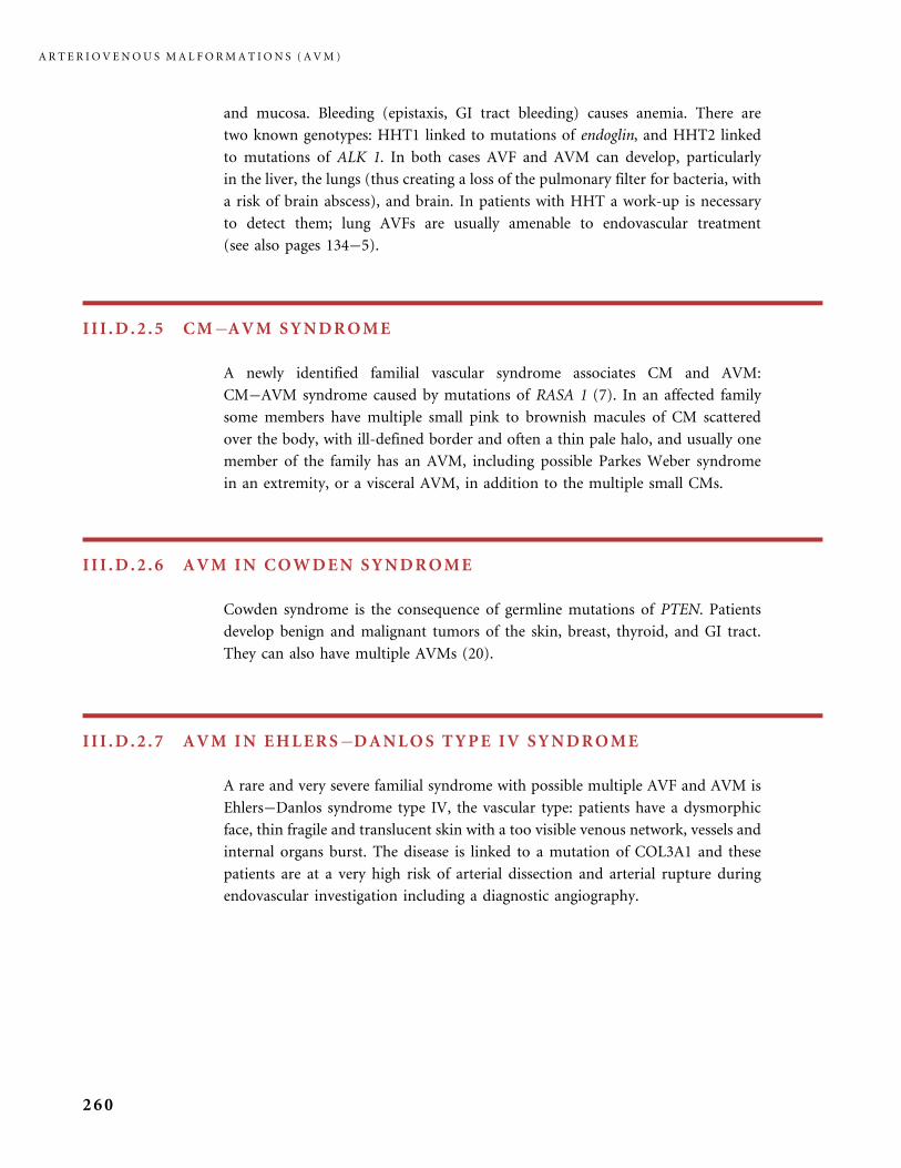
and mucosa. Bleeding (epistaxis, GI tract bleeding) causes anemia. There are
two known genotypes: HHT1 linked to mutations of endoglin, and HHT2 linked
to mutations of ALK 1. In both cases AVF and AVM can develop, particularly
in the liver, the lungs (thus creating a loss of the pulmonary filter for bacteria, with
a risk of brain abscess), and brain. In patients with HHT a work-up is necessary
to detect them; lung AVFs are usually amenable to endovascular treatment
(see also pages 134�5).
I I I .D .2 . 5 CM�AVM SYNDROME
A newly identified familial vascular syndrome associates CM and AVM:
CM�AVM syndrome caused by mutations of RASA 1 (7). In an affected family
some members have multiple small pink to brownish macules of CM scattered
over the body, with ill-defined border and often a thin pale halo, and usually one
member of the family has an AVM, including possible Parkes Weber syndrome
in an extremity, or a visceral AVM, in addition to the multiple small CMs.
I I I .D .2 . 6 AVM IN COWDEN SYNDROME
Cowden syndrome is the consequence of germline mutations of PTEN. Patients
develop benign and malignant tumors of the skin, breast, thyroid, and GI tract.
They can also have multiple AVMs (20).
I I I .D .2 .7 AVM IN EHLERS�DANLOS TYPE IV SYNDROME
A rare and very severe familial syndrome with possible multiple AVF and AVM is
Ehlers�Danlos syndrome type IV, the vascular type: patients have a dysmorphic
face, thin fragile and translucent skin with a too visible venous network, vessels and
internal organs burst. The disease is linked to a mutation of COL3A1 and these
patients are at a very high risk of arterial dissection and arterial rupture during
endovascular investigation including a diagnostic angiography.
A R T E R I O V E N O U S M A L F O R M A T I O N S ( A V M )
260
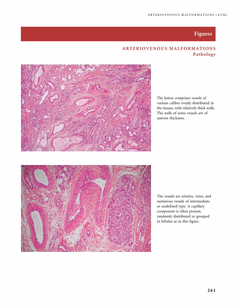
Figures
ARTERIOVENOUS MALFORMATIONS
Pathology
The lesion comprises vessels of
various caliber evenly distributed in
the tissues, with relatively thick walls.
The walls of some vessels are of
uneven thickness.
The vessels are arteries, veins, and
numerous vessels of intermediate
or undefined type. A capillary
component is often present,
randomly distributed or grouped
in lobules as in this figure.
A R T E R I O V E N O U S M A L F O R M A T I O N S ( A V M )
261

Pathology
Orcein staining also highlights the presence of direct
arteriovenous communications (fistulae) between an artery
with thick undulating internal elastic lamina (upper left)
and a venous-type vessel (lower right).
In this case of AVM, the capillary component dissects
between the skeletal-muscle cells, respecting the borders
of the fascicle.
On elastic tissue (orcein) staining, the architecture of the
malformed vessels is better seen. Some are veins with
thickened elastic fibers (upper vessel), some are arteries
of normal structure (middle), and others lack any elastic
component (lower right) being a malformed vessel without
features of an artery or of a vein.
A R T E R I O V E N O U S M A L F O R M A T I O N S ( A V M )
262

Specific Locations, Problems Created, Investigations, and Treatments
Schobinger staging of AVMs, based on clinical signs/symptoms, gives an
indication of the evolution: stage I is the quiescent initial period with blush,
stain, and local warmth. The vascular lesion may be misdiagnosed in infancy
as CM (a1, a2) and later as involuting hemangioma (b).
A R T E R I O V E N O U S M A L F O R M A T I O N S ( A V M )
263
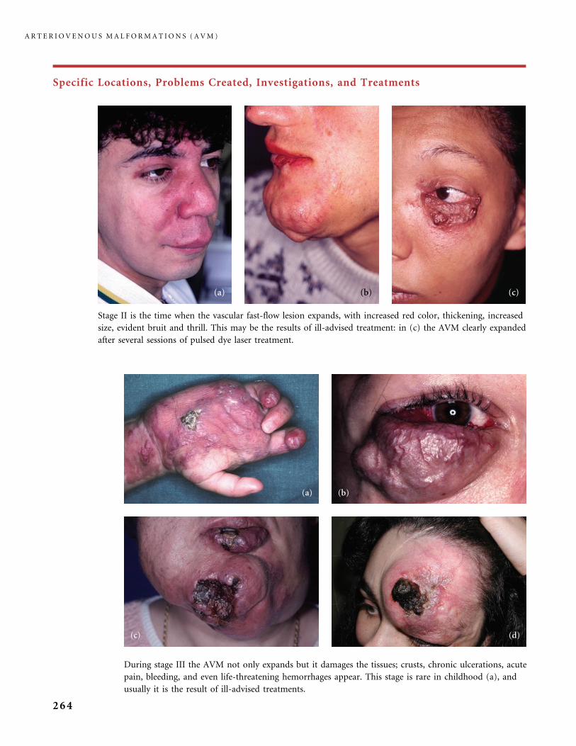
Specific Locations, Problems Created, Investigations, and Treatments
Stage II is the time when the vascular fast-flow lesion expands, with increased red color, thickening, increased
size, evident bruit and thrill. This may be the results of ill-advised treatment: in (c) the AVM clearly expanded
after several sessions of pulsed dye laser treatment.
During stage III the AVM not only expands but it damages the tissues; crusts, chronic ulcerations, acute
pain, bleeding, and even life-threatening hemorrhages appear. This stage is rare in childhood (a), and
usually it is the result of ill-advised treatments.
A R T E R I O V E N O U S M A L F O R M A T I O N S ( A V M )
264
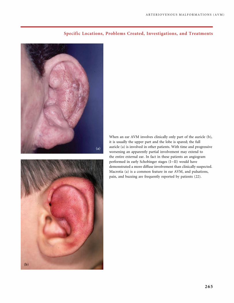
Specific Locations, Problems Created, Investigations, and Treatments
When an ear AVM involves clinically only part of the auricle (b),
it is usually the upper part and the lobe is spared; the full
auricle (a) is involved in other patients. With time and progressive
worsening an apparently partial involvement may extend to
the entire external ear. In fact in these patients an angiogram
performed in early Schobinger stages (I�II) would have
demonstrated a more diffuse involvement than clinically suspected.
Macrotia (a) is a common feature in ear AVM, and pulsations,
pain, and buzzing are frequently reported by patients (22).
A R T E R I O V E N O U S M A L F O R M A T I O N S ( A V M )
265
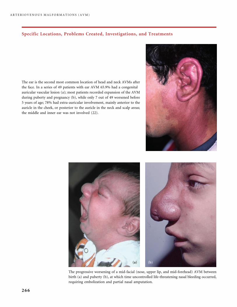
Specific Locations, Problems Created, Investigations, and Treatments
The ear is the second most common location of head and neck AVMs after
the face. In a series of 49 patients with ear AVM 65.9% had a congenital
auricular vascular lesion (a); most patients recorded expansion of the AVM
during puberty and pregnancy (b), while only 7 out of 49 worsened before
5 years of age; 78% had extra-auricular involvement, mainly anterior to the
auricle in the cheek, or posterior to the auricle in the neck and scalp areas;
the middle and inner ear was not involved (22).
The progressive worsening of a mid-facial (nose, upper lip, and mid-forehead) AVM between
birth (a) and puberty (b), at which time uncontrolled life-threatening nasal bleeding occurred,
requiring embolization and partial nasal amputation.
A R T E R I O V E N O U S M A L F O R M A T I O N S ( A V M )
266
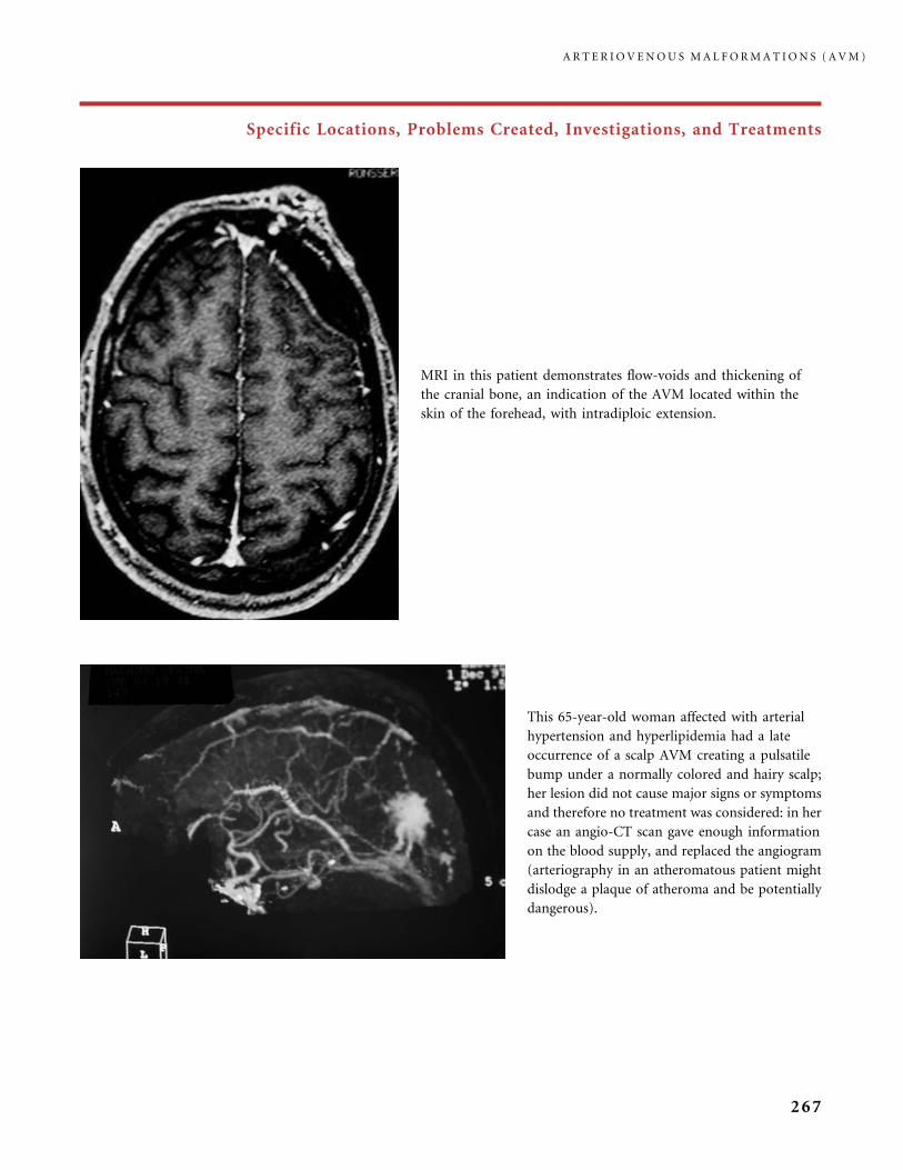
Specific Locations, Problems Created, Investigations, and Treatments
This 65-year-old woman affected with arterial
hypertension and hyperlipidemia had a late
occurrence of a scalp AVM creating a pulsatile
bump under a normally colored and hairy scalp;
her lesion did not cause major signs or symptoms
and therefore no treatment was considered: in her
case an angio-CT scan gave enough information
on the blood supply, and replaced the angiogram
(arteriography in an atheromatous patient might
dislodge a plaque of atheroma and be potentially
dangerous).
MRI in this patient demonstrates flow-voids and thickening of
the cranial bone, an indication of the AVM located within the
skin of the forehead, with intradiploic extension.
A R T E R I O V E N O U S M A L F O R M A T I O N S ( A V M )
267

Specific Locations, Problems Created, Investigations, and Treatments
This patient had noticed worsening of the hand AVM.
Arteriography remains the best investigative tool to depict
the arterial feeders, the AVM nidus and the venous
drainage, and decide if endovascular embolization is an
appropriate therapeutic option.
A patient with distal AVM of the upper extremity at the
beginning of Schobinger stage III AVM, with recent
discoloration of the second finger, an indication of
ischemia, and progressively enlarging distorted draining
veins.
Six years of progression of an AVM involving the first and second fingers (a). No relief was provided by palliative
embolizations. Puberty occurred during this six-year period and seemed to have triggered the expansion. Distal necrotic
skin changes in the thumb, with ulcer, infection, bleeding, and intense pain occurred (b). It was impossible to reverse
miniaturization of the nail and distal phalanx by arterial embolization, and phalanx amputation was the final outcome.
Superselective arterial embolization, to eradicate the nidus of an extremity AVM, has been considered the treatment
A R T E R I O V E N O U S M A L F O R M A T I O N S ( A V M )
268
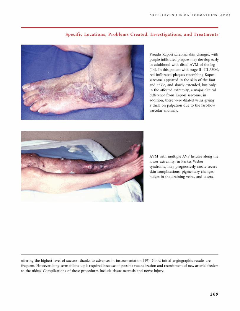
Specific Locations, Problems Created, Investigations, and Treatments
Pseudo Kaposi sarcoma skin changes, with
purple infiltrated plaques may develop early
in adulthood with distal AVM of the leg
(14). In this patient with stage II�III AVM,
red infiltrated plaques resembling Kaposi
sarcoma appeared in the skin of the foot
and ankle, and slowly extended, but only
in the affected extremity, a major clinical
difference from Kaposi sarcoma; in
addition, there were dilated veins giving
a thrill on palpation due to the fast-flow
vascular anomaly.
AVM with multiple AVF fistulae along the
lower extremity, in Parkes Weber
syndrome, may progressively create severe
skin complications, pigmentary changes,
bulges in the draining veins, and ulcers.
offering the highest level of success, thanks to advances in instrumentation (19). Good initial angiographic results are
frequent. However, long-term follow-up is required because of possible recanalization and recruitment of new arterial feeders
to the nidus. Complications of these procedures include tissue necrosis and nerve injury.
A R T E R I O V E N O U S M A L F O R M A T I O N S ( A V M )
269
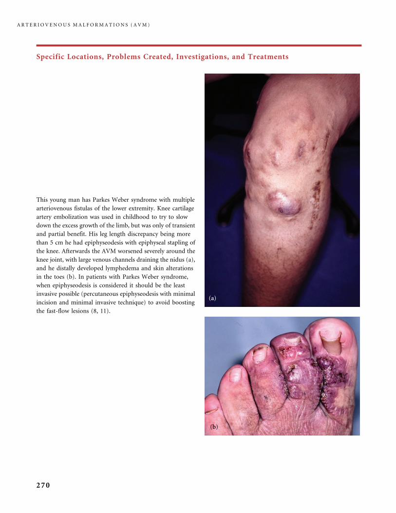
Specific Locations, Problems Created, Investigations, and Treatments
This young man has Parkes Weber syndrome with multiple
arteriovenous fistulas of the lower extremity. Knee cartilage
artery embolization was used in childhood to try to slow
down the excess growth of the limb, but was only of transient
and partial benefit. His leg length discrepancy being more
than 5 cm he had epiphyseodesis with epiphyseal stapling of
the knee. Afterwards the AVM worsened severely around the
knee joint, with large venous channels draining the nidus (a),
and he distally developed lymphedema and skin alterations
in the toes (b). In patients with Parkes Weber syndrome,
when epiphyseodesis is considered it should be the least
invasive possible (percutaneous epiphyseodesis with minimal
incision and minimal invasive technique) to avoid boosting
the fast-flow lesions (8, 11).
A R T E R I O V E N O U S M A L F O R M A T I O N S ( A V M )
270

Specific Locations, Problems Created, Investigations, and Treatments
Parkes Weber syndrome of the arm with cardiac failure. This is a rare situation
observed in less than 2% of patients with AVMs in our experience but when
this complication develops it may be uncontrolled and lethal (9, 10). Tolerance
of the heart to the high flow is sometimes surprising when the AVM has
gradually worsened in a slow protracted course over childhood and
adolescence. It is not uncommon to see a patient with diffuse lower limb AVM,
who has a femoral output 10 times higher in the affected extremity than in the
normal one, as measured by pulsed Doppler, and who has a relatively good
cardiac tolerance of the malformation, with acceptable cardiac output as
measured with a Doppler probe, and moderate hypertrophy of the left
ventricle.
Parkes Weber syndrome in these two infants was manifest at birth. In both cases lymphedema of the lower
extremity, large red cutaneous stains and abnormal warmth of the limb were present; AV fistulas were shown by
US/Doppler. One infant (a) had cardiac failure at birth: a palliative embolization reduced the cardiac output.
A R T E R I O V E N O U S M A L F O R M A T I O N S ( A V M )
271
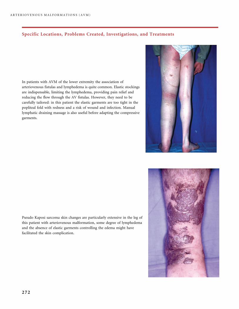
Specific Locations, Problems Created, Investigations, and Treatments
In patients with AVM of the lower extremity the association of
arteriovenous fistulas and lymphedema is quite common. Elastic stockings
are indispensable, limiting the lymphedema, providing pain relief and
reducing the flow through the AV fistulas. However, they need to be
carefully tailored: in this patient the elastic garments are too tight in the
popliteal fold with redness and a risk of wound and infection. Manual
lymphatic draining massage is also useful before adapting the compressive
garments.
Pseudo Kaposi sarcoma skin changes are particularly extensive in the leg of
this patient with arteriovenous malformation, some degree of lymphedema
and the absence of elastic garments controlling the edema might have
facilitated the skin complication.
A R T E R I O V E N O U S M A L F O R M A T I O N S ( A V M )
272
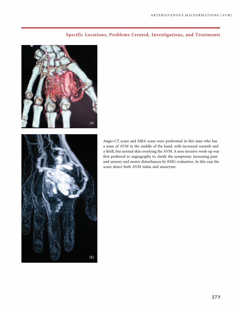
Specific Locations, Problems Created, Investigations, and Treatments
Angio-CT scans and MRA scans were performed in this man who has
a mass of AVM in the middle of the hand, with increased warmth and
a thrill, but normal skin overlying the AVM. A non-invasive work-up was
first preferred to angiography to clarify the symptoms: increasing pain
and sensory and motor disturbances by EMG evaluation. In this case the
scans detect both AVM nidus and aneurysm.
A R T E R I O V E N O U S M A L F O R M A T I O N S ( A V M )
273
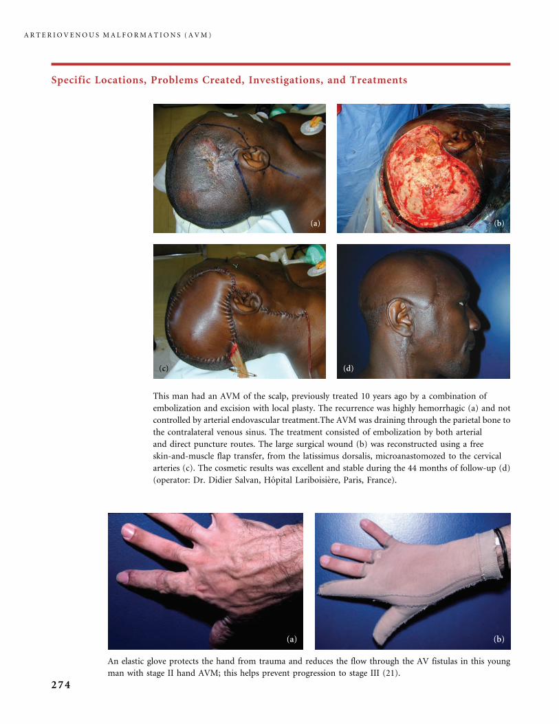
Specific Locations, Problems Created, Investigations, and Treatments
This man had an AVM of the scalp, previously treated 10 years ago by a combination of
embolization and excision with local plasty. The recurrence was highly hemorrhagic (a) and not
controlled by arterial endovascular treatment.The AVM was draining through the parietal bone to
the contralateral venous sinus. The treatment consisted of embolization by both arterial
and direct puncture routes. The large surgical wound (b) was reconstructed using a free
skin-and-muscle flap transfer, from the latissimus dorsalis, microanastomozed to the cervical
arteries (c). The cosmetic results was excellent and stable during the 44 months of follow-up (d)
(operator: Dr. Didier Salvan, Hopital Lariboisiere, Paris, France).
An elastic glove protects the hand from trauma and reduces the flow through the AV fistulas in this young
man with stage II hand AVM; this helps prevent progression to stage III (21).
A R T E R I O V E N O U S M A L F O R M A T I O N S ( A V M )
274

Specific Locations, Problems Created, Investigations, and Treatments
This woman had from birth a red stain of the upper eyelid. Growth of the AVM progressively occluded the visual axis.
Excision, after embolization, was performed, with a graft to reconstruct the unit (operators: Dr. Dominique Deffrennes and
Dr. Didier Salvan, Hopital Lariboisiere, Paris, France). Pre-operative embolization is always delicate in this location as the
ophthalmic artery is also feeding the AVM nidus.
This painful arteriovenous malformation of the foot
received palliative arterial embolization, to deliver an
ablative embolic material (absolute ethanol) to the
nidus and try to occlude it. However, inadvertent
migration of ethanol and embolization of adjacent
tissue during the procedure resulted in local
complications: necrosis developed and the patient
underwent amputation of two toes. Ethanol
embolization for ablating AVM involves similar
complications to those described for VM sclerotherapy,
and complications are more frequent than with VM,
due to the rapid flow. In a series of 450 patients
(2055 procedures) with either slow-flow or fast-flow
lesions, according to Yakes and Yee, minor local
complications included blistering (8.2%), infection
(2.2%), and temporary or permanent loss of sensation
(1.1%); major local complications were facial nerve
weakness (0.2%), decreased motor strength in an
extremity (0.5%), amputation (0.1%), tissue injuries
(0.36%), and cardiopulmonary arrest (0.06%);
however, the conclusion of this report is that, to be
safe, the procedure requires experienced operators
and dedicated facilities (25). In a group of 61 patients
(232 procedures) Burrows et al. (3) found a much
higher rate of complications (minor and self-limited
in 36.06%, and severe in 32.78% of patients) for
ethanol embolization of AVM.
A R T E R I O V E N O U S M A L F O R M A T I O N S ( A V M )
275

Specific Locations, Problems Created, Investigations, and Treatments
Stage III AVM, with macrotia, pain and bleeding, and very large draining veins (a). This
clinical situation led to auriculectomy, after preoperative embolization to minimize
intraoperative bleeding (b, c). Embolization was achieved with in situ injection of an acrylic
glue filling both arteries and veins. The large draining veins shrank to normal size after
amputation of the ear (d) and resection of the AVM nidus (operator: Dr. Benoit Faucon,
Hopital Lariboisiere, Paris, France). Depending on the size of the resection (the ear only or
the ear plus adjacent skin) the surgical wound is closed either by direct closure, or a skin graft,
or a flap transfer. Arterial embolization alone, even ethanol embolization as close as possible
to the AVM nidus (24, 25) cannot cure a ear AVM. In our experience it gives some transient
relief of the symptoms, which is helpful in young patients, then re-expansion occurs (22).
Two to five years after ear amputation, in a stable patient, ear reconstruction
may be considered (16) or a prosthetic ear can be offered.
A R T E R I O V E N O U S M A L F O R M A T I O N S ( A V M )
276
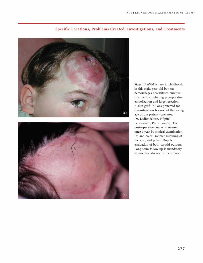
Specific Locations, Problems Created, Investigations, and Treatments
Stage III AVM is rare in childhood:
in this eight-year-old boy (a)
hemorrhages necessitated curative
treatment, combining pre-operative
embolization and large resection.
A skin graft (b) was preferred for
reconstruction because of the young
age of the patient (operator:
Dr. Didier Salvan, Hopital
Lariboisiere, Paris, France). The
post-operative course is assessed
once a year by clinical examination,
US and color Doppler screening of
the scar, and pulsed Doppler
evaluation of both carotid outputs.
Long-term follow-up is mandatory
to monitor absence of recurrence.
A R T E R I O V E N O U S M A L F O R M A T I O N S ( A V M )
277
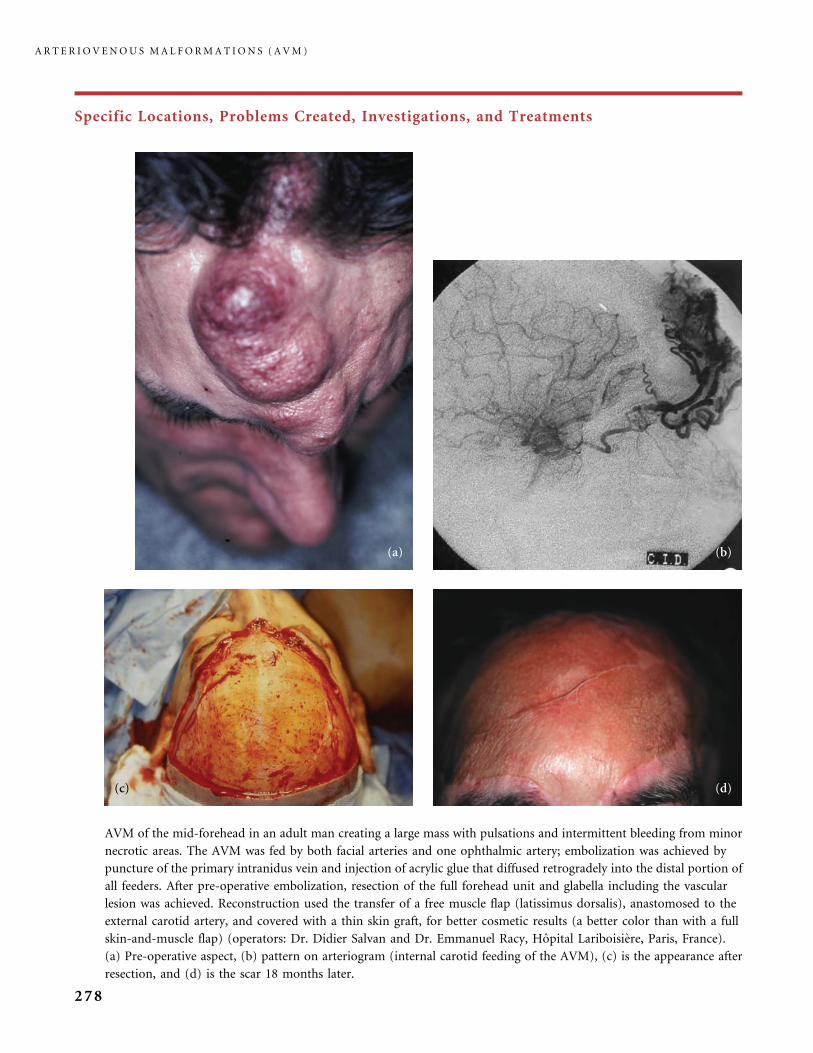
Specific Locations, Problems Created, Investigations, and Treatments
AVM of the mid-forehead in an adult man creating a large mass with pulsations and intermittent bleeding from minor
necrotic areas. The AVM was fed by both facial arteries and one ophthalmic artery; embolization was achieved by
puncture of the primary intranidus vein and injection of acrylic glue that diffused retrogradely into the distal portion of
all feeders. After pre-operative embolization, resection of the full forehead unit and glabella including the vascular
lesion was achieved. Reconstruction used the transfer of a free muscle flap (latissimus dorsalis), anastomosed to the
external carotid artery, and covered with a thin skin graft, for better cosmetic results (a better color than with a full
skin-and-muscle flap) (operators: Dr. Didier Salvan and Dr. Emmanuel Racy, Hopital Lariboisiere, Paris, France).
(a) Pre-operative aspect, (b) pattern on arteriogram (internal carotid feeding of the AVM), (c) is the appearance after
resection, and (d) is the scar 18 months later.
A R T E R I O V E N O U S M A L F O R M A T I O N S ( A V M )
278
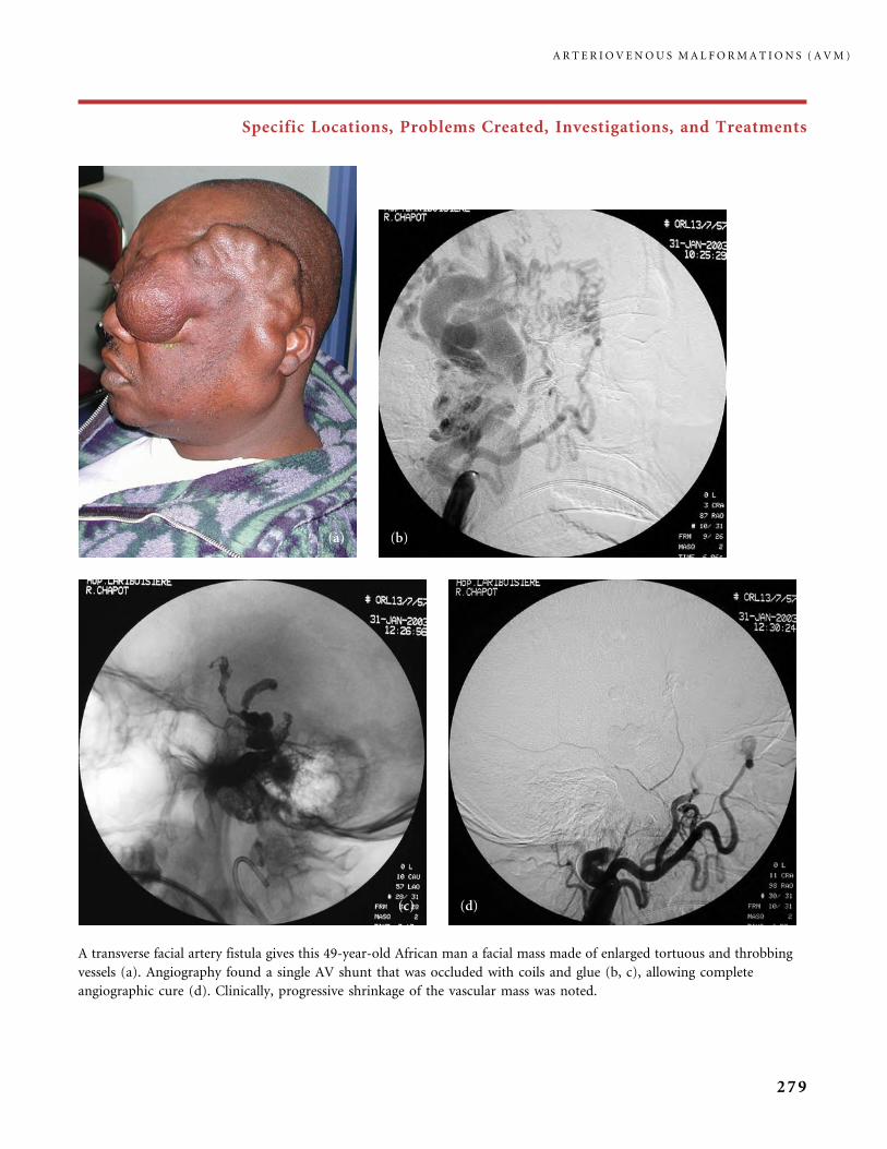
Specific Locations, Problems Created, Investigations, and Treatments
A transverse facial artery fistula gives this 49-year-old African man a facial mass made of enlarged tortuous and throbbing
vessels (a). Angiography found a single AV shunt that was occluded with coils and glue (b, c), allowing complete
angiographic cure (d). Clinically, progressive shrinkage of the vascular mass was noted.
A R T E R I O V E N O U S M A L F O R M A T I O N S ( A V M )
279
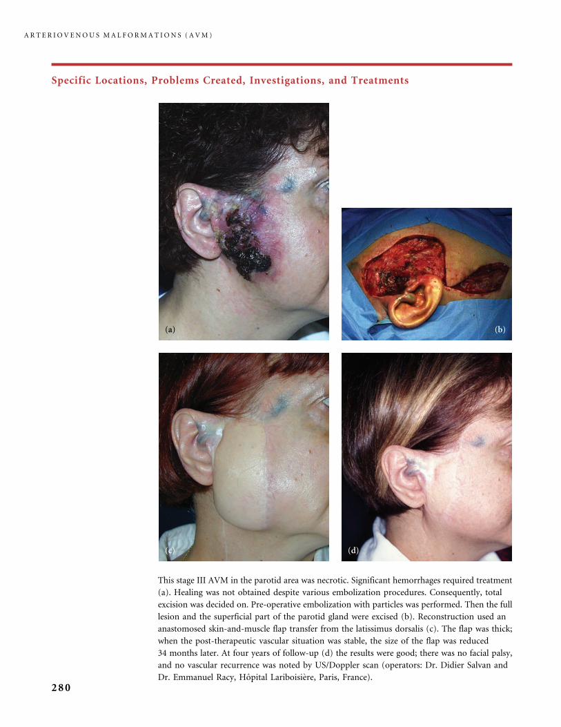
Specific Locations, Problems Created, Investigations, and Treatments
This stage III AVM in the parotid area was necrotic. Significant hemorrhages required treatment
(a). Healing was not obtained despite various embolization procedures. Consequently, total
excision was decided on. Pre-operative embolization with particles was performed. Then the full
lesion and the superficial part of the parotid gland were excised (b). Reconstruction used an
anastomosed skin-and-muscle flap transfer from the latissimus dorsalis (c). The flap was thick;
when the post-therapeutic vascular situation was stable, the size of the flap was reduced
34 months later. At four years of follow-up (d) the results were good; there was no facial palsy,
and no vascular recurrence was noted by US/Doppler scan (operators: Dr. Didier Salvan and
Dr. Emmanuel Racy, Hopital Lariboisiere, Paris, France).
A R T E R I O V E N O U S M A L F O R M A T I O N S ( A V M )
280
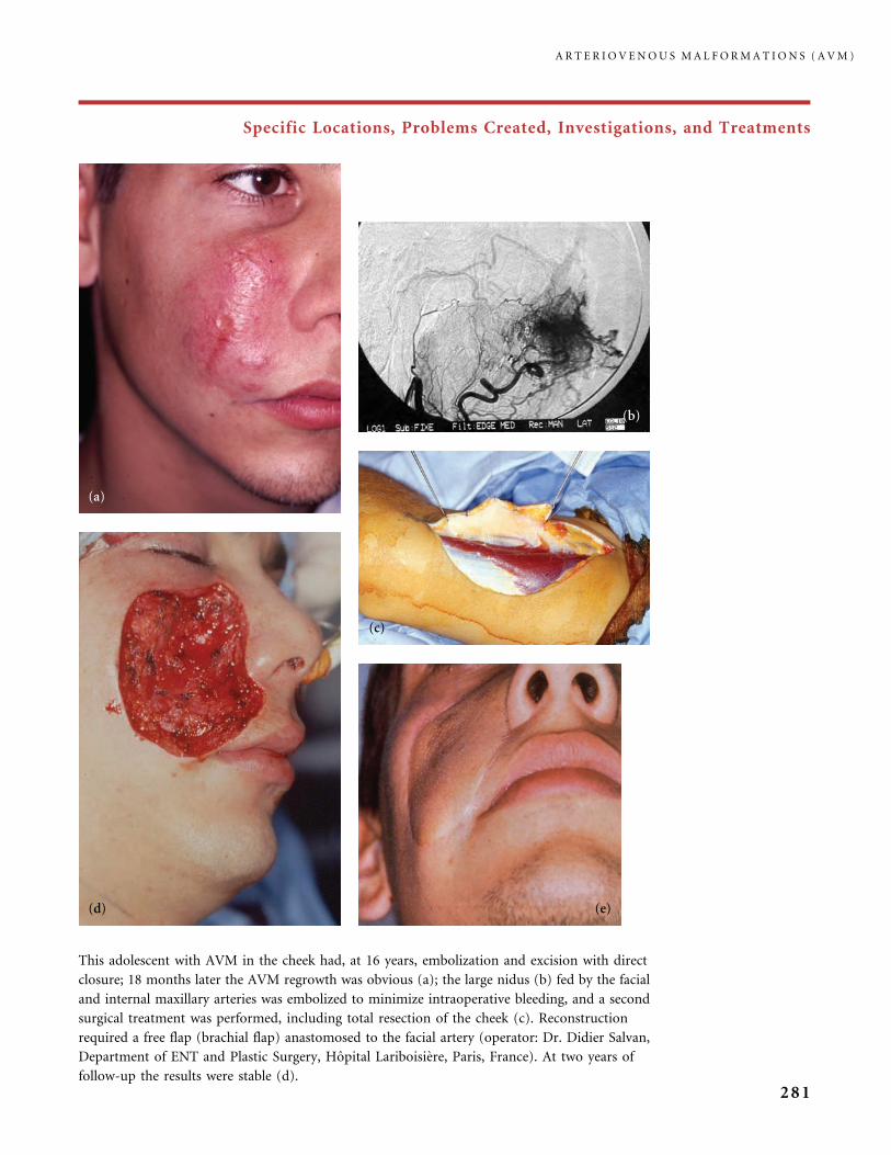
Specific Locations, Problems Created, Investigations, and Treatments
This adolescent with AVM in the cheek had, at 16 years, embolization and excision with direct
closure; 18 months later the AVM regrowth was obvious (a); the large nidus (b) fed by the facial
and internal maxillary arteries was embolized to minimize intraoperative bleeding, and a second
surgical treatment was performed, including total resection of the cheek (c). Reconstruction
required a free flap (brachial flap) anastomosed to the facial artery (operator: Dr. Didier Salvan,
Department of ENT and Plastic Surgery, Hopital Lariboisiere, Paris, France). At two years of
follow-up the results were stable (d).
A R T E R I O V E N O U S M A L F O R M A T I O N S ( A V M )
281
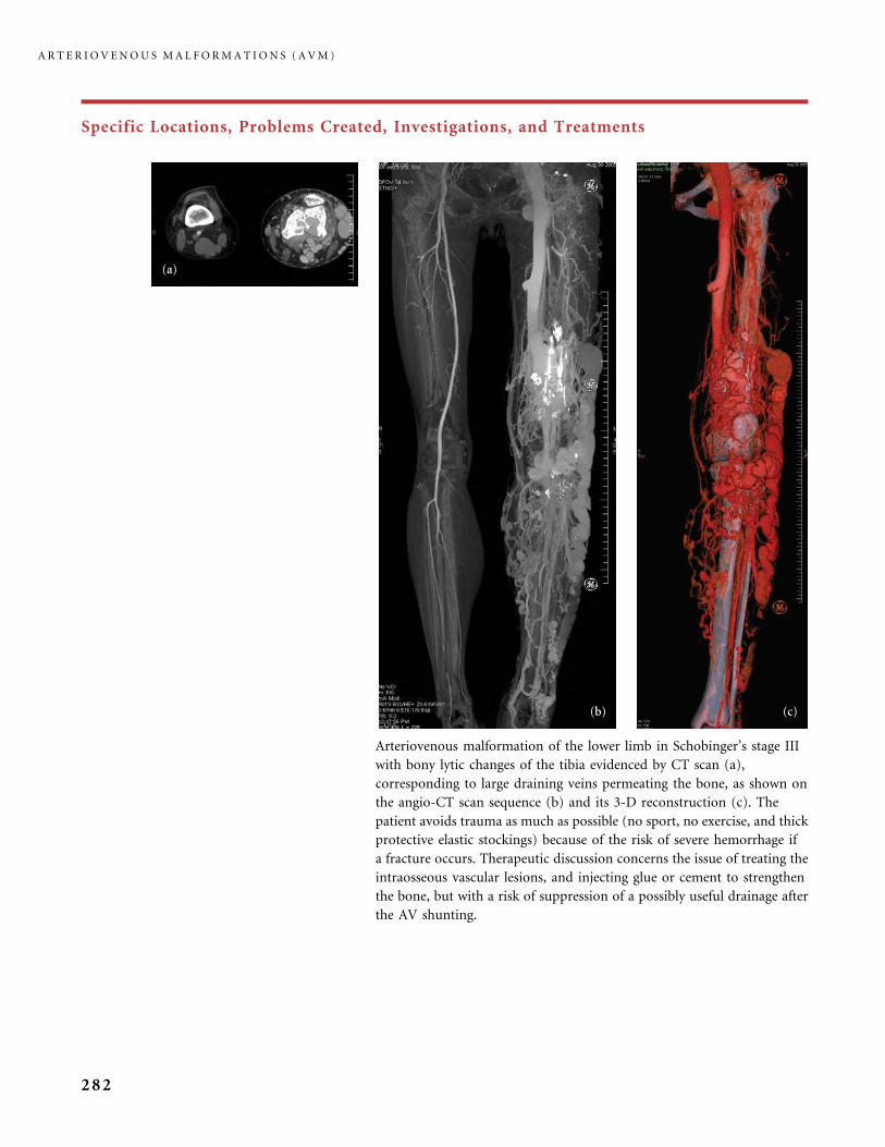
Specific Locations, Problems Created, Investigations, and Treatments
Arteriovenous malformation of the lower limb in Schobinger’s stage III
with bony lytic changes of the tibia evidenced by CT scan (a),
corresponding to large draining veins permeating the bone, as shown on
the angio-CT scan sequence (b) and its 3-D reconstruction (c). The
patient avoids trauma as much as possible (no sport, no exercise, and thick
protective elastic stockings) because of the risk of severe hemorrhage if
a fracture occurs. Therapeutic discussion concerns the issue of treating the
intraosseous vascular lesions, and injecting glue or cement to strengthen
the bone, but with a risk of suppression of a possibly useful drainage after
the AV shunting.
A R T E R I O V E N O U S M A L F O R M A T I O N S ( A V M )
282

Specific Locations, Problems Created, Investigations, and Treatments
This woman has Cobb syndrome. She also has a diffuse extremity
AVM with overgrowth of the affected arm, which constitutes
Parkes Weber syndrome: a number of our patients with spinal
lesions of Cobb syndrome also have such involvement of the
extremity located in the same metamere. This young woman
developed neurological signs from the spinal dorsal AVM; arterial
embolization was not effective and she became paraplegic.
MRI of the brain involvement in a patient with
Bonnet�Dechaume�Blanc syndrome. The Schobinger stage III
skin AVM in the mid-forehead was resected after arterial
embolization, and the long-term results were stable and good.
As the cerebral lesion was not symptomatic and difficult to access,
no treatment was offered.
A R T E R I O V E N O U S M A L F O R M A T I O N S ( A V M )
283
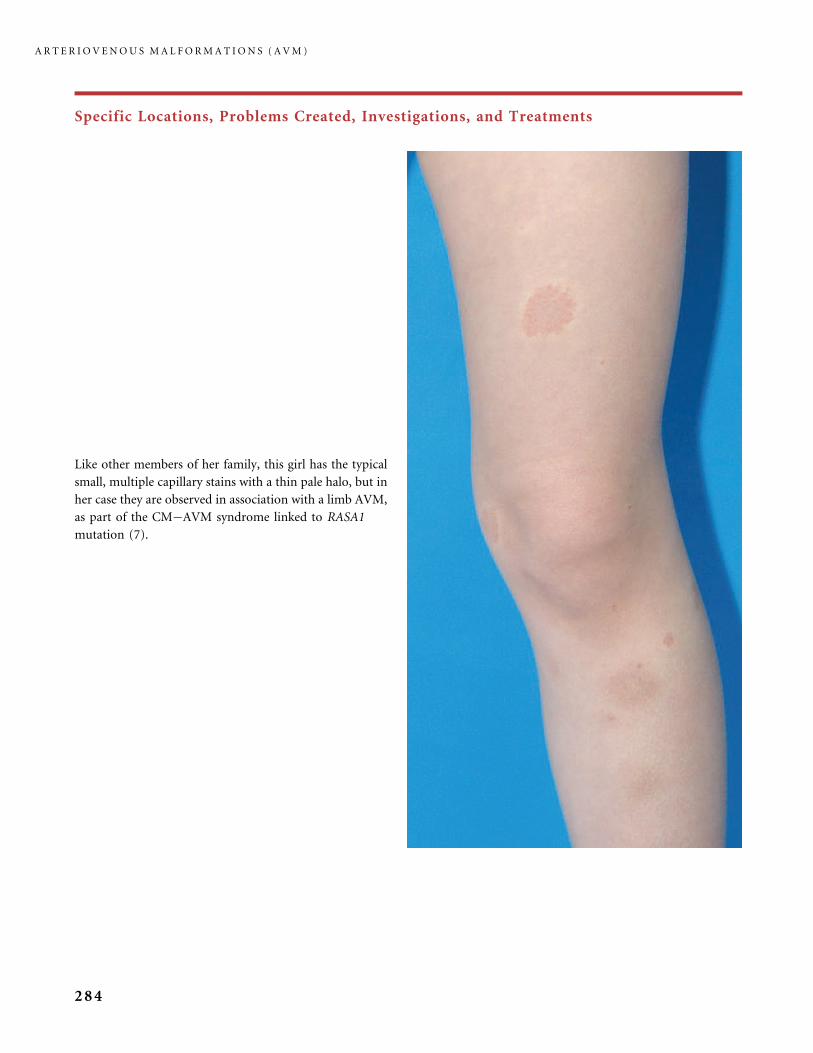
Specific Locations, Problems Created, Investigations, and Treatments
Like other members of her family, this girl has the typical
small, multiple capillary stains with a thin pale halo, but in
her case they are observed in association with a limb AVM,
as part of the CM�AVM syndrome linked to RASA1
mutation (7).
A R T E R I O V E N O U S M A L F O R M A T I O N S ( A V M )
284
References
1 Bhattacharya JJ, Luo CB, Suh D. Wyburn-Mason or Bonnet�Dechaume�Blanc as
cerebrofacial arteriovenous metameric syndromes (CAMS). Intervent Neuroradiol 2001;
7: 5�17.
2 Bonnet P, Dechaume J, Blanc E. L’anevrisme cirsoide de la retine (anevrisme racemeux).
Ses relations avec l’anevrisme cirsoide du cerveau. Le Journal Medical de Lyon 1937;
18: 165�78.
3 Burrows PB, Bisdorff A, Karian V, Mason K. Complications of ethanol embolization of
arteriovenous malformations. Communication T21 in 15th ISSVA Workshop.
Wellington, NZ. 22�25 February 2004.
4 Burrows PE, Laor T, Paltiel H, Robertson RL. Diagnostic imaging in the evaluation
of vascular birthmarks. Dermatol Clin 1998; 16: 455�88.
5 Do YS, Yakes WF, Shin SW, Lee BB, Kim DI, Liu WC et al. Ethanol embolization
of arteriovenous malformations: interim results. Radiology 2005; 235: 674�82.
6 Dubois J, Garel L, Grignon A, David M, Laberge L, Filiatrault D et al. Imaging of
hemangiomas and vascular malformations in children. Acad Radiol 1998; 5: 390�400.
7 Eerola I, Boon LM, Mulliken JB, Burrows PE, Dompmartin A, Watanabe S et al. Capillary
malformation�arteriovenous malformation, a new clinical and genetic disorder caused
by RASA1 mutations. Am J Hum Genet 2003; 73: 1240�9.
8 Enjolras O, Chapot R, Merland JJ. Vascular anomalies and the growth of limbs: a review.
J Pediatr Orthop B 2004; 13: 349�57.
9 Enjolras O, Logeart I, Gelbert F, Lemarchand-Venencie F, Reizine D, Guichard JP et al.
Arteriovenous malformations: a study of 200 cases. Ann Dermatol Venereol 2000; 127:
17�22.
10 Enjolras O, Mulliken JB. Vascular tumors and vascular malformations (new issues).
Adv Dermatol 1997; 13: 375�423.
11 Gladbach B, Pfeil J, Heijens E. Percutaneous epiphyseodesis. Correction of leg length
inequalities and frontal plane deformities. Orthopade 2000; 29: 2�8.
12 Khong PL, Burrows PE, Kozakewich HP, Mulliken JB. Fast-flow lingual vascular
anomalies in the young patient: is imaging diagnostic? Pediatr Radiol 2003; 33: 118�22.
13 Kohout MP, Hansen M, Pribaz JJ, Mulliken JB. Arteriovenous malformations of the head
and neck: natural history and management. Plast Reconstr Surg 1998; 102: 643�54.
14 Larralde M, Gonzalez V, Marietti R, Nussembaum D, Peirano M, Schroh R. Pseudo-
Kaposi sarcoma with arteriovenous malformation. Pediatr Dermatol 2001; 18: 325�7.
15 Lee BB, Do YS, Yakes W, Kim DI, Mattassi R, Hyon WS. Management of arteriovenous
malformations: a multidisciplinary approach. J Vasc Surg 2004; 39: 590�600.
16 Park S, Tepper OM, Galiano RD, Capla JM, Baharestani S, Kleinman ME et al. Selective
recruitment of endothelial progenitor cells to ischemic tissues with increased neo-
vascularization. Plast Reconstr Surg 2004; 113: 284�93.
17 Robertson RL, Robson CD, Barnes PD, Burrows PE. Head and neck vascular anomalies
of childhood. Neuroimaging Clin N Am 1999; 9: 115�32.
18 Rodesch G, Hurth M, Alvarez H, Tadie M, Lasjaunias P. Classification of spinal
cord arteriovenous shunts: proposal for a reappraisal � the Bicetre experience with
155 consecutive patients treated between 1981 and 1999. Neurosurgery 2002; 51: 374�9;
discussion 379�80.
19 Sofocleous CT, Rosen RJ, Raskin K, Fioole B, Hofstee DJ. Congenital vascular
malformations in the hand and forearm. J Endovasc Ther 2001; 8: 484�94.
20 Takaya N, Iwase T, Maehara A, Nishiyama S, Nakanishi S, Yamana D et al. Transcatheter
embolization of arteriovenous malformations in Cowden disease. Jpn Circ J 1999;
63: 326�9.
R E F E R E N C E S
285
21 Upton J, Coombs CJ, Mulliken JB, Burrows PE, Pap S. Vascular malformations of the
upper limb: a review of 270 patients. J Hand Surg Am 1999; 24: 1019�35.
22 Wu JK, Bisdorff A, Gelbert F, Enjolras O, Burrows PE, Mulliken JB. Auricular
arteriovenous malformation: evaluation, management, and outcome. Plast Reconstr Surg
2005; 115: 985�95.
23 Wyburn-Mason R. Arteriovenous aneurysm of midbrain and retina, facial nevi, and
mental changes. Brain 1943; 66: 163�203.
24 Yakes WF, Luethke JM, Merland JJ, Rak KM, Slater DD, Hollis HW et al. Ethanol
embolization of arteriovenous fistulas: a primary mode of therapy. J Vasc Interv Radiol
1990; 1: 89�96.
25 Yakes WF, Yee DC. Safety of ethanol embolization for the treatment of vascular
malformations. Communication T20 in 15th ISSVA Workshop. Wellington, NZ. 22�25
February 2004.
A R T E R I O V E N O U S M A L F O R M A T I O N S ( A V M )
286
Conclusion

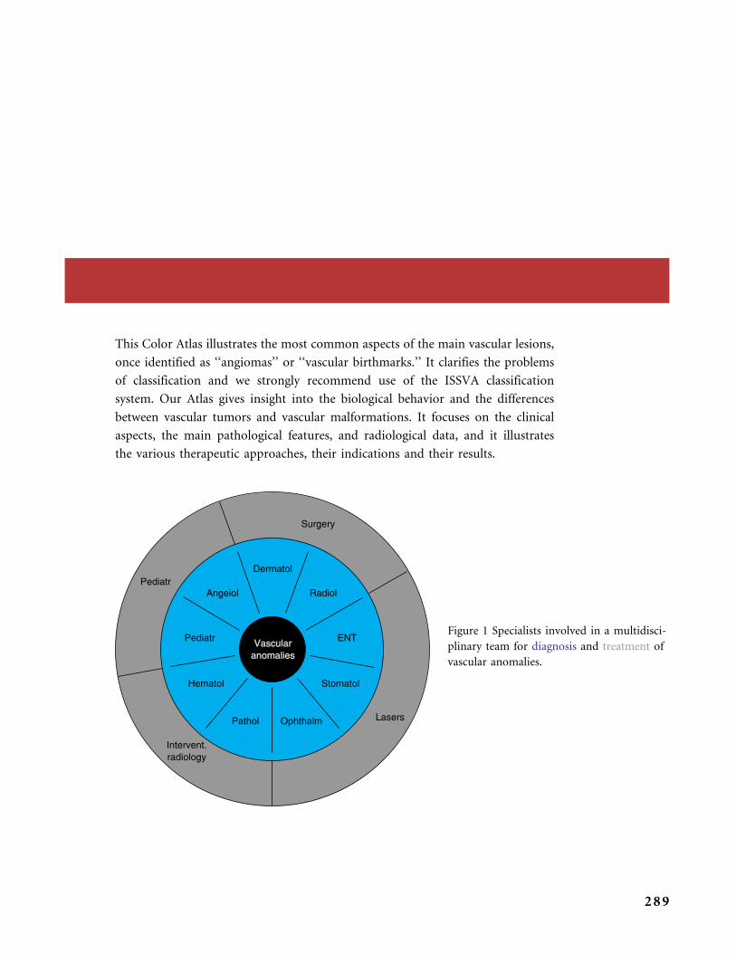
This Color Atlas illustrates the most common aspects of the main vascular lesions,
once identified as ‘‘angiomas’’ or ‘‘vascular birthmarks.’’ It clarifies the problems
of classification and we strongly recommend use of the ISSVA classification
system. Our Atlas gives insight into the biological behavior and the differences
between vascular tumors and vascular malformations. It focuses on the clinical
aspects, the main pathological features, and radiological data, and it illustrates
the various therapeutic approaches, their indications and their results.
Figure 1 Specialists involved in a multidisci-
plinary team for diagnosis and treatment of
vascular anomalies.
289
We do not attempt to offer an exhaustive book on the subject of vascular
lesions. We only try to offer a comprehensive review of those previously called
‘‘angiomas’’ or ‘‘vascular birthmarks.’’ Therefore this Atlas does not address in
detail the many genetic syndromes that cause vascular anomalies, and does not
display a dermatological pot-pourri of small uncommon dermatological
vascular lesions that are clearly benign (such as pyogenic granuloma, hobnail
hemangioma, epithelioid or histiocytoid hemangioma, etc.). Nor does it cover
vascular tumors of possibly intermediate malignancy, such as the composite
hemangioendothelioma. The vascular malignancies (angiosarcomas, lymphan-
giosarcomas) are beyond our scope (they were never considered ‘‘angiomas’’
or ‘‘birthmarks’’).
And finally, we want to stress the fact that identifying the diverse clinical,
pathological, and radiological aspects of the various vascular tumors and malfor-
mations, as well as selecting their optimal therapeutic management, requires
a multidisciplinary approach in the vast majority of cases.
C O N C L U S I O N
290
Index
Aagenaes syndrome 228
acrylic polymers
cf. Ethibloc�, 172
VM 172–3
Adams-Oliver syndrome 8, 134
AKC; see angiokeratoma circumscriptum
amyotrophy, VM 194
Anderson-Fabry disease, angiokeratoma
corporis diffusum 136
anetoderma, IH 39
angio-CT scans 6
angioblastoma of Nakagawa; see tufted
angioma
angiogenesis-dependent vascular anomalies,
pathogenesis 9
angiography 6
IH 24
angiokeratoma circumscriptum (AKC)
158–9
angiokeratoma corporis diffusum 160
Anderson-Fabry disease 136
angiokeratomas 135–6
Fordyce 159
angioma, nomenclature 3
angioma serpiginosum of Hutchinson 160
ankle
KMP 114
RICH 94
arm
IH 38, 40, 49
NICH 96
PWS 151
RICH 91–2
VM 195–6
arterial superselective embolization 7
arteriovenous fistulas (AVF), Rendu-Osler-
Weber syndrome (HHT) 259
arteriovenous malformations (AVM) 255–84
clinical features 255–6
CM-AVM syndrome 260
common 255–8
Cowden syndrome 260
CT angiography 257
CT scanning 257
digital arteriography 257
ear 265–6
Ehlers-Danlos type IV syndrome 260
eye 275
face 266, 278–9, 281
foot 275
FPDL 264
hand 268–9, 273, 274
investigations 263–84
leg 269–70, 272
MRA 257
MRI 257
overview 255
Parkes Weber syndrome 270–1
parotid area 280
pathology 261–2
problems created 263–84
pseudo Kaposi sarcoma skin changes 269,
272
pulsed Doppler 256
radiological investigations 256–7
Rendu-Osler-Weber syndrome (HHT)
259
scalp 267, 274
Schobinger staging 256, 263, 282
specific locations 263–84
stage III 276–7
syndromic 258–60
treatment 257–8, 263–84
ultrasonography/Doppler 256
291

aspirin
KMP 113, 118, 120
telangiectasia 118
ataxia telangiectasia (AT) 8, 10, 135
neurological risks 129
AVF; see arteriovenous fistulas
AVM; see arteriovenous malformations
back, KMP 115–16
Bannayan-Riley-Ruvalcaba syndrome 8, 10
Bean (blue rubber bleb nevus) syndrome 8,
173–4
clinical aspects 211–12
pathology
‘beard’ infantile hemangioma 53
benign lymphangioendothelioma 232, 246
biopsy 6
IH 24
birthmark, nomenclature 3
bleomycin treatment, IH 28
blue rubber bleb naevus (BRBN) syndrome 8,
173–4
clinical aspects 211–12
pathology
Bonnet-Dechaume-Blanc syndrome 258
MRI 283
neurological risks 129
brain anomalies, SWS 148
BRBN; see blue rubber bleb naevus
breast, IH 49
buttocks
IH 50–1, 60
KHE 112
capillary hemangioma; see infantile
hemangioma
capillary malformations (CM) 125–32
Adams-Oliver syndrome 8, 134
angiokeratomas 135–6
associations 127–8
AT 135
clinical aspects
CM-AVM syndrome 10, 260
CMTC 133
EAH 127
Ethibloc� 157
face 163
KTS 129–31
lumbar 151
macrocephaly-cutis marmorata syndrome
134
neurological risks 129
nevus anemicus 149
overview 125
pathology 137–9
PPV 127, 149
proteiform syndromes 131–3
Proteus syndrome 131–3
PWS 125–7, 140
pyogenic granulomas 162
Rendu-Osler-Weber syndrome (HHT) 8,
10, 134–5
reticulate diffuse CM 134, 157
spina-lipoma, attached cord 151
spinal cord, attached syringomyelia 150
SWS 128–9
syndromic 128–33
telangiectasia 133–5
thigh 140
cardiac assessment, PHACE(S) syndrome
57
causes
IH 8–9, 21–2
vascular malformations 8–9
cavernous hemangioma; see infantile
hemangioma
cephalic area
IH 26, 41
neurological risks 129
cephalic VM
clinical patterns 181
neurological risks 129
cerebral cavernous malformations 10
cerebral developmental venous anomaly
(DVA) 191
cholestasis-lymphedema syndrome 228
classification, ‘‘biological’’ 4
fast-flow vascular malformations 4
slow-flow vascular malformations 4
classification, ISSVA 3–10
fast-flow vascular malformations 6
molecular biology 9
slow-flow vascular malformations 6
updated 6
vascular malformations 6
vascular tumors 6
CM-AVM syndrome 10, 260
CM; see capillary malformations
CMTC; see cutis marmorata telangiectatica
congenita
CMVM; see cutaneous and mucosal venous
malformations
Cobb syndrome 258
computed tomography (CT) 6, 16
AVM 257
CT angiography, AVM 257
IH 24, 56
LM 226
VM 170
conclusion 287–90
I N D E X
292
congenital hemangiomas 78
features 78–80
missing links 82, 96
NICH 81–2
RICH 80–2
conventional vascular imaging 17
conventional X-rays 15
corticosteroid, topical, IH 27
Cowden syndrome, AVM 260
cryosurgery
see also surgical excision/resection
IH 25
CT; see computed tomography
cutaneous and mucosal venous
malformations (CMVM), familial 10,
174
cutis marmorata-macrocephaly syndrome 8,
134, 156
neurological risks 129
cutis marmorata telangiectatica congenita
(CMTC) 8, 133, 156
developmental venous anomaly (DVA),
cerebral 191
diagnostic imaging devices 13
IH 6, 24
vascular malformations 6
vascular tumors 6
diffuse reticulate CM 134, 157
digital arteriography, AVM 257
digital computed arteriogram, VM 170
direct puncture sclerotherapy 7
LM 226, 251
disseminated neonatal hemangiomatosis
(DNH), IH 22, 55
DNH; see disseminated neonatal
hemangiomatosis
Doppler
see also ultrasonography/Doppler
pulsed Doppler 256
DVA; see cerebral developmental venous
anomaly (DVA)
dyspnea
IH 54
laryngeal 66–7
VM 201
EAH; see eccrine angiomatous
hamartoma
ear
AVM 265–6
IH 56, 70–1
RICH 98
eccrine angiomatous hamartoma (EAH) 127,
160–1
Ehlers-Danlos type IV syndrome, AVM 260
elastic garments, VM 192, 195–6
embolization, IH 25
endoscopy, IH 26
endothelial cells, markers 7–8
epilepsy, SWS 128–9, 141
ethanol, VM 200, 203–4
Ethibloc�
cf. acrylic polymers 172
CM 157
LM 248, 252
VM 202, 205, 206
evanescent (or fading) macule, clinical
aspects 140–1
eye
AVM 275
IH 44–6, 57, 61–3, 65, 68–9
LM 225, 235, 246
PHACE(S) syndrome 57
TA 110
VM 183, 190, 207
face
AVM 266, 278–9, 281
CM 163
FPDL 163
IH 42, 58, 59, 64, 71
KMP 116, 117
LM 236
PWS 143–7
RICH 98
VM 206
fading (or evanescent) macule, clinical
aspects 140–1
familial cutaneous and mucosal venous
malformations (CMVM) 10, 174
fast-flow vascular malformations
classification, ‘‘biological’’ 4
classification, ISSVA 6
fingers, VM 193
flashlamp pumped-pulsed dye laser
(FPDL)
AVM 264
CM 163
face 163
PWS 126–7
foot, AVM 275
Fordyce angiokeratomas 159
forearm/hand, IH 40, 67
FPDL; see flashlamp pumped-pulsed dye
laser
genetic defects
see also inherited vascular malformations
IH 24
I N D E X
293
genetic defects (Contd.)
vascular malformations 10
genitalia, VM 194–5
glomuvenous malformations (GVM) 174–5
clinical aspects 216–18
glomangioma 10
pathology
glucocorticosteroid (GS) treatment, IH 25–7,
61, 65, 67
Gorham-Stout syndrome 8
Gorham syndrome 228
growth
hemangiomas 8–9
IH 3, 8, 35–7
vascular malformations 3–4
vascular tumors 3
GS treatment; see glucocorticosteroid
treatment
GVM; see glomuvenous malformations
hand
AVM 268–9, 273, 274
IH; see forearm/hand
KHE 111
VM 193
hearing tests, IH 26
hemangiomas
see also infantile hemangioma
characteristics 7–9
growth 8–9
nomenclature 3
pathogenesis 8
regression 7
hemarthrosis, VM 200
Hennekam syndrome 228
hereditary hemorrhagic telangiectasia
(HHT); see Rendu-Osler-Weber
syndrome
HHT; see Rendu-Osler-Weber syndrome
Hutchinson angioma serpiginosum 135
IFN; see interferon alpha 2a or 2b
IH; see infantile hemangioma
imaging; see diagnostic imaging devices
immature hemangioma; see infantile
hemangioma
incidence, IH 21
infantile hemangioma (IH) 21–6
anetoderma 39
angiography 24
arm 38, 40, 49
‘beard’, 53
biopsy 24
bleomycin treatment 28
breast 49
buttocks 50–1, 60
causes 8–9, 21–2
cephalic area 26, 41
clinical aspects 35–72
clinical examination 26
corticosteroid, topical 27
cryosurgery 25
CT scanning 24, 56
diagnosis 24
diagnostic imaging devices 6, 24
DNH 22, 55
dyspnea 54
ear 56, 70–1
embolization 25
endoscopy 26
eye 44–6, 57, 61–3, 65, 68–9
face 42, 58, 59, 64, 71
forearm/hand 38, 40, 67
genetic defects 24
glucocorticosteroid (GS) treatment 25–7,
61, 65, 67
growth 3, 8, 35–7
hearing tests 26
IFN 27–8, 62, 64, 66–7
incidence 21
intralesional glucocorticosteroid 27
investigations 35–72
laryngeal dyspnea 66–7
lasers 25
lips 47, 52, 72
mesenchymal stem cells 40
MRI 24, 26, 43, 56
nerves 40
neurological risks 129
cf. NICH 79
nomenclature 21
nose 48, 70
ophthalmological examination 26, 44–6,
57
oral glucocorticosteroid (GS) treatment
25–7, 65
parotid hemangioma 70–1
pathology 24–5, 30–4
PHACE(S) syndrome 23–4, 57, 58, 62
phases 22, 38
precursors 36–7, 58
radiotherapy 25
regression 22, 38, 39
cf. RICH 79
scalp 38
shoulder 39
sternal fusion defects 58
subcategories 22
surgical excision/resection 28–9
thorax 49, 58
I N D E X
294
topical corticosteroid 27
treatment 25–9, 35–72
ulceration 40, 49–52, 70–1
ultrasonography/Doppler 24, 26, 43
cf. vascular malformations 5
VCR 28, 66–7
X-linked defect 24
inherited vascular malformations 9
see also genetic defects
interferon alpha 2a or 2b (IFN), IH 27–8, 62,
64, 66–7
intralesional bleomycin treatment, IH 28
intralesional glucocorticosteroid, IH 27
investigations, IH 35–72
investigations tools 13
see also diagnostic imaging devices
kaposiform hemangioendothelioma (KHE)
102
buttocks 112
hand 111
KMP 102, 112
leg 111
LM 111
pathology 107–8
TA 112
treatment 102
Kasabach-Merritt phenomenon (KMP)
102–5, 112–20
ankle 114
aspirin 113, 118, 120
back 115–16
face 116, 117
KHE 102, 112
cf. LIC 171
neck 115–16, 119
nosology 103
pentoxyfillin 113
platelet infusions 116
presentation differences 113–14
TA 101, 102, 109, 112
telangiectasia 120
thorax 120
thrombocytopenia 115–16, 120
ticlopidine 113, 118, 120
treatment 103–4
VCR 112, 115–17, 119
Kasabach-Merritt syndrome (KMS) 102–3
cf. LIC 171
KHE; see kaposiform hemangioendothelioma
(KHE)
Klippel-Trenaunay syndrome (KTS) 8,
129–31
clinical characteristics 130, 259
leg 152, 153
thigh 152, 153
KMP; see Kasabach-Merritt phenomenon
KMS; see Kasabach-Merritt syndrome
knee
RICH 87–8, 90
VM 198, 206
KTS; see Klippel-Trenaunay syndrome
laryngeal dyspnea, IH 66–7
lasers 7
FPDL 126–7, 163, 264
IH 25
LM 227
PWS 162
leg
AVM 269–70, 272
KHE 111
KTS 152, 153
RICH 97
VM 198
LIC; see localized intravascular coagulopathy
lips
IH 47, 52, 72
LM 235
ulceration 52
VM 183, 203–4
LM; see lymphatic malformations
localized intravascular coagulopathy (LIC)
cf. KMP/KMS 171
VM 192, 200
lumbar CM 151
lymphangioma, nomenclature 3
lymphatic malformations (LM) 224–52
bony 225
clinical aspects 224
common 224–7
CT scanning 226
DIC 242
direct puncture 226, 251
Ethibloc� 248, 252
eye 225, 235, 246
face 236
investigations 226, 233–52
KHE 111
lasers 227
lips 235
macrocystic 224–6
mandible 237
microcystic 224, 225
mouth 236
MRI 226, 240–1, 245
neck 238, 244, 249–50
overview 224
pathology 230–2
pelvic 242–3
I N D E X
295
lymphatic malformations (LM) (Contd.)
problems created 233–52
regression 226–7, 248
sclerotherapy 248
specific locations 233–52
surgical excision/resection 227
syndromic 227–9
treatment 226–7, 233–52
ultrasonography/Doppler 224, 226, 239
visceral 226
lymphedema 247
lymphedema of Milroy 10
lymphedemadistichiasis 10
lymphedemas, syndromic 227–8
lymphoscintigraphy 6
LYVE-1/CD 31 double staining 7
macrocephaly-cutis marmorata syndrome 8,
134, 156
neurological risks 129
macrocystic LM 224–6
Maffucci syndrome 8, 175–6
clinical aspects 221
pathology 219–20
magnetic resonance angiography (MRA) 6
AVM 257
magnetic resonance imaging (MRI) 6, 16–17
AVM 257
Bonnet-Dechaume-Blanc syndrome 283
IH 24, 26, 43, 56
LM 226, 240–1, 245
PWS 148
RICH 80
VM 169, 170, 189, 197
Wyburn-Mason syndrome 283
magnetic resonance venography (MRV) 6
mandible, LM 237
markers
endothelial cells 7–8
vascular tumors cf. vascular malformations
5
maxilla, PWS 144
mesenchymal stem cells, IH 40
microcystic LM 224, 225
missing links, congenital hemangiomas 82,
96
molecular biology, classification, ISSVA 9
mouth
LM 236
VM 182, 183, 186–9, 201–5, 208
MRA; see magnetic resonance angiography
MRI; see magnetic resonance imaging
MRV; see magnetic resonance venography
multifocal lymphangioendotheliomatosis
247
multiple miliary-type hemangiomas; see
disseminated neonatal hemangiomatosis
neck
KMP 115–16, 119
LM 238, 244, 249–50
PWS 150
VM 184–6
nerves, IH 40
neurological risks, CM 129
nevus anemicus, CM 149
NICH; see Non-Involuting Congenital
Hemangioma
nodular hyperplasia, PWS 143
nomenclature 3
angioma 3
birthmark 3
hemangioma 3
IH 21
lymphangioma 3
Non-Involuting Congenital
Hemangioma (NICH) 81–2
arm 96
features
cf. IH 79
pathological features 99
pathology
cf. RICH 79
telangiectasia 95
nose, IH 48, 70
occipital RICH 93
ophthalmological examination
IH 26, 44–6, 57
PHACE(S) syndrome 57
oral glucocorticosteroid (GS) treatment, IH
25–7, 65
orbital LM, neurological risks 129
Parkes Weber syndrome 259
AVM 270–1
clinical characteristics 130, 259
parotid area, AVM 280
parotid hemangioma, IH 70–1
pathogenesis
angiogenesis-dependent vascular
anomalies 9
hemangiomas 8
vascular malformations 9–10
(F)PDL; see flashlamp pumped-pulsed dye
laser
pelvis, LM 242–3
pentoxyfillin
KMP 112
VCR 112
I N D E X
296

PHACE(S) syndrome
cardiac assessment 57
eye 57
IH 23–4, 57, 58, 62
ophthalmological examination 57
phakomatosis pigmentovascularis (PPV) 127,
149
pharmacological therapies 7
phlebography, VM 170–1
platelet infusions, KMP
port-wine stains (PWS) 125–7
arm 151
clinical aspects 125–6, 140, 142
diagnosis 126
dissemination 141
face 143–7
FPDL 126–7
lasers 162
maxilla 144
MRI 148
neck 150
nodular hyperplasia 143
proteiform syndromes 154–5
Proteus syndrome 154–5
regression 140
spinal cord, attached syringomyelia 150
tissular hyperplasia 144
treatment 126–7
PPV; see phakomatosis pigmentovascularis
precursors, IH 36–7, 58
pregnancy, telangiectasia 161
proteiform syndromes 131–3
PWS 154–5
Proteus syndrome 8, 131–3
diagnostic criteria 132
neurological risks 129
PWS 154–5
pseudo Kaposi sarcoma skin changes, AVM
269, 272
pulsed Doppler, AVM 256
PWS; see port-wine stains
pyogenic granulomas, CM 162
radiographs, plain 6
radiological investigations, AVM 256–7
radiological tools 13
see also diagnostic imaging devices
radiotherapy, IH 25
Rapidly Involuting Congenital Hemangioma
(RICH) 80–2
ankle 94
arm 91–2
ear 98
face 98
features 80–1, 87–94
cf. IH 79
knee 87–8, 90
leg 97
management 81
MRI 80
cf. NICH 79
occipital 93
pathology 83–4
scalp 94, 97
thigh 97
ultrasonography/Doppler 80
regional cerebral blood flow (rCBF), SPECT
149
regression
hemangiomas 7
IH 22, 38, 39
LM 226–7, 248
PWS 140
TA 110
vascular malformations 4
vascular tumors 4
Rendu-Osler-Weber syndrome (HHT) 8, 10,
134–5, 157
AVF 259
AVM 259
neurological risks 129
reticulate diffuse CM 134, 157
RICH; see Rapidly Involuting Congenital
Hemangioma
salmon patch, clinical aspects 140–1
scalp
AVM 267, 274
IH 38
RICH 94, 97
VM 190
Schobinger staging, AVM 256, 263, 282
shoulder
IH 39
VM 197
Single Photon Emission Computed
Tomography (SPECT), rCBF 149
sleep apnea syndrome, VM 182
slow-flow vascular malformations 8
classification, ‘‘biological’’ 4
classification, ISSVA 6
SPECT; see Single Photon Emission
Computed Tomography
spina-lipoma, attached cord, CM 151
spinal cord, attached syringomyelia, PWS 150
sternal fusion defects, IH 58
strawberry mark; see infantile hemangioma
Sturge-Weber syndrome (SWS) 128–9
brain anomalies 148
epilepsy 128–9, 141
I N D E X
297

Sturge-eber syndrome (SWS) (Contd.)
neurological risks 129
surgical excision/resection 7
see also cryosurgery
IH 28–9
LM 227
SWS; see Sturge-Weber syndrome (SWS)
syndromes 8
syndromic AVM 258–60
syndromic CM 128–33
syndromic LM 227–9
syndromic lymphedemas 227–8
syndromic VM, nosology 173–6
syringomyelia, attached spinal cord, PWS 150
TA; see tufted angioma
telangiectasia 118, 133–5
see also Rendu-Osler-Weber syndrome
aspirin 118
KMP 120
NICH 95
pregnancy 161
syndromes with 133–5
ticlopidine 118
therapeutic strategies
see also treatment
vascular malformations 7
vascular tumors 7
thigh
KTS 152, 153
RICH 97
thorax
IH 49, 58
KMP 120
thrombocytopenia, KMP 115–16, 120
ticlopidine
KMP 113, 118, 120
telangiectasia 118
tissular hyperplasia, PWS 144
tongue, VM 183, 189
topical corticosteroid, IH 27
treatment
see also therapeutic strategies
AVM 257–8, 263–84
IH 25–9, 35–72
KHE 102
KMP 103–4
LM 226–7, 233–52
PWS 126–7
TA 101
vascular malformations 9–10
vascular tumors 9–10
VM –208, 172
tufted angioma (TA) 101
clinical aspects 109–20
eye 110
KHE 112
KMP 101, 102, 109, 112
pathology 105–6
regression 110
treatment 101
ulceration
IH 40, 49–52, 70–1
lips 52
ultrasonography/Doppler 6, 15
AVM 256
IH 24, 26, 43
LM 224, 226, 239
RICH 80
VM 169
updated classification, ISSVA 6
Van Lohuizen syndrome; see cutis
marmorata telangiectatica congenita
vanishing bone syndrome 228
vascular anomalies; see vascular
malformations; vascular tumors
vascular endothelial growth factor (VEGF),
vascular tumors cf. vascular
malformations 7
vascular imaging, conventional 17
vascular malformations
causes 8–9
classification, ISSVA 6
diagnostic imaging devices 6
fast-flow 4, 6
genetic defects 10
growth 3–4
cf. IH 5
inherited 9
pathogenesis 9–10
regression 4
slow-flow 8
subcategories 4–5
therapeutic strategies 7
treatment 9–10
cf. vascular tumors 3–10
vascular tumors
see also infantile hemangioma
classification, ISSVA 6
diagnostic imaging devices 6
growth 3
regression 4
therapeutic strategies 7
treatment 9–10
cf. vascular malformations 3–10
VCR; see vincristine (VCR)
VEGF; see vascular endothelial growth factor
venous malformations (VM) 168–71
I N D E X
298

acrylic polymers 172–3
amyotrophy 194
arm 195–6
cephalic area 181
clinical patterns 168–9, 181–208
common 168–73
CT scanning 170
digital computed arteriogram 170
DVA 191
dyspnea 201
elastic garments 192, 195–6
ethanol 200, 203–4
Ethibloc� 202, 205, 206
eye 183, 190, 207
face 206
fingers 193
genitalia 194–5
hand 193
hemarthrosis 200
investigations 169, 181–208
knee 198, 206
leg 198
LIC 192, 200
lips 183, 203–4
mouth 182, 183, 186–9, 201–5, 208
MRI 169, 170, 189, 197
neck 184–6
overview 168–9
pathology 169, 177–80
phlebography 170–1
scalp 190
shoulder 197
sleep apnea syndrome 182
syndromic, nosology 173–6
tongue 183, 189
treatment 172, 181–208
ultrasonography/Doppler 169
vincristine (VCR)
IH 66–7
KMP 112, 115–17, 119
VM; see venous malformations (VM)
Wyburn-Mason syndrome 258
MRI 283
neurological risks 129
X-linked defect, IH 24
X-rays, conventional 15
I N D E X
299


