34
Combined Optical and SPM Microscopy Alex Berquand Jan. 2012

Combined Optical and SPM Microscopy
Alex Berquand Jan. 2012

Combining IOM and AFM
• What is optical microscopy?
• What is atomic force microscopy
• How do they combine together?
• What type of information does such combination provide?
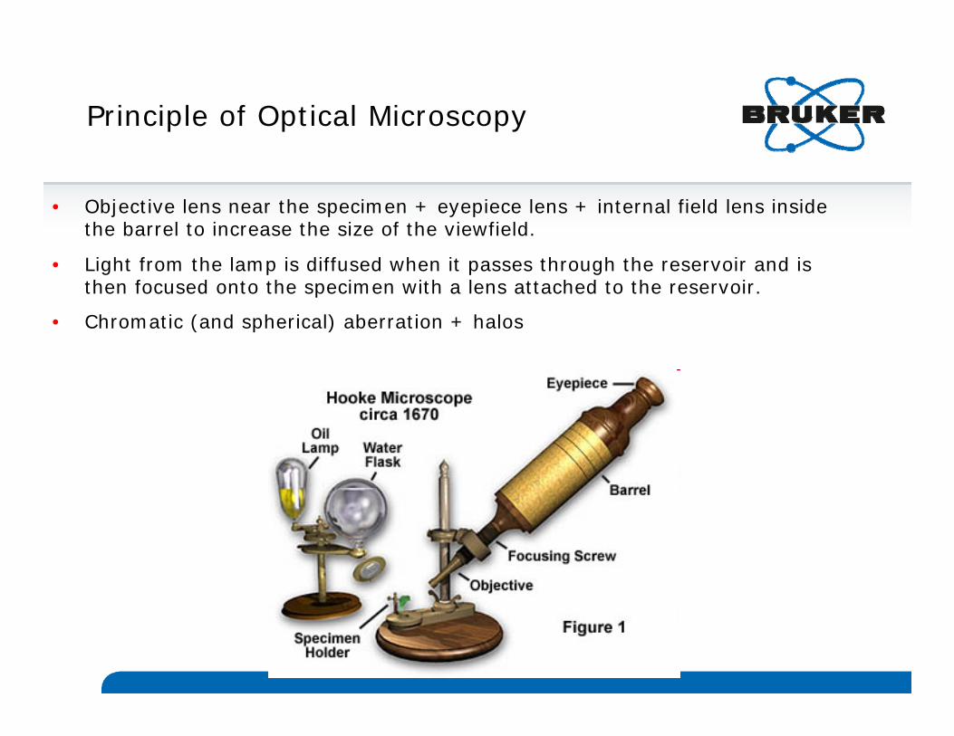
Principle of Optical Microscopy
• Objective lens near the specimen + eyepiece lens + internal field lens inside the barrel to increase the size of the viewfield.
• Light from the lamp is diffused when it passes through the reservoir and is then focused onto the specimen with a lens attached to the reservoir.
• Chromatic (and spherical) aberration + halos

Inverted Optical Microscopy
Inverted microscope stands incorporate the vertical illuminator within the body of the microscope.
Many types of objectives can be used with inverted reflected light microscopes.
All modes of reflected light illumination may be possible: brightfield, darkfield, polarized light, differential interference contrast, and fluorescence.

Fluorescence Microscopy
The absorption and subsequent re-radiation of light by organic and inorganic specimens is typically the result of fluorescence or phosphorescence. The emission of light through the fluorescence process is nearly simultaneous with the absorption of the excitation light due to a relatively short time delay between photon absorption and emission, ranging usually less than a microsecond in duration.
The basic function of a fluorescence microscope is to irradiate the specimen with a desired and specific band of wavelengths, and then to separate the much weaker emitted fluorescence from the excitation light. Only the emission light should reach the eye or detector so that the resulting fluorescent structures are superimposed with high contrast against a very dark (or black) background.
The limits of detection are generally governed by the darkness of the background, and the excitation light is typically 105 to 106x brighter than the emitted fluorescence.

Confocal Microscopy
Used to increase micrograph contrastand/or to reconstruct three-dimensional images by using a spatial pinhole to eliminate out-of-focus light in specimens that are thicker than the focal plane.
As much of the light from sample fluorescence is blocked at the pinhole this increased resolution is at the cost of decreased signal intensity so long exposures are often required.
As only one point in the sample is illuminated at a time, 2D or 3D imaging requires scanning over a regular raster (i.e. a rectangular pattern of parallel scanning lines) in the specimen.

Total Internal Reflection Microscopy
Uses evanescent wave to selectively illuminate and excite fluorophores in a restricted region of the specimen immediately adjacent to the glass-water interface. The evanescent wave is generated only when the incident light is totally reflected at the glass-water interface. The evanescent electromagnetic field decays exponentially from the interface, and thus penetrates to a depth of only approximately 100 nm into the sample medium. Thus the TIRFM enables a selective visualization of surface regions such as the basal plasma membrane.
Can also be used to observe the fluorescence of a single molecule.

Atomic Force Microscopy
1/18/2012 8BRUKER CONFIDENTIAL
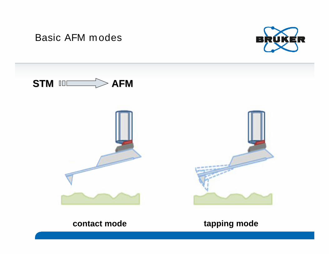
Basic AFM modes
contact mode tapping mode
STM AFMSTM AFM

Force Spectroscopy
distance (nm)fo
rce
(nN
)
0
1
2
-15000
In force mode during an approach-retract cycle, the tip is bought to the surface and contact with the sample, indenting into it. When the tip is pulled away, an hysteresis appears, reflecting the adhesion force existing between the tip apex and the sample.
This adhesion can be expressed as a force (measuring the height difference between the approach and retracted curves) or as an energy (integrating the area defined between the two curves).
If the tip is functionalized, the specific rupture/unbinding event can also be sensed on the retract curve.
Single force
Force volume
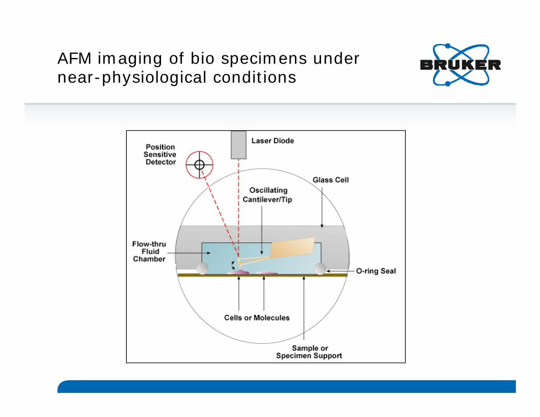
AFM imaging of bio specimens under near-physiological conditions
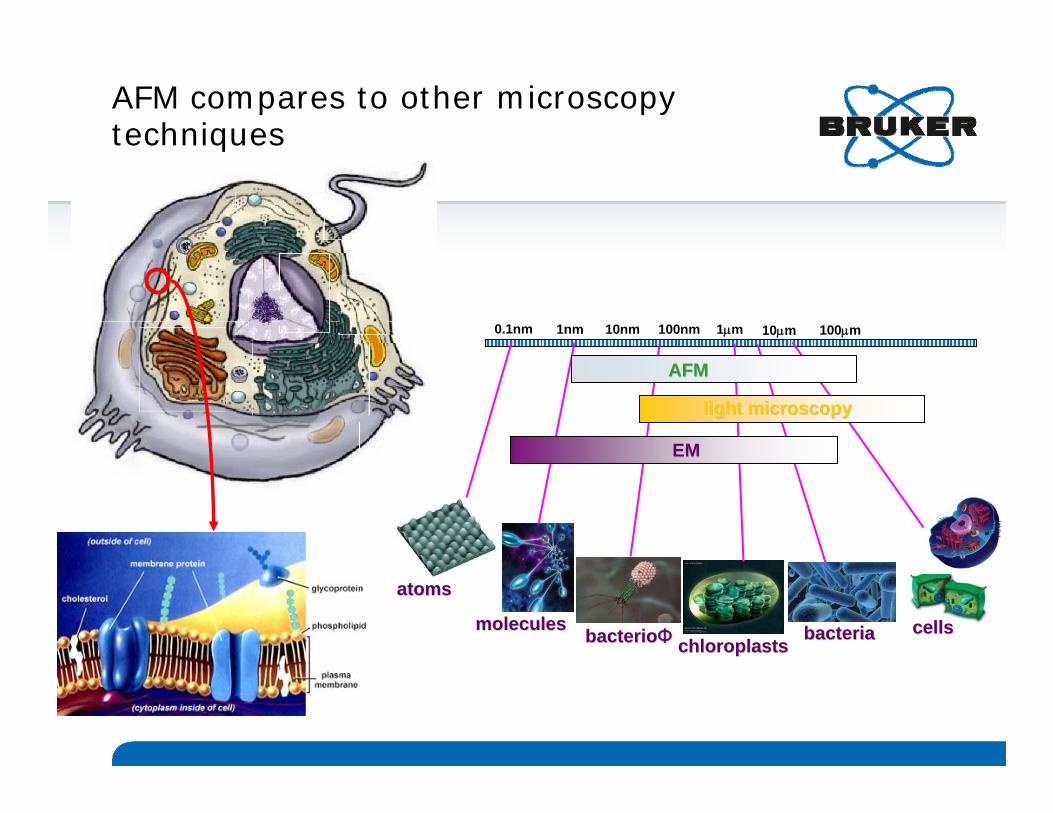
AFM compares to other microscopy techniques
atomsatoms
moleculesmoleculesbacteriobacterioΦΦ chloroplastschloroplasts bacteriabacteria cellscells
0.1nm 1nm 10nm 100nm 1μm 10μm 100μm
AFMAFM
light microscopylight microscopy
EMEM

AFM combined to IOM
1/18/2012 13BRUKER CONFIDENTIAL
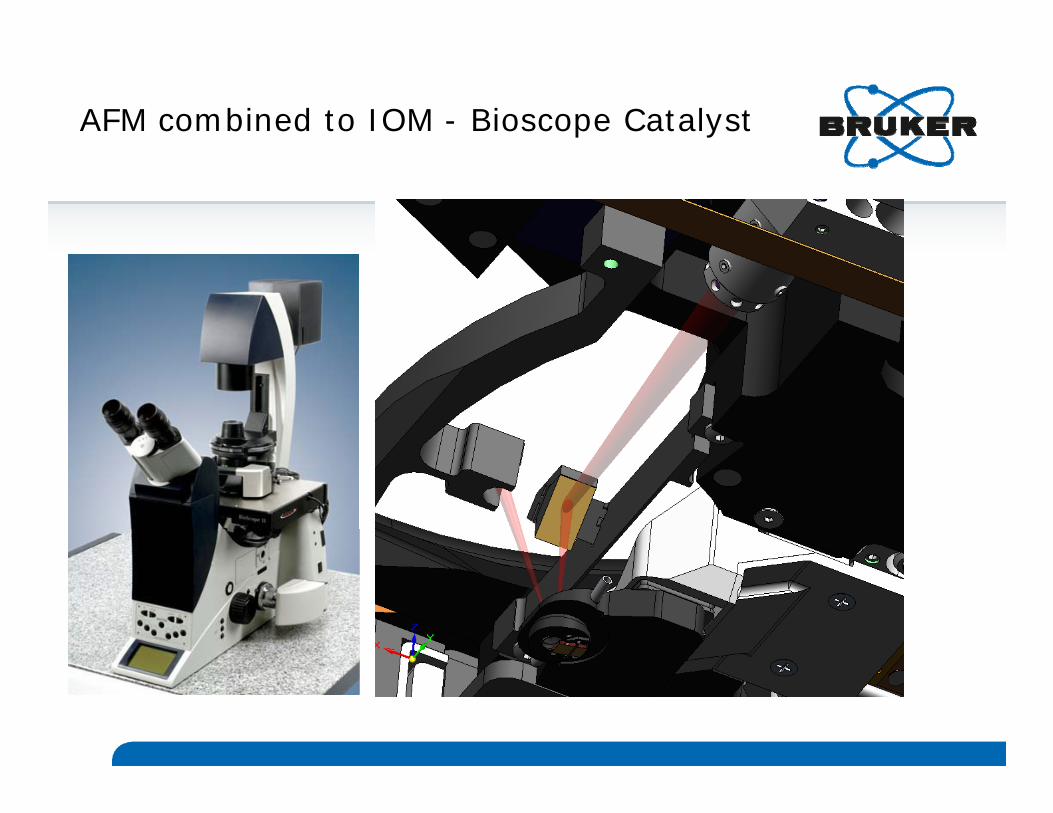
AFM combined to IOM - Bioscope Catalyst

AFM/IOM compatibility
AFM + …
Sample courtesy Zeiss
Sample courtesy NIH
Sample courtesy SWMC Stanford
Sample courtesy INRA Nantes
Sample courtesy Nat. Univ. Singapore
Bright Field
DIC
Epifluorescence
Confocal
RAMAN
TIRF
Sample courtesy Univ. of New South Wales

AFM/IOM combination: automatic overlay of AFM and optical images with MIRO
3) Overlay optical and AFM images
1) Import optical image into Nanoscope
2) Target a location for the AFM scan
Capture 3 samples images and 1 tip image… Locating the tip is mandatory!

1/18/2012 17BRUKER CONFIDENTIAL
+++
+++
+
+
+
+
+
+
+
240 240 μμmm
75 75 μμmm
FV scan
AFM image
SF curve
Example of overlay on living cells

Application: investigation of the effect of 2 disrupting agents on cell elasticity and morphology
Hela cells OS cells
- Nocodazole (destroys tubulin)- Latrunculin (destroys actin)
(Hochschule Mannheim)
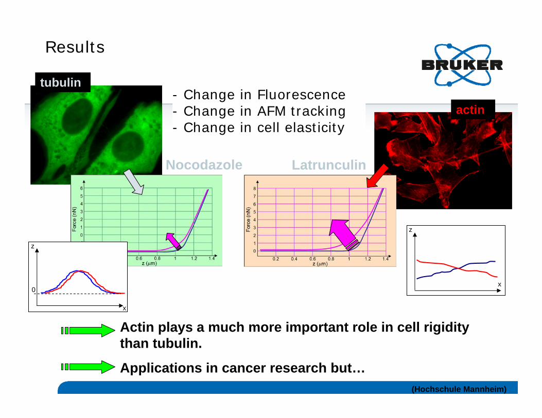
Results
Actin plays a much more important role in cell rigidity than tubulin.
Applications in cancer research but…
tubulin
actin
Nocodazole Latrunculin
0
z
x
z
x
- Change in Fluorescence- Change in AFM tracking- Change in cell elasticity
(Hochschule Mannheim)

1/18/2012 20BRUKER CONFIDENTIAL
Correlating fluorescence imaging and measurement of specific unbinding events - Living Helas
(Institut Pasteur Lille)

OHHO
O-CH3
O-CH3
H3C-O
H 3C-O
Receptor
DA
AFM tip
Plasma membrane
cytosol
YFP
~223 pN
Correlating fluorescence imaging and measurement of specific unbinding events - Living CHOs
(Hochschule Mannheim)
1/18/2012 22BRUKER CONFIDENTIAL
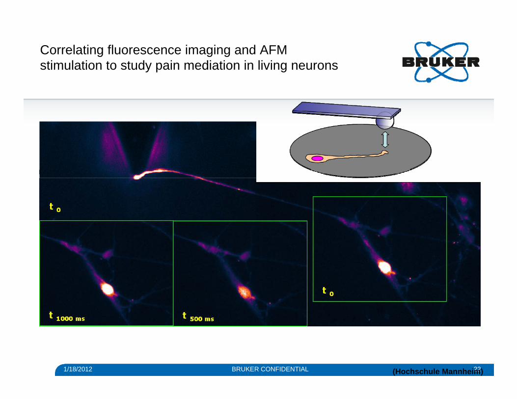
Correlating fluorescence imaging and AFM stimulation to study pain mediation in living neurons
(Hochschule Mannheim)

1/18/2012 23BRUKER CONFIDENTIAL
Combining AFM and Confocal Microscopy
(Bruker/Leica)

1/18/2012 24BRUKER CONFIDENTIAL
Combining AFM and TIRF Microscopy
(Roderick Lim)

1/18/2012 25BRUKER CONFIDENTIAL
Combining AFM and FLIM
(Bruker/PicoQuant)

1/18/2012 26BRUKER CONFIDENTIAL
Combining AFM and FLIM
(Bruker/PicoQuant)
3d-height representation + Fluorescence intensity skin
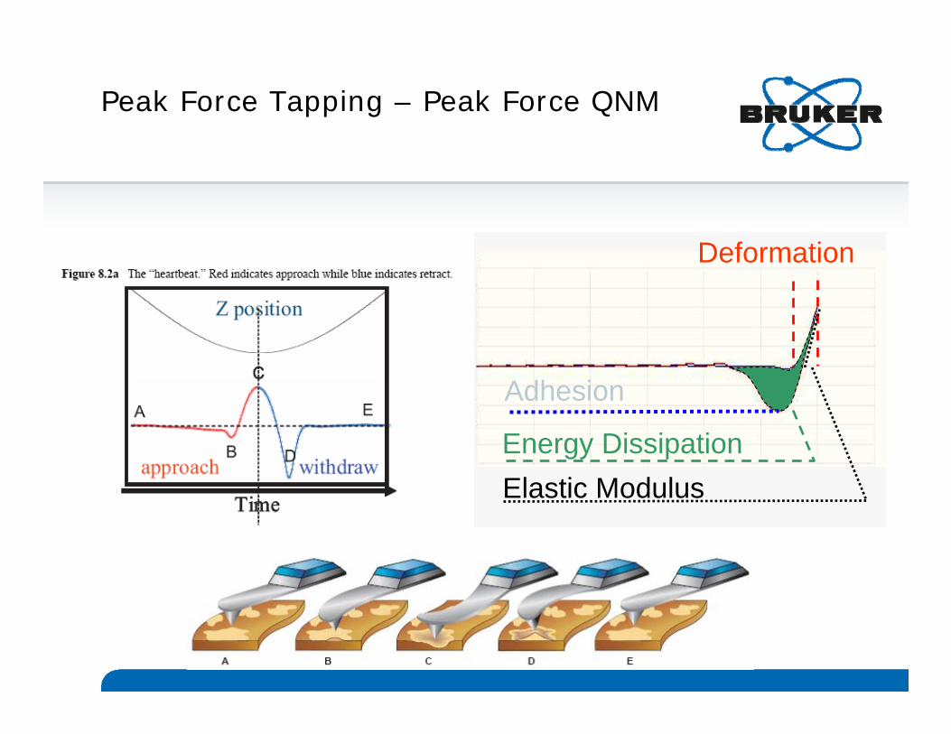
Peak Force Tapping – Peak Force QNM
Deformation
Energy Dissipation
Adhesion
Elastic Modulus
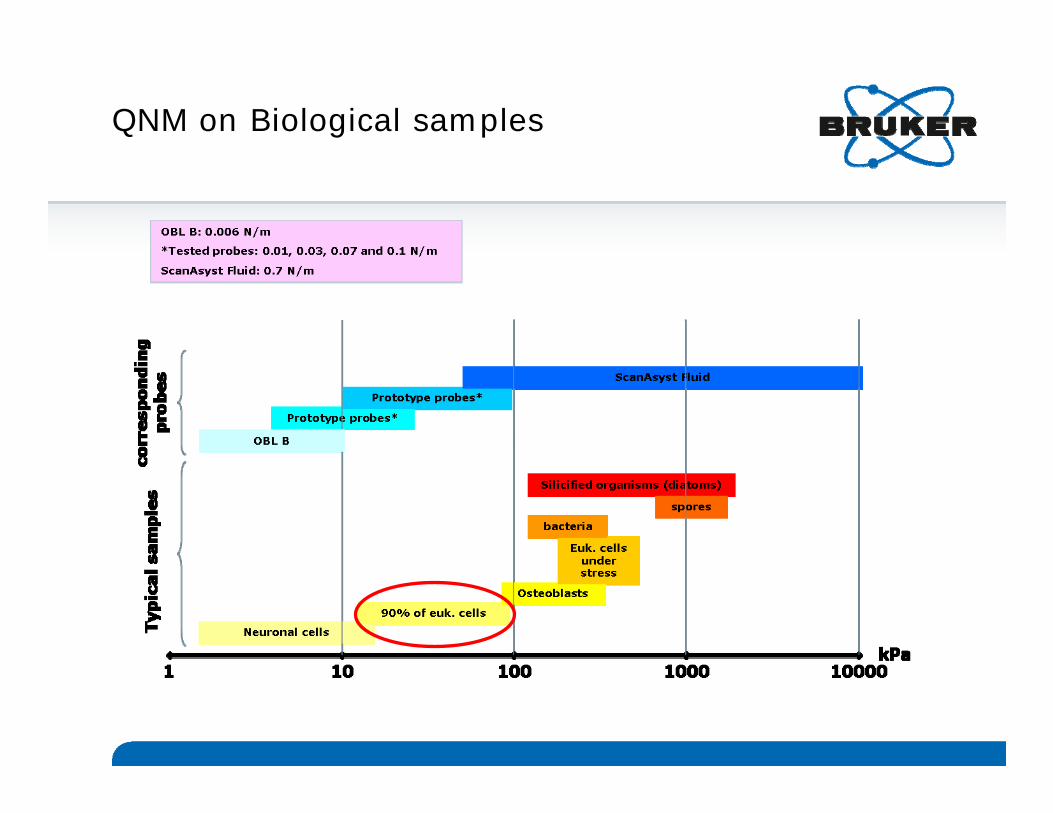
QNM on Biological samples

Combining MIRO and QNM on living HUVECs
(Hochschule Mannheim)
Epifluorescence (actin+nucleus)
PF error
Young’s modulus
Deformation
PF error / Young’s modulus (50% opacity)
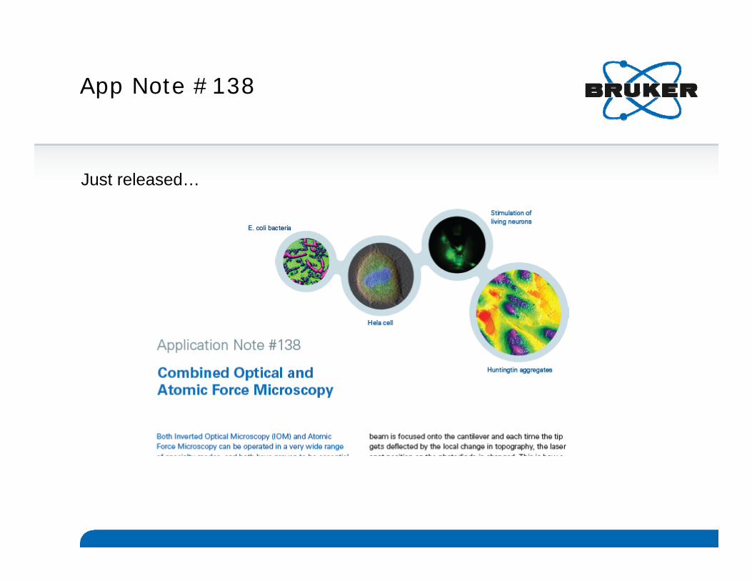
App Note #138
Just released…

1/18/2012 31BRUKER CONFIDENTIAL
Alexandre Berquand
Life Science Applications Scientist
+49 174 333 94 62 (mobile)
+49 621 842 10 66 (land)
Thanks for your attention!...

1/18/2012 32BRUKER CONFIDENTIAL
Seeing at the Nanoscale 201209th-11 July,2012 – Bristol, UK Exploring the future of nanotechnology using SPM and related techniques
• Focus: nanostructural imaging, characterization and technique development in Biology, Energy, and Material Science Applications using Scanning Probe Microscopy and related techniques
• Conference Chairman: Prof. Mervyn Miles (University of Bristol, UK)
• Co-Chairman: Prof. Heinrich Hörber
(University of Bristol, UK)
• Conference Keynote: Toshio Ando
(Kanazawa University, Japan)
• Important deadlines:
• Abstract submission deadline: 12th March 2012
• Early-Bird Registration deadline: 11th May 2012

1/18/2012 33BRUKER CONFIDENTIAL
Seeing at the Nanoscale 201209th-11 July,2012 – Bristol, UK Scientific programme
Share your research at the world’s leading SPM conference!Abstract submission deadline: 12th March 2012
www.bruker-axs.com/seeing-2012

www.bruker.com
© Copyright Bruker Corporation. All rights reserved.


