Instructions For Research Use Only. Not For Use In Diagnostic Procedures CometAssay ® Silver Kit Reagents for Comet Assay and Staining with Silver Catalog # 4251-050-K IFU0126 Rev 2 Status: RELEASED printed 2/1/2017 11:51:08 AM by Trevigen Document Control
Transcript
E01/17/17
Instructions For Research Use Only. Not For Use In Diagnostic Procedures
CometAssay® Silver Kit
Reagents for Comet Assay and Staining with Silver
Catalog # 4251-050-K
IFU0126 Rev 2 Status: RELEASED printed 2/1/2017 11:51:08 AM by Trevigen Document Control
i
CometAssay® Silver Kit
Reagents for Comet Assay and Staining with Silver
Catalog # 4251-050-K
Table of Contents Page Number
I. Background 1 II. Precautions and Limitations 2 III. Materials Supplied 2 IV. Materials Required But Not Supplied 2 V. Reagent Preparation 3 VI. Sample Preparation and Storage 5 VII. Assay Protocol 7 VIII. Warning/ Safety 10 IX. Data Analysis 10 X. References 13 XI. Related Products Available From Trevigen 14 XII. Appendices 15 XIII. Troubleshooting Guide 18
IFU0126 Rev 2 Status: RELEASED printed 2/1/2017 11:51:08 AM by Trevigen Document Control
1
I. Background Trevigen’s CometAssay®, or single cell gel electrophoresis assay, provides a
simple and effective method for evaluating DNA damage in cells. The principle of the assay is based upon the ability of denatured, cleaved DNA fragments to migrate out of the nucleoid under the influence of an electric field, whereas undamaged DNA migrates slower and remains within the confines of the nucleoid when a current is applied. Evaluation of the DNA “comet” tail shape and migration pattern allows for assessment of DNA damage. The Neutral CometAssay® is typically used to detect double-stranded breaks, whereas the Alkaline CometAssay® is more sensitive, and is used to detect smaller amounts
of damage including single and double-stranded breaks. Trevigen’s CometAssay® uses our exclusive CometSlide™ that is specially
treated to promote adherence of low melting point agarose. This eliminates the time consuming and unreliable traditional method of preparing base layers of agarose. The use of Trevigen’s CometSlide™ shortens assay time and allows the rapid and reliable analysis of large numbers of samples. Trevigen’s CometAssay® Silver Kit provides all the reagents for silver staining of the
processed CometSlide™ allowing visualization by standard light microscopy and providing permanent staining for sample archiving. In comet assays, cells are immobilized in a bed of low melting point agarose, on a Trevigen CometSlide™. Following gentle cell lysis, and for the Alkaline CometAssay®, samples are treated with alkali to unwind and denature the DNA
and hydrolyze sites of damage. For both assays, cells are lysed and the remaining nucleoids are subjected to electrophoresis and subsequent staining with a fluorescent DNA intercalating dye and/or silver stain. Trevigen recommends using Alkaline CometAssay® Control Cells (cat#
4256-010-CC) when performing alkaline electrophoresis, and Neutral CometAssay® Control Cells (cat# 4257-010-NC) when performing the neutral
comet assay, to monitor assay conditions and verify reproducibility between separate runs. SYBR® Gold for DNA visualization and quantitation by epifluorescence microscopy is recommended. Silver staining can replace or follow fluorescent analysis. We recommend the use of Trevigen’s CometAssay® Electrophoresis System
(cat# 4250-050-ES) designed to eliminate known causes of assay variability. The electrophoresis step is performed using an Alkaline Electrophoresis Solution pH>13, for the alkaline version, whereas a Neutral Electrophoresis Buffer is recommended for the neutral version. Quantitative and statistical data can readily be generated by fluorescent analysis of the results using Trevigen’s Comet Analysis Software (4260-000-CS) to calculate tail length, percent DNA in the tail, and tail moment. The CometAssay® may be coupled with Trevigen’s FLARE™ (Fragment
Length Analysis using Repair Enzymes) Assay that provides the added ability to probe for specific types of DNA damage using DNA repair glycosylases. Contact Trevigen for more details about analysis of DNA damage and repair.
IFU0126 Rev 2 Status: RELEASED printed 2/1/2017 11:51:08 AM by Trevigen Document Control
2
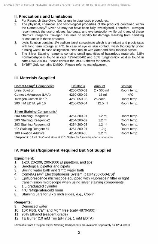
II. Precautions and Limitations 1. For Research Use Only. Not for use in diagnostic procedures. 2. The physical, chemical, and toxicological properties of the products contained within
the CometAssay® Silver Kit may not have been fully investigated. Therefore, Trevigen recommends the use of gloves, lab coats, and eye protection while using any of these chemical reagents. Trevigen assumes no liability for damage resulting from handling or contact with these products.
3. Lysis Solution contains 1% sodium lauryl sarcosinate which is an irritant and precipitates with long term storage at 4°C. In case of eye or skin contact, wash thoroughly under running water. In case of ingestion, rinse mouth with water and seek medical advice.
4. The Silver Staining reagents contains small quantities of hazardous materials: 2.8% Formaldehyde is found in cat# 4254-200-02 and 10% tungstosilicic acid is found in cat# 4254-200-03. Please consult the MSDS sheets for details.
5. SYBR® Gold contains DMSO. Please refer to manufacturer.
III. Materials Supplied
CometAssay® Components Catalog # Amount Storage
Lysis Solution 4250-050-01 2 x 500 ml Room temp.
Comet LMAgarose (LMA) 4250-050-02 15 ml 4°C
Trevigen CometSlide™ 4250-050-03 25 each Room temp.
20X Staining Reagent #2 4254-200-02 1.2 ml Room temp.
20X Staining Reagent #3 4254-200-03 1.2 ml Room temp. ±2X Staining Reagent #4 4254-200-04 1.2 g Room temp.
10X Fixation Additive 4254-200-05 2.2 ml Room temp. ±Suspend in 12 ml dH2O and store at 4°C. Stable for 3 months after suspension.
IV. Materials/Equipment Required But Not Supplied
Equipment: 1. 1-20, 20-200, 200-1000 μl pipettors, and tips 2. Serological pipettor and pipets 3. Boiling water bath and 37°C water bath 4. CometAssay® Electrophoresis System (cat#4250-050-ES)1 5. Epifluorescence microscope equipped with Fluorescein filter or light
transmission microscope when using silver staining components 6. 1 L graduated cylinder 7. 4°C refrigerator/cold room 8. Staining Jars for 3 x 2 inch slides, e.g., Coplin
Reagents: 9. Deionized water 10. 10X PBS, Ca++ and Mg++ free (cat# 4870-500)1 11. 95% Ethanol (reagent grade) 12. TE Buffer (10 mM Tris (pH 7.5), 1 mM EDTA)
1Available from Trevigen; Silver Staining Components are available separately as 4254-200-K.
IFU0126 Rev 2 Status: RELEASED printed 2/1/2017 11:51:08 AM by Trevigen Document Control
3
For alkaline assays: 13. NaOH Pellets 14. 0.5 M EDTA (pH 8.0)
For neutral assays: 15. Tris Base 16. Ammonium Acetate 17. Sodium Acetate 18. Glacial Acetic Acid
For Silver Staining:
19. Methanol 20. Glacial Acetic Acid Optional reagents: 21. 10,000X SYBR® Gold in DMSO (see Appendix C: DNA Stains) 22. Dimethylsulfoxide
V. Reagent Preparation
1. 1X PBS, Ca++ and Mg++ free
Dilute 10X PBS with deionized water to prepare 1X PBS and store at room temperature. (10X PBS is available from Trevigen, cat# 4870-500.)
2. Lysis Solution
For up to 10 slides (2 samples per slide) prepare:
Lysis Solution (cat# 4250-050-01) 40 ml DMSO (optional) 4 ml
Cool to 4°C for at least 20 minutes before use. The addition of DMSO is optional and is required only for samples containing heme, such as blood cells or tissue samples. The buffer formulation is proprietary.
3. Comet LMAgarose
The Comet LMAgarose is ready to use once molten. Loosen the cap to allow for expansion then heat the bottle in a 90-100°C water bath for 5 minutes, or until the agarose is molten (Caution: Microwaving is not recommended). Place the bottle in a 37°C water bath for at least 20 minutes to cool. The LMAgarose will remain molten at 37°C for sample preparation indefinitely. The LMAgarose formulation is proprietary.
IFU0126 Rev 2 Status: RELEASED printed 2/1/2017 11:51:08 AM by Trevigen Document Control
4
For Alkaline Comet Assay: 4. Alkaline Unwinding Solution, pH>13 (200 mM NaOH, 1 mM EDTA)
Wear gloves when preparing and handling the Alkaline Unwinding Solution. Per 50 ml of Alkaline Solution combine: NaOH Pellets 0.4 g 200 mM EDTA (cat # 4250-050-04) 250 µl dH2O 49.75 ml Stir until fully dissolved. The solution will warm during preparation. Allow to
cool to room temperature before use.
5. Alkaline Electrophoresis Solution pH >13 (200 mM NaOH, 1 mM EDTA) for the CometAssay® ES:
Prepare a stock solution of 500 mM EDTA, pH 8. For 1 liter of electrophoresis solution: NaOH pellets 8 g 500 mM EDTA, pH 8 2 ml dH2O (after NaOH is dissolved) add to: 1 liter Use of freshly made solution is recommended. Cool to 4°C. For Neutral Comet Assay: 6. 1X Neutral Electrophoresis Buffer To prepare 10X Neutral Electrophoresis Buffer:
Tris Base (mol. wt. = 121.14) 60.57 g Sodium Acetate (mol. wt. = 136.08) 204.12 g
Dissolve in 450 ml of dH2O. Adjust to pH = 9.0 with glacial acetic acid. Adjust volume to 500 ml and filter sterilize and store at room temperature. Dilute the 10X stock to 1X in dH2O to prepare 1 liter working strength buffer and cool to 4°C.
7. DNA Precipitation Solution
Prepare a 10 ml stock solution of 7.5M Ammonium Acetate: NH4Ac (mol. wt. = 77.08) 5.78 g dH2O (after NH4Ac is dissolved) add to: 10 ml
For 50 ml of DNA precipitation solution combine: 7.5 M NH4Ac (mol. wt. = 77.08) 6.7 ml 95% EtOH (reagent grade) 43.3 ml
Fluorescent Staining (optional): 8. SYBR® Gold Staining Solution (see Section IV: Materials Not Supplied)
The diluted stock is stable for several weeks stored at 4°C in the dark.
10,000 SYBR® Gold in DMSO 1 µl
TE Buffer, pH 7.5 30 ml (TE: 10 mM Tris-HCl pH 7.5, 1 mM EDTA) Note: Alternative dyes are described in Appendix C: DNA Stains.
IFU0126 Rev 2 Status: RELEASED printed 2/1/2017 11:51:08 AM by Trevigen Document Control
5
Silver Staining: 9. Fixation solution Prepare immediately before fixation. Mix per sample:
Mix by tapping tube and add 500 μl 2X Staining Reagent #4.
12. Stop solution
Prepare a 5% acetic acid solution. 100 μl per sample area is required.
VI. Sample Preparation and Storage
Cell samples should be prepared immediately before starting the assay, although success has been obtained using cryopreserved cells (see below). Cell samples should be handled under dimmed or yellow light to prevent DNA damage from ultraviolet light. Buffers should be cooled to 4°C to inhibit endogenous damage occurring during sample preparation and to inhibit repair in cells. PBS must be calcium and magnesium free to inhibit endonuclease activities. The appropriate controls should also be included (see below). Optimal results in the CometAssay® are usually obtained with 500-1000 cells per CometSlide™ sample area. Using 50 μl of a cell suspension at 1 x 10⁵ cells per ml combined with 500 μl of LMAgarose will provide the correct agarose concentration and cell density for optimal results when plating 50 μl per sample.
IFU0126 Rev 2 Status: RELEASED printed 2/1/2017 11:51:08 AM by Trevigen Document Control
6
Suspension Cells Cell suspensions are harvested by centrifugation. Suspend cells at 1 x 10⁵
cells/ml in ice cold 1X PBS (Ca++ and Mg++ free). Media used for cell culture can reduce the adhesion of LMAgarose to the CometSlide™.
Adherent Cells Gently detach cells from flask surface. Transfer cells and medium to centrifuge tube, perform cell count, and then pellet cells. Wash once in ice cold 1X PBS (Ca++ and Mg++ free). Suspend cells at 1 x 10⁵ cells/ml in ice cold 1X PBS (Ca++ and Mg++ free). If high level of damage is seen in healthy population, reduce cell exposure to Trypsin or try alternative detachment methods such as scraping using a rubber policeman. Trypsin-EDTA (0.25% Trypsin, 1 mM EDTA) Protocol: Wash the monolayer of cells with sterile PBS, warmed to 37°C. Add minimal amount Trypsin-EDTA to coat entire monolayer. Incubate flask at 37°C for 2 minutes or when cells easily detach upon tapping of flask. Add 10 ml of complete media (containing fetal bovine serum) to inactivate trypsin. Transfer cells and medium to centrifuge tube, perform cell count, and pellet cells (200xg). Wash once in ice cold 1X PBS (Ca++ and Mg++ free). Suspend cells at 1 x 10⁵ cells/ml in ice cold 1X PBS (Ca++ and Mg++ free).
Tissue Preparation Place a small piece of tissue into 1-2 ml of ice cold 1X PBS (Ca++ and Mg++ free), 20 mM EDTA. Using small dissecting scissors mince the tissue into very small pieces and let stand for 5 minutes. Recover the cell suspension, avoiding transfer of debris. Count cells, pellet by centrifugation, and suspend at 1 x 10⁵ cells/ml in ice cold 1X PBS (Ca2+ and Mg2+ free). For blood rich organs (e.g., liver, spleen), chop tissue into large pieces (1-2 mm3), let settle for 5 minutes then aspirate and discard medium. Add 1-2 ml of ice cold 20 mM EDTA in 1X PBS (Ca++ and Mg++ free), mince the tissue into very small pieces and let stand for 5 minutes. Recover the cell suspension, avoiding transfer of debris. Count cells, pellet, and suspend at 1 x 10⁵ cells/ml in ice cold 1X PBS (Ca++ and Mg++ free).
Controls A sample of untreated cells should always be processed to control for assay variability, endogenous levels of damage within cells, and for additional damage that may occur during sample preparation. Control cells and treated cells should be handled in an identical manner. If UV damage is being studied; the cells should be kept in low level yellow light during processing. Trevigen offers two sets of suspension cell preparations containing different levels of DNA damage to standardize methods between individual users, different runs, and laboratories for alkaline (cat# 4256-010-CC) and neutral (cat# 4257-010-NC) electrophoresis conditions, respectively.
Note: To generate samples positive for comet tails, treat cells with 100 µM hydrogen peroxide or 25 µM KMnO4 for 20 minutes at 4°C. Treatment will generate significant oxidative damage in most cells, thereby providing a positive control for each step in the alkaline comet assay.
IFU0126 Rev 2 Status: RELEASED printed 2/1/2017 11:51:08 AM by Trevigen Document Control
7
Method for Cryopreservation of Cells Prior to CometAssay™ Certain cells, e.g. lymphocytes, may be successfully cryopreserved prior to performing CometAssay® (Visvardis et al.). A pilot study should be performed to determine if cryopreservation is appropriate for the cells in use.
1. Centrifuge cells at 200 x g for 5 minutes. 2. Suspend cell pellet at 3 x 10⁵ cells/ml in 10% (v/v) dimethylsulfoxide, 40% (v/v) medium, 50% (v/v) fetal calf serum. 3. Transfer 50 µl aliquots into freezing vials.
4. Freeze at -70°C with -1°C per minute freezing rate overnight.
5. Transfer to liquid nitrogen for long term storage. 6. Recover cells by submerging in 37°C water bath until the last trace of ice has melted.
7. Add 500 µl ice cold 1X PBS (Ca++ and Mg++ free) to tube.
8. Centrifuge at 200 x g for 10 minutes at 4°C. 9. Suspend in 100 µl ice cold 1X PBS (Ca++ and Mg++ free) at ~1x10⁵ cells/ml
and proceed with CometAssay®. VII. Assay Protocol The electrophoresis conditions used will determine the sensitivity of the assay. Neutral CometAssay® will detect double-stranded DNA breaks, whereas Alkaline CometAssay® will detect single and double-stranded DNA breaks, and the majority of abasic sites as well as alkali labile DNA adducts (e.g. phosphoglycols, phosphotriesters). The comet assay has been reported to detect DNA damage associated with low doses (0.6 cGy) of gamma irradiation, providing a simple technique for quantitation of low levels of DNA damage. Prior to performing the comet assay, a viability assay should be performed to determine the dose of the test substance that gives at least 90% viability. False positives may occur when high doses of cytotoxic agents are used. For cryopreservation of cells, fixing the CometSlide™ samples, and storage, refer to Section VI: Sample Preparation and Storage. The Alkaline CometAssay® requires approximately 2–3 hours to complete, whereas the Neutral CometAssay® requires 4 hours, including the incubations
and electrophoresis. Once the cells or tissues have been prepared the procedure is not labor intensive. The Lysis Solution may be cooled and the LMAgarose melted while the cell and tissue samples are being prepared. When dealing with large number of samples, a convenient stopping point is to perform cell lysis overnight (Alkaline step 5). In addition, cryopreservation allows experimental samples to be processed concurrently.
A. Alkaline CometAssay®
1. Prepare Lysis Solution (see Section V: Reagent Preparation) and cool at 4°C for at least 20 minutes before use.
2. Melt LMAgarose in a beaker of boiling water for 5 minutes, with the cap
loosened. Place bottle in a 37°C water bath for at least 20 minutes to cool. The temperature of the agarose is critical or the cells may undergo heat shock.
IFU0126 Rev 2 Status: RELEASED printed 2/1/2017 11:51:08 AM by Trevigen Document Control
8
3. Combine cells at 1 x 10⁵/ml with molten LMAgarose (at 37°C) at a ratio of
1: 10 (v/v) and immediately pipette 50 μl onto CometSlide™. If necessary, use side of pipette tip to spread agarose/cells over sample area to ensure complete coverage of the sample area. If sample is not spreading evenly on the slide, warm the slide at 37°C before application.
When working with many samples aliquot agarose into 37°C warmed
tubes, add cells, mix gently by inversion, and spread 50 μl onto sample area.
Comet LMAgarose (molten and at 37°C from step 2) 500 μl Cells in 1X PBS (Ca++ and Mg++ free) at 1 x 10⁵/ml 50 μl
4. Place slide flat at 4°C in the dark (e.g. place in refrigerator) for 10 minutes. A 0.5 mm clear ring appears at edge of CometSlide™ area. Increasing gelling time to 30 minutes improves adherence of samples in high humidity environments.
5. Immerse slide in 4°C Lysis Solution for 30 to 60 minutes. For added
sensitivity or convenience incubate overnight at 4°C. 6. Drain excess buffer from slides and immerse in freshly prepared Alkaline
Unwinding Solution, pH>13 (see Section V: Reagent Preparation). WEAR GLOVES WHEN PREPARING OR HANDLING THIS SOLUTION.
7. Immerse CometSlide™ in Alkali Unwinding Solution for 20 minutes at
room temperature or 1 hour at 4°C, in the dark. 8. For the CometAssay® ES unit, add ~850 ml 4°C Alkaline Electrophoresis
Solution, place slides in electrophoresis slide tray (slide label adjacent to black cathode) and cover with Slide Tray Overlay. Set power supply to 21 volts and apply voltage for 30 minutes. (If not using an ES unit, see Appendix B.)
9. Drain excess electrophoresis solution from slides and gently immerse twice in dH2O for 5 minutes each, then in 70% ethanol for 5 minutes. Do not pour liquid over slides.
10. Dry samples at 37°C for 10-15 minutes. Drying brings all the cells in a single plane to facilitate observation. Samples may be stored at room temperature, with desiccant prior to scoring at this stage. 11. Proceed to section C. Fluorescent Staining (optional) before silver staining or directly to section D. Silver Staining.
B. Neutral CometAssay® 1. Prepare Lysis Solution (see Section V: Reagent Preparation) and cool at
4°C for at least 20 minutes before use. 2. Melt LMAgarose in a beaker of boiling water for 5 minutes, with the cap
loosened, and then cool in a 37°C water bath for at least 20 minutes.
IFU0126 Rev 2 Status: RELEASED printed 2/1/2017 11:51:08 AM by Trevigen Document Control
9
3. Combine cells at 1 x 10⁵/ml with molten LMAgarose (at 37°C) at a ratio of
1:10 (v/v) and immediately pipette 50 µl onto CometSlide™. Use side of pipette tip to spread agarose/cells over sample area.
Comet LMAgarose (molten and at 37°C from step 2) 500 µl Cells in 1X PBS (Ca++ and Mg++ free) at 1 x 10⁵/ml 50 µl Note: If sample is not spreading evenly on the slide, warm the slide at
37 °C before application. 4. Place slides flat at 4°C in the dark (e.g. place in refrigerator) for 10
minutes. A 0.5 mm clear ring appears at edge of CometSlide™ area. Increasing gelling time to 30 minutes improves adherence of samples in high humidity environments.
5. Immerse slides in 4°C (Step 1) Lysis Solution for 1 hour or overnight for
added sensitivity. 6. Remove slides from Lysis Buffer, drain excess buffer from slide and gently
immerse in 50 ml of 4°C 1X Neutral Electrophoresis Buffer for 30 minutes (see Section V: Reagent Preparation).
7. For the CometAssay® ES unit, add ~850 ml 4°C 1X Neutral
Electrophoresis Buffer, place slides in electrophoresis slide tray (slide label adjacent to black cathode) and cover with Slide Tray Overlay. Set power supply to 21 volts and apply voltage for 1 hour at 4°C.
For other electrophoresis units, align slides equidistant from electrodes, add 1X Neutral Electrophoresis Buffer not to exceed 0.5 cm above slides, and apply voltage at 1 volt per cm (measured electrode to electrode).
8. Drain excess Neutral Electrophoresis Buffer and immerse slides in DNA Precipitation Solution for 30 minutes at room temperature.
9. Immerse slides in 70% ethanol for 30 minutes at room temperature. 10. Dry samples at 37°C for 10-15 minutes. Drying brings all the cells in a
single plane to facilitate observation. Samples may be stored at room temperature, with desiccant prior to scoring at this stage.
11. Proceed to section C. Fluorescent Staining (optional) before silver staining or directly to section D. Silver Staining. C. Fluorescent Staining (optional) 1. Place 100 µl of diluted SYBR® Gold (See Section V: Reagent Preparation) onto each circle of dried agarose and stain 30 minutes (room temperature) in the dark. Gently tap slide to remove excess SYBR solution and rinse briefly in water. Allow slides to dry completely at 37°C. 2. View slides by epifluorescence microscopy. (SYBR® Gold’s maximum excitation/emission is 496 nm/540 nm. Fluorescein filter is adequate).
IFU0126 Rev 2 Status: RELEASED printed 2/1/2017 11:51:08 AM by Trevigen Document Control
10
3. Proceed to section D: Silver Staining. D. Silver Staining
1. Cover the sample area with 100 μl of Fixation solution prepared in section V: Reagent Preparation.
2. Incubate for 20 minutes at room temperature.
3. Rinse in dH2O for 30 minutes. (Removal of all residual acetic acid is
essential.)
4. Cover sample area with 100 μl of Staining Solution prepared in section V: Reagent Preparation.
5. Incubate at room temperature for 5 to 20 minutes. (Intensity of staining can
be visualized under the microscope using 10X objective, and reaction stopped when comets are easily visible.)
6. Stop reaction by covering samples with 100 μl of 5% acetic acid and
incubate for 15 minutes.
7. Rinse in dH2O.
8. Air dry and store in the dark. VIII. Warning/Safety The final Silver Staining solution (prepared in section V, step 11) is considered hazardous material. Disposal should be performed per local and state regulations. It is recommended to tap solution off the slide into a container for safe disposal.
IX. Data Analysis Silver Staining of DNA generates a brown to black stain easily detectable by microscopy. In healthy cells, the stain is confined to the nucleoid (comprised of high molecular weight DNA): undamaged DNA is supercoiled and thus, does not migrate very far out of the nucleoid under the influence of an electric current. Whereas in cells that have accrued DNA damage, migrating fragments (comet tail) from the nucleoid (comet head) are observed. The negatively charged DNA migrates toward the anode and the extrusion length reflects increasing relaxation of supercoiling, which is indicative of damage. Common descriptors of DNA damage for alkaline comet assays are Percent DNA in the Tail, and Tail Moment. Percent DNA in the Tail is a normalized measure of the percent of total cell DNA found in the tail. Tail moment is a damage measure combining the amount of DNA in the tail with distance of migration. In neutral comet assays, Tail Moment is primarily used, since tail length continues to increase in contrast to alkaline comet tails which have finite lengths. Qualitative Analysis (Alkaline CometAssay®)
The comet tail can be scored per DNA content (intensity). The control (untreated
IFU0126 Rev 2 Status: RELEASED printed 2/1/2017 11:51:08 AM by Trevigen Document Control
11
cells) should be used to determine the characteristics of data for a healthy cell. Scoring can then be made per nominal, medium or high intensity tail DNA content. At least 50 cells should be scored per sample. Quantitative Analysis (Alkaline and Neutral CometAssay®)
Trevigen’ Comet Analysis Software (4260-000-CS) is suitable for quantitation of CometAssay® data to measure the length of DNA migration, nuclear size, and
calculate DNA damage parameters. At least 50 randomly selected cells should be analyzed per sample.
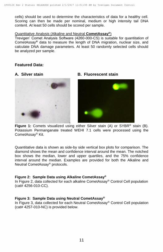
Featured Data: A. Silver stain B. Fluorescent stain
Figure 1: Comets visualized using either Silver stain (A) or SYBR® stain (B).
Potassium Permanganate treated WEHI 7.1 cells were processed using the CometAssay® Kit.
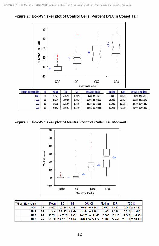
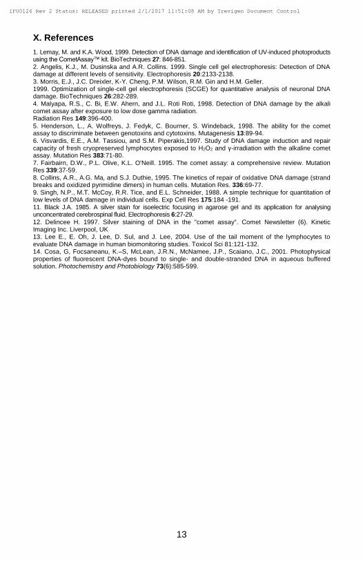
Quantitative data is shown as side-by side vertical box plots for comparison. The diamond shows the mean and confidence interval around the mean. The notched box shows the median, lower and upper quartiles, and the 75% confidence interval around the median. Examples are provided for both the Alkaline and Neutral CometAssay® protocols. Figure 2: Sample Data using Alkaline CometAssay®
In Figure 2, data collected for each alkaline CometAssay® Control Cell population (cat# 4256-010-CC). Figure 3: Sample Data using Neutral CometAssay®
In Figure 3, data collected for each Neutral CometAssay® Control Cell population (cat# 4257-010-NC) is provided below.
IFU0126 Rev 2 Status: RELEASED printed 2/1/2017 11:51:08 AM by Trevigen Document Control
12
Figure 2: Box-Whisker plot of Control Cells: Percent DNA in Comet Tail
Figure 3: Box-Whisker plot of Neutral Control Cells: Tail Moment
-10
10
30
50
70
90
CCO CC1 CC2 CC3
% D
NA
in
Tail
Control Cells
% DNA by Etoposide n Mean SD SE 75% CI of Mean Median IQR 75% CI of Median
CCO 50 5.757 7.7270 1.0928 4.485 to 7.029 1.640 8.925 1.290 to 2.230
CC1 50 28.374 14.0080 1.9810 26.068 to 30.680 28.990 20.313 25.180 to 31.840
CC2 50 39.736 21.8164 3.0853 36.144 to 43.328 37.050 32.183 27.790 to 44.630
CC3 50 56.800 23.5893 3.3360 52.916 to 60.683 51.905 40.240 45.460 to 64.390
IFU0126 Rev 2 Status: RELEASED printed 2/1/2017 11:51:08 AM by Trevigen Document Control
13
X. References
1. Lemay, M. and K.A. Wood, 1999. Detection of DNA damage and identification of UV-induced photoproducts using the CometAssay™ kit. BioTechniques 27: 846-851. 2. Angelis, K.J., M. Dusinska and A.R. Collins. 1999. Single cell gel electrophoresis: Detection of DNA damage at different levels of sensitivity. Electrophoresis 20:2133-2138. 3. Morris, E.J., J.C. Dreixler, K-Y. Cheng, P.M. Wilson, R.M. Gin and H.M. Geller. 1999. Optimization of single-cell gel electrophoresis (SCGE) for quantitative analysis of neuronal DNA damage. BioTechniques 26:282-289. 4. Malyapa, R.S., C. Bi, E.W. Ahern, and J.L. Roti Roti, 1998. Detection of DNA damage by the alkali comet assay after exposure to low dose gamma radiation. Radiation Res 149:396-400.
5. Henderson, L., A. Wolfreys, J. Fedyk, C. Bourner, S. Windeback, 1998. The ability for the comet assay to discriminate between genotoxins and cytotoxins. Mutagenesis 13:89-94. 6. Visvardis, E.E., A.M. Tassiou, and S.M. Piperakis,1997. Study of DNA damage induction and repair capacity of fresh cryopreserved lymphocytes exposed to H2O2 and γ-irradiation with the alkaline comet assay. Mutation Res 383:71-80. 7. Fairbairn, D.W., P.L. Olive, K.L. O'Neill. 1995. The comet assay: a comprehensive review. Mutation Res 339:37-59. 8. Collins, A.R., A.G. Ma, and S.J. Duthie, 1995. The kinetics of repair of oxidative DNA damage (strand breaks and oxidized pyrimidine dimers) in human cells. Mutation Res. 336:69-77. 9. Singh, N.P., M.T. McCoy, R.R. Tice, and E.L. Schneider, 1988. A simple technique for quantitation of low levels of DNA damage in individual cells. Exp Cell Res 175:184 -191. 11. Black J.A. 1985. A silver stain for isoelectric focusing in agarose gel and its application for analysing unconcentrated cerebrospinal fluid. Electrophoresis 6:27-29.
12. Delincee H. 1997. Silver staining of DNA in the "comet assay". Comet Newsletter (6). Kinetic Imaging Inc. Liverpool, UK 13. Lee E., E. Oh, J. Lee, D. Sul, and J. Lee, 2004. Use of the tail moment of the lymphocytes to evaluate DNA damage in human biomonitoring studies. Toxicol Sci 81:121-132. 14. Cosa, G, Focsaneanu, K.–S, McLean, J.R.N., McNamee, J.P., Scaiano, J.C., 2001. Photophysical properties of fluorescent DNA-dyes bound to single- and double-stranded DNA in aqueous buffered solution. Photochemistry and Photobiology 73(6):585-599.
IFU0126 Rev 2 Status: RELEASED printed 2/1/2017 11:51:08 AM by Trevigen Document Control
14
XI. Related Products Available From Trevigen Contact Trevigen for details of our unique product line for studying DNA damage and repair. All Trevigen kits include highly qualified enzymes, substrates, buffers, full instructions for use, and a synopsis specific for your kit.
CometAssay® Kits:
Catalog # Description Size
4250-050-ESK CometAssay® Starter Kit each
4250-050-ES CometAssay® ES each
4250-050-K CometAssay® Kit (25 x 2 well slides) 50 samples
4252-040-K CometAssay® HT (2 x 20 well slides) 40 samples
The CometAssay® may be performed using neutral conditions that employ 1X TBE. Without treatment with Alkaline Buffer, this Neutral CometAssay® will also
detect mainly double-stranded breaks.
1. Prepare Lysis Solution (see Section V: Reagent Preparation) and cool to 4°C for at least 20 minutes before use. 2. Melt LMAgarose in a beaker of boiling water for 5 minutes, with the cap loosened, and then cool in a 37°C water bath for at least 20 minutes. 3. Combine cells at 1 x 10⁵/ml with molten LMAgarose (at 37°C) at a ratio of 1:10 (v/v) and immediately pipette 50 µl onto CometSlide™. Use side of pipette tip to spread agarose/cells over sample area. Comet LMAgarose (molten and at 37°C from step 2) 500 µl Cells in 1X PBS (Ca++ and Mg++ free) at 1 x 10⁵/ml 50 µl Note: If sample is not spreading evenly on the slide, warm the slide at 37 °C
before application. 4. Place slides flat at 4°C in the dark (e.g. place in refrigerator) for 10 minutes. A 0.5 mm clear ring appears at edge of CometSlide™ area. Increasing gelling time to 30 minutes improves adherence of samples in high humidity environments. 5. Immerse slides in 4°C Lysis Solution for 1 hour or overnight for added sensitivity. 6. Remove slides from Lysis Buffer, drain excess buffer from slide and wash slide by immersing in 50 ml of 4°C 1X TBE buffer for 15 minutes. To prepare 10X TBE: Tris Base 108 g Boric Acid 55 g EDTA (disodium salt) 9.3 g
IFU0126 Rev 2 Status: RELEASED printed 2/1/2017 11:51:08 AM by Trevigen Document Control
16
Dissolve in 900 ml dH2O. Adjust volume to 1 liter and filter sterilize, and store at room temperature. Dilute the 10X TBE to 1X in dH2O to prepare 1 liter working strength buffer and cool to 4°C. 7. For the CometAssay® ES unit, add 4°C ~850 ml 4°C 1X TBE Buffer, place
slides in electrophoresis slide tray and cover with Slide Tray Overlay. Set power supply to 21 volts and apply voltage for 40 minutes. Note: For other electrophoresis units, align slides equidistant from electrodes, add 1X TBE Buffer not to exceed 0.5 cm above slides, and apply voltage at 1 volt per cm (measured electrode to electrode).
8. Drain excess TBE, immerse slides in dH2O for 5 minutes.
9. Immerse slides in 70% ethanol for 5 minutes. 10.Dry samples at 37°C for 10-15 minutes. Drying brings all the cells in a single plane to facilitate observation. Samples may be stored at room temperature, with desiccant prior to scoring at this stage. 11.Proceed to VII. C. Fluorescent Staining (optional) before silver staining or directly to section D. Silver Staining.
Appendix B Instructions for alkaline comet assay with other electrophoresis units.
Since the Alkaline Electrophoresis Solution is a non-buffered system, temperature control is highly recommended. In-house testing has shown great temperature
fluctuations when conducting the alkaline electrophoresis at ambient temperature. To improve temperature control, the use of a large electrophoresis apparatus (20–30 cm between electrodes) is recommended. Performing the electrophoresis at cooler temperatures (e.g. 4°C) will diminish background damage, increase sample adherence at high pH and significantly improves reproducibility. Choose the method that is most convenient for your laboratory and always use the same conditions, Alkaline CometAssay® Control Cells (cat# 4256-010-CC), power supplies and electrophoresis chambers for comparative analysis. Alternative Reagents:
1. Alkaline Unwinding Solution, pH>13 (300 mM NaOH, 1 mM EDTA)
Wear gloves when preparing and handling the Alkaline Unwinding Solution. Per 50 ml of Alkaline Solution combine: NaOH Pellets 0.6 g 200 mM EDTA (cat # 4250-050-04) 250 µl dH2O 49.75 ml Stir until fully dissolved. The solution will warm during preparation. Allow to cool to room temperature before use.
IFU0126 Rev 2 Status: RELEASED printed 2/1/2017 11:51:08 AM by Trevigen Document Control
17
2. Alkaline Electrophoresis Solution pH >13 (300 mM NaOH, 1 mM EDTA) for other electrophoresis systems:
Prepare a stock solution of 500 mM EDTA, pH 8. For 1 liter of electrophoresis solution: NaOH pellets 12 g 500 mM EDTA, pH 8 2 ml dH2O (after NaOH is dissolved) adjust to: 1 liter Adjust the volume prepared based on the dimensions of your electrophoresis apparatus. Use of freshly made solution is recommended. Cool to 4°C.
Align slides equidistant from electrodes and carefully add the Alkaline Solution until level just covers samples. Set the voltage to about 1 Volt/cm. Add or remove buffer until the current is approximately 300 mA and perform electrophoresis for 20–40 minutes. Continue at step 9 on page 8. Appendix C DNA Stains
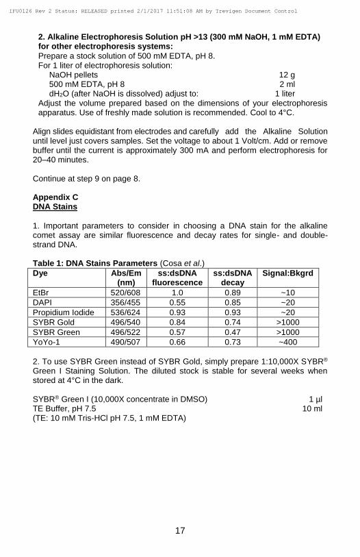
1. Important parameters to consider in choosing a DNA stain for the alkaline comet assay are similar fluorescence and decay rates for single- and double-strand DNA. Table 1: DNA Stains Parameters (Cosa et al.)
Dye Abs/Em (nm)
ss:dsDNA fluorescence
ss:dsDNA decay
Signal:Bkgrd
EtBr 520/608 1.0 0.89 ~10
DAPI 356/455 0.55 0.85 ~20
Propidium Iodide 536/624 0.93 0.93 ~20
SYBR Gold 496/540 0.84 0.74 >1000
SYBR Green 496/522 0.57 0.47 >1000
YoYo-1 490/507 0.66 0.73 ~400
2. To use SYBR Green instead of SYBR Gold, simply prepare 1:10,000X SYBR® Green I Staining Solution. The diluted stock is stable for several weeks when stored at 4°C in the dark. SYBR® Green I (10,000X concentrate in DMSO) 1 µl TE Buffer, pH 7.5 10 ml (TE: 10 mM Tris-HCl pH 7.5, 1 mM EDTA)
IFU0126 Rev 2 Status: RELEASED printed 2/1/2017 11:51:08 AM by Trevigen Document Control
18
XIII. Troubleshooting guide General Problems PROBLEM CAUSE ACTION
Unexpected and/or variety of tail shape.
LMAgarose too hot Cool LMAgarose to 37°C before adding cells.
Cells in LMAgarose did not remain attached to the CometSlide™.
Electrophoresis solution too hot.
Control temperature performing electrophoresis at 4°C.
Cells were not washed to remove medium before combining with LMAgarose.
The pH of medium and carry over serum proteins, etc., can reduce the adherence of the agarose. Suspend cells in 1X PBS.
Agarose percentage was too low.
Do not increase ratio of cells to molten agarose by more than 1 to 10.
LMAgarose was not fully set before samples were processed.
Ensure 0.5 mm dried ring due to agarose disc retraction is seen at the edge of the CometSlide area.
LMAgarose unevenly set on the slide. Rinsing steps too harsh.
Spread the agarose with the side of a pipette tip to ensure uniformity of agarose disc and better adherence. Gently place slides into solutions. Do not pour solutions over slides.
Specific to Alkaline Comet Assay PROBLEM CAUSE ACTION
Majority of cells in untreated control sample have large comet tails.
Unwanted damage to cells occurred in culture or in sample preparations
Check morphology of cells to ensure healthy appearance.
Handle cells or tissues gently to avoid physical damage.
Electrophoresis solution too hot
Control temperature by performing electrophoresis at 4°C.
Intracellular activity Keep cells on ice and prepare cell samples immediately before combining with molten LMAgarose.
Majority of cells in untreated control sample have small to medium comet tails.
Endogenous oxidative damage or endonuclease activity after sample preparation is damaging DNA.
Ensure Lysis solution was chilled before use.
Add DMSO to any cell sample that may contain heme groups.
Ensure PBS used is calcium and magnesium free.
Work under dimmed light conditions or under yellow light.
In positive control (e.g. 100 µM hydrogen peroxide for 30 minutes on ice) no evidence of comet tail.
No damage to DNA.
Sample was not processed correctly.
Use fresh hydrogen peroxide to induce damage.
Ensure each step protocol step was performed correctly. Failure to lyse, denature in alkali, or to properly perform electrophoresis may generate poor results.
Comet tails present but not significant in positive control.
Insufficient denaturation in Alkaline Solution.
Insufficient electrophoresis time.
Increase time in Alkaline Solution up to 1 hour.
Increase time of electrophoresis up to up to 1 hour for alkaline electrophoresis. Increase time of electrophoresis when running at cold temperatures.
IFU0126 Rev 2 Status: RELEASED printed 2/1/2017 11:51:08 AM by Trevigen Document Control
19
Please Recycle
Specific to Neutral Comet Assay PROBLEM CAUSE ACTION
In positive control, no evidence of comet tail.
Damaging agent doesn’t cause double-strand breaks.
Confirm damage by Alkaline Comet.
Run Neutral Control Cells to confirm electrophoresis conditions.
Increase treatment with damaging agent.
In positive control, comet tails are too long and do not fit analysis window.
Cells are necrotic or apoptotic.
Electrophoresis time too long.
Verify 90% viability.
Decrease treatment with damaging agent.
Decrease electrophoresis time to 15-30 minutes.
The product accompanying this document is intended for research use only and is not intended for diagnostic purposes or for use in humans.
Trevigen, Inc. 8405 Helgerman Ct. Gaithersburg, MD 20877