Brigham Young University Brigham Young University BYU ScholarsArchive BYU ScholarsArchive Theses and Dissertations 2013-05-10 Comfort and Compatibility of Silicone Hydrogel Contact Lenses Comfort and Compatibility of Silicone Hydrogel Contact Lenses Ngai Keung Tam Brigham Young University - Provo Follow this and additional works at: https://scholarsarchive.byu.edu/etd Part of the Chemical Engineering Commons BYU ScholarsArchive Citation BYU ScholarsArchive Citation Tam, Ngai Keung, "Comfort and Compatibility of Silicone Hydrogel Contact Lenses" (2013). Theses and Dissertations. 4031. https://scholarsarchive.byu.edu/etd/4031 This Thesis is brought to you for free and open access by BYU ScholarsArchive. It has been accepted for inclusion in Theses and Dissertations by an authorized administrator of BYU ScholarsArchive. For more information, please contact [email protected], [email protected].
Transcript
Brigham Young University Brigham Young University
BYU ScholarsArchive BYU ScholarsArchive
Theses and Dissertations
2013-05-10
Comfort and Compatibility of Silicone Hydrogel Contact Lenses Comfort and Compatibility of Silicone Hydrogel Contact Lenses
Ngai Keung Tam Brigham Young University - Provo
Follow this and additional works at: https://scholarsarchive.byu.edu/etd
Part of the Chemical Engineering Commons
BYU ScholarsArchive Citation BYU ScholarsArchive Citation Tam, Ngai Keung, "Comfort and Compatibility of Silicone Hydrogel Contact Lenses" (2013). Theses and Dissertations. 4031. https://scholarsarchive.byu.edu/etd/4031
This Thesis is brought to you for free and open access by BYU ScholarsArchive. It has been accepted for inclusion in Theses and Dissertations by an authorized administrator of BYU ScholarsArchive. For more information, please contact [email protected], [email protected].
Comfort and Compatibility of Silicone Hydrogel Contact Lenses
Ngai Keung Tam Department of Chemical Engineering, BYU
Master of Science
Silicone Hydrogel (SiHy) contact lenses are highly successful compared to previous soft lenses; they were developed to provide superior oxygen permeability. However, the hydrophobic natures of the silicone segments enhance lipid sorption which may diminish the lens surface wettability, clarity and comfort. While lens and lens care product are designed to remove lipid deposition, there is lack of experimental evidence to evaluate the actual performances with respect to lipid removal. An in vitro model using an artificial tear fluid containing radiolabeled lipids was employed in this thesis research to evaluate the efficacy of different multi-purpose lens care solutions in removing lipids from SiHy contact lenses. Additional rubbing with the lens care solution is often encouraged by professionals. Part of this research evaluated the effect of additional rubbing process on lipid removal.
Overall, a multi-purpose solution (MPS) for lens care, Opti-Free PureMoist®, removed
the most lipid deposition from lenses (senofilcon A, comfilcon A, and balafilcon A and one conventional hydrogel lens polymacon). The overall removal percentages were approximately 55% of DPPC and 28% of cholesterol from a conventional hydrogel. However, the MPSs did not remove lipids effectively from SiHy lenses. The highest percentages of removal were 3.08% of DPPC and 0.76% of cholesterol from SiHy lotrafilcon B lenses with Opti-Free PureMoist. The rubbing process increased the amount of removal in some MPSs, but the effects were small. The lack of removal of lipid suggests that the surfactants in the MPSs are not hydrophobic enough to remove lipids from SiHy lenses. Apparently a majority of deposited lipids absorbed into the lens matrix as rubbing did not enhance removal significantly. Future study on determining the concentration profile of lipid sorption throughout the lens thickness is encouraged.
Another topic in this research thesis is the use of hydrogel lenses to deliver comfort
agents or lubricating molecules from lenses. A screening study was performed in this research to select possible agents to be loaded into several SiHy macromer formulations. Experiments showed that comfort agents PNVP and Kollidon were the best candidates for such a procedure.
Preparation of Materials for Tear Solution (if not already prepared) .............................. 81
Preparation of Artificial Tear Fluid ................................................................................. 83
Protocol of the experiment of reduction of lipid deposition in chapter 4 ........................... 84
Protocol of the experiment of removal of sorbed lipid in chapter 5 ................................... 86
Protocol of the experiment of rubbing and peroxide-based solution in chapter 6 .............. 88
Protocol of the experiment of solubility of comfort agents in Chapter 8 ............................ 90
xi
LIST OF TABLES
Table 1: Characteristics of the contact lens materials. ............................................................ 14
Table 2: Components of the multi-purpose lens care solutions. ............................................. 15
Table 3: Composition of the artificial tear fluid. .................................................................... 16
Table 4: Percentages of removal of sorbed lipids by the multi-purpose lens care solutions. . 29
Table 5: Characteristics of the contact lens materials. ............................................................ 41
Table 6: Components of the multi-purpose lens care solutions. ............................................. 41
Table 7: Percentages of removal of the sorbed lipids by multi-purpose lens care solutions. . 44
Table 8: Summary of the estimated solubilities of comfort agents in DPGME ..................... 69
xiii
LIST OF FIGURES
Figure 1: Ocular tear film drug concentration based on delivery method.. ............................ 10
Figure 2: Chemical Structures of the lipids. CH and DPPC. .................................................. 13
Figure 3: A general flow diagram of experiments in chapter 4. ............................................. 18
Figure 4: Lipids sorption with preconditioning in lens care solutions.................................... 21
Figure 5: A general flow diagram of experiments in chapter 5. ............................................. 28
Figure 6: The elution profiles of sorbed lipids. DPPC and CH in solutions. .......................... 31
Figure 7: Initial sorption of DPPC and CH, and retained sorption. ........................................ 32
Figure 8: A general flow diagram of experiments in chapter 6. ............................................. 42
Figure 9: The proprietary vial for using the peroxide-based solution. ................................... 42
Figure 10: The amount of sorbed DPPC and CH removed inMPSs. ...................................... 45
Figure 11: The amount of sorbed DPPC and CH removed from lenses. ................................ 46
Figure 12: The elution profiles of sorbed lipids in solutions. ................................................. 47
Figure 13: Optical density vs concentration for agents with slight solubility. ....................... 57
Figure 14: Chemical Structure of dipropylene glycol methyl ether (DPGME). ..................... 58
Figure 15: Structures of comfort agents. ................................................................................. 58
Figure 16: Overlay plots of optical density of comfort agentsin DPGME ............................. 62
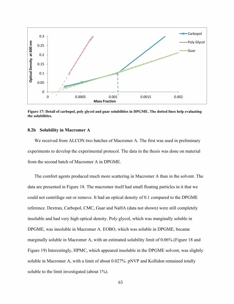
Figure 17: Detail of carbopol, poly glycol and guar solubilities in DPGME. ........................ 63
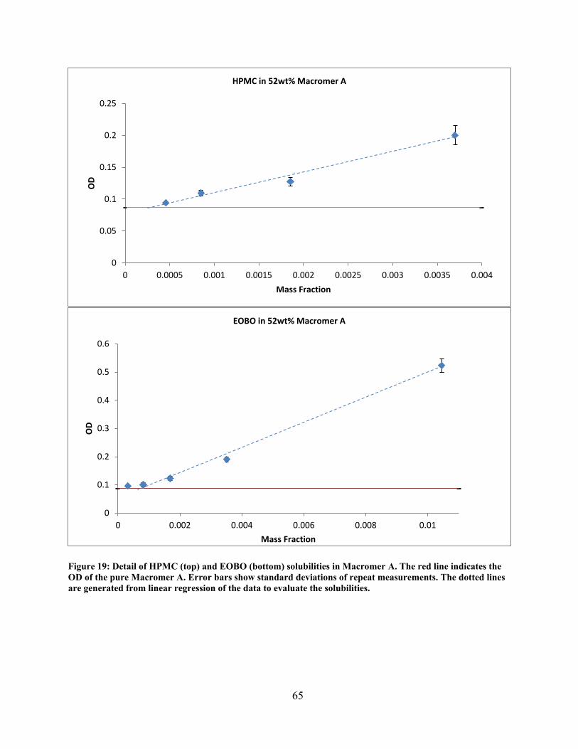
Figure 18: Overlay plots of optical densty of comfort agents in Macromer A. ...................... 64
Figure 19: Detail of HPMC and EOBO solubilities in Macromer A. ..................................... 65
Figure 20: Overlay plots of optical density of comfort agents in Macromer B. ..................... 67
Figure 21: Detail of Kollidon, pNVP and HPM solubilities in Macromer B.. ....................... 68
Figure 22: Overlay plots of optical density of comfort agents in Macromer C. ..................... 70
xiv
Figure 23: Detail of pNVP (top) and Kollidon (bottom) solubilities in Macromer C. ........... 71
1
CHAPTER 1 INTRODUCTION
The wearing of contact lenses is possibly the most ubiquitous application of a medical
hydrogel in our society. A hydrogel is a cross-linked polymer containing hydrophilic units which
normally dissolve in water. However, crosslinking prevents the polymer from dissolving. In
recent years, silicone hydrogel (SiHy) contact lenses have become the mainstream product in the
contact lens market because of their superior oxygen permeability, which is 3 to 6 times greater
than conventional poly-HEMA based hydrogel lenses 1. Since oxygen is able to transfer easily
through the silicone units in SiHy lenses, the oxygen permeability is enhanced significantly and
is not limited by the water content as conventional hydrogel lenses are. The improvement allows
a high level of comfort and improved corneal health during extended wear (even overnight wear)
by maintaining a sufficient level of oxygen concentration at the ocular surface. One study
estimated that the U.S. soft contact lens market in 2010 was $2.1 billion, the worldwide market
was about $6.1 billion, and it will reach $11.7 billion by 2015 2.
While SiHy lens are highly successful, the adsorption of tear components to contact lenses
remains a major challenge of contact lens wear that can lead to user discomfort and decreased
visual acuity 3-6, and ultimately leads to discontinuation of lens wear 6-9. Consequently, contact
lenses and lens-care products are often engineered to reduce deposition and improve wear
comfort. Of special note in this thesis, a multi-purpose solution (MPS) for lens care is often used
to store, to disinfect and to clean lenses by removing deposited proteins, lipids and other debris.
While many MPSs claim to be able to reduce or remove sorbed lipid, there is a lack of studies to
2
quantify their efficacy, particular with SiHy lenses. One of the objectives in this research is to
evaluate the efficiency of different MPSs in reducing lipid sorption and to compare their
performances with a simple buffered solution.
Unfortunately, the beneficial hydrophobic segments in SiHy lenses also reduce surface
wettability, which is highly correlated with wear comfort. Irritation and dry eye syndrome are
induced when tear films are unable to spread across the hydrophobic lens surfaces. Thus, the lens
material itself is a predominate factor to the level of comfort and the tendency to sorb tear
components. For example the surfaces of lotrafilcon B lenses are plasma coated a continuous
ultra-thin hydrophilic layer which improves the wettability 10, and which also lowers the lipid
deposition significantly.
While using hydrogel lenses to deliver therapeutic drugs to eyes is not a new idea, little
research has been done in controlled release of comfort agents or lubricants molecules from
lenses. A screening study was performed in this thesis research to select possible agents to be
loaded into several SiHy macromer formulations, which opens the door to produce lenses that
can deliver an additional level of comfort over many hours.
3
CHAPTER 2 LITERATURE REVIEW
This chapter reviews contact lenses, the sorption (adsorption and/or absorption) of lipids and
proteins, and the role of a multipurpose solution (MPS) for lens care. Contact lens materials are
named by a generic name of the material, such as lotrafilcon, senofilcon etc. Each material has a
unique chemistry. It is sometimes subcategorized as “A” or “B” representing small chemical or
processing differences. A manufacturer usually has a patent on the material (such as lotrafilcon
A) and produces a brand name contact lens from the material (such as CIBAVISION Focus
Night & Day). This thesis will usually use the generic chemical name.
2.1 Lipid deposition in silicone hydrogel contact lenses
The hydrophobic silicone components in silicone hydrogel (SiHy) lenses enhance oxygen
transfer but also enhance lipid deposition. In general, SiHy lenses are more apt to sorb lipids,
while traditional hydrogels are more likely to adsorb proteins 11,12. One study suggested that
initial deposition of phospholipid on SiHy lenses may stabilize the tear film and promote the
overall wettability; but eventually it may become deleterious as more lipid accumulates on the
lens surfaces, resulting in reductions in visual clarity, surface wettability, and comfort 13.
2.1a Reduction of lipid deposition in MPS
A general concern is the uncertainty in the effectiveness of lipid removal by MPSs. Several
studies have been performed to evaluate the efficiency of lens care solutions in reducing protein
4
sorption 14-19, but only a few evaluated lipid removal 20,21. For example, Lorentz et al. recently
reported that a hydrogen peroxide lens care solution containing Pluronic 17R4 (a di-functional
block copolymer surfactant with terminal secondary hydroxyl groups) removed more lipid from
lenses when compared to a non-surfactant hydrogen peroxide solution 20. One study analyzed
lipid sorption on several SiHy lenses and reported that lotrafilcon B lenses accumulated the least
amount of cholesterol (CH) regardless of the lens care solution used 21. This study also showed
that the lipid deposition process is dependent on lens materials and contact lens care solutions 21.
2.1b Removal of deposition in MPS with a rubbing process
MPSs are sterile, buffered solutions containing various surfactants and preservatives to clean,
store and disinfect contact lenses. While some MPSs emphasize that no rubbing is needed, two
MPSs used in this study recommend the consumer to rub both sides of the lens with the solution
for 20 seconds 22,23. Regarding the needs of rubbing for effective cleaning, the FDA stated,
“Several professional groups that represent optometrists and ophthalmologists recommend
rubbing each lens in the palm of the hand with a few drops of solution, even if using a “no rub”
product 24. Dr. Townsend mentioned in an article 25, “We're inclined to tell patients to rub their
lenses after removal for two reasons. First, some studies have demonstrated that even minimal
rubbing reduces the bacterial population by approximately 3 log units. Second, with the
increasing popularity of silicone hydrogel lenses, we're seeing more problems with lipid
deposits.” These statements suggested that a significant amount of tear deposit and other debris
could possibly be removed from the lens surface by rubbing with the MPSs. There are several
studies that investigated the effects of rubbing. Nichols reported that rubbing and rinsing lenses
decreased the amount of deposition in general 26. Previous studies have shown that rubbing in the
presence of MPS removes some proteins, and that protein removal by rubbing is greater on
5
conventional hydrogels than on SiHy lenses 14. Another study indicated that the use of a
manual rubbing step is more effective than rinsing or soaking alone in removing pathogenic
microbes from SiHy lenses 27. Many professionals suggested that rubbing will help remove lipid
deposits from SiHy lenses, but there are no specific evaluations reported in the literature. Thus,
the effect on lipid deposition by the addition of rubbing was quantified and is discussed in this
thesis.
2.2 Methods of quantifying lipid deposition
Lipid sorption is commonly quantified by chromatographic methods, as this allows for
simultaneous measurement of multiple classes of lipids from worn lenses or from complicated
lipid/protein solutions. Several studies of lipid sorption on contact lenses using chromatography
techniques have been reported. In 2003, Jones et al.11 confirmed that lipid deposition on SiHy
lenses (lotrafilcon A and balafilcon A) are significantly greater than on conventional hydrogel
(etafilcon) contact lenses. The composition of these lipid depositions was determined by a high-
performance liquid chromatography (HPLC) technique. In 2006, Maziarz et al.28 quantified the
sorption of oleic acid, oleic acid methyl ester, and cholesterol (CH) on commercial SiHy contact
lenses; they also compared two HPLC methods. In 2008, Iwata et al.29 claimed that
chromatography/mass spectrometry analytical methods are more accurate and sensitive than
standard HPLC techniques. In 2009, Zhao et al.21 used thin layer chromatography to measure CH
sorption on lotrafilcon B, balafilcon A, senofilcon A, and galyfilcon A lenses after 30 days of
wear and reported that both the lens type and the MPS had an effect upon sorption, which
averaged from 0.1 to 8.2 µg per lens. In 2011, Heynen et al.3 used HPLC to measure lipid
sorption on senofilcon A lenses and reported that less total lipid sorbed when a MPS was used
compared with another no-rub hydrogen peroxide system.
6
When comparing sorption data from various labs on similar lenses, often one sees similar
general trends but variations in the details of the amount sorbed. Lab-to-lab reproducibility is not
always obtained. For example, Jones et al.11 reported high levels of lipid adsorption to balafilcon
lenses. However, Maziarz et al.28 reported much lower levels and also demonstrated that small
differences in sample extraction and HPLC methods can yield substantially different results.
Lorentz et al.29,30 showed that when measuring sorption from laboratory solutions that simulate
tear, small variations in solution composition can produce statistical differences in amounts
sorbed.
In addition to chromatographic methods, radiolabeling techniques are sometimes used to
quantitate protein and lipid sorption from artificial tears. While radiolabels have excellent
accuracy and precision, they are less broadly applicable for studying simultaneous sorption of
multiple lipids and proteins because each species of lipid and/or protein requires a different
radioisotope (or a different experiment for each different species). Quantitation is independent of
complexation with proteins or other lipids that sometimes makes chromatography challenging
due to differences in retention between complexed and single lipids. Furthermore, radioisotope
methods can be used to validate and support chromatographic methods. There are several
radioisotopes such as 125I, 3H and 14C that were used in previous studies to investigate
lipid/protein deposition on contact lenses. With 125I-labeled chemicals, no extractions are
required as 125I emits highly penetrating gamma radiation, so the quantifying of such a labeled
substance can be accomplished with direct measurement methods, such as putting the lens
directly into scintillation fluid (SF). However, 125I labels are limited to proteins, and cannot be
easily applied to lipids. Other common radioactive elements, 3H and 14C, have a half-life of 12
years and 5730 years respectively, and their low penetrating beta radiation allows relatively
7
simpler experiments and reduces the risk of radiation hazards. However, the extraction process
becomes necessary as the lens itself will absorb some of beta particles so they cannot be
quantified by direct measurement techniques. In 1997 Prager and Quintana 30 reported uptake on
traditional hydrogels of 14C-dioleoyl phosphatidylcholine and 3H-cholesteryl oleate from a multi-
component artificial tear fluid (ATF). Since then, little has been published on the use of
radiolabels to study protein and lipid sorption to contact lenses. Only recently, Lorentz et al.31,32
quantified the deposition of lipids on SiHy contact lenses using 14C-labeled cholesterol (CH) and
phosphatidylcholine (PC). Their data showed that the quantities of CH and PC deposited on
balafilcon A and omafilcon A lenses are much less than reported in previous studies by Iwata et
al.29, Carney et al.12, and Pucker et al.33. Another key finding from Lorentz’s studies is that
variation in compositions of laboratory tear fluid had a large influence upon the amount of lipid
deposition 31,32; they reported that both CH and PC deposition significantly decreased when
lactoferrin and immunoglobulin G were not included in the ATF 31.
While chromatographic methods have become the norm, few such studies have also utilized
independent techniques such as radiochemistry to validate extraction procedures or
chromatographic results. The majority of chromatographic studies of deposition on contact
lenses required extraction steps prior to the quantification process, and chloroform:methanol
(Chlf:MeOH) solutions are often used as the extraction solvents. Zhao et al.21 reported that the
recovery percentage of their extraction technique with a 50% Chlf/50% MeOH solution was 72.7%
to 95.5%, and that the efficiencies were dependent on the lens type and the representative tear
components, which implied that the actual amount of lipid deposition may not have been
accurately quantified unless calibrations were done on each combination of lens and lipid type.
Lorentz et al. 31 extracted the lipid deposition 2 times with 2 mL of 66% Chlf/33% MeOH
8
solution for three hours at 37 °C while shaking on an orbital shaker. Jones et al.11 employed a
similar technique where lenses were placed in a 50% Chlf/50% MeOH extraction solvent, and
the extracted solution was evaporated and re-suspended in a HPLC buffer solution before
performing the quantitation procedure. Despite laboratory skills and careful extraction
procedures, solvent selection is the predominant factor for extraction efficiency, and it will
greatly alter the accuracy of an experimental result, with actual sorption higher than calculated
due to extraction inefficiency. Pitt et al.34 recently developed a 3 stage n-propanol extraction
method that captures 99% of dipalmitoylphosphatidylcholine (DPPC) deposition. Although the
extraction process is more laborious, the extraction efficiency is significantly higher than the
common one-stage Chlf/MeOH method. While considering using radiolabeling techniques, n-
propanol has an advantage over Chlf/MeOH extraction in that n-propanol does not interfere with
scintillation counting and thus does not need to be evaporated prior to performing the counting
procedure.
2.3 Controlled release from silicone hydrogel lenses
Besides providing vision correction, contact lenses are able to deliver drug molecules to the
ocular surface. In 1965, hydrogel contact lenses were invented by Otto Wycherley, and he
mentioned the potential for hydrogel contact lenses to act as a drug delivery platform 35. There
are various techniques that have been developed to control the release rate of a loaded drug from
a hydrogel polymer matrix. Among those methods, the molecular imprinting technique is capable
of increasing drug loading and extending the period of a relatively constant release rate 35,36. In
2008, Kim et al. developed SiHy contact lenses that deliver ophthalmic drugs (timolol,
dexamethasone, and dexamethasone 21-acetate) for an extended period of time from 6 days to 4
weeks; they proved that the variation of drug loading and elution kinetics greatly depend on the
9
compositions of hydrophobic and hydrophilic components of SiHy lenses 36. In 2012, Tieppo et
al. performed another successful in vivo study to extend the release of a therapeutic molecule
(ketotifen fumarate) from molecularly imprinted contact lenses 37. They stated that for
hydrophilic substrates, the solubility limit of the drug in solution is the major factor determining
the loading capacity.
Dry eye syndrome is a major contributor to discomfort during contact lens wear, and eye
drops which contain comfort agents are commonly applied to relieve dryness. However, ocular
tear flow reduces the residence time of comfort agents within the tear fluid, so repeated
application of eye drops becomes necessary. Applying novel techniques to embed and elute
comfort agents in SiHy lenses may make it possible to maintain consistent high levels of comfort
during wear. Nevertheless, no comparative studies are reported or found in the literature except
from Pitt et al. 38,39. The general ocular tear film drug concentration based on delivery methods
are shown in Figure 1, where A, B, and C represent the drug concentration profiles for applying
eye drops, using drug soaked lenses, and employing molecularly imprinted lenses, respectively 37.
In 2011, Pitt et al. reported that it is possible to polymerize a SiHy lenses containing the comfort
agent 1,2-dimyristoyl-sn-glycero-3-phosphocholine (DMPC) 39. They later discovered that the
DMPC concentration reached an equilibrium of 6 µg/lens at 122 oC while they were
investigating the effect of autoclaving and temperature on DMPC elution rate 38. Their findings
open future research opportunities in evaluating the possibility of loading DMPC during the
autoclaving process, and also identifying comfort agents that can be present during lens
polymerization. Ultimately, development of contact lenses that release comfort agents to the eye
with profitable manufacturing cost was achieved based on the above investigations.
10
Figure 1: Ocular tear film drug concentration based on delivery method. A, B, and C represent the drug concentration profiles for applying eye drops, drug soaked lenses, and imprinted lenses respectively. This figure is adapted from Tieppo et al. 37.
Time
Dru
g co
ncen
tratio
n in
tear
film
Missed dose Under-administered dose
Efficacy
Toxicity
A B C
Over-administered dose
A B C
11
CHAPTER 3 OBJECTIVES
The objective of this research is to improve comfort and compatibility of silicone hydrogel
(SiHy) contact lenses. Specifically, the objectives are 1) to evaluate the efficacy of lens care
multi-purpose solutions (MPS) in preventing lipid sorption to SiHy lenses; 2) to evaluate the
performance of MPSs in removing sorbed lipids; 3) to evaluate the performance of MPS
solutions in removing sorbed lipids with additional rubbing; 4) to determine the efficiency of an
experimental peroxide-based solution in removing lipid deposits; 5) to estimate the solubility
limits of various comfort agents in SiHy marcromer formulations. This last study will determine
the potential comfort agents that could be loaded into the macromer formulations without
significant decrease of visual clarity.
For the first four objectives, an in vitro model was used to mimic the actual lipid sorption
process in human tears. Lenses were incubated in an artificial tear fluid (ATF) containing
radiolabeled lipids. The lipid sorptions to lenses were extracted by n-propanol and quantified by
a liquid scintillation method. For objective 5, optical densities of several silicone macromer
solutions were determined when loaded with various wt% of comfort agents. The solubility
limits of the comfort agents were estimated by analyzing the optical density.
13
CHAPTER 4 PREVENTION OF LIPID DEPOSITION BY MPS
4.1 Experimental approach
In this study, we used a radiolabeling technique to evaluate the effectiveness of three multi-
purpose solutions (MPSs) – OPTI-FREE® PureMoist® (PureMoist), BiotrueTM and an
experimental MPS (BLS) developed by Bausch & Lomb – in preventing the depositions of
cholesterol (CH) and dipalmitoylphosphatidylcholine (DPPC) on various commercial silicone
hydrogel (SiHy) lenses and one conventional hydrogel lens. We also used borate buffered saline
(BBS) as a control having no surfactants. CH and DPPC are model components that represent
respectively non-polar and polar lipid components in human tear films (Figure 2).
HO
H
H
H
Cholesterol (CH)
O
OO
PO
N+
O
O
O
-O
Dipalmitoylphosphatidylcholine (DPPC)
Figure 2: Chemical Structures of the lipids. Cholesterol (CH) and dipalmitoylphosphatidylcholine (DPPC).
14
4.1a Materials
The active components of the three MPSs are presented in Table 2. Four commercial SiHy
lenses — senofilcon A, comfilcon A, lotrafilcon B ,and balafilcon A and one conventional
hydrogel lens polymacon — were used in this study (information of the lenses were obtained
from Jones et al. and Real et al.10,11, see Table 1).
Table 1: Characteristics of the contact lens materials.
Lens material Commercial name Manufacturer Principal components 10,11 Surface treatment
Senofilcon A Acuvue Oasys
Johnson & Johnson
mPDMS, DMA, HEMA, SiGMA, TEGDMA, PVP
No surface treatment. Internal wetting agent (PVP) throughout the matrix that also coats the surface
Proteins Chicken Egg White Lysozyme USB 18645 0.02648 Bovine Lactoferrin USB 18177 0.03584 Bovine Albumin USB 9048-46-8 0.087 Porcine Mucin Sigma M1778 0.1
Following sorption, a three-stage n-propanol extraction was used to extract the lipid sorbed
on the lenses, as a previous study confirmed the high extracting efficiency of this procedure 34.
Each lens was placed in a 20-mL glass scintillation vial containing 2 mL of n-propanol and
placed on a rotary table shaker at 60 rpm for one hour at 37°C. Lenses were then held with soft
tweezers above the extraction solution in the scintillation vial and rinsed with 1 mL of n-
propanol into the same vial; then the lens was transferred to a second extraction vial containing
another 2 mL of n-propanol. After one hour, it was rinsed again the same way and the lens was
transferred to a third extraction vial. Following the third one-hour extraction and rinsing
procedure, the lens was transferred to a final vial filled with 10 mL of scintillation fluid (SF,
Ecoscint™ A, National Diagnostics, Atlanta, Georgia) to measure any residual radioactivity in
the lens. All blanks, standards and samples were submitted in the same batch in identical
scintillation vials to a LS 6500 scintillation counter (Beckman Coulter) and simultaneously
counted twice using a program that counted both 14C and 3H.
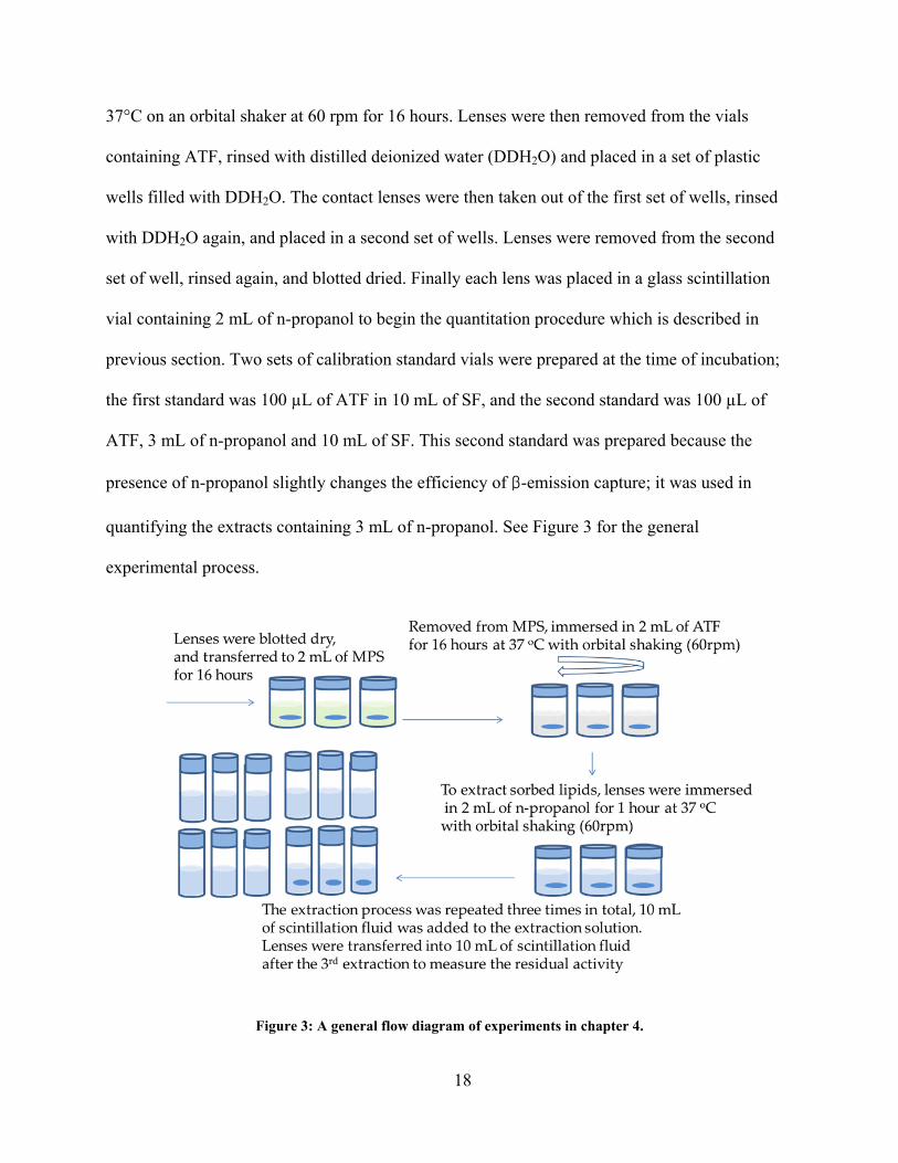
4.1d Pre-conditioning of lenses with MPS and mimicking the lipid sorption process
Senofilcon A, comfilcon A, balafilcon A, lotrafilcon B, and polymacon lenses were removed
from their blister packages and blotted dry on Kim Wipes®. The lenses were then placed into 5-
mL glass vials containing 2 mL of BBS, PureMoist or Biotrue at room temperature without
shaking or rubbing the lenses. After 16 hours, lenses were removed from vials, rinsed in BBS
and lightly blotted dry with a Kimwipe to remove excess liquid before being transferred to
another set of vials containing 2 mL of ATF containing radiolabeled lipids. A set of control
lenses (without pre-treatment) were removed from their blister packages, blotted dry, and placed
into ATF at the same time. To mimic the sorption process, lenses were placed in an incubator at
18
37°C on an orbital shaker at 60 rpm for 16 hours. Lenses were then removed from the vials
containing ATF, rinsed with distilled deionized water (DDH2O) and placed in a set of plastic
wells filled with DDH2O. The contact lenses were then taken out of the first set of wells, rinsed
with DDH2O again, and placed in a second set of wells. Lenses were removed from the second
set of well, rinsed again, and blotted dried. Finally each lens was placed in a glass scintillation
vial containing 2 mL of n-propanol to begin the quantitation procedure which is described in
previous section. Two sets of calibration standard vials were prepared at the time of incubation;
the first standard was 100 µL of ATF in 10 mL of SF, and the second standard was 100 µL of
ATF, 3 mL of n-propanol and 10 mL of SF. This second standard was prepared because the
presence of n-propanol slightly changes the efficiency of β-emission capture; it was used in
quantifying the extracts containing 3 mL of n-propanol. See Figure 3 for the general
experimental process.
Figure 3: A general flow diagram of experiments in chapter 4.
19
4.2 Results
In this study, we used a radiolabeling technique to evaluate the effectiveness of three MPSs –
PureMoist, Biotrue, and an experimental MPS made by Bausch and Lomb (BLS) – in reducing
the depositions of CH and DPPC on various commercial SiHy lenses and one conventional
hydrogel lens. CH and DPPC are model components that represent respectively non-polar and
polar lipid components in human tears.
After the 16 hours of incubation in the ATF, with or without any pre-soaking in an MPS, 4 to
6 µg of DPPC and 6 to 8 µg of CH were sorbed to senofilcon A, comfilcon A, and balafilcon A.
Approximately 1 µg of DPPC and 1 µg of CH were sorbed to lotrafilcon B, and about 0.2 µg of
DPPC and 0.1 µg of CH were sorbed to polymacon.
Figure 4 presents the amounts of DPPC and CH depositions on each lens material when pre-
conditioned with different solutions. In general, the DPPC sorption is consistently lower than the
CH sorption for all lenses except for lotrafilcon B lenses and polymacon lenses. Both DPPC and
CH depositions follow the same pattern for each combination of lens material and pre-soak
solution.
4.2a Analysis by lens type
There are significant differences in the amount of lipid sorbed on SiHy lenses while pre-
soaking with the same lens care solution. When lenses are preconditioned with PureMoist, DPPC
sorption on senofilcon A lenses is 20.3% lower than on balafilcon A lenses (p<0.001) and 18.7%
lower than on comfilcon A lenses (p<0.01). With the BBS pre-soak, the CH deposition on
senofilcon A is 13.2% higher than on balafilcon A lenses (p<0.001) and 15.7% higher than
comfilcon A lenses (p<0.0005). However, while statistically significant, these differences are not
20
large. The type of preconditioning solution made less difference in the amount of lipid sorbed to
lotrafilcon A and polymacon lenses. Both the DPPC sorption and CH sorption to these lenses
were significantly lower than the other lens types (p<0.0005), and polymacon lenses sorbed the
least amount of DPPC and CH overall (see Figure 4).
4.2c Analysis by presoaking solution
Lipid sorption on the same lens material is also somewhat dependent on the lens care
solution. For example DPPC sorption on senofilcon A lenses is 19.4% lower when the lenses are
preconditioned with PureMoist (p<0.001), and 25.1% lower when pre-conditioned with Biotrue
(p<0.00005) compared to those preconditioned with the BBS. CH sorption on senofilcon A is
also 20.3% lower when the lenses are presoaked in PureMoist (p<0.001), and 26.0% lower when
pre-conditioned in Biotrue (p<0.0005). Both DPPC and CH depositions on senofilcon A lenses
without prior exposure to any lens care solutions are 25.4% and 23.7% lower respectively than
lenses exposed to BBS (p<0.00005). However, there are no differences on DPPC and CH
sorptions (p>0.05) when senofilcon A was preconditioned with BLS compared with those
preconditioned in BBS.
There are no statistical differences (p>0.05) for both DPPC and CH sorptions when
balafilcon A, comfilcon A, and polmacon lenses were pre-conditioned with PureMoist, Biotrue,
BLS or BBS.
21
Figure 4: Lipids sorption with preconditioning in lens care solutions. Error bars represent the 95% confidence intervals (n>=6). * indicates statistical differences from BBS (p<0.05).
0
1
2
3
4
5
6
7
8
9
senofilcon A comfilcon A balafilcon A lotrafilcon B polymacon
µg/l
ens
DPPC BBS
B&L Proprietary solution
Opti-free® Pure Moist®
Biotrue
Control
0
1
2
3
4
5
6
7
8
9
senofilcon A comfilcon A balafilcon A lotrafilcon B polymacon
µg/l
ens
CH BBS
B&L Proprietary solution
Opti-free® Pure Moist®
Biotrue
Control
* * *
* * *
22
4.3 Discussion
The objective of this study presented in this chapter was to evaluate the efficacy of the MPSs
in reducing lipid sorption on SiHy lenses. The data show that only PureMoist and Biotrue
perform better than BBS in reducing lipid sorption to senofilcon A lenses, reducing the CH and
DPPC sorption by approximately 20 to 25% compared with lenses preconditioned in BBS.
Lens and solution manufactures hoped that MPSs would reduce lipid sorption. Their
proposed mechanism was that the polymeric surfactants in MPS would adsorb to the lens
material and block the hydrophobic sorption sites, thus reducing the level of lipid deposition.
However, sorption of DPPC and CH occurred generally to about the same amount to all lenses
irrespective of preconditioning the lenses with MPS or buffer or not at all. There were a few
statistically significant differences that may give hints as to mechanisms and directions for future
improvements. For example, there was some dependency on lens chemistry. This study showed
that both DPPC and CH sorption on senofilcon A are greater when lenses were preconditioned in
BBS instead of in the PureMoist and Biotrue; but there are no significant effects of solutions on
comfilcon A and balafilcon A lenses (Figure 4). A previous study also concluded that the
efficiencies of MPS on reduction of lipid deposition are somewhat dependent on lens material 21.
The non-surfaced-treated senofilcon A lenses are the only lens materials in this study that
used an internal wetting agent, polyvinyl pyrrolidone (PVP), to improve wettability and
hopefully reduce surface deposition 42. Our current speculation is that some of the PVP on the
senofilcon A lenses was removed during the 16 hour of preconditioning in BBS and the MPSs;
this may have reduced the hydrophilicity of the surface region and reduced the ability of the lens
surface to block lipid deposition with the very hydrophilic PVP. PureMoist and BioTrue both
contain various surfactants and wetting agents, and those substances may have replaced some of
23
the desorbed PVP in senofilcon A lenses, producing a reduction in sorption compared to sorption
with BBS pretreatment, but about the same sorption as non-pretreated control senofilcon A
lenses. Obviously this hypothesis needs to be substantiated in future studies. On the other hand,
comfilcon A material is inherently wettable, having more hydrophilic units in its chemistry and
thus requires no surface treatment 10. Balafilcon A lenses are treated with a plasma oxidation that
converts the TRIS structure on the surface into islands of hydrophilic silicate 42. Lotrafilcon B
lenses surface are coated with a uniform wettable layer which acts as a barrier to lipid sorption.
Since these materials are engineered to permanently enhance hydrophilicity of the surface, the
surfactants in the MPSs may have less additional influence on the surface chemistry with respect
to reducing lipid deposition.
To summarize, none of the MPSs (which all contain polymers and surfactants) are much
better than borate buffered saline in preventing lipid sorption. In fact in some cases
preconditioning with an MPS may increase lipid sorption most probably by removing
hydrophilic polymers from the contact lens.
25
CHAPTER 5 REMOVAL OF LIPID DEPOSITION BY MPS
5.1 Experimental approach
In this chapter, we report on using a radiolabeling technique to evaluate the effectiveness of
three multi-purpose solutions (MPSs) – OPTI-FREE® PureMoist® (PureMoist), BiotrueTM and an
experimental MPS (BLS) – in removing the depositions of cholesterol (CH) and
dipalmitoylphosphatidylcholine (DPPC) on various commercial silicone hydrogel (SiHy) lenses
and one conventional hydrogel lens. CH and DPPC are model components that represent
respectively non-polar and polar lipid components in human tear films.
5.1a Materials
The three MPSs are described in section 4.1a. Table 2 shows the active components of the
solutions. Four commercial SiHy lenses — senofilcon A, comfilcon A, lotrafilcon B, and
balafilcon A and one conventional hydrogel lens polymacon — were used in this study (see
Table 1).
5.1b Preparation of artificial tear solution
Table 3 of section 4.1a shows the composition of the artificial tear fluid (ATF) used in the
study of this chapter, which includes lipids, proteins and buffer. As mentioned previously, the
composition used herein differs somewhat from other published compositions 32,40,41. Compared
to others, it contains methyl-β-cyclodextrin (to achieve higher lipid concentrations), DPPC as the
26
phospholipid, and lower protein concentrations, and higher lipid concentrations compared to
natural tears, with the goal of accelerating the lipid spoliation. As there is currently no consensus
composition for tear film presented in the literature or used uniformly in industry, the
composition chosen for this work was selected because it deposits lipids and proteins on SiHy
lenses overnight at levels similar to deposits found on worn lenses after several weeks wear 34.
The preparation method of the ATF is presented in section 4.1b. Briefly, the aqueous base of
the ATF, borate buffered saline (BBS), was prepared by mixing boric acid, sodium borate and
sodium chloride in distilled deionized water (DDH2O), and the pH of the solution was adjusted
to 7.3 by adding NaOH or HCl. The ATF was prepared by adding appropriate amounts of
various lipids in organic solvent in a 100-mL volumetric flask. The solvent was evaporated under
a nitrogen stream for 2 hours and followed by 2 hours of vacuum drying. Then 50 mL of BBS
was added to the flask and magnetically stirred for 6 hours at room temperature (21°C) with a
1.5-cm Teflon stir bar at 650 rpm. Solid powdered protein components were added to the
solution. The mixture was stirred for additional 8 hours at room temperature. An additional 50
mL of BBS was added to the vial before final stirring for 20 minutes. The ATF was then used
immediately for incubation of lenses in the sorption studies. ATF stored for more than a few
hours tended to give less reproducible results.
5.1c Quantitation procedure
Following sorption, the three-stage n-propanol extraction was used to extract the lipid sorbed
on the lenses. The detailed extraction procedure is described in section 4.1c. In brief, each lens
was placed in a 20-mL glass scintillation vial containing 2 mL of n-propanol and placed on a
rotary table shaker at 60 rpm for one hour at 37°C. Lenses were then held above the extraction
27
solution and rinsed with 1 mL of n-propanol into the same vial. It was placed in another
container of n-propanol for 1 hour. Then, it was rinsed again the same way and the lens was
transferred to the third extraction vial. Following the third one-hour extraction and rinsing
procedure, the lens was transferred to a final vial filled with 10 mL of scintillation fluid to
measure any residual radioactivity in the lens. All blanks, standards and samples were submitted
in the same batch in identical scintillation vials to a LS 6500 scintillation counter (Beckman
Coulter) and simultaneously counted twice using a program that counted both 14C and 3H.
5.1d Soaking lenses with sorbed radiolabeled lipids in MPS
Lenses were removed from their blister packs, rinsed in BBS, lightly blotted dry with a
Kimwipe and immersed in glass vials each containing 2 mL of ATF. The vials were placed in an
incubator at 37°C on an orbital shaker at 60 rpm for 16 hours. Lenses were then removed from
the vials containing ATF, rinsed with DDH2O as described above. After rinsing, the lenses were
placed into 5-mL glass vials containing 2 mL of PureMoist, 2 mL of Biotrue or 2 mL of BBS. To
measure the kinetics of the elution of CH and DPPC, 200 µL samples were transferred from each
glass vial into scintillation vials containing 10 mL of SF at 0, 1, 2, 4 and 8 hours. After the 8
hours of desorption, the lenses were transferred into scintillation vials with 2 mL of n-propanol
to be extracted as described above.
Since the radioactivity of the aqueous samples was very low, accuracy was ensured by
preparing blank solutions and calibration solutions that were adjusted to have the same amount
of BBS as the samples. Thus the capture efficiency of β-particles was the same in samples,
standards and blanks. See section 4.1d for the composition of each blank solution. See Figure 5
for the general experimental process which is described above.
28
Figure 5: A general flow diagram of experiments in chapter 5.
5.2 Results
In this study, we used a radiolabeling technique to evaluate the effectiveness of three MPSs –
PureMoist, Biotrue, and BLS – in removing the depositions of CH and DPPC on various
commercial SiHy lenses and one conventional hydrogel lens. As mentioned, CH and DPPC are
model components that represent respectively non-polar and polar lipid components in human
tears.
Overall, DPPC was desorbed slowly in all the MPSs and also in BBS. However, little to no
CH was eluted from lenses to the solutions and the BBS. The overall removal percentages that
compare the amount of lipid deposition before and after soaking in MPSs or BBS for 8 hours
were calculated (Table 4). PureMoist solution removed the highest percentage of both CH (0.1%
to 28.2%) and DPPC (1.0% to 54.8%), from all lenses. The highest fractions of lipid were
removed from polymacon lenses (28.2% of CH and 54.8% of DPPC).
29
Table 4: Percentages of removal of sorbed lipids by the multi-purpose lens care solutions. Values are the mean % ± 95% intervals which are calculated
by propagation of errors.
Percentage of removal DPPC
Senofilcon A Comfilcon A Balafilcon A Lotrafilcon B Polymacon BBS 0.37(±0.16) 0.45(±0.32) 0.59(±0.17) 1.52(±0.71) 14.29(±4.63) Experimental MPS (BLS) 0.47(±0.16) 0.63(±0.17) 0.82(±0.36) 1.85(±0.51) 24.07(±3.55)
Opti-free® Pure Moist® 1.03(±0.31) 1.3(±0.46) 1.61(±0.31) 3.08(±0.77) 54.81(±12.04)
Data of DPPC and CH elution profiles from the various lenses to MPSs are presented in
Figure 6. In general, the DPPC is slowly eluted from all lenses, but there are no statistically
significant differences between balafilcon A, senofilcon A and comfilcon A lenses. Lotrafilcon B
lenses eluted comparatively less DPPC (but it sorbed less to begin with), and polymacon lenses
eluted the highest amounts of DPPC and CH to the solution compared with other lens types, and
these lenses sorbed the least. This is noteworthy since lotrafilcon B and polymacon lenses sorbed
less lipids than the other three lenses.
5.2b Analysis by MPS type
The elution data of sorbed DPPC and CH from lenses are solution dependent. For example
PureMoist removed the greatest amount of DPPC and CH, Biotrue and BLS removed both lipids
at similar levels, and BBS removed the least (Figure 6). The percentage of removal of DPPC and
30
CH deposition after 8 hours in various solutions are presented in Table 4. The percentages were
calculated by comparing the amounts of lipids desorbed from the lenses at the end of the soaking
procedure with the total amounts of sorbed lipid in control sets (control lenses were extracted
directly after incubation in ATF without any presoaking in MPSs or BBS, n = 7). After 8 hours
of soaking in MPSs, PureMoist removed greater amount of DPPC from senofilcon A, comfilcon,
balafilcon A , lotrafilcon B and polymacon lenses than did BBS statistically (p<0.05). The
differences were 0.03μg, 0.05μg, 0.04μg, 0.02μg and 0.7μg respectively. Biotrue and BLS both
statistically removed greater amounts of DPPC from polymacon lenses compared with BBS, and
the removals were 0.02μg more for both solutions.
Significant amount of CH deposits were removed from all lenses in PureMoist (confidence
intervals are above zero) during the 8 hours soaking period. The average removals from
senofilcon A, comfilcon A, balafilcon A, lotrafilcon B and polymacon were 0.0087 µg, 0.0286
µg, 0.0115µg, 0.0002 µg and 0.0324 µg respectively. For polymacon lenses, BLS and Biotrue
were also able to remove some of the CH sorption (Figure 6), but the amounts were significantly
less than removal by PureMoist (p<0.05).
31
Figure 6: The elution profiles of sorbed lipids. DPPC (left) and CH (right) in solutions. Error bars represent the 95% confidence intervals (n>=6). *indicates statistical difference from BBS at the same the point (p<0.05).
-0.02
0.03
0.08
0.13
hr 0 hr 1 hr 2 hr 4 hr 8 hr 0 hr 1 hr 2 hr 4 hr 8
ug/l
ens
DPPC CH
Senofilcon A BBSB&L proprietary MPSOpti-free® Pure Moist®Biotrue
-0.02
0.03
0.08
0.13
hr 0 hr 1 hr 2 hr 4 hr 8 hr 0 hr 1 hr 2 hr 4 hr 8
ug/l
ens
DPPC CH
Comfilcon A
-0.02
0.03
0.08
0.13
hr 0 hr 1 hr 2 hr 4 hr 8 hr 0 hr 1 hr 2 hr 4 hr 8
ug/l
ens
DPPC CH
Balafilcon A
-0.02
0.03
0.08
0.13
hr 0 hr 1 hr 2 hr 4 hr 8 hr 0 hr 1 hr 2 hr 4 hr 8
ug/l
ens
DPPC CH
Lotrafilcon B
-0.02
0.03
0.08
0.13
hr 0 hr 1 hr 2 hr 4 hr 8 hr 0 hr 1 hr 2 hr 4 hr 8
ug /
lens
DPPC CH
Polymacon
* *** *** ** * *** * ** **
** ** * * *
* * * *
* * * * * *
* *
32
Figure 7: Initial sorption of DPPC and CH, and retained sorption. Error bars represent the 95% confidence intervals (n=6).
0
2
4
6
8
10
12
Sorbed DPPC Retained DPPC Sorbed CH Retained CH
ug/l
ens
BLS senofilcon A
comfilcon A
balafilcon A
lotrafilcon B
polymacon
0
2
4
6
8
10
12
Sorbed DPPC Retained DPPC Sorbed CH Retained CH
ug/l
ens
PureMoist senofilcon A
comfilcon A
balafilcon A
lotrafilcon B
polymacon
0
2
4
6
8
10
12
Sorbed DPPC Retained DPPC Sorbed CH Retained CH
ug/l
ens
Biotrue senofilcon A
comfilcon A
balafilcon A
lotrafilcon B
polymacon
33
5.3 Discussion
The MPSs were generally able to remove more lipids than BBS. PureMoist removed the
largest amount of DPPC and CH from all lenses. However, even with PureMoist, less than 4% of
DPPC and less than 1% of CH were removed from SiHy lenses (senofilcon A, comfilcon A,
balafilcon A, and lotrafilcon B). PureMoist removed approximately 55% of DPPC and 28% of
CH from polymacon lenses, which are non-hydrophobic conventional hydrogel lenses.
SiHy materials have some very hydrophobic silicone segments so lipids are
thermodynamically driven to partition from the aqueous solution to the hydrophobic polymer
segments of the SiHy lenses. On the other hand, no such hydrophobic segments are present in
poly-HEMA based hydrogels; lipid deposits are small and are effectively removed by the
surfactants in the MPS. The chemicals and surfactants present in MPSs have to be mild enough
to be compatible with and comfortable to the eyes, so it is a difficult task to develop a multi-
purpose solution to remove lipid and protein deposits and yet remain biocompatible with human
eyes at the same time. Apparently the surfactants in the MPSs are not hydrophobic enough to
remove lipids, especially removal of non-polar lipids from the hydrophobic segments in SiHy
lenses.
5.3a Removal of sorbed lipids by the MPS
If one only examines the amounts of lipids that remain on the lenses before and after the
attempt to remove DPPC and CH by soaking in MPS, the scatter intrinsic to these experiments
hides the fact that there is a small but detectable desorption of DPPC. Thus while it appears that
there are no significant differences between the initial deposition and the retained sorption on
each lens type for both of the MPSs, some desorption actually occurs. For example, see Figure 7
34
which shows the amount of sorbed lipid and the retained lipid for lenses with preconditioning in
BLS, PureMoist and Biotrue. Thus it falsely appears that there are no significant differences
between the initial deposition and the retained sorption on these lens type for the MPSs. Less
sensitive techniques, such as chromatography might also lead to the incorrect assumption that
these MPSs do not remove sorbed lipid. However, because of the sensitivity of the radiolabeling
technique used, the DPPC and the CH elution into the solutions could be accurately measured,
showing that DPPC and CH elute slightly from some of the lenses under the conditions of these
experiments. However, the composition of the eluting solution, whether BBS or an MPS, appears
to make little difference on removing lipids from SiHy lenses (See Figure 6 and Table 4). While
some marketing schemes might tout the fact that PureMoist removes 3% of DPPC and 0.8% CH
from lotrafilcon B lenses, while statistically significant, there is little to cheer about. The
observation that the multi-purpose solutions clean only slightly better than BBS suggests that the
surfactants employed are not particularly effective for SiHy lenses. The small fraction of sorbed
DPPC was being extracted slowly with time in all solutions. In contrast, little to no significant
amount of CH was eluted in both MPSs and BBS after 8 hours of soaking. The difference in
removal might be attributed to the more polar nature of the DPPC compared to the non-polar CH.
This lack of desorption of CH hints that other non-polar tear components, such as CH esters,
fatty acid esters and triolein may also have little to no desorption from a lens by soaking in an
MPS.
Despite the small removal of lipids, there are other essential benefits using MPSs to clean
and store contact lenses. All soft contact lenses – both SiHy lenses and conventional hydrogel
lenses – need to be stored in MPS lens care solutions while not in use for at least 2 reasons. First,
storing in MPS rehydrates the lenses and replenished the lubricants and wetting agents to keep
35
the lenses moist and comfortable. Second, the preservatives eliminate harmful microorganisms to
prevent infections 24,43,44. As mentioned, rubbing has been shown to remove microorganisms and
protein deposits 14,27. Such benefits should always be considered when evaluating the overall
performance of an MPS.
37
CHAPTER 6 EFFECTS OF RUBBING AND A PEROXIDE-BASED SOLUTION
While experiments in previous chapters were performed without rubbing, rubbing has been
shown to remove proteins and bacterial from contact lenses 14,27. Most commercial multi-purpose
solutions (MPSs) for lens care and some professionals indicate that rubbing should be included
during the process of cleaning silicone hydrogel (SiHy) lenses 24,25. However, there is currently
no experimental evidence in the literature to support the additional rubbing process removes
significant amounts of lipid from SiHy lenses. Thus, the effect of rubbing was investigated and is
discussed in this chapter.
Hydrogen peroxide-based solutions for lens care are sometimes used to disinfect lenses. The
cleaning process of such solutions often involves a neutralization step to convert all the peroxide
to water, so the lens is ready to be worn without causing irritation. The ∙OH radicals in hydrogen
peroxide solutions kill microorganisms, and it might also enhance lipid removal. A study
reported that a hydrogen peroxide lens care solution containing a surfactant removed more lipid
from lenses when compared to the non-surfactant hydrogen peroxide solution 20. This chapter
also reports data for a peroxide-based experimental solution that was investigated for its lipid
removal efficacy and compared with a simple borate buffered saline (BBS).
6.1 Experimental approach
In this study, the experimental setup including the artificial tear fluid (ATF) preparation was
identical with the pervious experiments of removal and reduction of lipids by the MPSs (chapter
38
4 and 5), except there is an additional rubbing step after the incubation in ATF and before
soaking lenses in MPS. The rubbing procedure was designed to mimic the effect on lipid
removal by rubbing the lens with the finger in the palm of the hand. The detail of the procedure
will be discussed in the follow sections.
6.1a Materials
Senofilcon A, balafilcon A and a proprietary SiHy lens (Zeta) in development by Bausch and
Lomb were used in this study. Zeta is an experimental lens material as a next-generation, non-
plasma modified silicone hydrogel that has a chemistry that does not require a second step to
apply a hydrophilic coatings such as is done with lotrafilcon B and balafilcon A lenses. Two
commercial MPSs, SEEDO Softcare (SEEDO) and Opti-free® Pure Moist® (PureMoist), and one
experimental peroxide-based solution (BLP) were evaluated and compared with the performance
of the BBS.
6.1b Removal of sorbed lipids in MPSs and BLP
Six different removal methods were evaluated in this chapter. They are 1) rub and soak in
SEEDO, 2) soak only in SEEDO, 3) rub and soak in PureMoist 4) soak only in BLP, 5) rub and
soak in BBS and 6) soak only in BBS. With three lens types (senofilcon A, Zeta and balafilcon
A), there were 18 combinations overall, and 6 replicates were performed in each combination.
The experimental procedure for combinations that only involve soaking in solutions is the
same as the pervious experiment which is presented in section (5.1d), except that those lenses
were soaked in BLP used the special vials containing platinum-plated neutralizers rather than
using the 5-ml glass vials (Figure 9). Since BLP is a peroxide-based solution, neutralizers were
39
placed in the vials to convert H2O2 to H2O during the cleaning process. Two lenses and 10 mL of
BLP were placed into each vial. Similar to other lenses, 200 µL of solution were taken from the
vials at 0, 1, 2, 4 and 8 hours to determine the desorption kinetics of CH and DPPC. After the 8
hours of desorption, the lenses were transferred into scintillation vials with 2 mL of n-propanol
to be extracted by the standard three stages of n-propanol extraction as described in section 4.1c
With those lenses that were rubbed prior to soaking in MPSs, the process is described as
follow. First, lenses were incubated in ATF like other lenses (section 5.1d). Following the 16-
hour incubation process, lenses were transferred from the vials containing ATF, and rinsed with
distilled deionized water (DDH2O) twice. After rinsing, 400 µL of SEEDO, PureMoist or BBS
were pipetted into 10-mL polyethylene zip lock bags. The bags were labeled correspondingly.
Lenses with sorbed radioactive lipids were placed into the center of the bags, and the bags were
closed completely with care to exclude nearly all air. The rubbing step began with placing the
bag on the left palm, with the palm facing up. The lenses were rubbed through the bag using the
index finger for 20 times in a circular motion, and the finger traced the peripheral region of the
lenses. 200 µL of MPS sample were taken from the bags to scintillation vials filled with 10 mL
of SF. The lenses were rinsed with DDH20 and blotted dry with a Kimwipe®, and placed in 5-
mL glass vials containing 2 mL of the various “removal” solutions. 200 µL samples were taken
from each glass vial into scintillation vials containing 10 mL of SF at 0, 1, 2, 4 and 8 hours to
determine the desorption kinetics of CH and DPPC after the rubbing procedure. After the 8 hours
of desorption, the lenses were transferred into scintillation vials with 2 mL of n-propanol to be
extracted by the standard three stages of n-propanol extraction as described in section 4.1c.
After transferring the lenses to MPS, the bags were rinsed with DDH2O two times to make
sure no lipids were left in the aqueous phase. Then 2 mL of n-heptanol was transferred to the
40
bags, the air trapped in the bags was carefully removed by softly pushing. The bags were then
closed and placed flat at room temperature on a clean paper towel. After an hour, the solvent in
the bags was transferred to scintillation vials filled with 10 mL of SF. The bags were rinsed with
additional 1mL of n-heptanol into the same scintillation vials. The second extraction was
performed by transferring another 2 mL of n-heptanol to the bags and repeat the process
described above. After the third extraction, the bags were filled with 10mL of SF and placed into
a set of new scintillation vials for counting any residual radioactivity on the bags. This is called
“direct counting” of the bags. N-heptanol was selected for the bag extraction after trying several
solvents such as n-propanol, octane, hexane, n-hexanol, n-heptanol, and tetrahydrofuran. It was
found that alkanes diffuse through the bags quickly, and thus are not ideal as extraction solvents,
and n-heptnaol extracted most of the sorbed lipids from the bags (lowest direct counting values).
The direct counting of the bags was still slightly above the background count but of the same
order of magnitude. Thus the amount of detected lipid β emission was multiplied by two while
quantifying the lipids from the direct counting stage. This adjustment was made by assuming
half of the beta emissions from lipid depositions were registered by the SF, and half of the lipid
depositions were sorbed into the bags polymer and were not registered by the SF.
Upon the completion of the lens extraction and the bag extraction, all blanks, standards and
samples were submitted to a LS 6500 scintillation counter (Beckman Coulter) and counted using
a program that counted both 14C and 3H simultaneously. This was a tremendously complex
experiment that required careful timing, much extraction (bags and lenses) and the consumption
of about 1500 scintillation vials that were each counted twice. See Figure 8 for the general flow
diagram of the experiment.
41
Table 5: Characteristics of the contact lens materials.
Lens material
Commercial name Manufacturer Principal
components Surface treatment
Senofilcon A Acuvue Oasys
Johnson & Johnson mPDMS, DMA, HEMA, SiGMA, TEGDMA, PVP
No surface treatment. Internal wetting agent (PVP) throughout the matrix that also coats the surface
Balafilcon A PureVision Bausch & Lomb NVP, TPVC, NVA, PBVC
*EOBO: poly (oxyethylene)-poly (oxybutylene); BLP: Properiatory peroxide based solution ** This experimental solution is not yet in commercial production.
42
Figure 8: A general flow diagram of experiments in chapter 6.
Figure 9: The proprietary vial for using the peroxide-based solution.
43
6.2 Results
In this study, we used a radiolabeling technique to evaluate the effectiveness of three MPSs –
PureMoist, SEEDO and BLP – in removing the depositions of CH and DPPC on various SiHy
lenses. In additional to soaking in MPS, the effects of rubbing were determined with SEEDO,
PureMoist and BBS.
6.2a Effects by additional rubbing and the peroxide-based solution
There were no significant differences in the amount of removal of lipids between lenses.
Surprisingly the BLP did not remove any lipids from any lenses after 8 hours of soaking (Figure
10). Overall, more lipids were removed when lenses were treated with the additional rubbing
procedure compared with “soak only” procedure, and the removal is statistically independent of
solution types (p>0.05), as they all performed identically to BBS (Figure 10). The percentages of
removal of both lipids by rub and soak procedure were between 0.5% and 2.0% (Table 7).
The effect of the additional rubbing step in SEEDO and BBS were evaluated. Both solutions
statistically removed greater amounts of DPPC and CH from all lenses. While using SEEDO
with rubbing, approximately 5.3, 9.0 and 2.7 times more (p<0.05) DPPC and 8.4, 6.6 and 6.6
times more CH were removed respectively from senofilcon A, Zeta, and balafilcon A lenses
compared with lenses that were soaked only. While using BBS with rubbing, approximately 26.5,
9.9 and 7.2 fold more of DPPC sorption and 14.3, 14.1 and 34.5 fold more of CH was removed
(p<0.05) from lenses compared to those were only soaked in BBS (Figure 10).
The rubbing effect on senofilcon A and balafilcon A lenses when using PureMoist are also
evaluated and compared with other solutions (Figure 11) where the data of “PureMoist Soak only”
were generated from experiments of chapter 5. There were no significant differences in DPPC
44
removal between “PureMoist Rub” and “PureMoist soak only” (P>0.05) for both lenses.
Approximately 6.6 and 5.0 times more (p<0.05) CH was removed respectively form senofilcon
A and balafilcon A lenses when these lenses were cleaned with additional rubbing step.
The complete elution profiles of lipids of lenses in different solutions are showed in Figure
12. Additional rubbing did not affect the elution kinetic of lipids as there were no significant
differences (p>0.05) in removal along the 8 hours period between “SEEDO rub and soak” and
“SEEDO soak only”, and “BBS rub and soak” and “BBS soak only”.
Table 7: Percentages of removal of the sorbed lipids by multi-purpose lens care solutions. Parentheses indicate 95% intervals which are calculated
by propagation of errors, n=6.
Percentage of removal
DPPC CH Senofilcon A Zeta Balafilcon A Senofilcon A Zeta Balafilcon
Figure 10: The amount of sorbed DPPC (top) and CH (bottom) removed in multi-purpose lens care solutions and boric buffered saline, with or without rubbing. Error bars represent the 95% confidence intervals (n=6). All rubbing removes DPPC and CH at a greater level (p<0.05) than without rubbing.
-0.03
-0.01
0.01
0.03
0.05
0.07
0.09
0.11
0.13
0.15
SEEDO rub andsoak
SEEDO Soak only PureMoist Ruband Soak
BLP soak only BBS rub andsoak
BBS soak only
µg/l
ens
DPPC Senofilcon A
Zeta
Balafilcon A
-0.03
-0.01
0.01
0.03
0.05
0.07
0.09
0.11
0.13
0.15
SEEDO rub andsoak
SEEDO Soak only PureMoist Ruband Soak
BLP soak only BBS rub andsoak
BBS soak only
µg/l
ens
CH Senofilcon A
Zeta
Balafilcon A
46
Figure 11: The amount of sorbed DPPC (top) and CH (bottom) removed from senofilcon A and balafilcon A lenses in various solutions, comparing between rub and no rub. Error bars represent the 95% confidence intervals (n≥6). Data of “PureMoist Soak only” were generated from experiments of chapter 5. * indicates a significant improvement in removal when rubbing compared to removal in the same MPS without rubbing (p<0.05).
-0.01
0.01
0.03
0.05
0.07
0.09
0.11
0.13
0.15
SEEDO rub andsoak
SEEDO soak only PureMoist ruband soak
PureMoist soakonly
BBS rub andsoak
BBS soak only
µg/l
ens
DPPC Senofilcon A
Balafilcon A
-0.01
0.01
0.03
0.05
0.07
0.09
0.11
0.13
0.15
SEEDO rub andsoak
SEEDO soak only PureMoist ruband soak
PureMoist soakonly
BBS rub andsoak
BBS soak only
µg/l
ens
CH Senofilcon A
Balafilcon A
* * * * * *
* * * *
47
Figure 12: The elution profiles of sorbed lipids in solutions. DPPC (left) and CH (right). Error bars represent the 95% confidence intervals (n=6).
-0.02
0.03
0.08
0.13
Rubbingstep
hr 0 hr 1 hr 2 hr 4 hr 8 Rubbingstep
hr 0 hr 1 hr 2 hr 4 hr 8
µg/l
ens
DPPC CH
Senofilcon A SEEDO rub and soak
SEEDO Soak only
PureMoist Rub and Soak
BLP soak only
BBS rub and soak
BBS soak only
-0.02
0.03
0.08
0.13
Rubbingstep
hr 0 hr 1 hr 2 hr 4 hr 8 Rubbingstep
hr 0 hr 1 hr 2 hr 4 hr 8
µg/l
ens
DPPC CH
Zeta SEEDO rub and soak
SEEDO Soak only
PureMoist Rub and Soak
BLP soak only
BBS rub and soak
BBS soak only
-0.02
0.03
0.08
0.13
Rubbingstep
hr 0 hr 1 hr 2 hr 4 hr 8 Rubbingstep
hr 0 hr 1 hr 2 hr 4 hr 8
µg/l
ens
DPPC CH
Balafilcon A SEEDO rub and soak
SEEDO Soak only
PureMoist Rub and Soak
BLP soak only
BBS rub and soak
BBS soak only
48
6.3 Discussion
This particular study examined the effect of rubbing on lipid removal. Based on literature
reports of protein and bacterial removal by rubbing, we expected to see significant lipid removal
by rubbing. However, this was not observed in all cases. My current hypothesis as to why lipids
are not removed significantly by rubbing is that lipids are small compared to proteins. Thus the
majority of lipids could be absorbed into the lens material deeper than just the surface adsorption.
As rubbing should only affect the surface of the lens, less removal by rubbing was expected on
lenses with hydrophilic surfaces since less lipid should adsorb. Lipids that sorbed to the more
hydrophilic polymacon lenses could be easily be removed by just soaking in MPSs, as was
reported in chapter 5.
While it is true that additional rubbing in most cases removes more lipid deposition from the
SiHy lenses compared with the “soak only” treatment, the benefit is might not be significant on
SiHy lenses as the overall removal percentages of both DPPC and CH are less than 2% (Table 7) .
We should also consider that vigorous or repeated rubbing could possibly remove the thin
hydrophilic treated surface on particular lenses such as lotrafilcon and balafilcon, and thereby
expose the underlying hydrophobic polymer and accelerate the lipid deposition process. For
example, the average thickness of the hydrophilic surface layer of a lotrafilcon B lens is only 25
nm. To investigate the possible damage to the hydrophilic layer by rubbing, one could rub half of
a set of lenses before the incubation process in ATF and measure the total lipid sorption. If the
hydrophilic layer of the lens is damaged, more lipid deposition would be observed in rubbed
lenses compared to lenses that are not rubbed.
In conclusion, rubbing did improve the lipid removal efficacy of the MPSs. However, the
overall removal percentages are still low (≤2%). Vigorous or repeat rubbing of lens might
49
destroy the hydrophilic layer in some surface-treated lenses such as lotrafilcon B lenses and
balafilcon A lenses. Before suggesting whether consumers to employ rubbing in their lens care
regimens, further studies are encouraged that investigate the long term effects on lipid sorption
and wettability of rubbing various surface-treated SiHy lenses.
51
CHAPTER 7 CLINICAL APPLICABILITY OF THE IN VITRO MODEL AND THE SOURCES OF ERROR
The result at chapters 4, 5 and 6 may be disappointing to the industrial sponsors of this
research who hoped to show that their lens care solutions could prevent or reverse lipid sorption.
To temper the bleak results of chapter 4, 5 and 6, we must acknowledge that these results
may not directly represent the actual effectiveness of these MPSs in actual clinical wear. It is
always a challenge to extrapolate the results of in vitro experiments to in vivo performance.
There are obvious differences between our experimental model and the conditions that exist on
the eye. First, tear fluid in a human eye is constantly flowing, and throughout the day there are
physical shear stresses on the tear film due to eye movement and blinking. Second, the
composition of tears changes with external stimuli and often varies for each individual. Third,
the actual tear film on an ocular surface is thought to be a multi-layer structure of a few microns
in thickness, rather than a homogenous solution in a beaker. On the eye a complex lipid layer
containing both polar and non-polar lipids forms the outer-most layer and serves to prevent
excessive evaporation and to stabilize the tear film. An aqueous middle layer consist of proteins,
salts, electrolytes, and a relatively thick mucin layer lies on the bottom adjacent to the corneal
cells 45-48. In addition, these tear film layers should not be segregated into various experiments
examining only one layer at a time because components from all these layers, including proteins
and lipids, were found in contact lens deposits 11,29,49. The system becomes even more complex
when including the interactions of different types of contact lenses; for example one study
52
confirms that wearing contact lenses leads to higher tear film evaporation rates for up to one day
after removal of the lenses 50.
Although there are challenges in mimicking the actual ocular deposition process, the use of
in vitro models allows researchers to control variables easily, and such studies require a less
complex experimental design. Many in vitro studies involve incubation of lenses in artificial tear
solutions at body temperature with gentle shaking to predict the amount of lipid deposition to
contact lenses. Much of the published data from in vitro experiments are somewhat different than
the clinical observations. However they do show consistency in the general trends, such as
lotrafilcon materials sorb less lipid than other commercial SiHy lenses 12,33,51. Other general
trends are that DPPC sorption is less than CH sorption for SiHy lenses, and SiHy lenses sorb
relatively more lipids and less proteins, while conventional poly-HEMA based hydrogels sorb
relatively more proteins and less lipids 11,12,31. These general trends, observed in clinically worn
lenses, are also observed in these in vitro studies using artificial tear fluids and simulated soaking
and care procedures. Thus we are confident that the main observations of these in vitro studies in
this thesis research reflect the general behavior of worn lenses: in a no-rub situation, polar lipids
may elute very slowly from SiHy lenses into MPSs, but non-polar lipids probably have little to
no desorption. While rubbing may remove bacteria and perhaps some proteins, our studies show
that very little lipid is removed by rubbing, particularly on lenses with hydrophobic character.
This study produced other valuable observations and data that were not previously
published. For example, all SiHy lenses in this study swelled in n-propanol due to their intrinsic
hydrophobic properties, reaching a maximum size (50% increases in diameter) in about 4
minutes. While all SiHy lenses swelled in n-propanol, the edges of lotrafilcon B lenses did not
flare in a “scallop” border, as did balafilcon A and senofilcon A lenses. Additionally, the
53
lotrafilcon B lenses were more robust than the other SiHy lenses, as they did not easily break
during handling while swollen in n-propanol. As lenses become fragile with swelling, they could
easily be broken into small pieces between extraction steps. The extraction efficiency is reduced
when some of the pieces of the lens do not get transferred to the next extraction vial, leading to
challenges in quantitation. Thus lotrafilcon B lenses were easiest to use in this study. Although
the conventional hydrogel polymacon lenses do not swell in the n-propanol solvent as do the
SiHy lenses, they have a tendency to stick on the inner surface of glass vials. As a result, those
lenses could be easily torn apart during extractions.
An unexpected and very useful observation made in this study relates to the sensitivity of
sorption to the preparation of the ATF. In our lab we found that sorption from ATF is very
sensitive to the composition and preparation of the fluid. Even seemingly minor differences, such
as bovine versus porcine mucin, stir bar size and stirring speed affect sorption in measurable
amounts. In view of the importance of artificial tear composition, we published a
recommendation that ophthalmological societies and institutions should collaborate in
formulating an appropriate ATF that can be easily and reproducibly made and used by various
investigators around the world to generate more consistent and directly comparable data 34.
Other elements such as variation in the lens surface treatment, the material composition,
and the differences in the lens thickness also contribute to the overall scatter in the data. We are
surprised that other publications have not reported such sensitivity in their results to minor
experimental variables. Perhaps the high accuracy and precision of our radiolabelled experiments
revealed this sensitivity that other methods could not detect.
55
CHAPTER 8 SOLUBILITY OF COMFORT AGENTS
The fouling of silicone hydrogel (SiHy) lenses by lipid sorption lowers the surface
wettability may contribute to dry eye syndromes. My thesis study has shown that lipid deposition
to silicone hydrogel (SiHy) lenses appears very difficult to be removed or prevented. An
alternative method to maintain wear comfort would be altering the lens materials itself; release of
comfort agents from the lens matrix could mitigate the irritation by fouling and sustain consistent
high levels of comfort, hopefully resulting in better customer satisfaction. This chapter reports an
initial study of which comfort agents might be added to SiHy lenses.
8.1 Estimating the solubility of polymeric comfort agents in various solutions
The potential to deliver polymeric comfort agents to the anterior eye from a SiHy contact
lens requires that the comfort agent reside in the lens and not significantly reduce the optical
clarity of the lens. The optical clarity of the agent in the monomer or macromer formulation
(prior to lens polymerization) is a screening method for potential candidates. Those agents that
scatter light before polymerization have a high probability of scattering light after polymerization.
Light scattering is usually caused by poor solubility of the comfort agent in the solution –
undissolved particles scatter light if their refractive index is different from that of the solvent.
In this research, the optical densities of a series of solutions of comfort agents in potential
contact lens macromer formulations were measured. Not only is this a convenient lab technique,
but the results pertain directly to the optical density of a macromer formulation, and potentially
56
the optical density of the polymerized contact lens. Optical density (OD) is defined as –
Log10(Iin/Iout), where Iin is the intensity of light entering a sample, and Iout is the intensity exiting
the sample.
There are 2 factors that contribute to optical density. First is absorption. Light may be
absorbed by molecules in a solution, even when the mixture forms one phase and no scattering
occurs. The second contribution is scattering in which insoluble particles scatter light at various
angles depending on their size, shape and refractive index, and thus less light is directed forward
along the path to the detector in vitro or to the eye retina in vivo. Although a single reading of
OD cannot distinguish the proportion of each factor contributing to a single reading of OD, a
series of readings at different concentrations can in theory distinguish the two. Light absorption
is linear with concentration at low concentrations, and thus a plot of OD vs concentration will go
through zero if only absorption occurs. If a material is totally insoluble, its plot of OD vs
concentration will also go through zero (and usually with a steep slope), because even the
smallest amount of solute causes light scattering. However, if the solute is soluble up to a certain
limit, there will be no contribution from light scattering until that concentration limit is reached,
and thereafter the OD will increase steeply with solute concentration, as shown schematically in
Figure 13.
In this research we measured the optical density of solutions of several comfort agents in a
solvent and 3 different proprietary macromer formulations provided by ALCON.
57
Figure 13: Optical density vs concentration for agents with slight solubility.
8.1a Materials
Dipropylene glycol methyl ether (DPGME) was purchased from Aldrich and stored at room
temperature in a solvent cabinet (Figure 14). Macromers A, B, and C were shipped from
ALCON in Johns Creek, GA. They were stored at room temperature. The comfort agents were
provided by ALCON in powdered form.They are poly(N-vinylpyrrolidone) (pNVP), carboxy
acetate copolymer) , poly glycol (PEO), Guar, and sodium hyaluronate (NaHA). Chemical
structures of the comfort agents are presented in Figure 15. A small marble mortar and pestle
was purchased from Fischer Scientific. UV-vis optical cuvettes were purchased from the BYU
Chemistry Stockroom, as were stir bars, spatulas, and other small lab supplies.
Concentration
Opt
ical
Den
sity
absorption
light scattering
solubility
58
OO
OH
Dipropylene glycol methyl ether (DPGME)
Figure 14: Chemical Structure of dipropylene glycol methyl ether (DPGME).
NO
n
poly(N-vinylpyrrolidone) (pNVP)
OO
ORRO
ORn
R = H or CH2CO2H
OO
ORRO
ORn
R = H or CH3 or CH2CH(OH)CH3
Carboxymethyl cellulose (CMC) or cellulose gum
Hydroxypropyl methylcellulose (HPMC)
O
O
CH2O
O
OH
CH2
OH
OH
OH
OH
O
n
m
Dextran
HO
OH
n
Polyethyleneglycol (PEO)Guar
O
O
OHOH
HO
OHO
HOOH
OO
OH
OHHO
OH n
Carbopol
Hyaluronate (NaHA)
Ethyleneoxide butyleneoxide (EOBO)
HO O n
OO
bOn m
OOO
NHOHHOO
O-Na+
HO
OH
O
On
NO
m
polyvinylpyrrolidone-polyvinyl acetate (Kollidon)
O
O
H3C
n
Figure 15: Structures of comfort agents.
59
8.1b Methods
For solubility in DPGME, this solvent was weighed on an analytical balance in a small
container, and a small mass of comfort agent powder was weighed and added to the container. It
was stirred with a magnetic stir bar for minutes (if it dissolved quickly) to hours. Often the
preparation and stirring occurred in an optical cuvette. Sometimes it was prepared in a glass vial,
in which case it was transferred to the optical cuvette. The cuvette was spun in a swinging bucket
centrifuge for 5 minutes to consolidate the liquid on the bottom and remove any air bubbles that
would have scattered light. The cuvette was transferred to a Beckman 550 UV-vis spectrometer
for measurement. The reference was pure DPGME. After the OD of a sample was measured,
more DPGME was weighed and added to the sample to create a lower concentration of solute.
Due to the abundant supply of DPGME, this was repeated several times to generate many data
points. Optical densities were recorded and plotted, from which solubilities were estimated
graphically.
For solubilities in macromer formulations, we found that the high viscosity of the macromers
prevented any magnetic stirring. Therefore we developed a technique in which known masses of
macromer and solute was mixed using a mortar and pestle, and then transferred to an optical
cuvette, centrifuged to remove air bubbles, and measured in the spectrometer. Because of the
limited supply of macromer, we could not make serial dilutions as were done for DPGME.
Instead we measured very small amounts of powdered comfort agent on a Mettler microbalance
(precision to 0.1 µg). These small amounts were added to previous mixtures, generating a series
that increased in concentration of comfort agent. The very small masses of comfort agent, and
the difficulty of removing all of the mixture from the mortar surface (with a rubber policeman),
60
produced more scatter in the measurement with macromers. Optical densities were recorded and
plotted, from which solubilities were estimated graphically.
8.2 Results and discussion
This study examined the solubility of 10 polymeric comfort agents in various solutions to
determine their potential to dissolve in macromer formulations for contact lenses. The comfort
agents were pNVP, Kollidon, EOBO, CMC, HPMC, Dextran, Guar, poly glycol, Carbopol, and
sodium hyaluronate. The solutions were DPGME solvent and 3 macromer formulations provided
by ALCON. Solubility was assessed by light scattering and optical density of solution with
various weight fractions of comfort agents.
Results indicated that pNVP, Kollidon and EOBO are very soluble in the solvent dipropylene
glycol methyl ether (DPGME), and Guar, poly glycol and Carbopol are slightly soluble. In
Macromer A, pNVP and Kollidon are very soluble, while EOBO and HPMC have slight
solubility. pNVP and HPMC are slightly soluble in Macromer B. In Macromer C, only Kollidon
is slightly soluble. We did not study the effect of temperature or of macromer concentration on
the solubility values.
8.2a Solubility in DPGME
Figure 16 shows the optical density of the comfort agents in DPGME. There are comfort
agents that have obviously very high optical density such poly glycol, Dextran, Carbopol and
Guar. The most probably causes are low solubility and poor match of refractive index, as
evidenced by the steep slope of the plots. Although these agents have high scattering, Guar and
poly glycol are slightly soluble with solubility limits of 0.01wt% and 0.02wt% respectively
61
(Figure 16 and Figure 17). Carbopol appears to have an intermediate solubility of about 0.11% as
evidenced by the break in the curve at that point. Even though NaHA appears to be insoluble, it
has a fairly low scattering and low overall optical density. Likewise, CMC appears to be
insoluble, but has very low light scattering.
Three comfort agents appear to have very high solubility, such that we could not see
evidence of insolubility at less than 1% by mass. These are pNVP, Kollidon, and EOBO. They
also have very low absorption.
Some general observations about the solubility of these agents in DPGME are as follows.
Poly glycol took a while to break up and become dispersed in the DPGME. Each dilution was
very cloudy and there were particulates that settled very quickly. CMC solutions seemed to have
distinct particles suspended with no real cloudiness. HPMC solution dilutions were cloudy and
the particles settled slowly. Dextran solutions were cloudy, but became less so with each
dilution. The particles stayed suspended for a while, but eventually settled out. The particles
seemed to aggregate on the sides of the glass vials. NaHA solutions were cloudy and the
particles seemed to go to the top of the solution after sitting for a while. Carbopol solutions were
cloudy, but became less so with each dilution. The last dilution seemed pretty clear. Guar was
very cloudy and yellow, and also had particles that settled out very quickly. EOBO dissolved
very slowly, going from a semisolid pellet, to slowly becoming a paste in solution; finally the
solution went completely clear. Kollidon dissolved very quickly and each solution was
completely clear. pNVP seemed to be very clear in all concentrations with almost no visible
particles suspended. The dilutions didn't seem to become clearer by visual inspection, but the
spectrophotometer showed decreasing absorbance.
62
Figure 16: Overlay plots of optical density of comfort agentsin DPGME over the entire range (top), at low mass fraction (bottom).
0
0.2
0.4
0.6
0.8
1
1.2
1.4
1.6
1.8
0 0.002 0.004 0.006 0.008 0.01 0.012
Opt
ical
Den
sity
at 6
00 n
m
Mass Fraction
pNVP
CMC
HPMC
Dextran
Carbopol
EOBO
Kollidon
Poly Glycol
Guar
NaHA (2)
NaHA (1)
0
0.05
0.1
0.15
0.2
0.25
0.3
0 0.0005 0.001 0.0015 0.002
Opt
ical
Den
sity
at 6
00 n
m
Mass Fraction
pNVP
CMC
HPMC
Dextran
Carbopol
EOBO
Kollidon
Poly Glycol
Guar
NaHA (2)
NaHA (1)
63
Figure 17: Detail of carbopol, poly glycol and guar solubilities in DPGME. The dotted lines help evaluating the solubilities.
8.2b Solubility in Macromer A
We received from ALCON two batches of Macromer A. The first was used in preliminary
experiments to develop the experimental protocol. The data in the thesis was done on material
from the second batch of Macromer A in DPGME.
The comfort agents produced much more scattering in Macromer A than in the solvent. The
data are presented in Figure 18. The macromer itself had small floating particles in it that we
could not centrifuge out or remove. It had an optical density of 0.1 compared to the DPGME
reference. Dextran, Carbopol, CMC, Guar and NaHA (data not shown) were still completely
insoluble and had very high optical density. Poly glycol, which was marginally soluble in
DPGME, was insoluble in Macromer A. EOBO, which was soluble in DPGME, became
marginally soluble in Macromer A, with an estimated solubility limit of 0.06%.(Figure 18 and
Figure 19) Interestingly, HPMC, which appeared insoluble in the DPGME solvent, was slightly
soluble in Macromer A, with a limit of about 0.027%. pNVP and Kollidon remained totally
soluble to the limit investigated (about 1%).
0
0.05
0.1
0.15
0.2
0.25
0.3
0 0.0005 0.001 0.0015 0.002
Opt
ical
Den
sity
at 6
00 n
m
Mass Fraction
Carbopol
Poly Glycol
Guar
64
Figure 18: Overlay plots of optical density of comfort agents in Macromer A at high mass fractions (top) and at low mass fractions (bottom). Error bars show standard deviations of repeat measurements.
0
0.2
0.4
0.6
0.8
1
1.2
0 0.005 0.01 0.015 0.02
OD
Mass Fraction
pNVP
Kollidon
EOBO
CMC
HPMC
Dextran
Carbopol
Guar
Poly Glycol
0
0.2
0.4
0.6
0.8
1
0 0.001 0.002 0.003 0.004 0.005
OD
Mass Fraction
pNVP
Kollidon
EOBO
CMC
HPMC
Dextran
Carbopol
Guar
Poly Glycol
65
Figure 19: Detail of HPMC (top) and EOBO (bottom) solubilities in Macromer A. The red line indicates the OD of the pure Macromer A. Error bars show standard deviations of repeat measurements. The dotted lines are generated from linear regression of the data to evaluate the solubilities.
We received from ALCON sample Macromer B in DPGME.This macromer did not have
suspended particles. It had an optical density of 0.03 compared to the DPGME reference. All of
the comfort agents appeared to be insoluble in Macromer B with the exception of pNVP and
HPMC. Even Kollidon appeared to be insoluble, in that the plot of optical density vs
concentration intersected the y-axis above the OD of the Macromer B (See Figure 20 and Figure
21). However, the OD of Kollidon remained very low, even at high concentrations. HPMC, with
an estimated solubility limit of 0.006% had somewhat high OD beyond the solubility limit.
pNVP had a solubility limit of 0.2% (Figure 21) .
The solubility of NaHA was not measured in this macromer by instruction of ALCON
personnel who indicated that since it was not soluble in the DPGME or the 60% Macromer A,
we did not need to measure the solubility in the other macromers.
8.2d Solubility in Macromer C
We received from ALCON sample Macromer C in DPGME. This macromer did not have
suspended particles. It had a low optical density of 0.0126 compared to the DPGME reference.
All of the comfort agents appeared to be insoluble in Macromer C with the exception of Kollidon
(See Figure 22 and Figure 23). Even pNVP (Figure 23) and HPMC were not soluble in this
macromer. Although Kollidon was slightly soluble, beyond the solubility limit of about 0.00026
mass fraction, the OD of Kollidon increased very rapidly, suggesting a poor match in refractive
index (Figure 23). The solubility of NaHA was not measured in this macromer by instruction of
ALCON personnel who indicated that since it was not soluble in the DPGME or the 60%
Macromer A, we did not need to measure the solubility in the other macromers.
67
Figure 20: Overlay plots of optical density of comfort agents in Macromer B over the entire range (top) and at low mass fractions and (bottom). Error bars show standard deviations of repeat measurements.
Figure 21: Detail of Kollidon (top), pNVP (middle) and HPM (bottom) solubilities in Macromer B. The red line indicates the OD of the pure Macromer B. Error bars show standard deviations of repeat measurements. Error bars show standard deviations of repeat measurements. The dotted lines are generated from linear regression of the data to evaluate the solubilities.
A summary of the estimated solubilities are given in Table8. It appears that pNVP, Kollidon,
HPMC and perhaps HPMC may be candidates for agents in SiHy contact lenses. While pNVP
and Kollidon both contain polyvinylpyrrolidone chains, Kollidon is a copolymer with additional
polyvinylacetate units, it is not surprised that they behave similarly, and the slight difference in
solubility between them probably due to the acetate groups.
The next step of this research would be to determine if the polymerization process reduces
the solubility of these agents.
Table 8: Summary of the estimated solubilities of comfort agents in DPGME and macromer formulations. An infinite solubility indicates no
significantincrease in OD compared with the control.
Comfort Agent DPGME Mac A Mac B Mac C pNVP Infinite Infinite 0.17% Insoluble Kollidon Infinite Infinite Insoluble 0.030% EOBO Infinite 0.060% Insoluble Insoluble CMC Insoluble Insoluble Insoluble Insoluble HPMC Insoluble 0.027% 0.006% Insoluble Dextran Insoluble Insoluble Insoluble Insoluble Guar 0.010% Insoluble Insoluble Insoluble Poly glycol 0.020% Insoluble Insoluble Insoluble Carbopol 0.11% Insoluble Insoluble Insoluble HA Insoluble Insoluble not measured not measured
70
Figure 22: Overlay plots of optical density of comfort agents in Macromer C over the entire range (top) and at low mass fraction (bottom). Error bars show standard deviations of repeat measurements and are sometimes smaller than the data symbols.
0
0.2
0.4
0.6
0.8
1
1.2
0 0.001 0.002 0.003 0.004 0.005 0.006
OD
Mass Fraction
Kollidon
pNVP
HPMC
CMC
EOBO
Guar
Dextran
Poly Glycol
Carbopol
0
0.2
0.4
0.6
0.8
1
1.2
0 0.0005 0.001 0.0015 0.002
OD
Mass Fraction
Kollidon
pNVP
HPMC
CMC
EOBO
Guar
Dextran
Poly Glycol
Carbopol
71
Figure 23: Detail of pNVP (top) and Kollidon (bottom) solubilities in Macromer C. The red line indicates the OD of the pure Macromer C. Error bars show standard deviations of repeat measurements. Error bars show standard deviations of repeat measurements. The dotted lines are generated from linear regression of the data to evaluate the solubilities.
3. Heynen, M., Lorentz, H., Srinivasan, S., Jones, L. Quantification of Non-Polar Lipid Deposits on Senofilcon A Contact Lenses. Optometry & Vision Science 2011;88(10):1172-1179 10.1097/OPX.0b013e31822a5295.
4. Fonn, D., Dumbleton, K. Dryness and Discomfort with Silicone Hydrogel Contact Lenses. Eye & Contact Lens 2003;1S:S101-S104.
5. Gellatly, K.W., Brennan, N.A., Efron, N. Visual decrement with deposit accumulation of HEMA contact lenses. American journal of optometry and physiological optics 1988;65(12):937-41.
6. Jones, L., Franklin, V., Evans, K., Sariri, R., Tighe, B. Spoilation and clinical performance of monthly vs. three monthly Group II disposable contact lenses. Optom Vis Sci 1996;73(1):16-21.
7. Pritchard, N., Fonn, D., Weed, K. Ocular and subjective responses to frequent replacement of daily wear soft contact lenses. CLAO J 1996;22(1):53-9.
8. Fonn, D., Pritchard, N., Brazeau, D., Michaud, L. Discontinuation of contact lens wear: the number, reasons and patient profiles. Invest Ophthalmol Vis Sci 1995;36:S312.
9. Lorentz, H., Jones, L. Lipid deposition on hydrogel contact lenses: how history can help us today. Optom Vis Sci 2007;84(4):286-95.
11. Jones, L., Senchyna, M., Glasier, M.A., Schickler, J., Forbes, I., Louie, D., May, C. Lysozyme and lipid deposition on silicone hydrogel contact lens materials. Eye & Contact Lens 2003;29(1 Suppl):S75-9; discussion S83-4, S192-4.
76
12. Carney, F.P., Nash, W.L., Sentell, K.B. The adsorption of major tear film lipids in vitro to various silicone hydrogels over time. Investigative Ophthalmology & Visual Science 2008;49(1):120-124.
13. Lorentz, H., Rogers, R., Jones, L. The impact of lipid on contact angle wettability. Optom Vis Sci 2007;84(10):946-53.
14. Luensmann, D., Heynen, M., Liu, L., Sheardown, H., Jones, L. The efficiency of contact lens care regimens on protein removal from hydrogel and silicone hydrogel lenses. Mol Vis 2010;16:79-92.
15. Franklin, V.J. Cleaning efficacy of single-purpose surfactant cleaners and multi-purpose solutions. Cont Lens Anterior Eye 1997;20(2):63-8.
16. Jung, J., Rapp, J. The efficacy of hydrophilic contact lens cleaning systems in removing protein deposits. CLAO J 1993;19(1):47-9.
17. Green-Church, K.B., Nichols, J.J. Mass spectrometry-based proteomic analyses of contact lens deposition. Mol Vis 2008;14:291-7.
18. Kidane, A., Szabocsik, J.M., Park, K. Accelerated study on lysozyme deposition on poly(HEMA) contact lenses. Biomaterials 1998;19(22):2051-5.
19. Zhao, Z., Wei, X., Aliwarga, Y., Carnt, N.A., Garrett, Q., Willcox, M.D. Proteomic analysis of protein deposits on worn daily wear silicone hydrogel contact lenses. Mol Vis 2008;14:2016-24.
20. Lorentz, H., Heynen, M., Tran, H., Jones, L. Using an in vitro model of lipid deposition to assess the efficiency of hydrogen peroxide solutions to remove lipid from various contact lens materials. Curr Eye Res 2012;37(9):777-86.
21. Zhao, Z., Carnt, N.A., Aliwarga, Y., Wei, X., Naduvilath, T., Garrett, Q., Korth, J., Willcox, M.D.P. Care Regimen and Lens Material Influence on Silicone Hydrogel Contact Lens Deposition. Optometry & Vision Science 2009;86(3):251-259.
22. Biotrue Mylti-Purpose Solution directions for use. Bausch & Lomb Incorporated. Available at: http://solution.biotrue.com/biotrue-packaging-insert.pdf; 2013.
23. Opti-Free PureMoist Multi-Purpose Distinfecting Solution direction for use. Alcon Laboratories, Inc. Available at: https://www.myalcon.com/education/docs/OFPureMoist_us_en.pdf; 2011.
24. Clayton, H.D. Looking Good: Safe Use and Care of Contact Lenses. U.S. Food and Drug Adminstration. Volume May 2010, FDA. United States: U.S. Food and Drug Adminstration; May 2010.
25. Jones, L., Epstein, A., Townsend, W., Lebow, K., Kading, D. THE SCIENCE OF COMFORT - Keeping Lenses Clean. Contact Lens Spectrum. Volume April http://www.clspectrum.com; 2009.
26. Nichols, J.J. Deposition rates and lens care influence on galyfilcon A silicone hydrogel lenses. Optom Vis Sci 2006;83(10):751-7.
27. Kilvington, S., Lonnen, J. A comparison of regimen methods for the removal and inactivation of bacteria, fungi and Acanthamoeba from two types of silicone hydrogel lenses. Cont Lens Anterior Eye 2009;32(2):73-7.
28. Maziarz, E.P., Stachowski, M.J., Liu, X.M., Mosack, L., Davis, A., Musante, C., Heckathorn, D. Lipid deposition on silicone hydrogel lenses, part I: quantification of oleic Acid, oleic Acid methyl ester, and cholesterol. Eye & Contact Lens 2006;32(6):300-7.
29. Iwata, M., Ohno, S., Kawai, T., Ichijima, H., Cavanagh, H.D. In vitro evaluation of lipids adsorbed on silicone hydrogel contact lenses using a new gas chromatography/mass spectrometry analytical method. Eye Contact Lens 2008;34(5):272-80.
30. Prager, M.D., Quintana, R.P. Radiochemical studies on contact lens soilation. II. Lens uptake of cholesteryl oleate and dioleoyl phosphatidylcholine. Journal of Biomedical Materials Research 1997;37(2):207-11.
31. Lorentz, H., Heynen, M., Kay, L.M., Dominici, C.Y., Khan, W., Ng, W.W., Jones, L. Contact lens physical properties and lipid deposition in a novel characterized artificial tear solution. Molecular Vision 2011;17:3392-405.
32. Lorentz, H., Heynen, M., Trieu, D., Hagedorn, S.J., Jones, L. The impact of tear film components on in vitro lipid uptake. Optometry and vision science : official publication of the American Academy of Optometry 2012;89(6):856-67.
33. Pucker, A.D., Thangavelu, M., Nichols, J.J. In Vitro Lipid Deposition on Hydrogel and Silicone Hydrogel Contact Lenses. Investigative Ophthalmology & Visual Science 2010;51(12):6334-6340.
34. Pitt, W.G., Perez, K.X., Tam, N.K., Handly, E., Chinn, J., Liu, X.M., Maziarz, E.P. Quantitation of Cholesterol and Phospholipid Sorption on Silicone Hydrogel Contact Lenses Journal of Biomedical Materials Research: Part B - Applied Biomaterials 2013;Article In Press.
35. White, C.J., Tieppo, A., Byrne, M.E. Controlled drug release from contact lenses: a comprehensive review from 1965-present. Journal of drug delivery seience and technology 2011;21(5):369-384.
36. Kim, J., Conway, A., Chauhan, A. Extended delivery of ophthalmic drugs by silicone hydrogel contact lenses. Biomaterials 2008;29(14):2259-2269.
37. Tieppo, A., White, C.J., Paine, A.C., Voyles, M.L., McBride, M.K., Byrne, M.E. Sustained in vivo release from imprinted therapeutic contact lenses. J Control Release 2012;157(3):391-7.
38. Zhao, Y., Pitt, W.G., Jones, P.W., Nelson, J.L., Pruitt, J.D. Effect of Avtoclaving and Temperature on Phospholipid Release from Contact Lenses. Contact Lens and Anterior Eye Article in Press.
39. Zhao, Y., Pitt, W.G., Jones, P.W., Nelson, J.L., Pruitt, J. Polymerization of a silicone hydrogel contact lens containing elutable phospholipid. Article in preparation.
40. Mirejovsky, D., Patel, A.S., Rodriguez, D.D., Hunt, T.J. Lipid Adsorption onto Hydrogel Contact-Lens Materials - Advantages of Nile Red over Oil Red-O in Visualization of Lipids. Optometry and Vision Science 1991;68(11):858-864.
41. Pitt, W.G., Jack, D.R., Zhao, Y., Nelson, J.L., Pruitt, J.D. Loading and Release of DMPC from Contact Lenses. Optometry and Vision Science 2011;88(4):502-506.
42. Jadi, S., Heynen, M., Luensmann, D., Jones, L. Composition of incubation solution impacts in vitro protein uptake to silicone hydrogel contact lenses. Mol Vis 2012;18:337-47.
43. Campbell, R., Kame, G., Leach, N., Paul, M., White, E., Zigler, L. Clinical benefits of a new multipurpose disinfecting solution in silicone hydrogel and soft contact lens users. Eye Contact Lens 2012;38(2):93-101.
44. Stiegemeier, M.J., Cedrone, R., Evans, D., Friederichs, G., Holle, D., Jenkins, W., Lutzi, D., Roberson, R., Schenker, H., White, E. and others. Clinical performance of 'no rub' multi-purpose solutions. Cont Lens Anterior Eye 2004;27(2):65-74.
45. Bron, A.J., Tiffany, J.M., Gouveia, S.M., Yokoi, N., Voon, L.W. Functional aspects of the tear film lipid layer. Exp Eye Res 2004;78(3):347-60.
46. McCulley, J.P., Shine, W.E. The lipid layer: the outer surface of the ocular surface tear film. Biosci Rep 2001;21(4):407-18.
47. Wang, J., Fonn, D., Simpson, T.L., Jones, L. Precorneal and pre- and postlens tear film thickness measured indirectly with optical coherence tomography. Invest Ophthalmol Vis Sci 2003;44(6):2524-8.
48. King-Smith, P.E., Fink, B.A., Fogt, N., Nichols, K.K., Hill, R.M., Wilson, G.S. The thickness of the human precorneal tear film: evidence from reflection spectra. Invest Ophthalmol Vis Sci 2000;41(11):3348-59.
49. Subbaraman, L.N., Woods, J., Teichroeb, J.H., Jones, L. Protein deposition on a lathe-cut silicone hydrogel contact lens material. Optom Vis Sci 2009;86(3):244-50.
79
50. Guillon, M., Maissa, C. Contact lens wear affects tear film evaporation. Eye Contact Lens 2008;34(6):326-30.
51. Kilvington, S., Huang, L., Kao, E., Powell, C.H. Development of a new contact lens multipurpose solution: Comparative analysis of microbiological, biological and clinical performance. Journal of Optometry 2010;3(3):134-142.
81
APPENDIX
Protocol of preparing artificial tear fluid
Materials Needed: 100 mL Volumetric Flask, 500mL Volumetric Flask, Hot/Stir Plate, 1.5cm Stir Bar, Sharpie Marker, Double Distilled Water (500 mL), Boric Acid (5 g), Sodium Borate (0.6 g), Sodium Chloride (2 g), Cholesterol Hot (0.54 mL ~0.03wt% Chol in Ethanol), DPPC Hot (0.108 mL ~0.00025wt% DPPC in Ethanol), Cholesterol Cold (0.19mL of 16mg/mL in chloroform), DPPC Cold (0.2mL of 16mg/mL in chloroform), Methyl-β-cyclodextrin (0.2mL of 40mg/mL in chloroform), Cholesteryl Linoleate (0.2mL of 33mg/mL in chloroform), Oleic Acid (0.2mL of 2mg/mL in chloroform), Methyl Oleate (0.2mL of 44mg/mL in chloroform), Triolein (0.2mL of 5mg/mL in chloroform), Lysozyme (2.68 mg), Lactoferrin (3.58 mg), Albumin (8.7 mg), Mucin (10 mg)
Purpose: To prepare artificial tear solution containing radiolabeled Cholesterol (14C) and DPPC (3H).
Preparation of borate buffered saline
1. Prepare 1 volumetric flask by thoroughly cleaning out the flask. Fill flask to 500 mL with deionized water and add a large stir bar. Mark the level on the glass using a sharpie marker. Empty out the flasks and clean thoroughly before placing stir bar back in the flask. Autoclave both flasks for 10 minutes at 121°C. Autoclave tape can be used to ensure proper autoclaving.
2. Fill flask with 500 mL double distilled water. Carefully measure out 5 grams Boric Acid, 0.6 gm Sodium Borate and 2 grams Sodium Chloride and add to flask while stirring. Stir until the chemicals are completely dissolved. Test pH of the water and add NaOH or HCl until the pH is 7.3. Place mixture in the fridge with a lid on top.
Preparation of materials for tear solution (if not already prepared)
Cold cholesterol preparation (16mg/mL)
1. Pipette 10 mL of Chloroform into Scintillation Vial
2. Carefully weight out 160 mg Cold Cholesterol and add to vial
82
3. Label with black marker and store in freezer
Cold DPPC preparation (16mg/mL)
1. Pipette 10 mL of Chloroform into Scintillation Vial
2. Carefully weight out 160 mg Cold DPPC and add to vial
3. Label with black marker and store in freezer
Methyl-β-cyclodextrin preparation (40mg/mL)
1. Pipette 10 mL Chloroform into Scintillation vial
2. Weigh out 400 mg Methyl-β-cyclodextrin and add to vial
3. Label with black marker and store in freezer
Cholesteryl oleate esters preparation (33mg/mL)
1. Pipette 10 mL Chloroform into Scintillation vial
2. Weigh out 330 mg Cholesteryl Linoleate and add to vial
3. Label with black marker and store in freezer
Oleic acid preparation (2mg/mL)
1. Pipette 10 mL Chloroform into Scintillation vial
2. Weigh out 20 mg Oleic Acid and add to vial
3. Label with black marker and store in freezer
Methyl oleate preparation (44mg/mL)
1. Pipette 10mL Chloroform into Scintillation vial
2. Weigh out 440 mg Methyl Oleate and add to vial
3. Label with black marker and store in freezer
Triolein preparation (5mg/mL)
1. Pipette 10mL Chloroform into Scintillation vial
2. Weigh out 50 mg Triolein and add to vial
3. Label with black marker and store in freezer
(Cold cholesterol, methyl-β-cyclodextrin, cholesteryl oleate esters, oleic acid, methyl oleate, triolein in fridge, DPPC in freezer. Hot Cholesterol in fridge)
83
Preparation of artificial tear fluid
Lipids addition
1. Mark the liquid level on a 100mL volumetric flask with 100 ml of water and a 1.5 cm stir bar.
2. Wash the volumetric flask with the 1.5cm stir bar with chloroform 3 to 4 times, and dry the flask completely with nitrogen.
3. ln the volumetric flask with no stir bar, pipette 0.54 mL from the Hot Cholesterol and 0.108 mL from the Hot DPPC Vial
4. Pipette 0.2 mL of the Cold DPPC, Methyl-β-cyclodextrin, Cholesteryl oleate Esters, Oleic Acid, Methyl Oleate, Triolein solutions into vial.
5. Pipette 0.19 mL Cold Cholesterol solutions into vial.
6. Rinse the vial with 1 mL chloroform
7. Place in hood under nonreactive gas to evaporate for 2 hours
8. Vacuum until dry for 2 hours
9. Add 50 mL BBS and the stir bar to rehydrate lipids for at least 5-6 hours at 750 rpm
Proteins addition
1. Add sequentially 2.68 mg Lysozyme, 3.58 mg Lactoferrin, 8.7 mg Albumin, 10 mg Mucin to solution in vial. (The scale takes time to weight such small substances, so add the proteins in small portions and each time close the scale window and wait for 20 seconds to show the actual weight.)
2. Stir at the same speed until completely dissolve. (Overnight, at least 8 hrs.)
3. Now add rest of BBS until you reach the new 100 mL mark
4. Label Volumetric Flask “Tear Solution” and keep stirring until the use of the soltuion
84
Protocol of the experiment of reduction of lipid deposition in chapter 4
*First Follow “Protocol to Prepare ATF” to prepare BBS and ATF solutions
Materials Needed: Borate Buffered Saline, 100 mL of Radiolabeled Tear Solution, Experimental Solutions, Contact Lenses, 5 mL vials (24 + 1 for each contact lens that will be resting in solution), Pipette and Tips, Kim Wipes, Well Plates (2), Forceps, Sharpie Markers (Red/Black), Deionized Distillated H2O
Purpose: To measure Effect of Solutions on Cholesterol and Phospholipid uptake on hydrogel lenses
Soaking in solution
1. Label 2 sets of 5 mL vials with the contact brand and number
2. Pipette 2 mL of solution (A,B,C, or D) into each vial keeping track of which solution is in each vial
3. Gently blot lens on kimwipe
4. Place contact in one of the first set of vials corresponding to its number and brand
5. Repeat placement for each lens (Steps 3-6)
6. Once complete for all lenses, let lenses sit for 16 hours at room temperature
7. Label an additional set of vials for a set of lenses that will not soak (serve as control)
Sorption of lipids
1. Pipette 2mL of Tear solution into second set of labeled vials
2. Remove lens that have been soaking for 16 hours from first set of vials and rinse with BBS
3. Dab dry with Kimwipe.
4. Place lens in corresponding vial that contains Tear Solution.
5. Remove set of “control” (no soak) lenses from blister pack and rinse with BBS, blot dry with kimwipe, place lens in tear solution
6. Place all vials in shaker at body temperature (37°C) for 16 hours
1. Pipette 2 mL of solutions into each vial (use neutralization vials when using peroxide-based solution) keeping track of which solution is in each vial.
2. For lenses that does not required rubbing, skip step 2 to 11
3. Transfer 400 µL of MPS into zipper bags
4. Remove vials from shaker after 16 hours has expired
5. Fill 2 sets of 24 count wells with DDH20
6. Remove contact lens from tear solution vial, rinse with DDH2O into rinse cup and place in first well
7. Repeat until all the contacts are resting in wells
8. Take the contact lenses out of the first set of wells, rinse again, and place in second set of wells
89
9. Remove lenses from second set of well, rinse again, and blot dry with kimwipe
10. Place lenses into the center of the bags and close the bags. Place the bag on the left palm, use the right thumb to rub the lens 20 times in circular motion, focus on the peripheral region.
11. Take 200 µL of MPS sample after rubbing to scintillation vials filled with 10 mL of scintillation fluid
12. Rinse the lens with DDH20 and blotted dry with kimwipe
13. Place contacts in one of the first set of vials corresponding to its number and brand
14. Take 0.2 mL samples of the lens in solution
15. Repeat rinse and placement for each lens (Steps 3-6)
16. Take 0.2 mL samples of the lens in solution at times 1,2 and 4hrs
17. At 8hrs soaking, transfer lens into 2 mL n-propanol for standard extraction removal.
18. Take 0.2 mL sample from the left over lens solution.
Extraction of bags
1. Rinse the bags with DDH2O three times and transfer 2 mL of heptanol to the bags
2. Close the bag and keep the bag horizontal and let it sit at room temperature for one hour
3. Transfer the heptanol in bags to scintillation vials containing 10 mL of scintillation fluid
4. Rinse the bags with additional 1mL of heptanol to the scintillation vials
5. Repeat the extraction procedure by transfer another 2 mL of heptanol to the bags and wait for an hour, then use 1 mL of heptanol to rinse the bags into the same vial
6. Extract the bags three times in total
7. Place 10 mL of Scintillation fluid in the bags and place them in scintillation vials (Direct Counting)
8. Submit all generated vials to liquid scintillation counter, use program 1 that count both 3H and 14C to quantify the amount of DPPC and CH in each vial.
90
Protocol of the experiment of solubility of comfort agents in Chapter 8
Materials Needed: Comfort agents, DPGME,
Devices: Beckman 550UV-Vis spectrometer, Mettler microbalance, centrifuge extractor, optical cuvette (UV-light compatible), a motar and pestle
Purpose: To measure the optical densities of a series of solutions of comfort agents in potential contact lens macromers.
Solubility in DPGME
1. Weigh the solvent on an analytical balance in a cuvette
2. Weigh small amount of comfor agent powder and add to the cuvette to achieve desired wt%
3. Stir the solution with a megnetic stir bar until completely dissolved
4. Centrifuge the cuvette for 5 minutes to remove any gas bubble that would scatter light
5. Prepare the reference OD with pure DPGME
6. Meausre the optical density of the cuvette containing the solution at 600nm
7. Weight additional DPGME and add to the cuvette to create a lower wt% of comfort agent
8. Recorded the wt% and repeat step 3 to 6
9. Obtain enough data point and plot them to estimate the solubility limits.
Solubility in macromer fomulations
1. Transfer known masses of macromer to a mortar
2. Weight small amount of comfort agent on a small piece of aluminum foil with a microbalance, transfer comfort agent to the martor
3. Meausre the foil again to obtain the excat transferred weight (same as the macromer)
4. Mixed the solution with the pestle and transfer to an optical cuvette
5. Centriguge the cuvette to remove air bublles
6. Meausre the OD of the solution at 600nm
7. Repeat step 2 to 6 to obtain data point at higher wt% of the comfort agent
8. Plot the OD and estimate the solubility of the comfort agent graphicaly