Dr.Mohammed M. ZamzamProfessor & Consultant Pediatric Orthopedic Surgeon
Topics
1. Leg aches2. Limping3. In-toeing & out-toeing4. Leg length inequality5. Genu varus & valgus6. Proximal tibia vara7. Club foot8. L.L deformities in C.P patients
1) Leg Aches
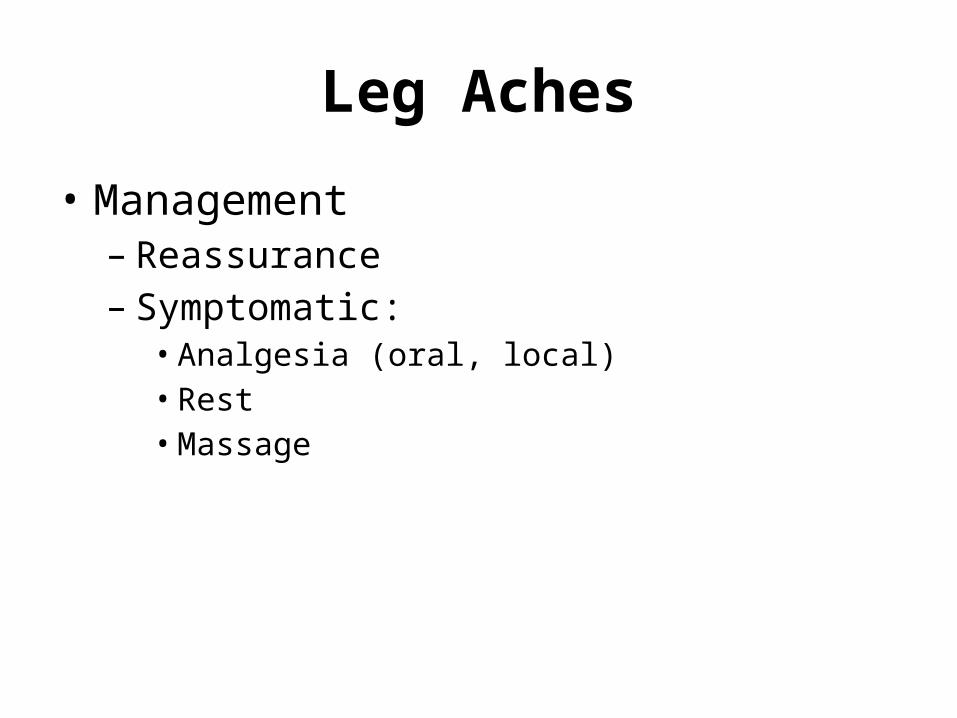
Leg Aches
• What is leg aches?– “Growing pain”– Benign– In 15 – 30 % of normal children– F > M– Unknown cause– No functional disability, or limping – Resolves spontaneously, over several years
Leg Aches
• Clinical features diagnosis by exclusion• H/O:
– At long bones of L.L (Bil)– Dull aching, poorly localized– Can be without activity– At night– Of long duration (months) – Responds to analgesia
• O/E:– Long bone tenderness nonspecific, large area, or none– Normal joints motion
(observational management)– Control child’s walking, sitting
or sleeping is extremely difficult and frustrating
– Shoe wedges or inserts are ineffective
– Bracing with twister cables limits child’s activities
– Night splints have no long term benefit
In-toeing and Out-toeing
• In-toeing:– Annual clinic F/U asses degree of deformity– Femoral anti-version sit cross legged – Tibial torsion spontaneous improvement– Forefoot adduction anti-version shoes, or proper
shoes reversal– Adducted big toe spontaneous improvement
In-toeing and Out-toeing
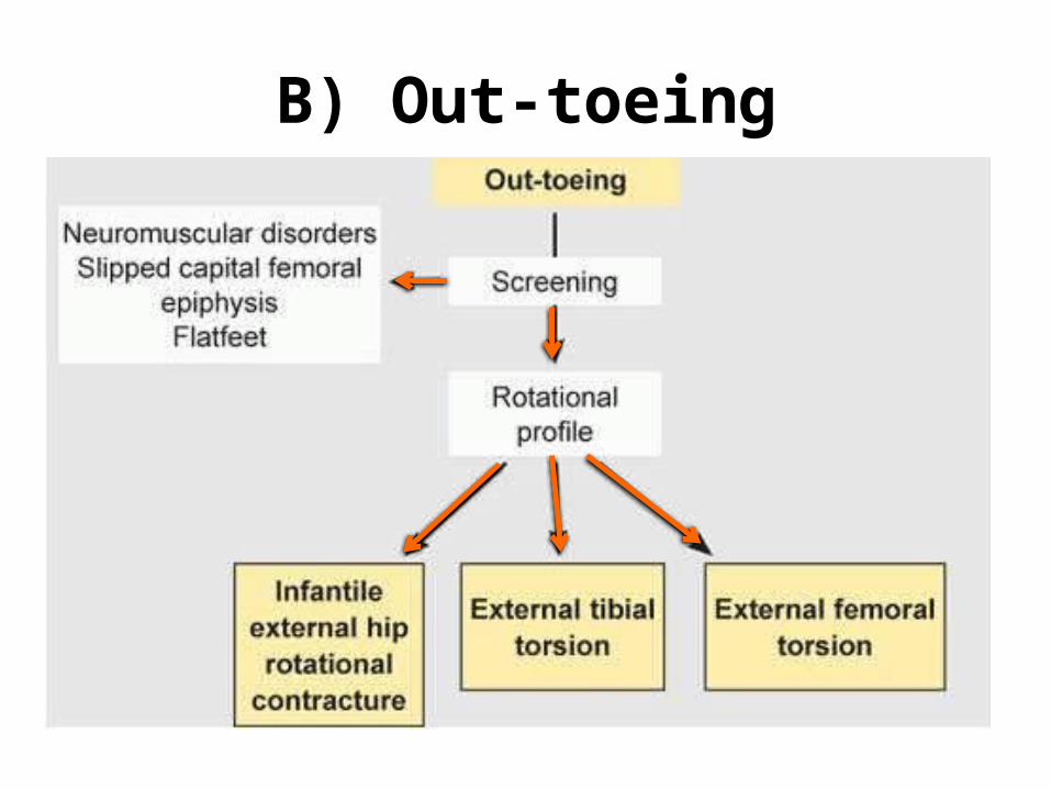
• Out-toeing:– Usually does not improve spontaneously– Will need an operation:• After the age 8y• Foot propagation angle >30°
In-toeing and Out-toeing
• Operative correction indicated for children:– (> 8) years of age– With significant cosmetic and functional deformity <1%
4) Limb Length Inequality
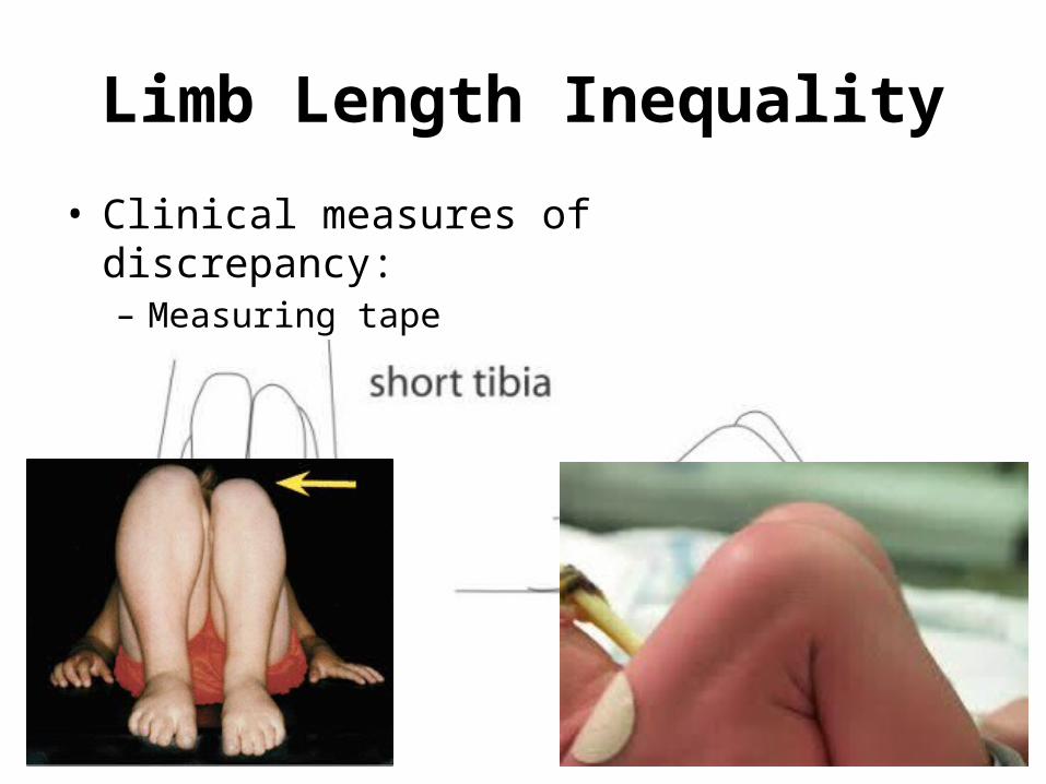
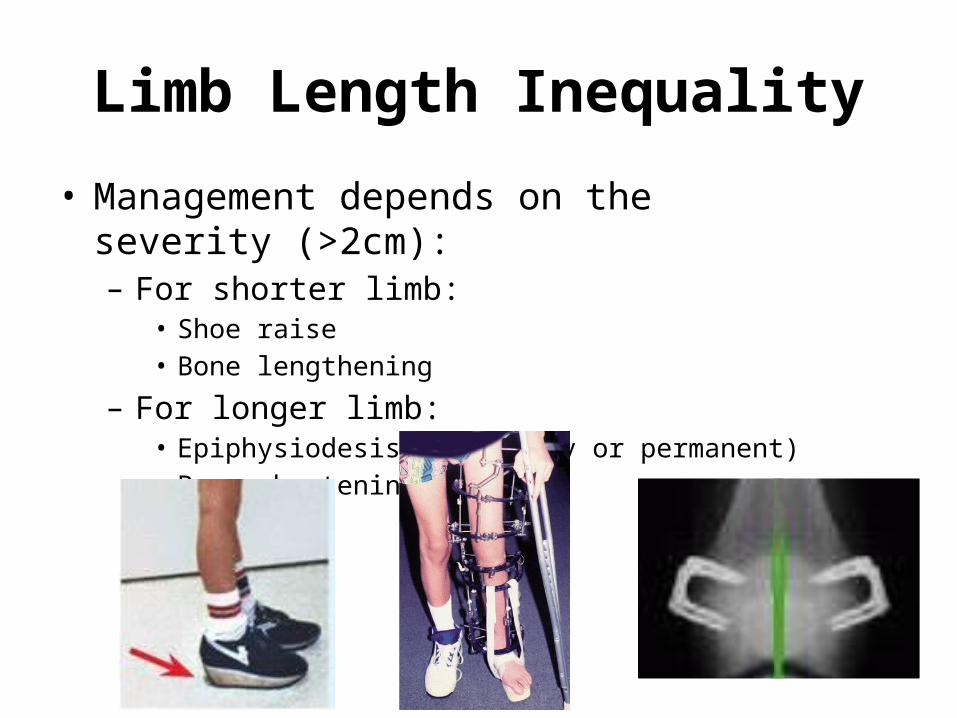
Limb Length Inequality
• True vs. apparent• Etiology:– Congenital as DDH– Developmental as Blount’s– Traumatic as oblique # (short), or multifragmented (long)– Infection stunted growth or dissolved part of bone– Metabolic as rickets (unilateral)– Tumor affecting physis
• Clinical examination:– Deformities don’t prevent walking– Calf muscles wasting– Internal torsion of the leg– Foot is smaller in unilateral affection– Callosities at abnormal pressure areas– Short Achilles tendon– Heel is high and small – No creases behind Heel– Abnormal crease in middle of the foot
Clubfoot
• Management:
The goal of treatment for is to obtain a foot that is plantigrade, functional, painless, and stable over time
A cosmetically pleasing appearanceis also an important goal sought by
surgeon and family
Clubfoot
• Manipulation and serial casts:– Validity up to 12 months soft tissue becomes more tight– Technique “Ponseti” 3 stages, weekly basis (usually by 6-8w)
Clubfoot
• Manipulation and serial casts:– Maintaining correction “Dennis Brown Splint” 3-4y old
Clubfoot
• Manipulation and serial casts:– Follow up watch and avoid recurrence, till 9y old– Avoid false correction by going in sequence – When to stop ? not improving, pressure ulcers
Clubfoot
• Indications of surgical treatment:– Late presentation (>12 months of age)– Complementary to conservative treatment
(residual forefoot adduction)– Failure of conservative treatment– Recurrence after conservative treatment
Clubfoot
• Types of surgery:– Soft tissue
Clubfoot
• Types of surgery:– Bony
Clubfoot
• Types of surgery:– If sever, rigid, and in an older child
Clubfoot
• Types of surgery:– If sever, rigid, and in an older child (salvage)
8) L.L Deformities inC.P Patients
Lower Limb Deformities in CP Child
• C.P is a non-progressive brain insult that occurred during the peri-natal period.
• Causes skeletal muscles imbalance that affects joint’s movements.
• Can be associated with:– Mental retardation (various degrees)– Hydrocephalus and V.P shunt– Convulsions
• Management is multidisciplinary:– Parents education– Pediatric neurology diagnosis, F/U, treat fits– P.T (home & center) joints R.O.M, gait training– Orthotics maintain correction, aid in gait– Social / Government aid– Others:• Neurosurgery (V.P shunt),• Ophthalmology (eyes sequent),• …etc.
Lower Limb Deformities in CP Child
• Indications of Orthopedic surgery:– Sever contractures preventing P.T– P.T plateaued due to contractures– Perennial hygiene (sever hips adduction)– In a non-walker to sit confortable in wheelchair – Prevent:• Neuropathic skin ulceration (as feet)• Joint dislocation (as hip)
Lower Limb Deformities in CP Child
• Options of Surgery: – Tenotomy– Tenoplasty– Muscle lengthening– Neurectomy – Tendon Transfer– Bony surgery Osteotomy/Fusion
Remember …
Conclusion1. Leg aches of exclusion, D.D, long bone, activity ±,
symptomatic treatment2. Limping due (pain- week- deformed), uni or bi, , proper
assessment
3. In & out toeing torsion vs. version, proper assessment to know cause & level, mainly observe, operate >8y old
4. L.L.I true vs. apparent, proper assessment to know cause & level, if not treated, >2cm, options of treat
5. Genu varus & valgus physiological vs. pathological, rickets, when operate
6. Blount medial physis, D.M.A, MRI, types, surgery
7. CTEV 3 types, diagnosis of exclusion, clinical picture, Ponseti, better to avoid surgery
8. L.L in C.P muscle imbalance, of different types, proper pt assessment, PT ± surgery