SOP Template V1 Q pulse 1/3/08 SOP 2014, Revision Number 1, Page 1 of 1 Printed versions of this document are uncontrolled unless signed by the Quality Department Community Reference Laboratory for Crustacean Diseases Standard Operating Procedure SOP 2014 Revision Number 1 Sampling of crustaceans for infectious diseases Cefas Weymouth Barrack Road The Nothe Weymouth Dorset DT4 8UB Copy No: Issued Location TM Signature QA Signature: Date:
Transcript
SOP Template V1 Q pulse 1/3/08
SOP 2014, Revision Number 1, Page 1 of 1
Printed versions of this document are uncontrolled unless signed by the Quality Department
Community Reference Laboratory
for Crustacean Diseases
Standard Operating Procedure SOP 2014
Revision Number 1
Sampling of crustaceans for infectious diseases
Cefas Weymouth Barrack Road The Nothe Weymouth Dorset DT4 8UB
Copy No:
Issued Location TM Signature QA Signature: Date:
SOP Template V1 Q pulse 1/3/08
SOP 2014, Revision Number 1, Page 2 of 2
Printed versions of this document are uncontrolled unless signed by the Quality Department
1 Introduction
The Community Reference Laboratory (CRL) routinely samples crustacean tissues; their health status examined and evaluated using wax histology methods, molecular biology and electron microscopy.
2 Scope
This SOP describes the procedure for sampling of crustaceans to survey for notifiable and infectious diseases via histology, electron microscopy and molecular biology. It covers the opening and dissecting crabs, lobsters and crayfish, dissecting and cassetting these tissues for histology and sampling of tissues for molecular biology and electron microscopy. It also covers injecting, fixing and cassetting of whole crabs, lobsters, crayfish and shrimp for testing for infectious diseases by histology.
3 Training (Identify any specific training linked to the SOP)
This procedure may only be carried out by staff who have received training in this SOP.
4 Safety Precautions
Many hazardous chemicals are used during the sampling and processing of samples, before performing this procedure staff should have read and understood all relevant safety documentation. The use of fume hoods, where available, is advised for making of solutions and fixation of samples. Where a fume hood is not available ensure chemicals are used in a well-ventilated area. Staff should wear personal protective equipment, such as lab coat, eye protection and gloves, at all times. Material Safety Data Sheets (MSDS) can be found on the Internet or via the suppliers. Absolute Ethanol Eye: Causes severe eye irritation Skin: Causes moderate skin irritation Ingestion: May cause gastrointestinal irritation with nausea, vomiting and diarrhoea Inhalation: Vapours may cause dizziness or suffocation Decalcifier II Eye: There may be irritation and redness Skin: There may be irritation and redness at the site of contact Ingestion: Nausea and stomach pain may occur. There may be vomiting.
Highly Flammable Flash point 16.6ºC
Corrosive
SOP Template V1 Q pulse 1/3/08
SOP 2014, Revision Number 1, Page 3 of 3
Printed versions of this document are uncontrolled unless signed by the Quality Department
Inhalation: Nausea and stomach pain may occur. There may be vomiting. Drowsiness or mental confusion may occur. There may be loss of conciousness. Formaldehyde – Ensure sufficient ventilation Eye: Causes irritation. May result in corneal injury Skin: Causes skin irritation. Harmful if absorbed through the skin Ingestion: Cause gastrointestinal irritation with nausea, vomiting and diarrhoea. Harmful if swallowed Inhalation: Harmful if inhaled. Causes respiratory tract irritation. Glacial Acetic Acid Eye: Causes severe irritation Skin: Causes skin burns. May be harmful if absorbed through the skin Ingestion: May cause severe and permanent damage to the digestive tract Inhalation: Effects may be delayed. Causes chemical burns to the respiratory tract. Glutaraldehyde – Ensure sufficient ventilation Eye: Causes severe eye irritation. Skin: Causes burns. May cause sensitisation by skin contact. Ingestion: Toxic if swallowed. Inhalation: Toxic by inhalation. May cause sensitisation by inhalation. IMS Eye: Causes severe eye irritation Skin: Causes moderate skin irritation. May be absorbed through the skin in harmful amounts Ingestion: May be fatal or cause blindness if ingested. May cause gastrointestinal irritation with nausea, vomiting and diarrhoea Inhalation: Inhalation of high concentrations may cause central nervous system effects characterised by nausea, headache, dizziness, unconsciousness and coma. Vapours may cause dizziness or suffocation. Sodium Cacodylate Buffer Eye: Causes severe eye irritation Skin: Causes skin irritation. May be absorbed through the skin in harmful amounts Ingestion: Toxic if swallowed. Inhalation: Toxic by inhalation. May cause sensitisation by inhalation.
Toxic
Highly Flammable
Flash point 18ºC Harmful
Dangerous for the environment
Toxic
Dangerous for the environment
Toxic
Irritant
Flammable Corrosive
SOP Template V1 Q pulse 1/3/08
SOP 2014, Revision Number 1, Page 4 of 4
Printed versions of this document are uncontrolled unless signed by the Quality Department
6 Equipment /Apparatus
Ice
Data recording sheet
Safety glasses
Dissection kit (to include heavy duty scissors for opening shells and sprung
scissors for dissection)
Labelled cassettes
Suitably sized fix pot, this will normally be 1 Litre size three quarters filled with
Davidson’s fixative, maximum of 30 cassettes per pot.
Labelled EM tubes filled with 2.5 % Glutaraldehyde in Sodium Cacodylate
buffer
Labelled Molecular tubes filled with Absolute Ethanol
Needles and syringes
7 Ingredients/Reagents/Media
Davidson’s Seawater Fixative
Stock Solution
Filtered sea water 3340ml
95% Ethanol 3330ml
36 - 40% Formaldehyde 2220ml
Glycerin 1110ml
Working Solution
Stock solution 720ml
Glacial acetic acid 80ml
Davidson’s Freshwater Fixative
To be used for fixation of freshwater crustacean samples, for example crayfish
Stock Solution
Distilled water 3350ml
95% Ethanol 3300ml
36 - 40% Formaldehyde 2200ml
SOP Template V1 Q pulse 1/3/08
SOP 2014, Revision Number 1, Page 5 of 5
Printed versions of this document are uncontrolled unless signed by the Quality Department
Working Solution
Stock solution 885ml
Glacial Acetic Acid 115ml
Neutral Buffered Formalin
Can be used for fixation of freshwater crustacean samples, for example crayfish
Distilled Water 9L
36-40% Formaldehyde 1L
Sodium dihydrogen orthophosphate dihydrate 40g
di-Sodium hydrogen orthophosphate anhydrous 65g
Glutaraldehyde fixative
25% Glutaraldehyde 10ml
0.1M Sodium Cacodylate Buffer (pH 7.4) 90ml
8 Procedure - Crabs
8.1.1 Check the Visit data sheet corresponds to the label on the sample container. If there is any discrepancy refer to the Inspectorate Technical Manager or Deputy prior to proceeding.
8.1.2 Crabs are anaesthetised on ice for at least 30 minutes before dissection.
8.1.3 Check how many crabs are present within sample and label cassettes with Laboratory Reference Number and animal number followed by A, B and C. Label Glutaraldehyde tubes and Ethanol tubes with Laboratory Reference Number, animal number and tissue type.
8.1.4 Label Davidson’s Fixative pot and lid with a paper label with the Laboratory Reference Number written in pencil.
8.1.5 Examine crabs for any deformities or abnormalities, shell fouling, shell disease, missing appendages and claws. Log onto data recording sheet (see appendix 1).
SOP Template V1 Q pulse 1/3/08
SOP 2014, Revision Number 1, Page 6 of 6
Printed versions of this document are uncontrolled unless signed by the Quality Department
8.1.6 Carapace width (CW) is measured across the widest part of the carapace, as shown below. Record carapace width onto data recording sheet.
8.1.7 Sex of the sample is determined by examining the abdomen, female crabs have a wide oval shaped abdomen, male crabs have thin narrow V shaped abdomen. Record sex of sample onto data recording sheet.
8.1.8 Remove legs and claws, legs can be discarded but claws should be kept.
8.1.9 Wearing safety glasses open shell by cutting around the edge of the carapace on the underside taking care not to damage internal organs. Remove top of shell to reveal organs underneath. Note any abnormalities onto data recording sheet.
8.1.10 Remove heart, section and place in cassette A.
8.1.11 Remove two small sections (1mm3) of heart and place one section in labelled glutaraldehyde tube and second section in labelled Ethanol tube.
8.1.12 Remove section of hepatopancreas and place in cassette A.
8.1.13 Remove two small sections (1mm3) of hepatopancreas and place one section in labelled glutaraldehyde tube and second section in labelled Ethanol tube.
8.1.14 Remove section of gonad and place in cassette A.
8.1.15 Remove section of body muscle and place in cassette A.
8.1.16 Remove section of gill and place in cassette B.
SOP Template V1 Q pulse 1/3/08
SOP 2014, Revision Number 1, Page 7 of 7
Printed versions of this document are uncontrolled unless signed by the Quality Department
8.1.17 Remove two small sections (1mm3) of gill and place one section in labelled glutaraldehyde tube and second section in labelled Ethanol tube.
8.1.18 Open claw, remove section of claw muscle and place in cassette B.
8.1.19 Remove section of epidermis and place in cassette B. Epidermis can be found under the surface of the carapace, easily identifiable in pre-moult samples.
8.1.20 Remove two small sections (1mm3) of epidermis; place one section in labelled glutaraldehyde tube and second section in labelled Ethanol tube.
8.1.21 If epidermis is not available (post-moult sample) remove both mouthpieces from underneath the crab, place one mouthpiece in cassette C and place lid on cassette C and place cassette into separate labelled pot of Davidson’s fixative. Place second mouthpiece into labelled Ethanol tube.
8.1.22 Place lids on cassettes A and B and place in Davidson’s fixative.
8.1.23 Record the time that the last cassette was placed into fixative on the histology work sheet (see appendix 2).
8.1.24 Allow fixation to proceed for at least 24 hours after the last cassette was placed in fixative before transferring cassettes A and B into 70% IMS. If taken, transfer C cassettes into Decalcifier II for 30-40 minutes (times may need to be increased depending on sample).
8.1.25 Place C cassettes into 70% IMS.
8.1.26 Crabs, which are too small to dissect, can be fixed whole. Open carapace to allow fixative to penetrate into sample and drop sample into Davidson’s fixative.
8.1.27 Record the time that the last crab was placed into fixative on the histology work sheet (see appendix 2).
8.1.28 Discard all waste tissue into an incineration bag. All sharps must be disposed of in sharps bin.
8.1.29 Allow fixation to proceed for at least 24 hours after the last crab was placed in fixative before transferring crabs into Decalcifier II for 30-40 minutes (times may need to be increased depending on sample).
8.1.30 Check how many crabs are present within sample and label cassettes.
8.1.31 Place crabs into cassettes and place lids on cassettes.
8.1.32 Place cassettes into 70% IMS.
SOP Template V1 Q pulse 1/3/08
SOP 2014, Revision Number 1, Page 8 of 8
Printed versions of this document are uncontrolled unless signed by the Quality Department
8.2 Procedure – Lobsters and Crayfish
8.2.1 Check the Visit data sheet corresponds to the label on the sample container. If there is any discrepancy refer to the Inspectorate Technical Manager or Deputy prior to proceeding.
8.2.2 Lobsters are anaesthetised on ice for at least 30 minutes before dissection.
8.3.1 Check how many lobsters or crayfish are present within sample and label cassettes with Laboratory Reference Number and animal number followed by A, B and C. Label Glutaraldehyde tubes and Ethanol tubes with Laboratory Reference Number, animal number and tissue.
8.2.3 Label Davidson’s Fixative pot and lid with a paper label with the Laboratory Reference Number written in pencil.
8.1.33 Lobsters and crayfish are examined for any deformities or abnormalities, shell fouling, shell disease, missing appendages and claws. Log onto data recording sheet (see appendix 1).
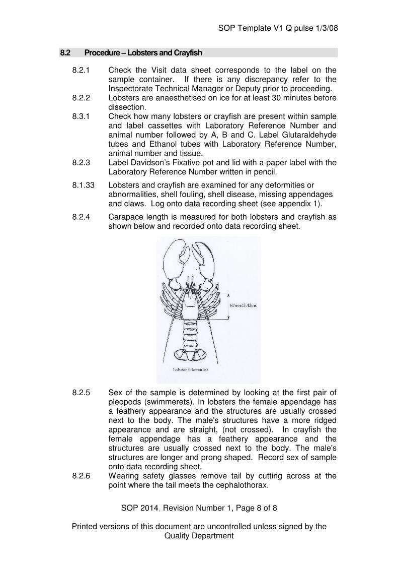
8.2.4 Carapace length is measured for both lobsters and crayfish as shown below and recorded onto data recording sheet.
8.2.5 Sex of the sample is determined by looking at the first pair of pleopods (swimmerets). In lobsters the female appendage has a feathery appearance and the structures are usually crossed next to the body. The male's structures have a more ridged appearance and are straight, (not crossed). In crayfish the female appendage has a feathery appearance and the structures are usually crossed next to the body. The male's structures are longer and prong shaped. Record sex of sample onto data recording sheet.
8.2.6 Wearing safety glasses remove tail by cutting across at the point where the tail meets the cephalothorax.
SOP Template V1 Q pulse 1/3/08
SOP 2014, Revision Number 1, Page 9 of 9
Printed versions of this document are uncontrolled unless signed by the Quality Department
8.2.7 Wearing safety glasses open shell by cutting along the side of the carapace and down the centre of the carapace taking care not to damage internal organs. Remove shell to reveal organs underneath. Note any abnormalities onto data recording sheet.
8.2.8 Remove heart, section and place in cassette A. 8.2.9 Remove two small sections (1mm3) of heart and place one
section in labelled glutaraldehyde tube and second section in labelled Ethanol tube.
8.2.10 Remove section of hepatopancreas and place in cassette A. 8.2.11 Remove two small sections (1mm3) of hepatopancreas and
place one section in labelled glutaraldehyde tube and second section in labelled Ethanol tube.
8.2.12 Remove section of gonad and place in cassette A. 8.2.13 Remove section of body muscle and place in cassette A. 8.2.14 Remove section of gill and place in cassette B. 8.2.15 Remove two small sections (1mm3) of gill and place one section
in labelled glutaraldehyde tube and second section in labelled Ethanol tube.
8.2.16 Open claw, remove section of claw muscle and place in cassette B.
8.2.17 Cut down the centre of the underside of the tail and open to reveal central nerve cord. Section central nerve cord and place in cassette B.
8.2.18 Remove section of epidermis and place in cassette B. Epidermis can be found under the surface of the carapace, easily identifiable in pre-moult samples.
8.2.19 Remove two small sections (1mm3) of epidermis; place one section in labelled glutaraldehyde tube and second section in labelled Ethanol tube.
8.2.20 If epidermis is not available (post-moult sample) remove two pleopods from underneath the lobster, place one pleopod in cassette C, place lid on cassette C and place cassette into separate labelled pot of Davidson’s fixative. Place second pleopod into labelled Ethanol tube.
8.2.21 Place lids on cassettes A and B and place in Davidson’s fixative.
8.2.22 Record the time that the last cassette was placed into fixative on the histology work sheet (see appendix 2).
8.2.23 Discard all waste tissue into an incineration bag. All sharps must be disposed of in sharps bin.
8.2.24 Allow fixation to proceed for at least 24 hours after the last cassette was placed in fixative before transferring cassettes A and B into 70% IMS. If taken, transfer C cassettes into Decalcifier II for 30-40 minutes (times may need to be increased depending on sample).
8.2.25 Place C cassettes into 70% IMS 8.2.26 Lobsters and crayfish, which are too small to dissect, can be
fixed whole. Open carapace to allow fixative to penetrate into sample and drop sample into Davidson’s fixative.
SOP Template V1 Q pulse 1/3/08
SOP 2014, Revision Number 1, Page 10 of 10
Printed versions of this document are uncontrolled unless signed by the Quality Department
8.2.27 Record the time that the last lobster or crayfish was placed into fixative on the histology work sheet (see appendix 2).
8.2.28 Allow fixation to proceed for at least 24 hours after the last lobster or crayfish was placed in fixative before transferring crabs into Decalcifier II for 30-40 minutes (times may vary depending on sample).
8.2.29 Check how many lobsters or crayfish are present within sample and label cassettes with Lab Reference number and animal number.
8.2.30 Place lobsters or crayfish into cassettes and place lids on cassettes.
8.2.31 Place cassettes into 70% IMS.
8.3 Procedure - Shrimp
8.3.2 Check the Visit data sheet corresponds to the label on the sample container. If there is any discrepancy refer to the Inspectorate Technical Manager or Deputy prior to proceeding.
8.3.3 Label Davidson’s Fixative pot and lid with a paper label with the Laboratory Reference Number written in pencil.
8.3.4 Shrimp are examined for any deformities or abnormalities, shell markings, colouration. Log onto data recording sheet (see appendix 1).
8.3.5 Carapace length is measured as for lobsters and crayfish. Record length onto data recording sheet.
8.3.6 Wearing safety glasses and taking extreme care inject shrimp with Davidson’s fixative, ensure that the area around the cephalothorax is injected several times with small amounts of fixative to ensure good fixation of the hepatopancreas region.
8.3.7 Place in Davidson’s fixative. 8.3.8 Record the time that the last shrimp was placed into fixative on
the histology work sheet (see appendix 2). 8.3.9 Discard all sharps in sharps bin. 8.3.10 Allow fixation to proceed for at least 24 hours after the last shrimp
was placed in the fixative before sectioning samples. 8.3.11 Check how many shrimp are present within sample and label
cassettes with Laboratory Reference Number and animal number.
8.3.12 In a fume hood, wearing gloves and safety glasses remove shrimp from fixative and place on chopping board.
8.3.13 Using razor blade carefully remove antennae and pereiopods, leaving the base of appendages attached to enable any protozoan infections to be identified in section, see below red lines indicate cut.
8.3.14 Remove eyes, rostrum and mandible, see below blue line indicates cut.
SOP Template V1 Q pulse 1/3/08
SOP 2014, Revision Number 1, Page 11 of 11
Printed versions of this document are uncontrolled unless signed by the Quality Department
8.3.15 Remove tail from shrimp and cut shrimp along the mid-sagittal line, see below red lines indicate cut. Place half of the cephalothorax into the cassette.
8.3.16 Remove pleopods from tail and section 1st, 3rd and 6th abdominal section, see below red lines indicate cut. Longitudinally cut 6th segment in half as shown above to include midgut posterior caecum and hindgut in the block. Place one half of the 6th segment and the 1st and 3rd segment into the cassette.
SOP Template V1 Q pulse 1/3/08
SOP 2014, Revision Number 1, Page 12 of 12
Printed versions of this document are uncontrolled unless signed by the Quality Department
8.3.17 Take the second half of the cephalothorax and section across the middle as shown below, red lines indicate cut, to include the lymphoid organ in the block. Place section into cassette. Remove the carapace from the remaining cephalothorax and section gills, place section in the cassette.
8.3.18 Place lid on cassette and place into Decalcifier II solution for 30-40 minutes (times may vary depending upon samples).
8.3.19 Place cassettes into 70% IMS. 8.3.20 Discard all waste tissue into an incineration bag.
9 Review
This procedure will be reviewed as a minimum on the time scales given in the review / amendment programme. A record of the review will be made on a separate Review / Amendment Sheet which will be added to the Master Copy file of this SOP. Any amendments arising from such review or from operating requirements will result in the issue of the entire amended procedure as a new Issue.
10 Records
This procedure, its review sheets and its subsequent revisions constitute records in themselves and each master copy will be retained in a file as arranged by the Quality Manager. Records will be retained for a minimum of five years unless otherwise specified.
(List specific record sheets/books)
SOP Template V1 Q pulse 1/3/08
SOP 2014, Revision Number 1, Page 13 of 13
Printed versions of this document are uncontrolled unless signed by the Quality Department
Appendix 1. An example of the data-recording sheet for dissection.
Laboratory Reference Number
Date SampleNumber
Site Species CW (mm)
Sex Shell Disease
Fouling Claws Missing
Legs Missing
Notes
1
2
3
4
5
6
7
8
9
10
11
12
13
14
15
16
17
18
19
20
SOP Template V1 Q pulse 1/3/08
SOP 2014, Revision Number 1, Page 14 of 14
Printed versions of this document are uncontrolled unless signed by the Quality Department
Appendix 2. An example of the process sheet for histology
Group(s) 1 2 3
Notes
Cassettes printed
Sample cassetted (date, time of last cassette, initials)