Université de Bourgogne
UFR des Sciences de Santé
Circonscription Médecine
ANNEE 2017
N°
Comparaison de différentes modalités de recueil en tomographie
par cohérence optique Spectral-Domain dans la détection de
pathologies maculaires et de l’interface vitréorétinienne : l’étude
MONTRACHET
THESE
présentée
à l’UFR des Sciences de Santé de Dijon
Circonscription Médecine
et soutenue publiquement le 4 octobre 2017
pour obtenir le grade de Docteur en Médecine
par Ghislaine ASSAD
Née le 20 janvier 1986
A Paris 14ème
Université de Bourgogne
UFR des Sciences de Santé
Circonscription Médecine
AVERTISSEMENT
Ce document est le fruit d’un long travail approuvé par le jury de soutenance et mis à la
disposition de la communauté universitaire élargie.
Il est soumis à la propriété intellectuelle de l’auteur.
Ceci implique une obligation de citation et de référencement dans la rédaction de vos travaux.
D’autre part, toutes contrefaçons, plagiats, reproductions illicites encourt une poursuite
pénale.
Université de Bourgogne
UFR des Sciences de Santé
Circonscription Médecine
ANNEE 2017
N°
Comparaison de différentes modalités de recueil en tomographie
par cohérence optique Spectral-Domain dans la détection de
pathologies maculaires et de l’interface vitréorétinienne : l’étude
MONTRACHET
THESE
présentée
à l’UFR des Sciences de Santé de Dijon
Circonscription Médecine
et soutenue publiquement le 4 octobre 2017
pour obtenir le grade de Docteur en Médecine
Par Ghislaine ASSAD
Née le 20 janvier 1986
A Paris 14èm
Université de Bourgogne
UFR des Sciences de Santé
Circonscription Médecine
Année Universitaire 2017-2018
au 1er Septembre 2017
Doyen : M. Frédéric HUET
Assesseurs : M. Marc MAYNADIE M. Pablo ORTEGA-DEBALLON
PROFESSEURS DES UNIVERSITES – PRATICIENS HOSPITALIERS
Discipline
M. Marc BARDOU Pharmacologie clinique M. Jean-Noël BASTIE Hématologie - transfusion M. Emmanuel BAULOT Chirurgie orthopédique et traumatologie M. Yannick BEJOT Neurologie M. Alain BERNARD Chirurgie thoracique et cardiovasculaire Mme Christine BINQUET Epidémiologie, économie de la santé et prévention M. Bernard BONIN Psychiatrie d’adultes M. Philippe BONNIAUD Pneumologie M. Alain BONNIN Parasitologie et mycologie M. Bernard BONNOTTE Immunologie M. Olivier BOUCHOT Chirurgie cardiovasculaire et thoracique M. Belaid BOUHEMAD Anesthésiologie - réanimation chirurgicale M. Alexis BOZORG-GRAYELI ORL M. Alain BRON Ophtalmologie M. Laurent BRONDEL Physiologie Mme Mary CALLANAN Hématologie type biologique M. Patrick CALLIER Génétique M. Jean-Marie CASILLAS-GIL Médecine physique et réadaptation Mme Catherine CHAMARD-NEUWIRTH Bactériologie - virologie; hygiène hospitalière M. Pierre-Emmanuel CHARLES Réanimation M. Pascal CHAVANET Maladies infectieuses M. Nicolas CHEYNEL Anatomie M. Alexandre COCHET Biophysique et médecine nucléaire M. Luc CORMIER Urologie M. Yves COTTIN Cardiologie M. Charles COUTANT Gynécologie-obstétrique M. Gilles CREHANGE Oncologie-radiothérapie Mme Catherine CREUZOT-GARCHER Ophtalmologie M. Frédéric DALLE Parasitologie et mycologie M. Serge DOUVIER Gynécologie-obstétrique Mme Laurence DUVILLARD Biochimie et biologie moléculaire M. Olivier FACY Chirurgie générale Mme Laurence FAIVRE-OLIVIER Génétique médicale Mme Patricia FAUQUE Biologie et Médecine du Développement Mme Irène FRANCOIS-PURSSELL Médecine légale et droit de la santé M. Pierre FUMOLEAU Cancérologie M. François GHIRINGHELLI Cancérologie M. Claude GIRARD Anesthésiologie – réanimation chirurgicale M. Vincent GREMEAUX Médecine physique et réadaptation
(Mise en disponibilité du 12 juin 2017 au 11 juin 2018) M. Frédéric HUET Pédiatrie M. Pierre JOUANNY Gériatrie
Université de Bourgogne
UFR des Sciences de Santé
Circonscription Médecine
M. Sylvain LADOIRE Histologie M. Gabriel LAURENT Cardiologie M. Côme LEPAGE Hépato-gastroentérologie M. Romaric LOFFROY Radiologie et imagerie médicale M. Luc LORGIS Cardiologie M. Jean-Francis MAILLEFERT Rhumatologie M. Cyriaque Patrick MANCKOUNDIA Gériatrie M. Sylvain MANFREDI Hépato-gastroentérologie M. Laurent MARTIN Anatomie et cytologie pathologiques M. David MASSON Biochimie et biologie moléculaire M. Marc MAYNADIE Hématologie – transfusion M. Marco MIDULLA Radiologie et imagerie médicale M. Thibault MOREAU Neurologie M. Klaus Luc MOURIER Neurochirurgie Mme Christiane MOUSSON Néphrologie M. Paul ORNETTI Rhumatologie M. Pablo ORTEGA-DEBALLON Chirurgie Générale M. Jean-Michel PETIT Endocrinologie, diabète et maladies métaboliques M. Christophe PHILIPPE Génétique M. Lionel PIROTH Maladies infectieuses Mme Catherine QUANTIN Biostatistiques, informatique médicale M. Jean-Pierre QUENOT Réanimation M. Patrick RAT Chirurgie générale M. Jean-Michel REBIBOU Néphrologie M. Frédéric RICOLFI Radiologie et imagerie médicale M. Paul SAGOT Gynécologie-obstétrique M. Emmanuel SAPIN Chirurgie Infantile M. Henri-Jacques SMOLIK Médecine et santé au travail M. Éric STEINMETZ Chirurgie vasculaire Mme Christel THAUVIN Génétique M. Benoit TROJAK Psychiatrie d’adultes ; addictologie M. Pierre VABRES Dermato-vénéréologie M. Bruno VERGÈS Endocrinologie, diabète et maladies métaboliques M. Narcisse ZWETYENGA Chirurgie maxillo-faciale et stomatologie
PROFESSEURS EN SURNOMBRE
M. Roger BRENOT Chirurgie thoracique et cardiovasculaire
(Surnombre jusqu’au 31/08/2018) M. Philippe CAMUS Pneumologie
(Surnombre jusqu’au 31/08/2019) Mme Monique DUMAS-MARION Pharmacologie fondamentale
(Surnombre jusqu’au 31/08/2018) M. Maurice GIROUD Neurologie
(Surnombre jusqu’au 21/08/2018)
Université de Bourgogne
UFR des Sciences de Santé
Circonscription Médecine
MAITRES DE CONFERENCES DES UNIVERSITES
PRATICIENS HOSPITALIERS DES DISCIPLINES MEDICALES
Discipline Universitaire
Mme Lucie AMOUREUX BOYER Bactériologie M. Sylvain AUDIA Médecine interne Mme Shaliha BECHOUA Biologie et médecine du développement M. Benjamin BOUILLET Endocrinologie Mme Marie-Claude BRINDISI Nutrition M. Jean-Christophe CHAUVET-GELINIER Psychiatrie, psychologie médicale Mme Marie-Lorraine CHRETIEN Hématologie Mme Vanessa COTTET Nutrition M. Alexis DE ROUGEMONT Bactériologie-virologie ; hygiène hospitalière M. Hervé DEVILLIERS Médecine interne Mme Ségolène GAMBERT-NICOT Biochimie et biologie moléculaire Mme Marjolaine GEORGES Pneumologie Mme Françoise GOIRAND Pharmacologie fondamentale M. Charles GUENANCIA Cardiologie Mme Agnès JACQUIN Physiologie M. Alain LALANDE Biophysique et médecine nucléaire M. Louis LEGRAND Biostatistiques, informatique médicale Mme Stéphanie LEMAIRE-EWING Biochimie et biologie moléculaire M Maxime SAMSON Médecine interne M. Paul-Mickaël WALKER Biophysique et médecine nucléaire
PROFESSEURS EMERITES M. Laurent BEDENNE (01/09/2017 au 31/08/2020) M. Jean-François BESANCENOT (01/09/2017 au 31/08/2020) M. François BRUNOTTE (01/09/2017 au 31/08/2020) M. Jean FAIVRE (01/09/2015 au 31/08/2018) M. Patrick HILLON (01/09/2016 au 31/08/2019) M. François MARTIN (01/09/2015 au 31/08/2018) M. Pierre POTHIER (01/09/2015 au 31/08/2018) M. Pierre TROUILLOUD (01/09/2017 au 31/08/2020)
PROFESSEURS DES UNIVERSITES DE MEDECINE GENERALE M. Jean-Noël BEIS Médecine Générale
PROFESSEURS ASSOCIES DE MEDECINE GENERALE M. Didier CANNET Médecine Générale M. Gilles MOREL Médecine Générale M. François MORLON Médecine Générale
MAITRES DE CONFERENCES ASSOCIES DE MEDECINE GENERALE Mme Anne COMBERNOUX -WALDNER Médecine Générale M. Clément CHARRA Médecine Générale M. Rémi DURAND Médecine Générale M. Arnaud GOUGET Médecine Générale
Université de Bourgogne
UFR des Sciences de Santé
Circonscription Médecine
MAITRES DE CONFERENCES DES UNIVERSITES M. Didier CARNET Anglais M. Jean-Pierre CHARPY Anglais Mme Catherine LEJEUNE Pôle Epidémiologie M. Gaëtan JEGO Biologie Cellulaire
PROFESSEURS DES UNIVERSITES Mme Marianne ZELLER Physiologie
PROFESSEURS AGREGES de L’ENSEIGNEMENT SECONDAIRE Mme Marceline EVRARD Anglais Mme Lucie MAILLARD Anglais
PROFESSEURS CERTIFIES Mme Anaïs CARNET Anglais M. Philippe DE LA GRANGE Anglais Mme Virginie ROUXEL Anglais (Pharmacie)
PROFESSEURS DES UNIVERSITES – PRATICIENS HOSPITALIERS DES DISCIPLINES
PHARMACEUTIQUES M. François GIRODON Sciences biologiques, fondamentales et cliniques
Mme Evelyne KOHLI Immunologie
MAITRES DE CONFERENCES DES UNIVERSITES
PRATICIENS HOSPITALIERS DES DISCIPLINES PHARMACEUTIQUES M. Mathieu BOULIN Pharmacie clinique M. Philippe FAGNONI Pharmacie clinique M. Frédéric LIRUSSI Toxicologie M. Marc SAUTOUR Botanique et cryptogamie M. Antonin SCHMITT Pharmacologie
Université de Bourgogne
UFR des Sciences de Santé
Circonscription Médecine
L’UFR des Sciences de Santé de Dijon, Circonscription Médecine, déclare que les opinions
émises dans les thèses qui lui sont présentées doivent être considérées comme propres à
leurs auteurs, et qu'elle n'entend ne leur donner ni approbation, ni improbation.
COMPOSITION DU JURY
Président : Madame le Professeur Catherine CREUZOT-GARCHER
Membres : Monsieur le Professeur Alain-Marie BRON
Monsieur le Docteur Hervé DEVILLIERS
Madame le Docteur Clémence DESCHASSE
A notre Président et Directeur de thèse,
Madame le Professeur Catherine CREUZOT-GARCHER,
Nous vous remercions de l’honneur que vous nous faites en acceptant de présider
notre jury.
Votre détermination, vos connaissances et votre dextérité chirurgicale nous ont
toujours inspiré une grande admiration.
Nous saluons votre persévérance, votre optimisme et votre patience dans la
recherche et la formation des internes, qui sont pour nous un magnifique exemple
à suivre.
Nous sommes très fières d’avoir été votre élève et nous vous remercions pour
votre aide et votre soutien tout au long de ces années.
Soyez assurée de notre sincère reconnaissance et notre plus profond respect.
A notre juge,
Monsieur le Professeur Alain BRON,
Nous vous remercions de l’honneur que vous nous faites en acceptant de juger
notre travail.
Nous admirons votre détermination et votre travail de longue haleine qui a permis
au service de Dijon de devenir une école prestigieuse de l’ophtalmologie.
Votre savoir immense et votre sens de la rigueur nous ont servi de modèle dans
l’apprentissage de l’ophtalmologie. Cela aura été un grand honneur d’avoir pu
apprendre à vos côtés.
Veuillez recevoir ici l’expression de notre gratitude et de notre plus profond
respect.
A notre juge,
Monsieur le Docteur Hervé DEVILLIERS,
Soyez vivement remercié de l’honneur que vous nous avez fait en acceptant de
juger notre travail.
Vos connaissances dans tous les domaines de la médecine, votre capacité de
travail et votre dynamisme nous épatent et font de vous un futur grand Professeur.
Nous vous remercions pour votre enseignement pendant ces quelques mois dans
le service de médecine interne et pour votre précieuse collaboration avec le
service d’ophtalmologie.
Veuillez recevoir l’expression de notre reconnaissance et de notre plus profond
respect.
A notre juge,
Madame le Docteur Clémence DESCHASSE,
Soyez vivement remerciée de l’honneur que vous nous avez fait en acceptant de
juger notre travail.
Nous saluons vos connaissances débordantes dans tous les domaines de
l’ophtalmologie. Neuro-ophtalmolgie, uvéite, rétine, paupière, glaucome, plus rien
ne vous arrête.
Nous vous remercions infiniment pour vos enseignements et votre patience au
bloc opératoire. Vous avez su nous transmettre la passion de la chirurgie de la
paupière. Nous avons beaucoup appris chirurgicalement, toujours avec un fond
musical de Disney ou Dalida. Votre bonne humeur, votre humour, vos playlists et
chorégraphies improbables ont fait de la « team-paupiette » un duo magique gravé
dans l’histoire du bloc.
Vous êtes devenue une amie et confidente précieuse. Nous sommes
reconnaissantes de vos conseils et votre soutien en toute circonstance. Nous
sommes ravies de partager encore des moments formidables à vos côtés.
Soyez assurée de notre reconnaissance et de notre profonde amitié.
Un grand MERCI à …
A mes anciens chefs et co-interne
Denis (#DD) : Mon premier mentor de la paupière, toujours speed et dynamique, tu m’impressionnes à
chaque bloc opératoire. En un tour de main et un peu de coagulation, tu viens à bout de toutes les
chirurgies de paupière. Je te remercie pour tout ce que tu m’as appris et je te souhaite plein de réussite
dans votre tout nouveau cabinet.
Yann et Philippe (#PK) : Merci pour vos enseignements et votre partage de l’amour de la rétine qui
m’ont beaucoup apporté au début de mon internat. Je vous souhaite que du bonheur et de la réussite
pour la suite, vous êtes la jeunesse florissante de l’ophtalmologie de ville.
Olivier (#Oliv’) : Toujours le sourire aux lèvres et de bonne humeur, tu as toujours plein de bons plans
et de belles idées de voyage. Je te remercie pour ton aide dans mes débuts de chirurgie réfractive.
J’espère aussi que tu pourras m’accompagner pour cette 2ème année du DU de paupière et continuer
avec la secte de la paupière cosmopolite.
Arthur (#Arthuro) : Quand je t’ai rencontré la première fois à Nevers, tu étais déjà très impliqué et
soigneux dans le travail et tu as continué à être un formidable cornéologue dans le service. Ton sourire,
ta bonne humeur et ton accent toulousain nous manquent. Je te souhaite une belle réussite dans ta
nouvelle vie parisienne.
Aurore, Rodica, Arnaud, Till, JC : Je n’ai malheureusement pas eu la chance de travailler avec vous,
mais c’est toujours un plaisir de vous revoir notamment à la SFO.
A mes chefs actuels
Cyril (#CM) : Que dire à part un immense merci. On ne souligne pas assez l’implication que tu as dans
le service, le bloc et le soutien envers tous les internes. Merci pour m’avoir toujours soutenue depuis
mon premier semestre au CHU où je me noyais dans les urgences du lundi matin jusqu’à la relecture
de ma thèse aujourd’hui. Merci pour ta patience et tes conseils au bloc au prix de nombreux spasmes
coronariens. Merci de m’avoir redonné confiance en moi et de m’avoir permis de réaliser ma première
cataracte. Merci pour tous ces fous rires et bons moments. Tu es une personne entière, sincère, pleine
de vie, forte et fragile à la fois, je suis heureuse de t’avoir rencontré.
Firas (#DucDeSemur #MercrediEtVendrediC’estSacré)
Charlotte : Je te remercie pour ta détermination et ton sérieux dans l’organisation du service et la
formation des externes. Tu es notre pilier de l’ophtalmologie pédiatrique et je suis fière d’apprendre à
tes côtés pendant encore quelques temps même si tu devras nous abandonner pour un magnifique
évènement. Je te souhaite une vie pleine de bonheur avec Julien et votre futur petit bout de chou
(j’espère que tu le mettras très tôt à la course à pied).
Jacques (#Jacky) : Je t’ai toujours connu calme, soigné et travailleur, mais aussi drôle et sympathique,
avec une petite touche d’humour bien placée et un sens de la répartie inégalée. C’est un réel plaisir de
travailler avec toi, ne change rien. Je te souhaite pleins de bonheur et de réussite avec Perrine.
A mes co-internes : les « vieux » et « moyens vieux »
Aurélie : Très sérieuse, impliquée et rigoureuse, tu as contribué à développer la contactologie dans le
service et je t’en félicite. Je te souhaite une belle continuation à Chalon et un bel épanouissement avec
Maxime dans ta future nouvelle grande maison.
Louis (#Luigi) : J’apprécie beaucoup ton calme et ta sérénité en toute circonstance, pour preuve, tu as
réussi à convaincre la troupe de remanger à l’internat sans effusion de sang, chapeau ! Je te félicite
surtout pour avoir eu le courage de rallonger cette aventure d’une année, mais je suis sure que le jeu
en valait la chandelle. Je te souhaite une belle réussite pour la suite.
Pierre-Henry (#PH #Pitch) : Grand et élancé, cheveux grisonnants bien peignés, une curiosité
débordante, une humilité indéniable et une touche d’humour pour couronner le tout…il n’y a aucun
doute, tu as toutes les qualités du futur Professeur Mamour de l’ophtalmologie dijonnaise. Tu es devenu
un véritable ami, je te souhaite tout le bonheur et une vie heureuse avec Clem et bientôt mini-PH.
Youssef (#Youyou) : Sérieux, travailleur, toujours de bonne humeur et parfois même taquin sur les
bords, tu es toujours là pour soutenir et aider les jeunes internes. Tu es devenu un grand ami et mon
acolyte des révisions EBO. On ne compte plus les week-end et soirées passées ensemble dans le
service et les appels mutuels de soutien et de motivation. Je suis heureuse de continuer cette route
avec toi.
Edouard (#Edou) : Tu fais partie d’une de mes meilleures rencontres de cette aventure. Ta
détermination et ta rigueur dans le travail, ton sens de la perfection et tes accès maniaques, ta
générosité, ta bonne humeur et tes coups de gueule font de toi une personne entière et un ami précieux.
Je te souhaite le meilleur mon Edou.
Sophia : J’ai rarement vu une fille aussi déterminée, volontaire et travailleuse. Deuxième jeune mariée
de la troupe, tu aimes ta vie planifiée et ordonnée. Mais derrière cette femme de caractère, se cache
une jeune fille aimante, sensible, pleine de bonté et de générosité. Je suis heureuse de t’avoir
rencontrée et je te souhaite une vie paisible avec Jocelyn.
Julie (#Juju #JB) : Ma Juju, on a passé des moments merveilleux à la fois joyeux, festifs et tristes en
sillonnant la Bourgogne ensemble de Macon à Nevers. Tu pourrais être juste grande blonde et jolie,
mais tu es aussi intelligente, travailleuse, impliquée dans ton travail (notamment les plannings) et tu
sais profiter de la vie autour d’une bière. Tu es devenue une amie et confidente qui m’est chère et sur
qui on peut compter. Tout plein de bonheur pour François et toi.
Elsa (#Kitty #PetiteBiche) : Toujours élégante, dynamique et consciencieuse, tu seras sans nul doute
une cornéologue redoutable. Tu vas pouvoir approfondir tes connaissances à Lyon le semestre
prochain, mais ta bonne humeur et ta joie de vivre vont nous manquer. Reviens nous vite !
Florian (#Flo) : Premier jeune marié et papa de la troupe, j’admire ton courage et ton aisance pour
assimiler à la fois ta vie de famille, ton implication sans faille à la recherche et ton dévouement en
contactologie. Petit génie de l’informatique, tu réponds toujours présent pour nous aider à maitriser
Excel et les statistiques. Tu reviens enfin après un an d’exil…welcome home !
A mes co-internes : les « jeunes » et « les piou-piou »
Solenne (#Soso) et Lucie (#NoFilter) les nouvelles pionnières de l’ophtalmo-pédiatriques, Mélanie
(#Mel) bosseuse mais chat noir, tu es bannie des urgences le prochain semestre, Lolita (#chikita) ma
future jeune Padawan de la paupière? Martin (#Martine) courage tu auras bientôt un vrai bloc, en
attendant rdv tous les jours même tram, même heure... Ines (#Nénès) la bosseuse de l’ombre, dernière
sortie du service, ton nouveau look te va à ravir !
Aux nouveaux piou-piou Alicia, Déa, Marc-Antoine, Chloé, Victoire, Mohammed : bon courage,
soyez fort, plein de nouvelles aventures vous attendent!
Aux secrétaires :
Françoise (perdue au combat), Annick (The boss), Anna et Emilie (merci pour tous les bonbons et
sucreries qui nous réconfortent en fin de journée), Amandine, Audrey, Aude, Christine : un grand
merci pour gérer tous ces appels incessants et ces rdv, désolée pour toutes ces consultations
déplacées. Vous êtes un des piliers du service.
Aux infirmières du service :
Anne-Claude (ironwoman, je rêverai pouvoir courir comme toi), Sarah (pas plus d’un verre!), Fabienne
(la grande Fafa, toujours souriante et de bonne humeur), Christelle (c’est toi avec moi aux angios ?),
Ghislaine VACHON (future ex-cadre d’ophtalmo, je vais perdre mon homonyme !) : un grand grand
merci pour toutes ces explos faites sans même ronchonner, vous êtes au top (#Oct mac et fibres svp
merci).
Aux aides-soignantes
Pascale (Pascalou, impératrice des IVT, mascotte du service et des patients, profites bien de ta
nouvelle vie de retraitée amplement méritée, tu vas nous manquer), Fabienne (la petite Fafa, au top,
promis on arrêtera d’embêter les jeunes internes).
Aux orthoptistes :
Aurélie (# Ça va ? Tu veux la vraie réponse ou la standard), Magalie, Clémence, Sophie et tous les
étudiants orthoptistes : un grand merci pour tous les champs-visuels, les multiples explos et les pré-
consults.
Aux ARCSs :
Emmanuella (#Manu#TenuesColorées), Hélène et Perrine : vous formez une équipe de choc ! La
petite carte des protocoles aux couleurs tendances est une très bonne idée !
A l’équipe de basse vision : Monique, Anne, Aline et Aurélie.
Aux filles de l’UPOC : Bénédicte, Emilie, Rachida, Jeanne, Valérie, Laetitia et Marie-Brigitte.
Aux filles de l’UCA : Babeth, Cindy, Véro, Pascale, Karine et les autres (# quelqu’un peut monter en
UCA faire la sortie ? Le taxi est là). Un grand merci pour les préparations des patients et les mydriasert).
Au bloc opératoire :
Nath (#17077, merci pour ton implication infaillible au bloc), Séverine (#On est bon pour l’implant ?),
Marie (#T’as un gaviscon ou un zophren pour Clemy ?), Delphine (# T’as fait ma tarte ?), Sophie (#Tu
me prêtes un stylo stp?), Cécile (#Tablette serrée à fond !), Sandrine et Marie-Claude (#Tic et Tac,
notre duo de choc, merci pour vos gâteaux entre deux blocs), Danielle (#Tablette de chocolat en douce
dans la poche) : un grand merci pour votre bonne humeur tous les matins, c’est un réel plaisir de
travailler avec vous.
Au service d’ophtalmologie de Mâcon :
Au Dominique REVY, merci d’avoir été le premier à m’enseigner l’ophtalmologie et d’avoir initié mes
premières cataractes, vous êtes un vrai modèle pour moi. A Jérome, Corinne, Julien, Isabelle,
Eléonore et les belles sécrétaires bien-sûr, un grand merci pour votre accueil et votre gentillesse.
Au service d’ophtalmologie de Nevers :
A Akram, Louhay, Martine et Aziz merci pour votre confiance au bloc et en consultation. A Marie-Thé
et Marie-France, vous êtes au top les filles ! Au trio de choc de secrétaires : Michèle, Sophie et Cécile.
Au service de médecine interne :
Au Professeur BESANCENOT, Géraldine, Alin, Hervé, Jean-Baptiste et Philip. Un grand merci pour
votre aide précieuse et désolée pour les comptes-rendus non transférés sur DxCare !
Au service de radiologie :
Au Professeur RICOLFI, aux Docteurs Martine COUDERT, Nathalie BAUDOUIN, Adrien
CHAVENT, Marie-Charlotte LELAURIN ROCHE. Un grand merci pour m’avoir fait découvrir le monde
souterrain de la neuro-radiologie !
Aux délégués médicaux : Arnaud, Benjamin, Audrey, Pat’, Aline, Nadia, Peggy, Fabienne, Sylvain,
Coco, Patricia, Floriane, Dorian. Merci !!
A Seydou : Je te remercie pour ton calme et ta patience malgré mon harcèlement de mails et les heures
supp infligées. Tu as été d’une aide précieuse, mais la route est encore longue !
Un énoooorme MERCI à ma famille !!!!
A ma chère Maman : J’ai tellement de chance de t’avoir, tu es une maman en or. Je sais combien tu
souffres depuis tant d’années d’être loin de nous, chaque séparation est un déchirement. Tous ces
sacrifices ont finalement porté leur fruit aujourd’hui, je te promets de rattraper le temps perdu. Je te
remercie pour tout ton amour, tes précieux conseils, ton optimisme et ta bonne humeur. Tu es le socle
de cette famille. Je t’aime tellement.
A mon cher Papa : Tu n’arrêtes pas de nous dire que tes enfants sont ta fierté. Moi, je suis fière d’avoir
un père comme toi. Je te remercie de nous avoir toujours protégés et défendus contre vents et marées.
Je te remercie pour tout ton amour, ton côté papa « poule », ton aide en toute circonstance et pour les
belles valeurs que tu nous as inculquées. Je t’aime fort.
A mon Ilieso : Tu es le frère que toute sœur rêverait d’avoir. Je te remercie de t’occuper si bien des
parents loin de nous. Tu as aussi passé des moments difficiles, je suis si fière de toi. Ces études nous
ont trop longtemps éloignés, j’aimerais qu’on puisse se retrouver et partager de bons moments comme
au bon vieux temps autour d’une crêpe et un thé ! Merci pour tout. Je t’aime fort. Tu me manques.
A ma Latis : ma jumelle, mon acolyte, mon âme sœur de toujours. On ne pouvait rien faire l’une sans
l’autre, toujours à se concerter « je mets quoi aujourd’hui ? », « j’ai grossi tu trouves pas ? », «et si on
faisait ophtalmo ?» et à tout partager jusqu’à même compter le nombre de pommes noisettes…Tout ça
pour dire que tu me manques énormément, je te souhaite tout le bonheur du monde avec Adri (mais
pas trop loin de moi). Je t’aime infiniment.
A mon Adri : Ma sœur n’aurait pas pu mieux tomber ! Déjà 11 ans que tu es obligé de me supporter
(#sorcière). Je te remercie pour m’avoir toujours soutenue et réconfortée, pour tous ces moments de
fous rire et de délire, pour les sous-conf où tu étais toujours le meilleur, pour ta générosité et ta modestie,
pour ta joie et ta bonne humeur…Merci d’être mon super Bof que j’adore. Et non on n’ira pas s’installer
à Lille !
A Claude, Daniel et Antoine : Merci de nous avoir aussi bien accueillies, ma sœur et moi, dans votre
si belle famille. Claude, merci pour ta gentillesse et ta douceur (# j’ai enfin les filles que je n’ai jamais
pu avoir). Daniel, merci pour tes plats succulents dont tu nous cacheras toujours la recette. Antoine,
merci pour toutes les fois où tu m’as laissé squatter ta chambre ! Bref…Une belle-famille en or !
A ma mimi : la triplette ! Ma meilleure amie de toujours. Ma belge marocaine (#on dit nonante !). Depuis
la 5ème qu’on se connait, tu n’as pas changé ! Toujours aussi belle et souriante, tu as toujours été une
amie entière, sincère et sur qui on peut compter. Je te remercie pour toutes ces années folles passées
ensemble. Je t’aime fort ma mimi, notre amitié n’est pas prête de s’arrêter !!
SERMENT D'HIPPOCRATE
« Au moment d'être admise à exercer la médecine, je promets et je jure d'être fidèle aux
lois de l'honneur et de la probité.
Mon premier souci sera de rétablir, de préserver ou de promouvoir la santé dans tous ses
éléments, physiques et mentaux, individuels et sociaux.
Je respecterai toutes les personnes, leur autonomie et leur volonté, sans aucune
discrimination selon leur état ou leurs convictions.
J'interviendrai pour les protéger si elles sont affaiblies, vulnérables ou menacées dans leur
intégrité ou leur dignité.
Même sous la contrainte, je ne ferai pas usage de mes connaissances contre les lois de
l'humanité.
J'informerai les patients des décisions envisagées, de leurs raisons et de leurs
conséquences.
Je ne tromperai jamais leur confiance et n'exploiterai pas le pouvoir hérité des
circonstances pour forcer les consciences.
Je donnerai mes soins à l'indigent et à quiconque me les demandera.
Je ne me laisserai pas influencer par la soif du gain ou la recherche de la gloire.
Admise dans l'intimité des personnes, je tairai les secrets qui me seront confiés. Reçue à
l'intérieur des maisons, je respecterai les secrets des foyers et ma conduite ne servira pas à
corrompre les mœurs.
Je ferai tout pour soulager les souffrances. Je ne prolongerai pas abusivement les agonies.
Je ne provoquerai jamais la mort délibérément.
Je préserverai l'indépendance nécessaire à l'accomplissement de ma mission. Je
n'entreprendrai rien qui dépasse mes compétences. Je les entretiendrai et les perfectionnerai
pour assurer au mieux les services qui me seront demandés.
J'apporterai mon aide à mes confrères ainsi qu'à leurs familles dans l'adversité.
Que les hommes et mes confrères m'accordent leur estime si je suis fidèle à mes
promesses ; que je sois déshonorée et méprisée si j'y manque ».
17
TABLE DES MATIERES
LISTE DES ABREVIATIONS ............................................................................................... 20
INTRODUCTION .................................................................................................................. 21
ARTICLE.............................................................................................................................. 24
Abstract ................................................................................................................... 25
Introduction ............................................................................................................. 26
Material end Methods ............................................................................................. 27
Results ..................................................................................................................... 31
Discussion ............................................................................................................... 33
Tables et Figures .................................................................................................... 37
CONCLUSIONS ................................................................................................................... 43
REFERENCES ..................................................................................................................... 44
18
TABLE DES TABLEAUX ET FIGURES
FIGURE 1. Detection of subretinal drusenoid with macular cube, a single horizontal B-scan
line and a single vertical line pattern centred on the fovea .................................................... 37
TABLE 1. Frequency of macular and vitreomacular interface diseases detected by different
OCT scan pattern ................................................................................................................. 38
TABLE 2. Detection of macular disease by different OCT scan pattern ............................... 39
TABLE 3. Detection of vitreomacular interface disease by different OCT scan pattern ........ 40
TABLE 4. Frequency of drusen according to their size detected by different OCT scan pattern
............................................................................................................................................. 41
TABLE 5. Detection of drusen according to their size by different OCT scan pattern ........... 41
TABLE 6. Detection of epiretinal membrane according to their stage by different OCT scan
pattern .................................................................................................................................. 42
19
LISTE DES ABREVIATIONS
SD-OCT: Spectral-domain optical coherence tomography
TD-OCT: Time-domain optical coherence tomography
MONTRACHET: Maculopathy Optic Nerve nuTRition neurovAsCular and HEarT diseases
NVAMD: Neovascular age-related macular degeneration
ILM: Internal limiting membrane
RPE: Retinal pigment epithelium
EZ: Ellipsoid zone
SDD: Subretinal drusenoid deposits
SRF: Subretinal fluid
IRF: Intraretinal cystoid fluid
SRT: Subretinal tissue
PED: Pigment epithelial detachement
VMI: Vitreomacular interface
PVD: Posterior vitreous detachment
VMA: Vitreomacular adhesion
VMT: Vitreomacular traction
ERM: Epiretinal membrane
FTMH: Full-thickness macular hole
LMH: Lamellar macular hole
MPH: Macular pseudo hole
VPP: Positive predictive value
VPN: Negative predictive value
20
INTRODUCTION
L'avènement de la tomographie par cohérence optique Spectral-Domain (SD-OCT) a
transformé la pratique de l'ophtalmologie. Cette technologie permet d’examiner, avec
précision, chacune des couches de la rétine et de détecter des pathologies pré-rétiniennes,
intra-rétiniennes et sous-rétiniennes.
L’examen par SD-OCT est devenu un outil diagnostique et de suivi essentiel dans la
pratique ophtalmologique courante. C’est le cas des patients atteints de dégénérescence
maculaire liée à l'âge où l’OCT est devenu essentiel en complément de l’examen clinique
(acuité visuelle et fond d’œil) afin d'identifier et localiser précisément l'exsudation active. Il est
ainsi plus facile d’évaluer objectivement la réponse au traitement, en comparant l'analyse OCT
de la visite actuelle avec la visite précédente, et de déterminer la nécessité d'injections intra-
vitréennes supplémentaires d’anti-VEGF (Vascular Endothelium Growth Factor) ou de définir
l’intervalle de retraitement. Par ailleurs, avec l’avènement de nouvelles techniques de
chirurgies vitréorétiniennes, dont le pelage de la membrane limitante interne et l'introduction
récente de l’ocriplasmine (Jetrea, Thrombogenics, New Jersey, États-Unis), le nombre de
patients atteints de pathologies de l’interface vitréomaculaire pouvant bénéficier d’un
traitement, augmente rapidement. Par conséquent, l'optimisation de plates-formes d'imagerie
SD-OCT efficaces est importante pour la détection de pathologies maculaires et
vitréorétiniennes.
De nombreux appareils SD-OCT de dernière génération ont été commercialisés,
chacun proposant de multiples modalités de recueil et d’analyse de l’image rétinienne. Bien
que l’examen SD-OCT soit sujet à de nombreuses études, aucune étude n’a permis de définir
un protocole standardisé d’acquisition et d’analyse approprié dans la détection et le suivi des
pathologies maculaires et vitréorétiniennes.
Le modèle de recueil standard en SD-OCT le plus couramment utilisé en pratique
courante est le cube maculaire qui fournit une analyse ligne-par-ligne de l’aire maculaire avec
21
un échantillonnage d’images plus dense, au détriment d’une qualité d’image moins précise et
détaillée.
Le but de cette étude est de comparer différents modèles d’acquisition en SD-OCT
pour détecter plusieurs pathologies maculaires et vitréorétiniennes fréquemment rencontrées
en pratique clinique.
23
Comparison of Spectral Domain Optical Coherence Tomography
(SD-OCT) Scan Patterns in Detection of Macular and Vitroretinal
Pathology: MONTRACHET Study (Maculopathy, Optic Nerve,
nuTRition, neurovasCular and HEarT diseases)
Ghislaine Assad,1 Seydou Alassane,2,3 Inès Ben Guezala,1 Cyril Meillon,1 Clémence
Deschasse,1 Pierre-Henri Gabrielle,1 Christine Binquet,2 Alain-Marie Bron,1,3 Catherine
Creuzot-Garcher1,3
1 Dijon University Hospital, Ophthalmology Department, Dijon, France
2 INSERM, CIC 1432, Clinical Epidemiology Unit, Dijon, France; University Hospital, Clinical
Center Investigation, Clinical Epidemiology/Clinical Trials Unit, Dijon, France
3 Centre des Sciences du Goût et de l’Alimentation, CNRS, INRA, University Bourgogne
Franche-Comté, Dijon, France
Word count: 3261
Number table and figure: 6 tables and 1 figure
Keywords: spectral-optical coherence tomography; vitreomacular interface; macular disease
24
Abstract
PURPOSE. To compare various spectral-optical coherence tomography (SD-OCT) scan
patterns to detect several macular and vitroretinal pathology routinely encountered in a daily
practice.
METHODS. The three-city study (3C) was a population-based study including 9294 patients
from three French cities (Dijon, Bordeaux and Montpellier) in 1999. After 10 years, the cohort
of Dijon benefited a complete ophthalmologic examination as part of the MONTRACHET study.
Each patient underwent a SD-OCT examination of the macula with 3 scan patterns: the
macular cube (19B-scans), a single horizontal B-scan line and a single vertical scan line, both
centred on the fovea. SD-OCT images were analyzed for the presence of drusen, subretinal
drusenoid deposits (SDD), intraretinal cystoid fluid (IRF), pigment epithelial detachement
(PED), vitreomacular adhesion (VMA), vitreomacular traction (VMT) and epiretinal membrane
(ERM).
RESULTS. A total of 2306 eyes of 1153 patients were included. The mean age was 82.2 3.8
years. Taking the macular cube as a reference pattern, the sensitivity of the combination of a
single horizontal/vertical line SD-OCT scan pattern, centred on the fovea, for detecting macular
lesion such as SDD, PED, drusen and IRF was 90%, 89%, 76% and 72%, respectively. For
vitreoretinal pathology such as VMA, VMT and ERM, the detection sensitivity increased to
99%, 99% and 91% respectively.
CONCLUSIONS. The combination of a single horizontal/vertical line SD-OCT scan pattern
centred on the fovea permits detection of multiple retinal pathology, especially
intravitreomacular interface disorders that nearly mirrors the macular cube, with higher-quality
retinal images.
25
Introduction
The advent of spectral-optical coherence tomography (SD-OCT) has transformed the practice
of ophthalmology. This technology has enhanced our ability to image the preretinal, intraretinal,
and subretinal space in vivo at near-histologic resolution.
SD-OCT has become an important diagnostic test in the evaluation of patients with
neovascular age-related macular degeneration (NVAMD), serving as the key means of
identifying active exudation in the clinic and in clinical trials.1 Therefore, physicians can
objectively assess response to treatment by comparing the current visit scan with the previous
visit and determine the need for additional anti- vascular endothelial growth factor (anti-VEGF)
injections.2, 3 Moreover, with the modern vitreoretinal techniques, including internal limiting
membrane (ILM) peeling and the recent introduction of ocriplasmin (Jetrea; Thrombogenics,
New Jersey, USA), the number of patient with treatable vitreoretinal disorders is rapidly rising.
Therefore, optimizing the efficient use of office-based imaging platforms is important for the
detection of retinal pathology.4, 5
A large number of commercial devices based on SD-OCT have been released, but no
single system has attained a pre-eminent position and each system offer multiples approaches
to scan strategy and review images.6 Despite the ubiquity of OCT in clinical practice, no
standardized protocol for scan acquisition or review exists.
The most commonly used SD-OCT acquisition scan pattern is the standard macular
cube that provides a line-by-line review but these scans are not typically averaged and will
frequently have more speckle noise, which may limit visualization.
The aim of this study was to compare various SD-OCT scan patterns to detect several
macular and vitreoretinal diseases routinely encountered in daily practice.
26
Material and Methods
The Montrachet (Maculopathy Optic Nerve nuTRition neurovAsCular and HEarT diseases)
Study is a population-based study designed to assess the relationships between vascular risk
factors and the onset of aging disorders.
Participants of the Montrachet Study were recruited from an ongoing population-based
study, the Three-City (3C) Study, which was designed to examine the relationship between
vascular diseases and dementia in 9294 community-dwelling persons aged 65 years and over,
in three French cities: Bordeaux, Dijon and Montpellier.
In Dijon, 4931 participants participated in the first run of the 3C Study in 1999. Ten
years later, from October 2009 to March 2013, a subgroup of 1153 volunteers was invited to
participate in the Montrachet Study in the Department of Ophthalmology of the Dijon University
Hospital France.
Each participant signed an informed consent. The study followed the tenets of the
Declaration of Helsinki and was approved by the regional ethics committee (Number 2009-
A00448-49).
Retinal photographs
Colour fundus photographs were obtained with a fundus camera (TRC NW6S, Topcon, Tokyo,
Japan) after pupil dilation with tropicamide 0.5% (Thea, Clermont-Ferrand, France).
Optical coherence tomography acquisition/ scanning protocol
High-resolution imaging was performed using a combined instrument (Spectralis HRA+OCT;
Heidelberg Engineering, Heidelberg, Germany) that allows for simultaneous recording of cSLO
and SD-OCT images. Certified ophthalmic photographers performed all macular SD-OCT
acquisitions after pupil dilatation with one drop of tropicamide 0.5% (Thea, Clermont-Ferrand,
France).
27
The SD-OCT acquisition consists of 3 scanning patterns centred on the fovea: macular
volume (19 B-scans equally spaced 240 µm apart, 512 A scans, volume 20°x15°, area of 6x6
mm, ART 15 frames, high speed acquisition mode), a single horizontal B-scan line and a single
vertical line, both centred on the fovea (768 A scans, 30°, scan length 8.3 mm, ART 51 frames,
high speed acquisition mode).
Optical coherence tomography analysis
Two experienced observers interpreted SD-OCT images independently. Interobserver
differences were resolved by a third interpreter. For each eye, the observer evaluated serial
SD-OCT scans, beginning with the single horizontal line and the single vertical line both
centred on the fovea, and then followed by the line-by-line review of the corresponding macular
cube from the same examination date. Scans were analyzed for the presence of macular
disorders and vitreomacular interface (VMI) diseases (Fig. 1). Scans with poor image
resolution were excluded. The number of eyes analyzed was different according to the
pathology because of poor image resolution or missing data.
Definitions of macular disorders
Drusen were defined as discrete areas of retinal pigment epithelium (RPE) elevation with
variable reflectivity. Each drusen was measured and classified as intermediate (>63 µm but
<125 µm), large (≥125 µm), and confluent drusen (>500 µm).7 Drusen measurements were
made with one-to-one correspondence between drusen on fundus photography and the RPE
deformations shown using SD-OCT images.
Subretinal drusenoid deposits (SDD), or reticular pseudodrusen, were seen as ≥5
hyperreflective mounds above the RPE or as spikes (lesions breaking through the epiretinal
limiting membrane) in ≥1 scan.8
28
Subretinal fluid (SRF) was defined by the hyporeflective space between the outer
photoreceptor border and the inner surface of RPE. Intraretinal cystoid fluid (IRF) appeared as
round or oval hyporeflective spaces.
Subretinal tissue (SRT) was defined by the hyperreflective space between the outer
surface of the photoreceptors and the inner surface of RPE. Pigment epithelial detachment
(PED) and ellipsoid zone (EZ) disruption were also notified.
Definitions of VMI diseases
Posterior vitreous detachment (PVD) was seen as a hyperreflectivity of the posterior hyaloid
induced by the separation of posterior vitreous cortex from the inner ILM.
Vitreomacular traction (VMT) was defined as a perifoveal vitreous cortex detachment
from the retina within 3 mm radius of the fovea with an association of attachment with distorsion
of the foveal surface, intraretinal structural changes, and/or elevation of the fovea above the
RPE with no interruption of all retinal layers.9 Vitreomacular adhesion (VMA) was defined as
perifoveal vitreous separation with remaining vitreomacular attachment and unperturbed
foveal morphologic features. VMA and VMT were graded by size of attachment area: focal
(≤1500 µm) or broad (>1500 µm), isolated or concurrent with pathological retinal conditions.9
Epiretinal membranes (ERM) were classified into three stages based on OCT
anatomical criteria: hyperreflectivity of the membrane, corrugation (ridges and grooves) along
the surface of ILM and bridging tissue that may connect the corrugated sections of the ILM.10
Full-thickness macular hole (FTMH), lamellar macular hole (LMH) and macular pseudo
macular hole (MPH) were also noticed.
Stastistical methods
Categorical data were expressed as number and proportion. Sensitivity, specificity, positive
predictive value (VPP) and negative predictive value (VPN) for detection of each lesion were
calculated and their 95% confidence intervals (CI) were determined. The macular cube of 19-
29
line OCT scan pattern was considered as reference standard. Sensitivity for each feature was
calculated as the proportion of true positives correctly identified as such [Sensitivity= True
positives/ (True positives + False negatives)]. Specificity was calculated as the proportion of
true negatives identified as such [Specificity= True negatives/ (True negatives + False
positives)]. Positive and negative predictive values were also calculated as [VPP= True
positives/ (True positives + False positives); VPN= True negatives/ (True negatives + False
negatives)] respectively. The agreement for each lesion between two eyes was calculated
using Kappa coefficient (k).
Statistical analysis was performed using SAS (version 9.4, SAS institute, INC, Cary,
NC,USA).
30
Results
A total of 2306 eyes of 1153 patients were reviewed in this retrospective study. The mean age
at inclusion was 82.2 ± 3.8 years. Eighty-three eyes (3.6%) were excluded because of poor
image quality.
Frequency for detection of each pathology by different scan patterns is listed in Table
1. For each SD-OCT scan pattern, and considering the macular cube as the reference
standard, the sensitivity, specificity and predictive values for detection of macular disorders
and VMI diseases are provided in Table 2 and Table 3, respectively.
All macular and VMI lesions were better detected with a combination of a single
horizontal/vertical line radial pattern centred on the fovea than with a single horizontal B-scan
line and a single vertical scan line analysed separately.
Macular disorders
The combination of a single horizontal/vertical line radial pattern centred on the fovea permitted
a good detection of SDD, EZ disruption, SRT and PED, with a detection sensitivity of 89.5%,
85.5%, 87.7% and 89.2%, respectively. The detection sensitivity for drusen, IRF and SRF were
76.5%, 72.2% and 66.7%, respectively.
Frequency of drusen detected according to their size is provided in Table 4.The
sensitivity, specificity and predictive values for the detection of drusen according to their size
are provided in Table 5. Intermediate drusen (>63 µm but <125 µm) were better detected than
large (≥125 µm) and confluent drusen (>500 µm), with a detection sensitivity of 83.1%, 69.4%
and 67.2% respectively.
31
VMI disorders
The combination of a single horizontal/vertical line radial pattern centred on the fovea permitted
an excellent detection of VMA, VMT and ERM with a detection sensitivity of 99.6%, 99.6% and
90.6%, respectively.
The single horizontal B-scan line centred on the fovea detected broad VMA (VMA>1500
µm) in 131 of 234 scans resulting in a sensitivity of 98.1% and a specificity of 80.5%. The
single vertical line centred on the fovea detected broad VMA in 141 of 241 scans resulting in
a sensitivity of 91.1% and a specificity of 76.1%. The combination of a single horizontal/vertical
line radial pattern centred on the fovea detected broad VMA in 158 of 243 scans resulting in a
sensitivity of 96.8% and a specificity of 68.6%.
The sensitivity, specificity and predictive values for detection of ERM according to their
classification in stages, based on OCT anatomical criteria, are provided in Table 6.
Hyperreflectivity of ERM was better detected than corrugation and bridging of ERM, with a
detection sensitivity of 90.6%, 70.1% and 66.4% respectively.
The combination of a single horizontal/vertical line radial pattern centred on the fovea
detected LMH in 12 eyes (0.5%) resulting in a sensitivity of 99.6%. MPH and FTMH were
detected only in 5 eyes (0.2%) and 2 eyes (< 0.1%), respectively.
Agreement between two eyes
Correlation between two eyes was higher for detection of SDD and EZ disruption. The macular
cube, the single vertical line centred on the fovea and the combination of a single
horizontale/vertical line pattern permit the same agreement between two eyes for detection of
SDD and EZ disruption, k=0.8 and k=0.7 respectively.
32
Discussion
Several studies have established the superiority of SD-OCT compared with earlier Time-
Domain (TD-OCT) platforms.11-13 However, there is a lack of data comparing the utility of
different SD-OCT scan densities and patterns in the detection of macular and VMI pathology.
The results of our study suggest that the combination of a single horizontal B-scan line and a
single vertical scan line, both centred on the fovea, report a better detection sensitivity for VMI
disorders than macular pathology, using the macular cube as the reference standard.
De Bats et al. found that SD-OCT macular cube has the highest sensitivity and
specificity to detect SDD compared to infrared reflectance and multicolor imaging.14 However,
to the best of our knowledge, no study has yet compared different SD-OCT scan pattern to
detect SDD. The results of our study show that a single vertical line pattern and the
combination of a single horizontal/vertical line radial pattern had higher sensitivity than the
horizontal B-scan pattern to detect SDD. These results can be explained by the fact that SDD
are localized prominently in the superior-temporal perifovea near the superior vascular
arcades.15, 16 Indeed, the single horizontal and vertical line pattern have a 30° scan angle and
can therefore detect SDD along the superior vascular arcades, not imaged by the classical
20°x15° SD-OCT cube.
In our study and contrary to SDD, the combination of a single horizontal/vertical line
scan pattern centred on the fovea was less efficient to detect drusen than the volumetric scan.
Indeed, drusen have no preferential macular location, thus, they were not visualized if they
were located outside the area screened by the combination of the single horizontal/vertical line
radial pattern. Moreover, large drusen were less detected than intermediate drusen, while they
were more frequent in the population. As a remainder, in our study, drusen measurements
were made with correspondence between drusen on fundus photography and the RPE
deformations shown using SD-OCT images. However, although drusen measurements made
with SD-OCT seem to correlate well with fundus photography,17 there need not be an exact
33
one-to-one correspondence between them.18 Fundus photography identifies abnormalities in
macular pigmentation. However, all deformations in the RPE geometry seen on OCT images
are not necessarily associated with obvious pigmentary changes shown on the colour fundus
photographs.18 Therefore, errors can be made in measurement and interpretation of drusen
on SD-OCT images.
As for drusen detection, the combination of horizontal/vertical scan pattern centred on
the fovea was less efficient to detect intraretinal or subretinal fluid than the volumetric scan.
Our result are similar to the study of Courtney et al. who compared horizontal/vertical five-line
raster and macular cube analysis of 39 patients with a new diagnosis of NVAMD in detection
of intraretinal or subretinal fluid. In their study, sensitivities for definite fluid detection for
combined horizontal/vertical and macular cube were 76% and 96% respectively.19
Furthermore, Baranano et al. analyzed the impact of OCT scan density for the detection
of qualitative features of NVAMD, such as PED and SRT, by using reduced sampling densities
of 64, 32, 16, 8 B-scans relative to the macular cube 128 B-scans as the reference standard.
The results of their first study suggested that PED detection was far less dependent on
sampling density than SRT, at the lowest scanning density (1/16, 8 B-scans), their sensitivities
were 94.9% and 64.7% respectively.20 In our study, sensitivity for PED detection was similar
to the previous report (89.2%), but sensitivity of SRT detection was better (87.7%) using
combination of a single horizontal/vertical line radial pattern.
The result of our study show that VMI disorders were better detected than macular
pathology with the combination of a single horizontal/vertical line OCT scan pattern. To our
knowledge, no study have yet compared different SD-OCT pattern in detection of VMI
disorders.
In our study, VMA and VMT were subclassified into either focal (≤1500 µm) or broad
(>1500 µm), depending on the width of vitreous attachment. VMT were focal in all OCT scan
pattern. Broad VMA detection was better with the single horizontal B-scan line than the
34
combination of a single horizontal/vertical scan line pattern, with a sensitivity of 98.1% and
96.8% respectively. It has been suggested that VMA may play a role in the progression of
diabetic retinopathy and aged related macular degeneration 21, 22 but it remains unclear
whether there is any prognostic difference between focal and broad VMA.9 The Beijing eye
study reported an association between broad size of VMA and best-corrected near visual
acuity. Author’s conclusion was that macula may function better if the vitreous is attached but
this explanation is actually uncertain.23
According to previous studies, 10, 24 corrugation of the ILM and bridging tissue were
considered, in our study, as an advanced stage of ERM, although they were less detected with
the combination of a single horizontal/vertical OCT scan pattern. Romano et al. found that
intraretinal changes varied according to the depth of traction and were better identified by
sagittal and transverse OCT scan.25 However, there is no standard classification of ERM in the
literature and the classification used in our study only described epiretinal changes related to
ERM, without taking into account intraretinal changes.
The combination of a single horizontal/vertical line radial pattern centred on the fovea
detected LMH in 12 eyes (0.5%) resulting in a sensitivity of 99.6%. Sensitivities for MPH and
FTMH detection were unable to study because of their low incidence in our sample. Indeed,
MPH and FTMH were detected only in 5 eyes (0.2%) and 2 eyes (<0.1%), respectively. No
study have yet compared different SD-OCT pattern in detection of LMH and MPH, however,
Schneider et al demonstrated that high-density 24-line radial pattern permitted superior
detection rates of small FTMH compared to standard raster volume scanning.5
Use of scanning SD-OCT protocols with a lower sampling density, such as the
combination of a single horizontal/vertical scan, allows multiple B-scans to be averaged, thus
reducing speckle noise and allowing detailed visualization of fine structures that can be missed
with the volumetric scan.26, 27 Improved axial resolutions enables better visualization of
intraretinal layers such as the photoreceptor layers, ganglion cell layer, and plexiform and
35
nuclear layers. Indeed, a single horizontal B-scan line or a single vertical line permits higher-
definition OCT images (768 A scans, ART 51 frames) compared to the macular cube (512 A
scans, ART 15 frames).
Strength of our study is that it is the first population-based study which compare various
SD-OCT scan patterns to detect at a time several macular and VMI diseases commonly
encountered in a daily practice and in a large patient sample size.
There are noteworthy limitations to this study including its retrospective design and the
use of a single SD-OCT platform (Heidelberg SD-OCT) that may limit the generalizability of
these findings to other OCT machines. Another limit is our assumption that the macular cube,
consisting of 19 B-scan, constituted a gold standard. In addition, manual review of the OCT
scans for detection of qualitative retinal lesion features was necessary due to lack of
commercially available automated algorithm. However by employing two independent graders
with a third grader in the event of any discrepancies, we minimized the chances of missing
pathology based on the manual interpretation.
In summary, the combination of a single horizontal/vertical line SD-OCT scan pattern,
centred on the fovea, permits detection of multiple retinal pathology that nearly mirrors the line-
by-line cube review, which is considered as the reference pattern. Therefore, the combination
of a single horizontal/vertical line SD-OCT scan pattern may be adequate to detect retinal
disorders with higher-quality retinal images and in a more time-efficient manner and guide
clinical decision in a daily practice in adjunct to the macular cube.
36
FIGURE 1. Detection of subretinal drusenoid (white arrow) with macular cube (A), a single
horizontal B-scan line (B) and a single vertical line (C) pattern centred on the fovea
37
TABLE 1. Frequency of macular and vitreomacular interface diseases detected by different OCT scan pattern (n=2306 eyes)
Macular Cube Horizontal line Vertical line Horizontal / Vertical line
n % n % n % n %
Macular lesions
Drusen 544 23.6 355 15.4 338 14.7 416 18
SDD 334 14.5 205 8.9 284 12.3 299 13.0
EZ disruption 572 24.8 428 18.6 434 18.8 491 21.3
IRF 97 4.2 61 2.7 54 2.3 70 3.0
SRF 33 1.4 18 0.8 17 0.7 26 1.1
SRT 57 2.5 46 2.0 42 1.8 50 2.2
PED 74 3.2 53 2.3 55 2.4 66 2.9
VMI lesions
PVD 185 8.0 165 7.2 173 7.5 180 7.8
VMA 244 10.6 234 10.2 241 10.5 243 10.5
VMT 25 1.1 24 1.0 23 1.0 22 1.0
FTMH 2 <0.1 1 <0.1 2 <0.1 0 0
LMH 14 0.6 10 0.4 10 0.4 12 0.5
PMH 5 0.2 4 0.2 5 0.2 6 0.3
ERM hyperreflectivity 642 27.8 778 33.7 545 23.6 887 38.5
ERM corrugation 308 13.4 167 7.2 204 8.9 248 10.8
ERM bridging 276 12.0 134 5.8 151 6.6 204 8.9
SDD= subretinal drusenoid; EZ= ellipsoid zone; IRF= intraretinal fluid; SRF= subretinal fluid; SRT= subretinal tissue; PED= pigment epithelial detachment; PVD= posterior vitreous detachment; VMA= vitreomacular adhesion; VMT= vitreomacular traction; FTMH= full-thickness macular hole; LMH= lamellar macular hole; PMH= pseudo macular hole; ERM= epiretinal membrane
38
TABLE 2. Detection of macular disease by different OCT scan pattern
Macular Cube*
Se Sp VPP VPN
Patterns % IC, 95% % IC, 95% % IC, 95% % IC, 95%
Drusen Horizontal line 65.1 61.1-69.1 99.9 99.8-100 99.7 99.2-100 89.8 88.8-91.2 Vertical line 62.1 58.1-66.2 100 - 100 - 89.1 87.6-90.5 Horizontal / vertical line 76.5 72.9-80.1 100 - 100 - 92.9 91.7-94.1 SDD Horizontal line 61,4 56.2-66.6 100 - 100 - 93.6 92.5-94.7 Vertical line 84.7 80.9-88.6 99.9 99.9-100 99.7 98.9-100 97.4 96.6-98.1 Horizontal / vertical line 89.5 86.2-92.8 100 - 100 - 98.2 97.6-98.8 EZ disruption Horizontal line 74.6 71.1-78.2 99.9 99.8-100 99.9 99.3-100 91.9 90.6-93.2 Vertical line 75.7 72.2-79.2 99.9 99.8-100 99.8 99.3-100 92.2 90.9-93.5 Horizontal / vertical line 85.5 82.6-88.4 99.9 99.7-100 99.6 99.1-100 95.2 94.2-96.2 IRF Horizontal line 62.9 53.3-72.5 100 - 100 - 98.3 97.8-98.9 Vertical line 55.7 45.8-65.6 100 100 98.1 97.5-98.6 Horizontal / vertical line 72.2 6.3-81.1 100 100 98.7 98.3-99.2 SRF Horizontal line 54.6 37.6-71.5 100 - 100 - 99.3 98.9-99.7 Vertical line 51.5 34.5-68.6 100 - 100 - 99.3 98.9-99.6 Horizontal / vertical line 66.7 50.6-82.8 99.8 99.6-1.0 84.6 70.8-98.5 99.5 99.2-99.8 SRT Horizontal line 80.7 70.5-90.9 100 - 100 - 98.9 98.4-99.3 Vertical line 73.7 62.3-85.1 100 - 100 - 98.9 98.8-99.3 Horizontal / vertical line 87.7 79.8-96.2 100 - 100 - 98.9 98.4-99.3 PED Horizontal line 71.6 61.4-81.9 100 - 100 - 99.1 98.6-99.4 Vertical line 74.3 64.4-84.3 100 - 100 - 99.1 98.7-99.5 Horizontal / vertical line 89.2 82.1-96.3 100 - 100 - 99.6 99.4-99.9
*Reference standard; Se= sensitivity; Sp= specificity; VPP= positive predictive values; VPN= negative predictive values; SDD= subretinal drusenoid; EZ= ellipsoid zone; IRF= intraretinal fluid; SRF= subretinal fluid; SRT= subretinal tissue; PED= pigment epithelial detachment
39
TABLE 3. Detection of vitreomacular interface disease by different OCT scan pattern
Macular Cube*
Se Sp VPP VPN
Patterns % IC, 95% % IC, 95% % IC, 95% % IC, 95%
PVD
Horizontal line 89.2 84.7-93.7 100 - 100 - 99.0 98.6-99.5
Vertical line 93.5 89.9-97.1 100 - 100 - 99.4 99.1-99.7
Horizontal / vertical line 97.3 94.9-99.6 100 - 100 - 99.8 99.5-99.9
VMA
Horizontal line 95.9 93.4-98.4 100 - 100 - 99.5 99.2-99.8
Vertical line 98.8 97.4-100 100 - 100 - 98.9 99.7-100
Horizontal / vertical line 99.6 98.8-100 100 - 100 - 99.9 99.9-100
VMT
Horizontal line 88.0 75.3-100 100 - 100 - 99.9 99.7-100
Vertical line 92.0 81.4-100 100 - 100 - 99.9 99.9-100
Horizontal / vertical line 99.1 98.8-100 100 - 100 - 99.9 99.9-100
LMH
Horizontal line 57.1 31.2-83.1 99.9 99.8-100 80.3 62.3-100 99.7 91.5-99.9
Vertical line 57.1 31.2-83.1 99.9 99.8-100 80.0 55.1-100 99.7 91.5-99.9
Horizontal / vertical line 71.4 47.8-95.1 99.9 99.8-100 88.8 62.3-100 98.8 99.6-100
*Reference standard; Se= sensitivity; Sp= specificity; VPP= positive predictive values; VPN= negative predictive values; PVD= posterior vitreous detachment; VMA= vitreomacular adhesion; VMT=vitreomacular traction; LMH= lamellar macular hole
40
TABLE 4. Frequency of drusen according to their size detected by different OCT scan pattern (n=568 eyes)
Drusen Macular cube Horizontal line Vertical line Horizontal / Vertical line
n % n % n % n %
63-124 µm 354 62.3 255 44.9 250 44.0 295 51.9
≥125 µm 422 74.3 234 41.2 241 42.4 293 51.6
>500 µm confluent 119 21.0 68 12.0 71 12.5 80 14.1
TABLE 5. Detection of drusen according to their size by different OCT scan pattern
Macular Cube*
Se Sp VPP VPN
Patterns % IC, 95% % IC, 95% % IC, 95% % IC, 95%
Drusen 63-125 µm
Horizontal line 71.8 67.1-76.4 99.5 98.6-100 99.6 98.8-100 67.2 61.9-72.5
Vertical line 70.6 65.9-75.4 100 - 100 - 66.4 61.2-71.7
Horizontal / vertical line 83.1 79.1-86.9 99.5 98.6-100 99.9 98.8-100 96.7 99,0-100
Drusen >125 µm
Horizontal line 55.5 50.7-60.2 100 - 100 - 52.5 47.0-57.9
Vertical line 57.1 52.4-61.8 100 - 100 - 52.9 47.5-58.5
Horizontal / vertical line 69.4 65.1-73.8 100 - 100 - 57.3 51.4-63.2
Confluent drusen >500 µm
Horizontal line 57.1 48.3-66.0 100 - 100 - 89.6 86.9-92.3
Vertical line 59.7 50.9-68.5 100 - 100 - 90.2 87.6-92.8
Horizontal / vertical line 67.2 58.8-75.7 100 - 100 - 91.9 89.4-94.3
*Reference standard; Se= sensitivity; Sp= specificity; VPP= positive predictive values; VPN= negative predictive values
41
TABLE 6. Detection of epiretinal membrane according to their stage by different OCT scan pattern
Macular Cube*
Se Sp VPP VPN
Patterns % IC, 95% % IC, 95% % IC, 95% % IC, 95%
ERM hyperreflectivity
Horizontal line 81.9 78.9-84.9 83.9 82.1-85.7 67.2 63.8-70.5 92.0 90.6-93.4
Vertical line 72.5 69.0-75.9 95.1 93.9-96.1 85.5 82.6-88.5 89.5 88.1-91.0
Horizontal / vertical line 90.6 88.3-92.9 80.8 78.9-82.8 65.7 62.5-68.8 92.5 94.4-96.6
ERM corrugation
Horizontal line 50.2 44.6-55.8 99.3 98.9-99.7 92.2 88.2-96.3 92.5 91.4-93.7
Vertical line 59.8 54.3-65.3 99.1 98.6-99.5 91.0 87.1-94.9 93.9 92.8-94.9
Horizontal / vertical line 70.0 64.9-75.2 98.4 97.9-98.9 87.8 83.7-91.9 95.3 94.4-96.2
ERM bridging
Horizontal line 46.4 40.5-52.3 99.7 99.5-99.9 96.2 92.9-99.5 92.9 91.9-94.0
Vertical line 49.5 43.5-55.4 99.3 98.9-99.7 91.2 86.7-95.8 93.3 92.2-94.4
Horizontal / vertical line 66.4 60.8-72.0 99.1 98.6-99.5 91.0 87.0-94.9 95.4 94.5-96.3
*Reference standard; Se= sensitivity; Sp= specificity; VPP= positive predictive values; VPN= negative predictive values; ERM= epiretinal membrane
42
UNIVERSITE DE BOURGOGNE
THESE SOUTENUE PAR Mme Ghislaine ASSAD
CONCLUSIONS
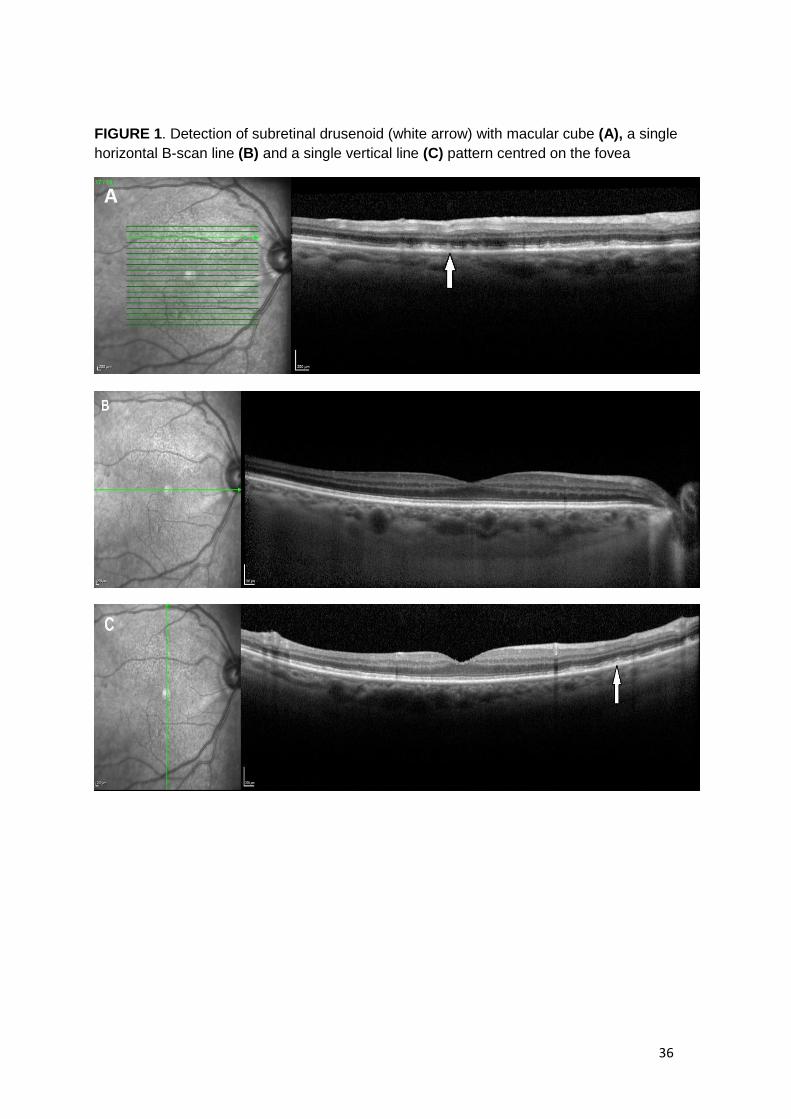
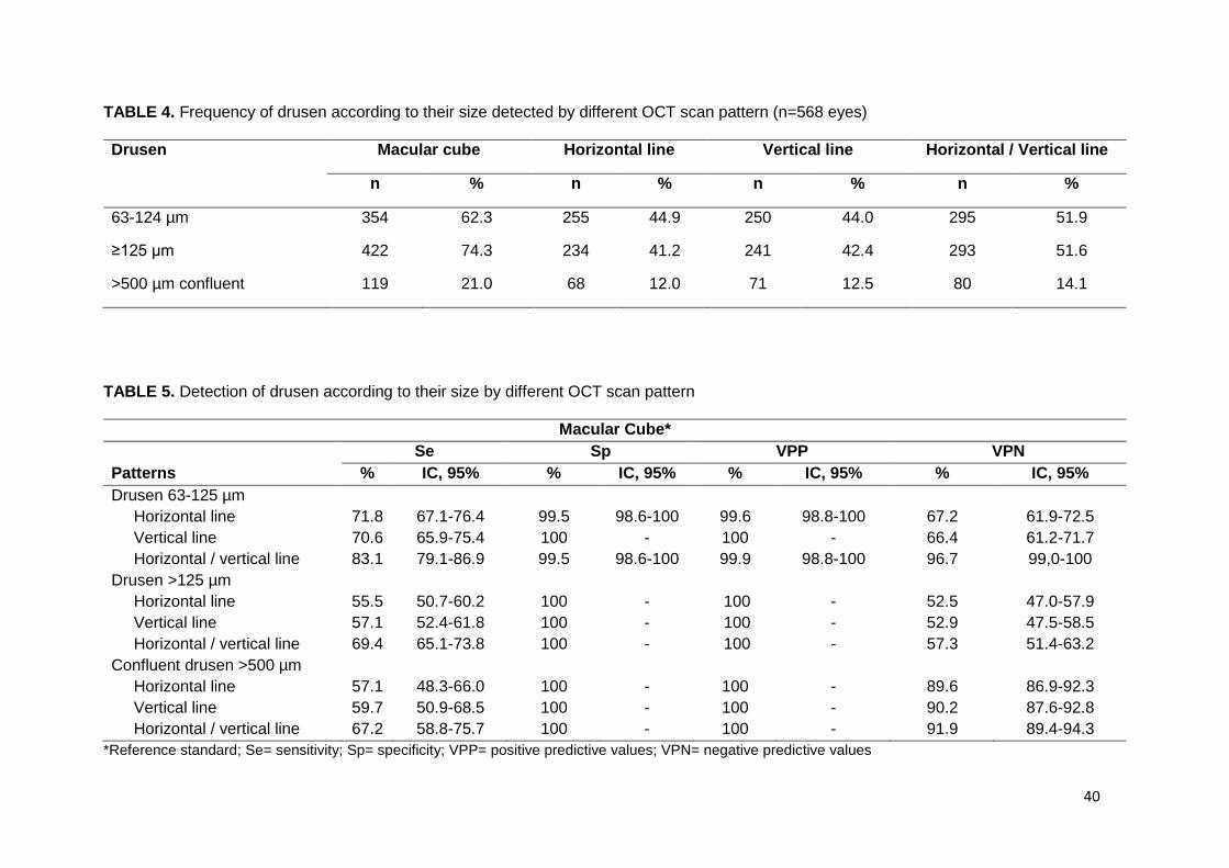
L’examen par cohérence optique Spectral-Domain (SD-OCT) permet d’examiner, avec précision,
chacune des couches de la rétine. Il est devenu un outil diagnostique et de suivi essentiel des
pathologies maculaires et de l’interface vitréorétinienne dans la pratique ophtalmologique courante.
Cependant les appareils SD-OCT proposent de multiples modalités de recueil mais aucune étude n’a
permis de définir un protocole standardisé d’acquisition et d’analyse approprié dans la détection et le
suivi des pathologies rétiniennes. Le but de cette étude était de comparer différentes modalités de
recueil en SD-OCT pour détecter des pathologies maculaires et vitréorétiniennes rencontrées en
pratique clinique.
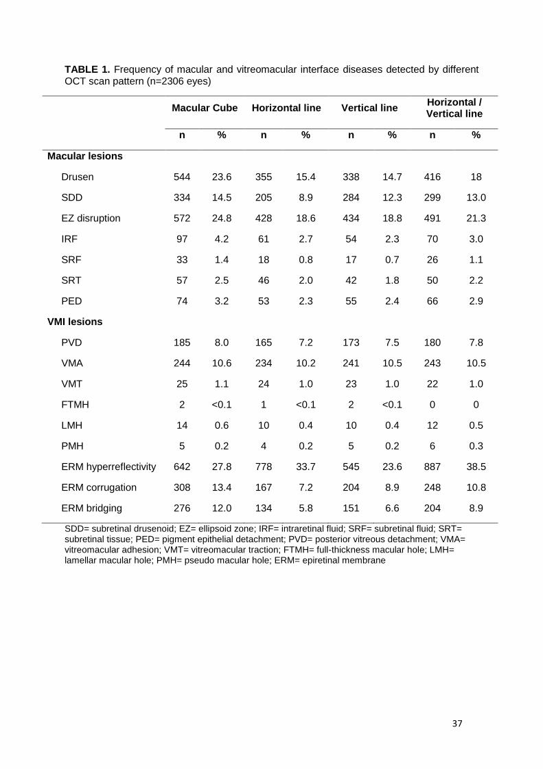
Deux-mille trois cent six yeux de 1153 patients d’âge moyen de 82,2 ± 3,8 ans ont été inclus dans
l’étude de MONTRACHET (Maculopathy, Optic Nerve, nuTRition, neurovasCular and HEarT diseases).
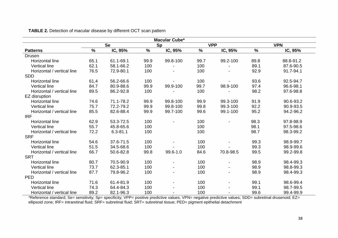
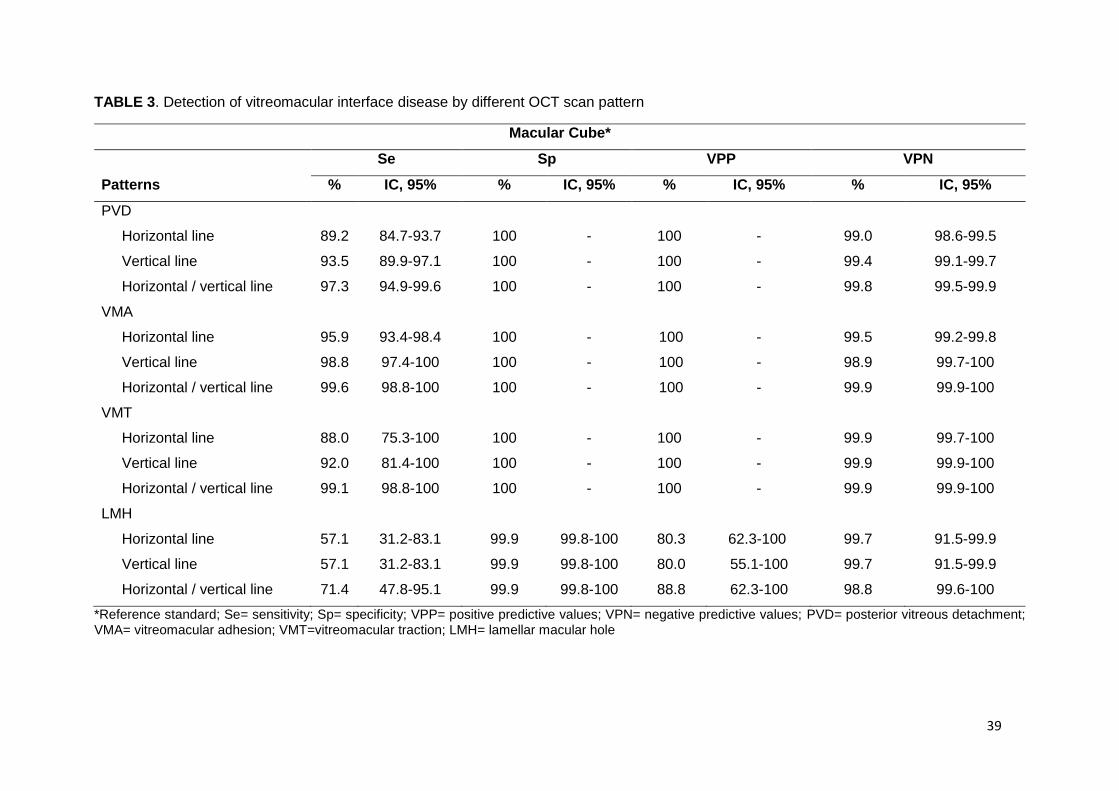
Chaque patient a bénéficié d’un examen SD-OCT incluant différentes modalités de recueil : le cube
maculaire (19 B-scans) considéré comme examen de référence, ainsi qu’une ligne horizontale et une
ligne verticale, toutes deux centrées sur la fovea. La détection des pathologies maculaires et
vitréorétiniennes était meilleure avec l’association d’une ligne horizontale/verticale rétrofovéolaire
qu’avec une ligne horizontale rétrofovéolaire. L’association d’une ligne horizontale/verticale
rétrofovéolaire a détecté les pathologies maculaires comme les pseudodrusens réticulés, les
décollements de l’épithélium pigmentaire, les drusens et les œdèmes intrarétiniens avec une sensibilité
de 90%, 89%, 76% et 72%, respectivement. Les pathologies de l’interface vitréorétinienne comme les
adhésions et tractions vitréomaculaires et les membranes épirétinienne ont été détectées avec une
sensibilité de 99%, 99% et 91% respectivement.
Cette étude a montré que l’association d’une ligne horizontale/verticale rétrofovéolaire en SD-OCT est
fortement corrélée au modèle de référence du cube maculaire dans la détection de pathologies
maculaires et vitréorétiniennes. Elle permet une analyse simple et rapide des clichés SD-OCT avec une
bonne qualité d’image.
43
References
1. Hoerster R, Muether PS, Hermann MM, et al. Subjective and functional deterioration in recurrences of neovascular AMD are often preceded by morphologic changes in optic coherence tomography. Br J Ophthalmol. 2011;95:1424-1426. 2. Lalwani GA, Rosenfeld PJ, Fung AE, et al. A variable-dosing regimen with intravitreal ranibizumab for neovascular age-related macular degeneration: year 2 of the PrONTO Study. Am J Ophthalmol. 2009;148:43-58 e41. 3. Comparison of Age-related Macular Degeneration Treatments Trials Research G, Martin DF, Maguire MG, et al. Ranibizumab and bevacizumab for treatment of neovascular age-related macular degeneration: two-year results. Ophthalmology. 2012;119:1388-1398. 4. Stalmans P, Benz MS, Gandorfer A, et al. Enzymatic vitreolysis with ocriplasmin for vitreomacular traction and macular holes. N Engl J Med. 2012;367:606-615. 5. Schneider EW, Todorich B, Kelly MP, Mahmoud TH. Effect of optical coherence tomography scan pattern and density on the detection of full-thickness macular holes. Am J Ophthalmol. 2014;157:978-984. 6. Kiernan DF, Mieler WF, Hariprasad SM. Spectral-domain optical coherence tomography: a comparison of modern high-resolution retinal imaging systems. Am J Ophthalmol. 2010;149:18-31. 7. Ferris FL, Davis MD, Clemons TE, et al. A simplified severity scale for age-related macular degeneration: AREDS Report No. 18. Arch Ophthalmol. 2005;123:1570-1574. 8. Zweifel SA, Imamura Y, Spaide TC, Fujiwara T, Spaide RF. Prevalence and significance of subretinal drusenoid deposits (reticular pseudodrusen) in age-related macular degeneration. Ophthalmology. 2010;117:1775-1781. 9. Duker JS, Kaiser PK, Binder S, et al. The International Vitreomacular Traction Study Group classification of vitreomacular adhesion, traction, and macular hole. Ophthalmology. 2013;120:2611-2619. 10. Meuer SM, Myers CE, Klein BE, et al. The epidemiology of vitreoretinal interface abnormalities as detected by spectral-domain optical coherence tomography: the beaver dam eye study. Ophthalmology. 2015;122:787-795. 11. Major JC, Jr., Wykoff CC, Mariani AF, et al. Comparison of spectral-domain and time-domain optical coherence tomography in the detection of neovascular age-related macular degeneration activity. Retina. 2014;34:48-54. 12. Sayanagi K, Sharma S, Kaiser PK. Comparison of retinal thickness measurements between three-dimensional and radial scans on spectral-domain optical coherence tomography. Am J Ophthalmol. 2009;148:431-438. 13. Sayanagi K, Sharma S, Yamamoto T, Kaiser PK. Comparison of spectral-domain versus time-domain optical coherence tomography in management of age-related macular degeneration with ranibizumab. Ophthalmology. 2009;116:947-955. 14. De Bats F, Mathis T, Mauget-Faysse M, et al. Prevalence of Reticular Pseudodrusen in Age-Related Macular Degeneration Using Multimodal Imaging. Retina. 2016;36:46-52. 15. Curcio CA, Messinger JD, Sloan KR, et al. Subretinal drusenoid deposits in non-neovascular age-related macular degeneration: morphology, prevalence, topography, and biogenesis model. Retina. 2013;33:265-276. 16. Schmitz-Valckenberg S, Alten F, Steinberg JS, et al. Reticular drusen associated with geographic atrophy in age-related macular degeneration. Invest Ophthalmol Vis Sci. 2011;52:5009-5015. 17. Freeman SR, Kozak I, Cheng L, et al. Optical coherence tomography-raster scanning and manual segmentation in determining drusen volume in age-related macular degeneration. Retina. 2010;30:431-435. 18. Gregori G, Wang F, Rosenfeld PJ, et al. Spectral domain optical coherence tomography imaging of drusen in nonexudative age-related macular degeneration. Ophthalmology. 2011;118:1373-1379.
44
19. Courtney RJ, McClintic JI, Ehlers JP. Comparison of Spectral Domain Optical Coherence Tomography Scan Patterns and Clinical Review Strategies in Neovascular Age-Related Macular Degeneration. Retina. 2015;35:1315-1322. 20. Baranano AE, Keane PA, Ruiz-Garcia H, Walsh AC, Sadda SR. Impact of scanning density on spectral domain optical coherence tomography assessments in neovascular age-related macular degeneration. Acta Ophthalmol. 2012;90:e274-280. 21. Johnson MW. Posterior vitreous detachment: evolution and complications of its early stages. Am J Ophthalmol. 2010;149:371-382 e371. 22. Mojana F, Cheng L, Bartsch DU, et al. The role of abnormal vitreomacular adhesion in age-related macular degeneration: spectral optical coherence tomography and surgical results. Am J Ophthalmol. 2008;146:218-227. 23. Shao L, Xu L, You QS, et al. Prevalence and associations of incomplete posterior vitreous detachment in adult Chinese: the Beijing Eye Study. PLoS One. 2013;8:e58498. 24. Wilkins JR, Puliafito CA, Hee MR, et al. Characterization of epiretinal membranes using optical coherence tomography. Ophthalmology. 1996;103:2142-2151. 25. Romano MR, Cennamo G, Amoroso F, et al. Intraretinal changes in the presence of epiretinal traction. Graefes Arch Clin Exp Ophthalmol. 2017;255:31-38. 26. Sakamoto A, Hangai M, Yoshimura N. Spectral-domain optical coherence tomography with multiple B-scan averaging for enhanced imaging of retinal diseases. Ophthalmology. 2008;115:1071-1078 e1077. 27. Spaide RF. Enhanced depth imaging optical coherence tomography of retinal pigment epithelial detachment in age-related macular degeneration. Am J Ophthalmol. 2009;147:644-652.
Université de Bourgogne
UFR des Sciences de Santé
Circonscription Médecine
TITRE DE LA THESE : Comparaison de différentes modalités de recueil en tomographie par
cohérence optique Spectral-Domain dans la détection de pathologies maculaires et de l’interface
vitréorétinienne : l’étude MONTRACHET
AUTEUR : Ghislaine ASSAD
RESUME
Objectifs : Comparer différentes modalités de recueil en tomographie par cohérence optique Spectral-
Domain (SD-OCT) pour détecter des pathologies maculaires et vitréorétiniennes rencontrées en
pratique courante.
Méthodes : Les patients de l’étude MONTRACHET (Maculopathy, Optic Nerve, nuTRition,
neurovasCular and HEarT diseases) ont bénéficié d’un examen SD-OCT maculaire incluant différentes
modalités de recueil : le cube maculaire (19 B-scans) ainsi qu’une ligne horizontale et une ligne verticale,
toutes deux centrées sur la fovea. L’analyse des clichés SD-OCT recherchait la présence de drusens
et pseudo-drusens réticulés (PDR), d’œdème intrarétinien (OIR), de décollement de l’épithélium
pigmentaire (DEP), d’adhésion vitréomaculaire (AVM) ou de traction vitréomaculaire (VMT) et de
membrane épirétinienne (MER).
Résultats : Deux-mille trois cent six yeux de 1153 patients ont été inclus. L’âge moyen était de 82,2 ±
3,8 ans. Le cube maculaire était considéré comme modèle de référence. La ligne horizontale
rétrofovéolaire a détecté les PDR, les DEP, les drusens et les OIR avec une sensibilité de 61%, 72%,
65% et 63%, respectivement. Les AVM, les TVM et les MER étaient détectées avec une sensibilité de
96%, 88% et 82% respectivement. L’association d’une ligne horizontale/verticale rétrofovéolaire a
détecté les PDR, les DEP, les drusens et les OIR avec une sensibilité de 90%, 89%, 77% et 72%,
respectivement. Les AVM, les TVM et les MER étaient détectées avec une sensibilité de 99%, 99% et
91% respectivement.
Conclusion : L’association d’une ligne horizontale/verticale rétrofovéolaire en SD-OCT est fortement
corrélée au modèle de référence du cube maculaire dans la détection de pathologies maculaires et
vitréorétiniennes, tout en permettant une bonne qualité d’image et une interprétation simple et rapide.
MOTS-CLES : tomographie par cohérence optique Spectral-Domain, pathologie maculaire, pathologie
de l’interface vitréorétinienne