Page 1
UNIVERSIDADE DE LISBOA
FACULDADE DE MEDICINA DENTÁRIA
COMPARATIVE ANALYSIS OF ROOT CANAL
INSTRUMENTATION USING PROTAPER NEXTTM,
RECIPROCTM & WAVEONEGOLDTM
SHAPE SYSTEMS
ANA RAQUEL TAVARES
Dissertação Orientada pelo
Professor Doutor António Ginjeira
MESTRADO INTEGRADO EM MEDICINA DENTÁRIA
LISBOA, 2016
Page 5
“Quero, terei –
Se não aqui,
Noutro lugar que ainda não sei.
Nada perdi,
Tudo serei.”
Fernando Pessoa
Page 7
II
INDEX
Agradecimentos…………………………………………………………………………………IV
Resumo………………………………………………………………………………………..VIII
Palavras-Chave………………………………………………………………………………..XIV
Abstract……………………………………………………………………………………….XVI
Keywords……………………………………………………………………………………...XVI
1. Introduction
1.1 Endodontics – Definition and aims………………………………………………….1
1.2 Evolution of mechanical rotary instruments………………………………………...1
1.2.1 M-Wire NiTi alloy……………………………………………………...2
1.2.2 Gold-Wire – The new supermetal………………………………………2
1.2.3 Reciprocating motion…………………………………………………...3
1.2.4 Single-file shaping technique…………………………………………...3
1.3 Rotary instruments…………………………………………………………………..4
1.3.1 WaveOne GoldTM……………………………………………………….4
1.3.2 ReciprocTM……………………………………………………………...5
1.3.3 ProTaper NextTM………………………………………………………..5
1.4 Mandibular first molar – Anatomy………………………………………………….6
1.5 Image Analysis – Computerized micro tomography………………………………..7
2. Aims……………………………………………………………………………………..8
3. Materials and methods
3.1 Sample preparation teeth……………………………………………………9
3.2 Tridimentional images with computerized micro tomography……………..9
3.3 Canal instrumentation……………………………………………………..11
3.4 Statistical analysis…………………………………………………………12
3.5 Bibliographic research……………………………………………………..13
Page 8
III
4. Results………………………………………………………………………………….14
4.1 Volume…………………………………………………………………….14
4.2 SMI………………………………………………………………………...15
4.3 Surface……………………………………………………………………..15
4.4 Area………………………………………………………………………...16
4.5 Diameter…………………………………………………………………...17
5. Discussion………………………………………………………………………………18
6. Conclusions…………………………………………………………………………….21
7. Acknowledgments……………………………………………………………………...22
References…………………………………………………………………………………...XVIII
Appendix…………………………………………………………………………………....XXVI
a) Abbreviations
b) Symbols
c) Units
d) Figure Index
e) Tables Index
f) Graphs Index
Appendix 2………………………………………………………………………………...XXVIII
Appendix 3………………………………………………………………………………..XXXIV
Page 9
IV
AGRADECIMENTOS
Porque não é a felicidade que nos faz gratos, mas sim a gratidão que nos faz felizes…
Ao Professor António Ginjeira, por ter acreditado neste projeto tanto como eu,
por nunca ter desistido de o pôr em prática e ter sempre as portas abertas para me
receber e apoiar em tudo o que foi necessário.
Ao Dr. Mário Rito Pereira pela sugestão deste estudo que seria pioneiro a testar
o sistema WaveOne GoldTM, e pela disponibilidade e incansável ajuda sempre que
necessário.
Á Margarida Franco e ao CDRsp – IP Leira, pela cooperação e pela paixão por
este projeto que se tornou “nosso”. Sem a sua colaboração nada disto seria possível.
Aos meus amigos e colegas: Maria Mouzinho, Sofia Araújo, João Oliveira,
Nuno Gonçalves, João Gil Morais, Catarina Gonçalves, Andreia Silva, Mariana Neves e
Luís Braz pelo companheirismo e amizade durante este percurso académico.
Ao Miguel Rosa, pela amizade, paciência e por ser o meu braço direito em tudo
e qualquer coisa na minha vida. Aconteça o que acontecer, sei que me apoiarás
incondicionalmente.
Ao Jorge Melo, meu Xico, que me acompanhou neste percurso, todos os dias, e
sempre, a cada segundo de todos os minutos e todas as horas, presente no meu
pensamento.
Á Bárbara Menezes, por ser uma irmã para mim, tão diferente, mas que me
completa tão bem. Pela presença na minha vida, dentro e fora da faculdade. Por todos os
momentos felizes, e até pelos menos bons, em que esteve sempre presente. Quero-te na
minha vida, hoje e sempre. Juntas, para o que der e vier.
Á minha dupla, Ivana Basso, por ter partilhado este projeto comigo, e por ser a
melhor aliada que poderia ter tido nestes últimos anos. Pela amizade, entreajuda e
presença constante. A nossa cumplicidade é única! Guardarei para sempre todos os
momentos, com saudade, gratidão e carinho.
Page 11
VI
Ao meu Godjinha, pela companhia de todas as horas, mesmo as mais tardias.
Quando precisei de uma mão, a vida deu-me 4 patas.
Á minha avó Rosa, por ser como uma mãe para mim, sem o seu apoio nada tinha
sido possível da forma como foi, tão tranquilo e tão feliz.
Por último, contudo, de maior importância, aos meus pais, que acreditam sempre
em mim e nas minhas capacidades, mesmo quando eu própria duvido. Que lutam
sempre, mesmo quando as minhas forças se esgotam. Que nunca me deixam desistir dos
meus sonhos, dos meus objetivos, da vida e de mim própria. Que nunca me deixam
sentir sozinha e que vão estar sempre junto a mim, no meu coração e naquilo que eu
sou. Tudo isso devo-lhes, acima de tudo e qualquer coisa, a Eles.
Eternamente grata.
Page 13
VIII
RESUMO
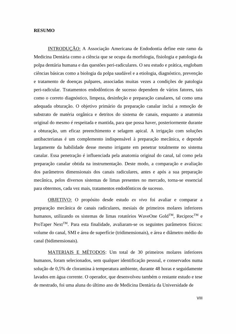
INTRODUÇÃO: A Associação Americana de Endodontia define este ramo da
Medicina Dentária como a ciência que se ocupa da morfologia, fisiologia e patologia da
polpa dentária humana e das questões peri-radiculares. O seu estudo e prática, englobam
ciências básicas como a biologia da polpa saudável e a etiologia, diagnóstico, prevenção
e tratamento de doenças pulpares, associadas muitas vezes a condições de patologia
peri-radicular. Tratamentos endodônticos de sucesso dependem de vários fatores, tais
como o correto diagnóstico, limpeza, desinfeção e preparação canalares, tal como uma
adequada obturação. O objetivo primário da preparação canalar inclui a remoção de
substrato de matéria orgânica e detritos do sistema de canais, enquanto a anatomia
original do mesmo é respeitada e mantida, para que possa haver, posteriormente durante
a obturação, um eficaz preenchimento e selagem apical. A irrigação com soluções
antibacterianas é um complemento indispensável à preparação mecânica, e depende
largamente da habilidade desse mesmo irrigante em penetrar totalmente no sistema
canalar. Essa penetração é influenciada pela anatomia original do canal, tal como pela
preparação canalar obtida na instrumentação. Deste modo, a comparação e avaliação
dos parâmetros dimensionais dos canais radiculares, antes e após a sua preparação
mecânica, pelos diversos sistemas de limas presentes no mercado, torna-se essencial
para obtermos, cada vez mais, tratamentos endodônticos de sucesso.
OBJETIVO: O propósito desde estudo ex vivo foi avaliar e comparar a
preparação mecânica de canais radiculares, mesiais de primeiros molares inferiores
humanos, utilizando os sistemas de limas rotatórios WaveOne GoldTM, ReciprocTM e
ProTaper NextTM. Para esta finalidade, avaliaram-se os seguintes parâmetros físicos:
volume do canal, SMI e área de superfície (tridimensionais), e área e diâmetro médio do
canal (bidimensionais).
MATERIAIS E MÉTODOS: Um total de 30 primeiros molares inferiores
humanos, foram selecionados, sem qualquer identificação pessoal, e conservados numa
solução de 0,5% de cloramina à temperatura ambiente, durante 48 horas e seguidamente
lavados em água corrente. O operador, que desenvolveu também o restante estudo e tese
de mestrado, foi uma aluna do último ano de Medicina Dentária da Universidade de
Page 15
X
Lisboa. As coroas dos dentes foram cortadas aproximadamente ao nível da junção
amelo-cementária, deixando apenas 2 milímetros da câmara pulpar, e as raízes distais
removidas com a ajuda de uma turbina. Cada raiz mesial foi colocada num tubo de
Eppendorf com 1 mililitro de solução salina, para que se mantivessem hidratadas.
Restos de tecidos ou calcificações foram removidos com uma ponta destartarizadora. Os
fatores de exclusão de possíveis amostras foram os seguintes: raízes com ápex aberto,
canais sem permeabilidade, raízes com ângulos acentuados ou canais calcificados,
canais com tratamento endodôntico realizado, fraturas e cáries radiculares, nódulos
pulpares e reabsorções internas. Todas as amostras foram analisadas antes e depois da
instrumentação canalar, por um aparelho de micro tomografia computorizada
(SKYSCAN 1174 v.2 - Kontich, Bélgica), em colaboração com o “Centro para o
Desenvolvimento Rápido e Sustentável do Produto – Instituto Politécnico de Leiria”, e
registados todos os dados e imagens. Após a primeira captação de imagens, as amostras
foram preparadas mecânica e aleatoriamente, em grupos de 10, de acordo com as
técnicas recomendadas pelos fabricantes. O comprimento de trabalho foi determinado
introduzindo limas manuais K-10 (Dentsply-Maillefer, Ballaigues, Switzerland) nos
canais até serem visíveis no forame apical. A esse valor foi retirado meio milímetro. A
via de permeabilidade foi conseguida com recurso ao sistema rotatório Proglider
(Dentsply-Maillefer, Ballaigues, Switzerland) e a irrigação feita com uma solução de
10% de ácido cítrico. A instrumentação foi concluída com as limas correspondentes ao
diâmetro apical de 0,25mm, em cada um dos sistemas: lima primária no WaveOne
GoldTM (grupo A), R25 no ReciprocTM (grupo B) e X2 no ProTaper NextTM (grupo C).
Na análise estatística, com recurso ao programa IBM SPSS versão 23.0, foi feita uma
análise descritiva inicial para cada grupo em estudo. As variáveis com distribuição
normal foram testadas com One-Way ANOVA e testes Bonferroni post-hoc. As
variáveis que rejeitaram distribuição normal foram testadas com Kruskall Wallis (não
paramétrico). O nível de significância foi padronizado em 0.05. A informação teórica
para este estudo foi encontrada em livros, da Biblioteca da Faculdade de Medicina
Dentária da Universidade de Lisboa, e em pesquisas online no motor de busca PubMed,
com acesso à base de dados MEDLINE, com as seguintes palavras-chave:
“endodontics”, “root canal shaping”, “WaveOne Gold”, “ProTaper Next”, “Reciproc”,
“rotary instruments” and “micro computerized tomography”. Foram encontrados cerca
Page 17
XII
de 2000 artigos, dos quais se revelaram com interesse e dentro dos fatores de inclusão
para o presente estudo, apenas 34. Os critérios de exclusão foram: artigos com acesso
limitado mediante pagamento, estudos em modelo animal, artigos em idiomas para além
do inglês ou português, e artigos publicados há mais de 40 anos.
RESULTADOS: Após a avaliação das preparações mecânicas feitas pelos 3
sistemas de limas em estudo, WaveOne GoldTM, ReciprocTM e ProTaper NextTM,
comparando os parâmetros: volume do canal, SMI, superfície, área do canal e diâmetro
médio, os resultados foram os seguintes: nos parâmetros tridimensionais (volume, SMI
e superfície canalares) não se verificaram diferenças estatisticamente significativas entre
os 3 sistemas (p-value > 0,05); nos parâmetros bidimensionais, sim: ao nível da área,
verificou-se um aumento estatisticamente significativo apenas no sistema Reciproc; ao
nível do diâmetro, o sistema Reciproc revelou, novamente, um aumento significativo
em relação ao sistema ProTaper Next, mas sem diferença estatística para o sistema
WaveOne Gold.
DISCUSSÃO: A utilização de dentes humanos naturais extraídos, em estudos de
natureza endodôntica, tem a vantagem de reproduzir as condições clínicas. Contudo, a
variabilidade morfológica dos sistemas de canais radiculares num mesmo grupo de
dentes, torna bastante complexa a sua padronização. A nossa amostra inicial, não
revelou, no entanto, na análise estatística, significantes diferenças anatómicas entre os 3
grupos em estudo, sendo, portanto, um bom ponto de partida. Para além disso,
perderam-se duas amostras durante o estudo: a amostra 16 fraturou na captação de
imagens na micro tomografia computorizada, e a amostra 20 não tinha permeabilidade
canalar. Assim sendo, a amostra total final foi de 28. Em geral, face à dificuldade em
obter dentes dentro dos critérios pretendidos, a amostra do nosso estudo é relativamente
reduzida. No entanto, outros estudos semelhantes, utilizaram amostras
quantitativamente semelhantes. Os resultados ao nível do SMI, parâmetro que permite
quantificar a forma tridimensional de uma estrutura, revelaram-se dentro do esperado,
em média próximos de 4, refletindo a conicidade dos instrumentos rotatórios utilizados,
que conferem características circulares às preparações canalares realizadas. O aumento
da área e diâmetro canalares verificados nos sistemas ReciprocTM e WaveOne GoldTM
(este último apenas ao nível do diâmetro), poderá constituir uma vantagem ao nível da
penetração do agente irrigante antibacteriano, que assim, mais facilmente percorrerá
Page 19
XIV
todo o sistema canalar e atuar eficazmente a nível químico, complementando a ação
mecânica das limas na fase de instrumentação canalar. Embora, as limas finais dos 3
sistemas em estudo tenham o mesmo diâmetro apical, ISSO 25, a sua conicidade é
diferente, sendo uma possível explicação para os resultados obtidos. A utilização da
técnica de captação de imagem com micro tomografia computorizada, teve como
vantagens a sua alta-resolução, imagens tridimensionais e de vários ângulos, medidas
detalhadas qualitativa e quantitativamente, e ser uma técnica não invasiva/destrutiva. No
entanto, é um método muito caro, que necessita de equipamentos e profissionais
tecnicamente habilitados, e cuja captação de imagem pode ser demorada. Este método é
uma referência para estudos ex vivo, contudo não é aplicável a nível clínico.
CONCLUSÃO: Em geral, pode admitir-se que o sistema ReciprocTM foi o que
produziu as alterações geométricas mais significativas ao nível dos canais radiculares
em estudo, seguido pelo sistema WaveOne GoldTM e ProTaper NextTM, respetivamente.
Não existem, até à data deste estudo, artigos que avaliem os modernos instrumentos
rotatórios WaveOne GoldTM, e assim sendo, as conclusões são limitadas. Estudos
adicionais envolvendo estes sistemas de limas endodônticas são necessários para que
mais e melhor informação possa ser adquirida e transmitida, permitindo aos Médicos
Dentistas, durante a sua prática clínica, adaptar cada sistema rotatório a um caso clínico
específico.
Palavras-chave: endodontia; instrumentos rotatórios; instrumentação canalar;
micro tomografia computorizada; WaveOne Gold; Reciproc; ProTaper Next.
Page 21
XVI
ABSTRACT
INTRODUCTION: Root canal therapy is one of the most widely accepted
treatment modality for pulpally involved teeth. Objective in root canal preparation is to
develop a shape that tapers from apical to coronal, maintaining the original canal
anatomy. Irrigation with antibacterial solutions is performed as complement to
mechanical preparation and it depends largely on the ability of the irrigant to penetrate
the full extent of the root canal system, influenced by the original anatomy as well as
the final shape created through mechanical preparation.
AIM: The purpose of this ex vivo study was to evaluate and compare the
mechanical preparation of three different rotary file systems: WaveOne GoldTM,
ReciprocTM and ProTaper NextTM. Analyzed with computerized micro tomography, the
following parameters were evaluated: canalar volume, SMI, surface, area and diameter
(average).
MATERIALS AND METHODS: A total of 30 extracted humans mandibular
first molars were selected. Each mesial root was, randomly, placed separately and
prepared, by the same operator, in three groups of 10 samples: Group A – WaveOne
GoldTM, Group B – ReciprocTM, Group C – ProTaper NextTM. An image analysis, and
data register, was made before and after the canal instrumentation, using micro-CT (in
collaboration with CDRsp - IP Leiria).
RESULTS: There are statistically differences in the post instrumentation
bidimensional parameters: the area increase with Reciproc files was significantly
greater than with WaveOne Gold (p = 0.026) or Protaper Next (p = 0.007); the diameter
increment after the preparation with Reciproc files was significantly higher than with
ProTaper Next (p = 0.032), however, the difference between Reciproc and Wave One
Gold was not statistically significant.
DISCUSSION AND CONCLUSION: The Reciproc files produce major changes
in the geometric conditions of the root canal systems, followed by WaveOne Gold and
ProTaper Next, respectively.
KEYWORDS: endodontics; root canal shaping; WaveOne Gold; ProTaper
Next; Reciproc; rotary instruments; micro computerized tomography.
Page 23
COMPARATIVE ANALYSIS OF ROOT CANAL INSTRUMENTATION USING PROTAPER NEXTTM, RECIPROCTM &
WAVEONEGOLDTM SHAPE SYSTEMS
2016
1
Raquel Tavares
1. INTRODUCTION
1.1 Endodontics – definition and aims
The American Association of Endodontics defines Endodontics as “the branch
of dentistry concerned with the morphology, physiology and pathology of the human
dental pulp and periradicular tissues; it’s study and practice encompass the basic and
clinical sciences including the biology of the normal pulp and the etiology, diagnosis,
prevention and treatment of diseases and injuries of the pulp and associated
periradicular conditions.” (AAE 2012)
Root canal therapy is one of the most widely accepted treatment for pulpally
involved teeth. Successful endodontic therapy depends on many factors like correct
diagnosis, effective cleaning, shaping and disinfection of the root canals, and on
adequate obturation (Mehran, 2008). The primary objective of canal preparation
includes removal of organic substrate from the canal system into a continuously
tapering preparation while maintaining the original outline and form of the canal
(Schelder, 1974). This is one of the most important steps in any root canal treatment,
following the original shape of the canal (Dhingra et al. 2015). The configuration for
prepared root canals should be a conical tapered canal with the smallest diameter and a
marked stop at the apical constriction. As root canal curvature increases, more difficult
is to have an adequate canal preparation (Shäfer et al. 1996).
1.2 Evolution of mechanical rotary instruments
Due to their flexibility and elasticity, nickel-titanium (NiTi) instruments have
been introduced into the endodontic armamentarium to facilitate the instrumentation of
curved canals and with rotary techniques improve root canal preparation, with their
unique properties: able to improve morphological characteristics and safety of canal
shaping (Bergmans et al. 2003). Rotary files can prepare root canals faster, easier and at
the same time, preserve the original canal shape with considerably less iatrogenic errors
(Capar et al. 2014). It has been reported that they can maintain the original shape of
Page 24
COMPARATIVE ANALYSIS OF ROOT CANAL INSTRUMENTATION USING PROTAPER NEXTTM, RECIPROCTM &
WAVEONEGOLDTM SHAPE SYSTEMS
2016
2
Raquel Tavares
canal with minimal transportation (Bryant et al. 2005). Irregularities in the root canal
system can restrict the complete debridement of root canal by mechanical
instrumentation. Irrigation serves as a flush to remove debris and smear layer, tissue
solvent, eliminate pulpal tissue, bacteria and endotoxins, and as a lubricant (Chan et al.
1996).
1.2.1 M-Wire NiTi alloy
Instruments manufactured from M-wire NiTi alloy improve file flexibility and
resist cyclic fatigue while retaining cutting efficiency (Alapati et al., 2009). They’re
instruments made of a special metal alloy, which undergoes alternate cycles of cold and
heat during manufacture, which provides a significant increase in their flexibility and
mechanical strength (Kim et al. 2012). NiTi rotary instruments are important in
endodontics because of their ability to shape root canals with minimum complications
(Young et al., 2007). As the rotary NiTi instruments maintained the original canal
curvature, particularly in the apical region of the root canal better than stainless steel
hand instruments, studies compared the shaping ability of different rotary NiTi systems
with different designs. With advent of instruments manufactured from nickel titanium
alloys (NiTi), there was significant improvement of quality of root canal shaping, with
predictable results and less iatrogenic damage, even in severely curved canals (Peters
OA, 2004).
1.2.2 Gold-Wire – The new supermetal
Engineers have identified the desired phase-transition point between martensite
and austenite that serves to produce a more clinically optimal metal than NiTi itself.
This thermal process and post-machining procedure have generated a new supermetal
that is commercially termed Gold-Wire. Specifically, the Primary WaveOne GoldTM file
is at least 80% more flexible, 50% more resistant to cyclic fatigue, and 23% more
efficient, compared to its Primary WaveOne M-Wire predecessor (data on file: Dentsply
Maillefer engineering and testing; Ballaigues, Switzerland, 2014). The new patented
cross section and supermetal serve to improve shaping results in anatomically longer,
Page 25
COMPARATIVE ANALYSIS OF ROOT CANAL INSTRUMENTATION USING PROTAPER NEXTTM, RECIPROCTM &
WAVEONEGOLDTM SHAPE SYSTEMS
2016
3
Raquel Tavares
narrower, and more apically curved canals, while decreasing the potential for iatrogenic
events (Clifford JR, 2016).
1.2.3 Reciprocating motion
The use of reciprocating motion may be considered as a recent innovation in
mechanized root canal instrumentation; with its differentiated kinematics being
described as an oscillatory movement in which the instrument turns in the clockwise
direction, and then counter-clockwise before completing a full 360º rotation cycle
(Gavini et al. 2012). Reciprocating instruments have been developed to reduce the stress
that rotary instruments suffer, particularly during the preparation of curved canals
(Varela-Patiño et al., 2010). The use of reciprocating motion can extend the lifespan of
a NiTi instrument and allow it to resist fatigue better than it can with continuous
rotation (You et al., 2010).
1.2.4 Single-file shaping technique
Single file rotary systems are gaining clinical acceptance as they reduce the time
required for biomechanical preparation, as well as reduce the number of failures related
to instrumentation (Bürklein et al., 2012). This single-file systems concept, used either
in a reciprocal motion or in continuous rotation, provide a shaping technique, regardless
of the length, diameter, or curvature of any given canal. In fact, it has been shown that a
single-file shaping technique is more than 4 times safer and almost 3 times faster than
using multiple rotary files to achieve the same final shape (Gambarini et al., 2010; You
SY, et al., 2010). In previously studies, single-file reciprocating system strongly
decrease the mean preparation time in comparison with multi-file rotational system
(Bürklein et al., 2012) and, so, thee time available for chemical disinfection of the root
canal system is also simultaneously increased.
Page 26
COMPARATIVE ANALYSIS OF ROOT CANAL INSTRUMENTATION USING PROTAPER NEXTTM, RECIPROCTM &
WAVEONEGOLDTM SHAPE SYSTEMS
2016
4
Raquel Tavares
1.3 Rotary instruments
The aim of this study was to evaluate the mechanical preparation of natural root
canals with three different rotary systems:
1.3.1 The WaveOne GoldTM (Dentsply Tulsa Dental Specialties) system is a
single-file and single-us technique. Through the convergence of an advanced design,
Gold-wire technology, and a unique reciprocating movement (170º counterclockwise
motion followed by 50º clockwise rotation), preparing canals is safer, easier and faster
than with WaveOne (at least 80% more flexible, 50% more resistant to cyclic fatigue,
and 23% more efficient, compared to the original Primary WaveOne M-wire file). There
are 4 WaveOne Gold files available in various lengths to more effectively address a
wider range of endodontic anatomy compared to its WaveOne predecessor. The 4 files
are termed: Small (yellow 20/07), Primary (red 25/07), Medium (green 35/06),
and Large (black 45/05). The Small 21/06 file has a fixed taper of 6% over its active
portion. Each file has a fixed taper from D1-D3, yet a progressively decreasing
percentage tapered design from D4-D16, which serves to preserve dentin. For example,
the Primary file has diameters of 0.85 mm and 1.0 mm at D9 and D12, respectively, or
the length this file typically extends below the orifice during canal preparation.
Fortuitously, the Primary 25/07 file is generally the only file required to fully shape
virtually any given canal. (Clifford J. Ruddle DDS - Advanced Endodontics)
Figure 1 – WaveOne GoldTM system
Page 27
COMPARATIVE ANALYSIS OF ROOT CANAL INSTRUMENTATION USING PROTAPER NEXTTM, RECIPROCTM &
WAVEONEGOLDTM SHAPE SYSTEMS
2016
5
Raquel Tavares
1.3.2 The ReciprocTM (VDW, Munique, Germany) system consists of 3 files
with a fixed taper over the first 3mm from the tip: R25 (size 25 tip and .08 taper) for
narrow canals, R40 (size 40 tip and .06 taper) for medium canals, and R50 (size 50 tip
and .05 taper) for wide canals, characterized by an S-Shape cross-section, specifically
designed for curved and narrow canals (Dhingra et al., 2015). Reciproc files provide
clockwise (150º) and counterclockwise (30º) rotation. As, the rotation in the cutting
direction is larger than reverse direction, it results in movement towards apex. Only one
instrument is used for the canal preparation depending on the initial size of the canal.
The instruments are made from an M-Wire nickel-titanium that offers greater flexibility
and resistance to cyclic fatigue than traditional nickeltitanium. They have an S-shaped
cross-section with sharp cutting edges (Bürklein et al., 2012). Only very light apical
pressure should be applied on the instrument, as it’s advancement would be almost
automatic.
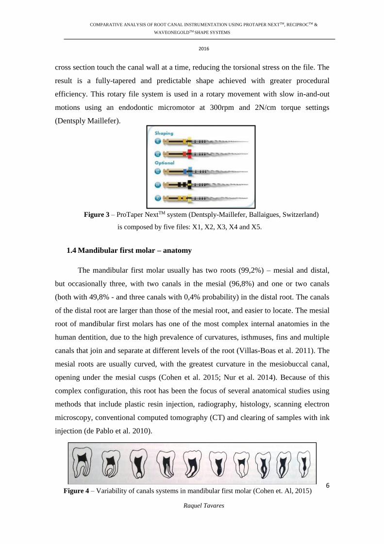
1.3.3. ProTaper NextTM (Dentsply-Maillefer, Ballaigues, Switzerland) is a NiTi
system manufactured using M-wire Ni-Ti alloy (Sportswire, Langley, OK). The files
have an off-centered rectangular cross-section, giving a snake-like swaggering
movement as it moves along the root canal, thus reducing the screw effect, the
unwanted taper lock and torque on any of the given file; thus decreasing the file-root
dentin contact (Ruddle CJ, 2001). The ProTaper Next system is composed by five files
with the same rectangular cross section, off-centered, intended to reduce torsional stress
on the instrument: X1, X2, X3, X4 and X5, all in different lengths – 21, 25 and 31mm
(Files X1 and X2 are used for shaping, and Files X3, X4 and X5 are optional). The off-
center rectangular cross section differs from the center of mass. Only two points of the
Figure 2 – ReciprocTM system (Dentsply-Maillefer, Ballaigues, Switzerland)
Page 28
COMPARATIVE ANALYSIS OF ROOT CANAL INSTRUMENTATION USING PROTAPER NEXTTM, RECIPROCTM &
WAVEONEGOLDTM SHAPE SYSTEMS
2016
6
Raquel Tavares
cross section touch the canal wall at a time, reducing the torsional stress on the file. The
result is a fully-tapered and predictable shape achieved with greater procedural
efficiency. This rotary file system is used in a rotary movement with slow in-and-out
motions using an endodontic micromotor at 300rpm and 2N/cm torque settings
(Dentsply Maillefer).
1.4 Mandibular first molar – anatomy
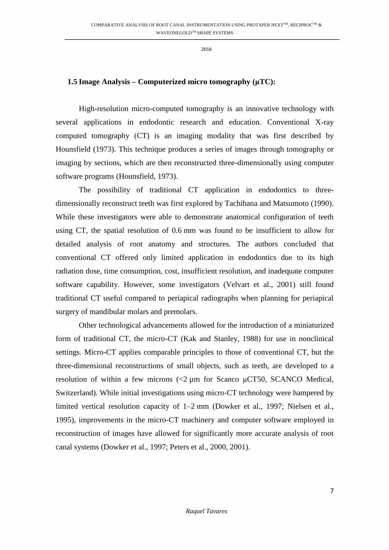
The mandibular first molar usually has two roots (99,2%) – mesial and distal,
but occasionally three, with two canals in the mesial (96,8%) and one or two canals
(both with 49,8% - and three canals with 0,4% probability) in the distal root. The canals
of the distal root are larger than those of the mesial root, and easier to locate. The mesial
root of mandibular first molars has one of the most complex internal anatomies in the
human dentition, due to the high prevalence of curvatures, isthmuses, fins and multiple
canals that join and separate at different levels of the root (Villas-Boas et al. 2011). The
mesial roots are usually curved, with the greatest curvature in the mesiobuccal canal,
opening under the mesial cusps (Cohen et al. 2015; Nur et al. 2014). Because of this
complex configuration, this root has been the focus of several anatomical studies using
methods that include plastic resin injection, radiography, histology, scanning electron
microscopy, conventional computed tomography (CT) and clearing of samples with ink
injection (de Pablo et al. 2010).
Figure 3 – ProTaper NextTM system (Dentsply-Maillefer, Ballaigues, Switzerland)
is composed by five files: X1, X2, X3, X4 and X5.
Figure 4 – Variability of canals systems in mandibular first molar (Cohen et. Al, 2015)
Page 29
COMPARATIVE ANALYSIS OF ROOT CANAL INSTRUMENTATION USING PROTAPER NEXTTM, RECIPROCTM &
WAVEONEGOLDTM SHAPE SYSTEMS
2016
7
Raquel Tavares
1.5 Image Analysis – Computerized micro tomography (μTC):
High-resolution micro-computed tomography is an innovative technology with
several applications in endodontic research and education. Conventional X-ray
computed tomography (CT) is an imaging modality that was first described by
Hounsfield (1973). This technique produces a series of images through tomography or
imaging by sections, which are then reconstructed three-dimensionally using computer
software programs (Hounsfield, 1973).
The possibility of traditional CT application in endodontics to three-
dimensionally reconstruct teeth was first explored by Tachibana and Matsumoto (1990).
While these investigators were able to demonstrate anatomical configuration of teeth
using CT, the spatial resolution of 0.6 mm was found to be insufficient to allow for
detailed analysis of root anatomy and structures. The authors concluded that
conventional CT offered only limited application in endodontics due to its high
radiation dose, time consumption, cost, insufficient resolution, and inadequate computer
software capability. However, some investigators (Velvart et al., 2001) still found
traditional CT useful compared to periapical radiographs when planning for periapical
surgery of mandibular molars and premolars.
Other technological advancements allowed for the introduction of a miniaturized
form of traditional CT, the micro-CT (Kak and Stanley, 1988) for use in nonclinical
settings. Micro-CT applies comparable principles to those of conventional CT, but the
three-dimensional reconstructions of small objects, such as teeth, are developed to a
resolution of within a few microns (<2 µm for Scanco μCT50, SCANCO Medical,
Switzerland). While initial investigations using micro-CT technology were hampered by
limited vertical resolution capacity of 1–2 mm (Dowker et al., 1997; Nielsen et al.,
1995), improvements in the micro-CT machinery and computer software employed in
reconstruction of images have allowed for significantly more accurate analysis of root
canal systems (Dowker et al., 1997; Peters et al., 2000, 2001).
Page 30
COMPARATIVE ANALYSIS OF ROOT CANAL INSTRUMENTATION USING PROTAPER NEXTTM, RECIPROCTM &
WAVEONEGOLDTM SHAPE SYSTEMS
2016
8
Raquel Tavares
2. AIM
The purpose of this in vitro study was to evaluate and compare the mechanical
preparation of mesial root canals of mandibular first molars, using three different file
systems: ProTaper NextTM (Dentsply Maillefer, Ballaigues, Switzerland), ReciprocTM
(VDW, Munich, Germany) and WaveOne GoldTM (Dentsply Tulsa Dental Specialties).
Using computerized micro tomography (μTC), the following parameters were
evaluated: 3D parameters: 1. Canal Volume, 2. SMI, 3. Canal Surface; 2D: 4. Canal
Area and 5. Average canal diameter. To achieve this objective, the following
experimental hypotheses were formulated:
H0: There is no statistically significant difference between the three
mechanical canalar instrumentation systems using mesial root canals of
mandibular first molars in the parameters in study;
H1: There are statistically significant differences between the three
mechanical canalar instrumentation systems using mesial root canals of
mandibular first molars in the parameters in study.
Page 31
COMPARATIVE ANALYSIS OF ROOT CANAL INSTRUMENTATION USING PROTAPER NEXTTM, RECIPROCTM &
WAVEONEGOLDTM SHAPE SYSTEMS
2016
9
Raquel Tavares
3. MATERIALS AND METHODS
3.1 Sample preparation teeth
A total of 30 extracted humans mandibular first molars were selected, without
identification, kept in a 0.5% chloramine solution at room temperature for 48h, and
next, washed in running water. The operator is a final year student of the course of
Dentistry at the University of Lisbon, who led the study and the master's thesis. The
crowns were eliminated by the lap, leaving 2mm of the pulp chamber, with an Isomet
1000 precision saw (Buehler, Lake Bluff, IL, USA) equipped with a 0,3mm diamond
disc (Buehler) and water cooling. The distal roots were split with a tapered drill turbine
at high speed and water. Each mesial root was placed in an Eppendorf vial with a saline
solution (1mL) to keep hydrated. To each root/vial was add a specific number (1 to 30).
Tissue fragments and calcified debris were removed from the teeth by ultrasonic
scaling. The following were determined as exclusion factors: roots with open apex,
without permeable canals, roots with right angles or calcified canals, with endodontic
treatment, root fractures, root caries, root restorations, pulp nodules and internal
resorption. An image analysis was made before and after the canal instrumentation,
using micro-CT.
3.2 Tridimensional images with Computerized Micro Tomography (μTC):
In collaboration with “Center for rapid and sustainable product development -
Instituto Politécnico de Leiria”, was used X-ray micro-CT SKYSCAN model 1174 v.2,
Software version 1.1 (SkyScan, Kontich, Belgium). This scanner uses an x-ray source
with adjustable voltage and a range of filters for versatile adaptation to different object
densities. A sensitive 1.3-megapixel x-ray camera allows scanning of your whole
sample volume in several minutes. Variable magnification (6-30 µm pixel size) is
combined with object positioning for easy selection of the object part to be scanned.
The scanner can run from any desktop or portable computer, requiring just one USB (or
serial) port and a FireWire (IEEE1394) input. The full range of SkyScan software is
Page 32
COMPARATIVE ANALYSIS OF ROOT CANAL INSTRUMENTATION USING PROTAPER NEXTTM, RECIPROCTM &
WAVEONEGOLDTM SHAPE SYSTEMS
2016
10
Raquel Tavares
supplied, including fast volumetric reconstruction, software for 2D/ 3D quantitative
analysis and for realistic 3D visualization (Brochure Bruker Micro CT Academy, 2015).
In this study micro-CT was used with appropriate parameters for scanning each
root, pre and post instrumentation: each image pixel size is 22.70 µm; image rotation is
0.2000; source Voltage 50kV and source Current 800µA; exposure 8500ms; sharpening
40%; rotational step of 1.500º (degrees); rotational angle of 187.50º; average exposure
time of 55 minutes.
The reconstruction of de images was made with NRecon program, version
1.6.8.0 (SkyScan, Kontich, Belgium), in standard reconstruction mode. Each image has
752 x 752 pixel; smoothing 0 (scale 0-10); ring artifact correction 6 (scale 0-20); beam
hardening correction 45% (scale 0-100); lower grey threshold between 58 and 70; and
upper grey threshold always 255.
At the end of each image analysis, a record was made of the values obtained for
each parameter studied. The tables are attached (see appendix 2).
Figure 6 – Computorized Micro Tomography (IPLeiria)
Page 33
COMPARATIVE ANALYSIS OF ROOT CANAL INSTRUMENTATION USING PROTAPER NEXTTM, RECIPROCTM &
WAVEONEGOLDTM SHAPE SYSTEMS
2016
11
Raquel Tavares
3.3 Canal instrumentation:
The mesial root canals of the mandibular first molars were prepared, by the same
operator, in three groups of 10 (A, B and C), randomly, using the technique
recommended by the manufacturer. Working length was determined by advancing a size
10K-file (Dentsply-Maillefer, Ballaigues, Switzerland) into the canal until it is just
visible at the apical foramen. Adjusted the stop to the top of the root and the value of the
working length was the measurement value of that length minus 0.5mm. The
permeability pathway was achieved using the Proglider rotary instruments (Dentsply-
Maillefer, Ballaigues, Switzerland). The Proglider NiTi rotary instrument is
manufactured using M-Wire NiTi alloy to enhance flexibility and cyclic fatigue
resistance as claimed by the manufacturer. The system consists of a single instrument,
with a variable progressive taper. The Proglider instrument is available in 21, 25
(used) and 31 mm length and tip size 16 with a taper of .02 (10). Canals was irrigated
during preparation using a citric acid 10% solution in a disposable plastic syringe.
Group A – WaveOne GoldTM (Dentsply Tulsa Dental Specialties)
instruments were used with a dedicated reciprocating motor – “WAVE
ONE ALL” mode with 170º counterclockwise motion followed by 50º
clockwise rotation with a speed of 350 rpm. The shaping procedure was
done with the Primary file (025/07 red, taper .08), using gentle inward
pressure and letting passively progress through any region of the canal
that has confirmed glide path. After shaping 2-3mm of any given canal,
the Primary file was removed and cleaned. Irrigate, recapitulate with a
size 010 hand file, and re-irrigate. Then, used the Primary file, and
continued in 2-3 passes, to the full working length.
Group B - ReciprocTM (VDW, Munique, Germany): R25 file was used
in a programmed reciprocating motion generated by the electric motor
(Tecnika, Dentsply Maillefer, Schools Grant Program) in the
“RECIPROC ALL” mode with 150º counterclockwise motion followed
by 30º clockwise rotation with a speed of 300rpm. The were used in a
Page 34
COMPARATIVE ANALYSIS OF ROOT CANAL INSTRUMENTATION USING PROTAPER NEXTTM, RECIPROCTM &
WAVEONEGOLDTM SHAPE SYSTEMS
2016
12
Raquel Tavares
pecking motion (amplitude less than 3mm, 3 pecks) according to the
manufacturer’s instructions. (R25: diameter 0,25mm; taper .08)
Group C - ProTaper NextTM (Dentsply-Maillefer, Ballaigues,
Switzerland): This rotary file system was used in a rotary movement with
slow in-and-out motions using an endodontic micromotor at 300rpm and
2N/cm torque settings. The sequence was: X1 instrument at working
length, X2 instrument at WL (taper .06). For each root canal, a new set of
ProTaper Next instruments was used.
3.3 Statistical analysis
The statistical analysis was carried out using the IBM SPSS 23.0 software.
Descriptive analysis was first performed for each group. Sample means and sample
standard deviations were calculated and boxplots were constructed. Normality and
homoscedasticity were tested using Shapiro-Wilk test and Levene’s test respectively.
Clearly defined outlier candidates which altered distribution were removed and sample
means and standard deviations adjusted.
Variables with normal distribution allowed testing for differences among group
means with One-Way ANOVA and Bonferroni post hoc-tests. In the cases where
homoscedasticity was not verified, a Welch ANOVA was carried out along with a
Games-Howell post-hoc test.
Figure 5 – Electric motor
(Tecnika, Dentsply Maillefer,
Schools Grant Program)
Page 35
COMPARATIVE ANALYSIS OF ROOT CANAL INSTRUMENTATION USING PROTAPER NEXTTM, RECIPROCTM &
WAVEONEGOLDTM SHAPE SYSTEMS
2016
13
Raquel Tavares
The statistical testing was performed on the differences registered after canal
preparation, as well as the measurements before canal preparation.
Non-parametric test Kruskall Wallis was performed on variables which rejected
normal distribution. Significance level was set at 0.05 throughout the analysis.
3.5 Bibliographic research
For this work, a literature search was carried out by scientific articles and books.
The online search was made in Pub-med, using the following keywords: “endodontics”,
“root canal shaping”, “WaveOne Gold”, “ProTaper Next”, “Reciproc”, “rotary
instruments” and “micro computerized tomography”. The selected articles were
published in the last 40 years and written in English. I met around 2000 articles of
which used 34 that met the inclusion criteria described above. Exclusion criteria were
paid articles, research in animal models or articles that did not cover directly the article
under consideration.
Page 36
COMPARATIVE ANALYSIS OF ROOT CANAL INSTRUMENTATION USING PROTAPER NEXTTM, RECIPROCTM &
WAVEONEGOLDTM SHAPE SYSTEMS
2016
14
Raquel Tavares
4. RESULTS
The results of the experimental procedure are shown in the following tables,
with the parameters analyzed in the study (1. Volume (µm3), 2. SMI, 3. Surface (µm2),
4. Area (µm2), 5. Diameter Average (µm)), before and after canal instrumentation, and
compared the results between the 3 files systems under study (1. WaveOne Gold, 2.
Reciproc, 3. ProTaper Next).
Table 1 - morphometric data of the root canal (mean ± standard deviation) before
instrumentation; Statistically significant differences between groups before the preparation
weren’t recorded.
4.1 Volume (µm3):
Table 2 - Absolute change and percentage of root canal volume (mean ± standard deviation).
There’s no statistically significant differences between groups in the volume after canal preparation.
Graph 1 – Volume box-plots; Left: after remove outliers;
right: before removing outliers.
Page 37
COMPARATIVE ANALYSIS OF ROOT CANAL INSTRUMENTATION USING PROTAPER NEXTTM, RECIPROCTM &
WAVEONEGOLDTM SHAPE SYSTEMS
2016
15
Raquel Tavares
4.2 SMI
Table 3 - Absolute change and percentage SMI root canal (mean ± standard deviation).
No statistically significant differences between groups in change SMI after canal preparation.
Graph 2 – Surface box-plot.
4.3 Surface (µm2):
Table 4 - Absolute change and percentage of root canal surface (mean ± standard deviation).
There’s no statistically significant differences between groups in the surface change after canal
preparation.
Page 38
COMPARATIVE ANALYSIS OF ROOT CANAL INSTRUMENTATION USING PROTAPER NEXTTM, RECIPROCTM &
WAVEONEGOLDTM SHAPE SYSTEMS
2016
16
Raquel Tavares
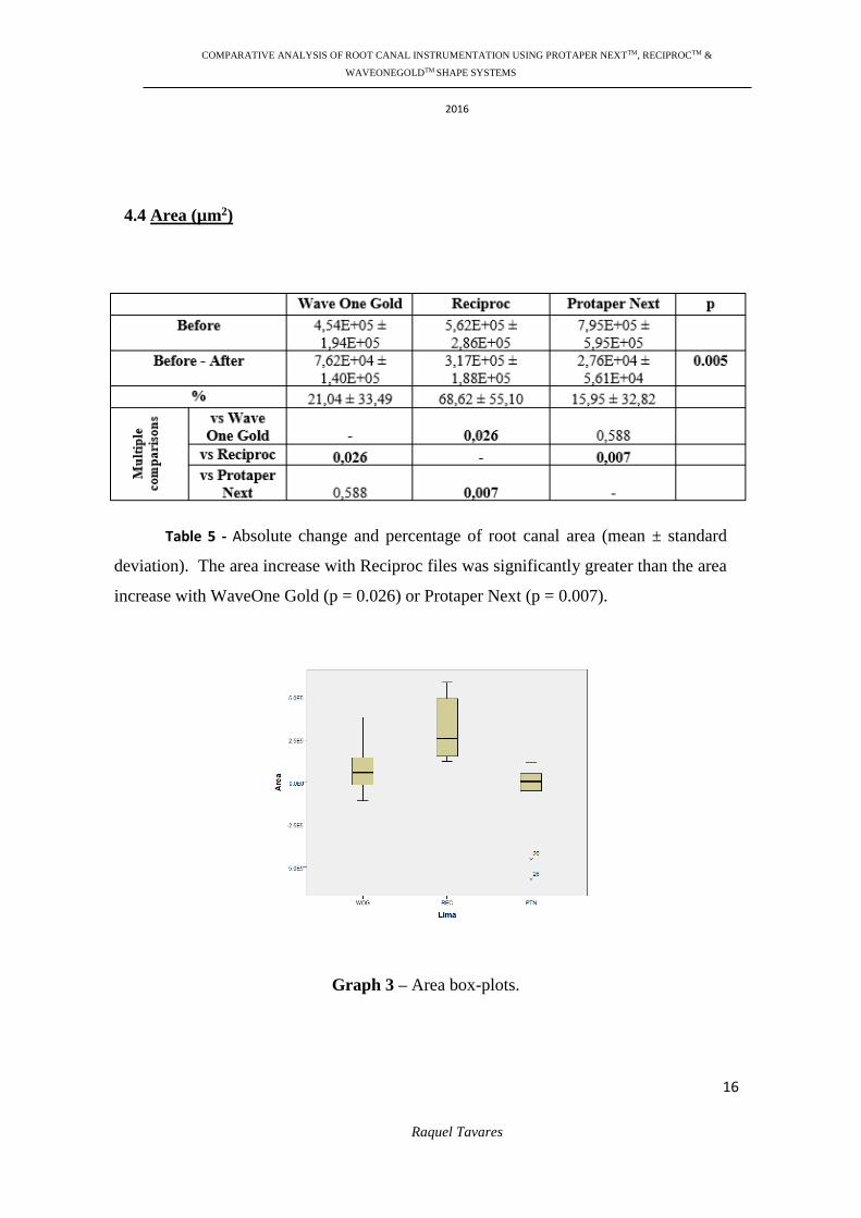
Graph 3 – Area box-plots.
4.4 Area (µm2)
Table 5 - Absolute change and percentage of root canal area (mean ± standard
deviation). The area increase with Reciproc files was significantly greater than the area
increase with WaveOne Gold (p = 0.026) or Protaper Next (p = 0.007).
Page 39
COMPARATIVE ANALYSIS OF ROOT CANAL INSTRUMENTATION USING PROTAPER NEXTTM, RECIPROCTM &
WAVEONEGOLDTM SHAPE SYSTEMS
2016
17
Raquel Tavares
Graph 4 – Diameter box-plots.
4.5 Diameter (µm)
Table 6 - Absolute change and percentage of root canal diameter (mean value ±
standard deviation). The increase of diameter after preparations with Reciproc files was
significantly higher than the increase with ProTaper Next (p = 0.032). However, the difference
between Reciproc and Wave One Gold was not statistically significant.
Page 40
COMPARATIVE ANALYSIS OF ROOT CANAL INSTRUMENTATION USING PROTAPER NEXTTM, RECIPROCTM &
WAVEONEGOLDTM SHAPE SYSTEMS
2016
18
Raquel Tavares
5. DISCUSSION
The aim of this ex vivo study was to evaluate the mechanical shaping ability of 3
different file systems, A- WaveOne GoldTM, B- ReciprocTM and C- ProTaper NextTM,
comparing 5 parameters: canalar volume, SMI, surface, canalar area and diameter
(average).
Our data revealed no differences between the three systems in the tridimensional
parameters: volume, SMI and surface of shaping (p-value > 0,05). However, there are
statistically differences in the canalar area and diameter:
Area: The area increment with ReciprocTM files was significantly greater
than with WaveOne GoldTM system (p = 0.026) or Protaper NextTM (p =
0.007).
Diameter: The increase of diameter after preparations with ReciprocTM
files was significantly higher than the increase with ProTaper NextTM (p
= 0.032). However, the difference between ReciprocTM and Wave One
GoldTM was not statistically significant.
Structure Model Index (SMI) parameter makes possible to quantify the
characteristic form of a three-dimensionally described structure in terms of the amount
of plates and rod composing the structure. The SMI is calculated by means of three-
dimensional image analysis based on a differential analysis of the triangulated bone
surface. For an ideal plate and rod structure the SMI value is 0 and 3, respectively,
independent of the physical dimensions. For a structure with both plates and rods of
equal thickness the value lies between 0 and 3, depending on the volume ratio of rods
and plates. The SMI parameter is evaluated by examining bone biopsies from different
skeletal sites. The bone samples were measured three-dimensionally with a micro-CT
system. Samples with the same volume density, but varying trabecular architecture can
uniquely be characterized with the SMI. Furthermore, the SMI values were found to
correspond well with the perceived structure type. The values range from 0 to 4, and the
values 0, 3 and 4 correspond, respectively, to a plan, a cylinder and a regular ball
(Hildebrand et al., 1997). Our SMI results (on average close to 4) reflected the
settlement of the root canal walls after mechanical preparation, with no statistically
Page 41
COMPARATIVE ANALYSIS OF ROOT CANAL INSTRUMENTATION USING PROTAPER NEXTTM, RECIPROCTM &
WAVEONEGOLDTM SHAPE SYSTEMS
2016
19
Raquel Tavares
significant differences between the systems. also reflecting the conical feature of the
instruments used.
In previously studies, single-file reciprocating system strongly decrease the
mean preparation time in comparison with multi-file rotational systems (Bürklein et al.,
2012) and, so, the time available for chemical disinfection of the root canal systems is
simultaneously increased, which is an advantage for ReciprocTM and Wave One GoldTM
files. To compensate the inferior irrigation time in multiple-files systems, as ProTaper
NextTM, utilization of larger volumes of irrigant or activation of the irrigants, has been
advised to improve chemical dissolution of residual debris (Bürklein et al., 2012).
Objective in root canal preparation is to develop a shape that tapers from apical
to coronal, maintaining the original canal shape (Gergi et al., 2010). The disrespect of
the original canal anatomy can lead the clinician to miss preparation objectives: remove
remaining pulp tissue, eliminate microorganisms, remove debris and shape the root
canal(s), so that the root canal system can be cleaned and filled (European Society of
Endodontology, 2006). Thus, irrigation with antibacterial solutions is performed as
complement to mechanical preparation and it depends largely on the
ability of the irrigant to penetrate the full extent of the root canal system (Salzgeber et
al., 1977). Irrigant penetration is influenced by the original anatomy of the root canal
system as well as the final shape created through mechanical preparation (Gulabivala et
al., 2005). Therefore, the size and taper of the apical instrumentation are important in
order for the needle and the irrigating solution to reach the working length (Ellen et al.,
2013). An increase in the taper of the root canal was shown to have a direct effect on
irrigant flow, resulting in more efficient replacement and debridement in the apical part
of the root canal, apart from allowing penetration of the needle closer to working length
(Albrecht et al., 2004), which could be an advantage for ReciprocTM and WaveOne
GoldTM systems that showed, in this study, an increase in the canalar diameter and area.
In addition, apical preparation size was found to affect the extent of irrigant
replacement, the shear stress on the canal wall and the pressure at the apical foramen.
Root canal enlargement to sizes larger than 25 improved the performance of syringe
irrigation. Adequate space between the needle and the canal wall should be ensured to
allow effective reverse flow of the irrigant towards the root canal orifice (Boutsioukis et
Page 42
COMPARATIVE ANALYSIS OF ROOT CANAL INSTRUMENTATION USING PROTAPER NEXTTM, RECIPROCTM &
WAVEONEGOLDTM SHAPE SYSTEMS
2016
20
Raquel Tavares
al., 2010). So in this case, although the 3 groups finished the mechanical preparations
with instruments with a tip diameter equivalent to size 25, the canalar diameter don’t
increases equally between them. Possibly, due to the different taper that the systems
have between each other: while ProTaper NextTM X2 file has a taper of 0.06 in the
initial 3mm, WaveOne GoldTM Primary file has a initial taper of 0.07 and ReciprocTM,
has the greater cronicity, with a taper of 0.08 in the apical 3 mm.
The use of extracted teeth in endodontics research has the advantage of enabling,
partially, the reproducibility of the clinical conditions (Nagy et al., 2008). However, the
morphological variability of the root canal system in the same group of teeth, makes the
sample standardization very complex (Hülsmann et al., 2008). Our sample, between the
3 groups, didn’t recorded any significant anatomic differences before the mechanical
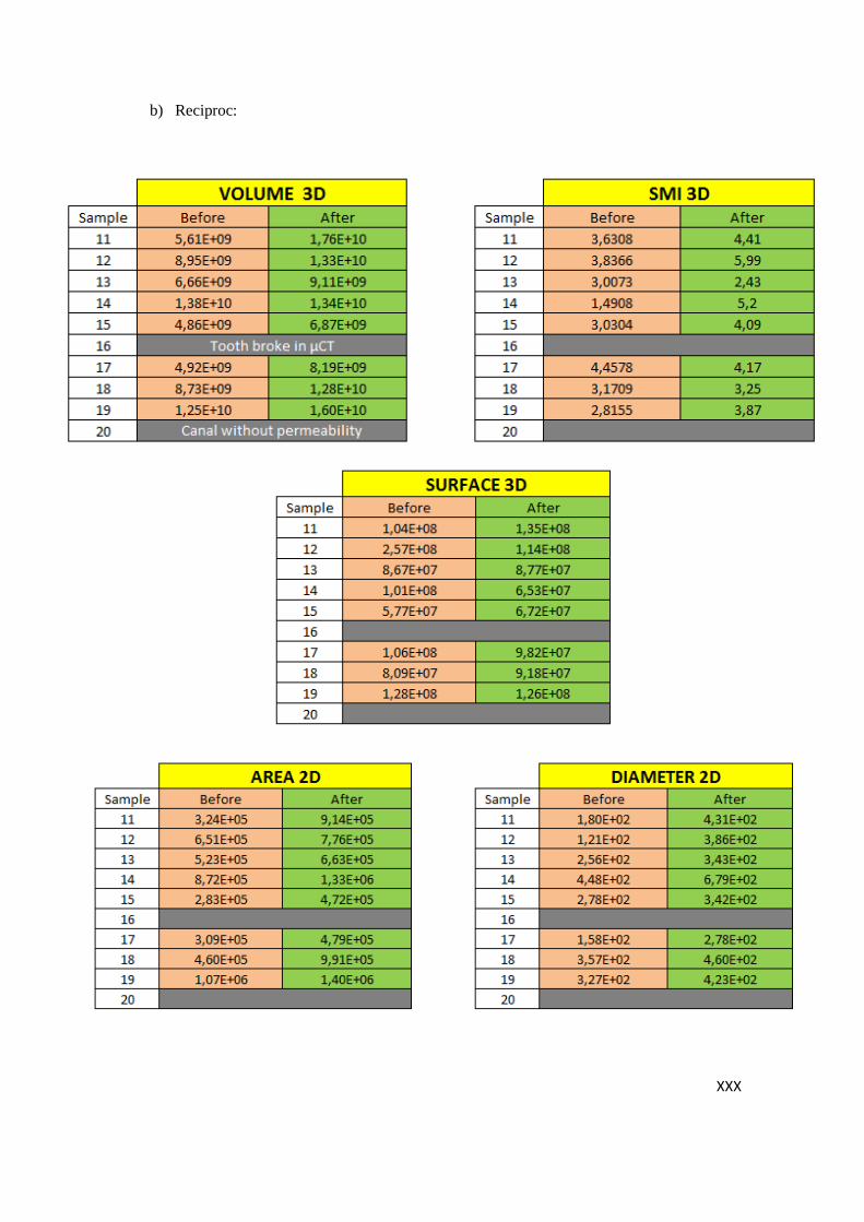
preparation. Furthermore, two of the samples were excluded: sample #16 fractured
during the analysis procedure in the micro tomography and the sample #20 had no
canalar permeability. Both samples were from the group B in study (the ReciprocTM
system), which has made this a shorter sample relative to the others (8 instead of 10
samples per group). In general, due to the difficulty in obtaining teeth within the
inclusion criteria, one potential limitation of this study can be a result of a relatively
small sample size (n total final = 28), however this is common to other µCT studies
(Ribeiro et al., 2013; Peters et al., 2003).
An ideal technique for the study of root canal anatomy would be the one that is
not only accurate, simple, nondestructive, but also and most importantly,
feasible and reproducible in an in vivo scenario (Neelakantan et al. 2010; Zhang et al.
2011). On the other hand, in an ex vivo scenario, nondestructive micro-computed
tomographic techniques (micro-CT) has gained popularity, because they provide
accuracy, high-resolution, and can be used for detailed quantitative and qualitative
measurements of root canal anatomy (Peters et al., 2000; Plotino et al., 2006). Micro-
CT is a powerful tool for research and preclinical education in fundamental procedures
of endodontic treatments, as well as for clinicians and researchers who desire to study
dental anatomy in great detail (Plotino et al., 2006). One of the advantages of this
method is that the dentist can observe the internal anatomy of teeth from different
angles and it can facilitate endodontic instrumentation. Furthermore, with this technique
Page 43
COMPARATIVE ANALYSIS OF ROOT CANAL INSTRUMENTATION USING PROTAPER NEXTTM, RECIPROCTM &
WAVEONEGOLDTM SHAPE SYSTEMS
2016
21
Raquel Tavares
it is possible to tilt and rotate the image while areas of interest were magnified (Grande
et al., 2012). Micro-CT provides a better assessment of fine anatomical structures
because of the possibility of using a higher exposure time (~40 min) and lower voxel
size (19.6 lm) than CBCT, for example, (exposure time: 20 sec; voxel size: 120– 150
lm) during the scanning procedure. Additionally, the possibility of micro-CT devices to
acquire imaging projections using a higher degree rotation of the specimen (360) in
comparison with Planmeca CBCT unit (200) allowed the development of a more
accurate and detailed 3D models of the root canal space. MicroCT offers exciting
potential; however, current imaging times, are around 2hours per sample, the equipment
is expensive and the 3-D reconstruction requires a high degree of computer expertise. In
addition, the technique is not suitable for clinical use; notwithstanding the limited
clinical applicability of micro-CT technology, this method has been proven to be the
current reference method for the ex vivo studies, like this one, of root canal anatomy.
Additional studies on endodontic techniques and instruments taking into
consideration the configuration of the root canals are required to provide more
information and better endodontic instrumentation.
6. CONCLUSIONS
According to the results of the present investigation, the null hypothesis was
rejected, because significant differences were obtained between the 3 file systems
regarding their shaping ability in mesial roots extracted from humans mandibular first
molars. Thus, based on the methodology used and the results obtained in this study, it
may be conclude that:
WaveOne GoldTM, ReciprocTM and ProTaper NextTM showed similar
shaping ability at the 3D canalar parameters: volume, SMI and surface;
Page 44
COMPARATIVE ANALYSIS OF ROOT CANAL INSTRUMENTATION USING PROTAPER NEXTTM, RECIPROCTM &
WAVEONEGOLDTM SHAPE SYSTEMS
2016
22
Raquel Tavares
About 2D parameters, there was significantly higher increase in canalar
area after preparation with ReciprocTM files comparing to WaveOne GoldTM and
ProTaper NextTM (without statistically significant difference between those two
systems). About the canalar diameter (the average), ReciprocTM and WaveOne GoldTM
revealed a significant increase post instrumentation, superior to the ProTaper NextTM
value;
In general, we can admit that the system ReciprocTM was which produced
major changes in the geometric conditions of the root canal, followed by WaveOne
GoldTM and ProTaper NextTM, respectively;
Since WaveOne GoldTM modern files are on market, there are no articles
or studies availing or comparing this system and so, the conclusions are limited.
7. ACKNOWLEDGMENTS
The author deny any financial affiliations related to this study or its sponsors.
Page 47
XVIII
REFERENCES
Alapati SB, Brantley WA, Iijma M, Clark WA, Kovarik L, Buie C, et al.
Metallurgical characterization of a new nickel-titanium wire for rotary
endodontic instruments. J Endod 2009;35:1589-1593
Bergmans L, Cleynenbreugel JV, Beullens M, Wavers M, Meerbeek BV.
Progressive versus constant tapered shaft design using NiTi rotary instruments.
IntEndod J. 2003;36:288-95
Brochure: Bruker (2015) Micro CT Academy. Innovation with integrity. 2015
August; 2(8):1-3
Bryant ST, Thompson SA, Al-Omari MAO, Dummer PMH. Shaping ability
of Profilerotary nickel-titanium instruments with ISO. IntEndod J.2005;5:256-78
Boutsiouki C, Gogos C, Verhaagen B, Versluis M, E. Kastrinakis & van der
Sluis L.W.M. The effect of apical preparation size on irrigant flow in root canals
evaluated using an unsteady Computational Fluid Dynamics model. Int Endod J. 2010
43, 874–881
Bürklein S, Hinschitza K, Dammaschke T, Schäfer E. Shaping ability and
cleaning effectiveness of two single-file systems in severely curved root canals of
extracted teeth: Reciproc and WaveOne versus Mtwo and ProTaper. Int Endod J. 2012
May;45(5):449- 61
Capar ID, Ertas H, Ok E, Arslan H, Ertas ET. Comparative study of different
novel nickel-titanium rotary systems for root canal preparation in severely curved root
canals. J Endod 2014;40:852-856
Chan AW, Cheung GS.A Comparisionstainlessteel and NiTi K files in curved
root canals. Int Endod J. 1996;29:170-5
Clifford J. Ruddle DDS - Advanced Endodontics: www.endoruddle.com
Cohen, S, Burns, R. Pathways of the pulp, 11th ed. Missouri: Ladig, D; 2015
Page 49
XX
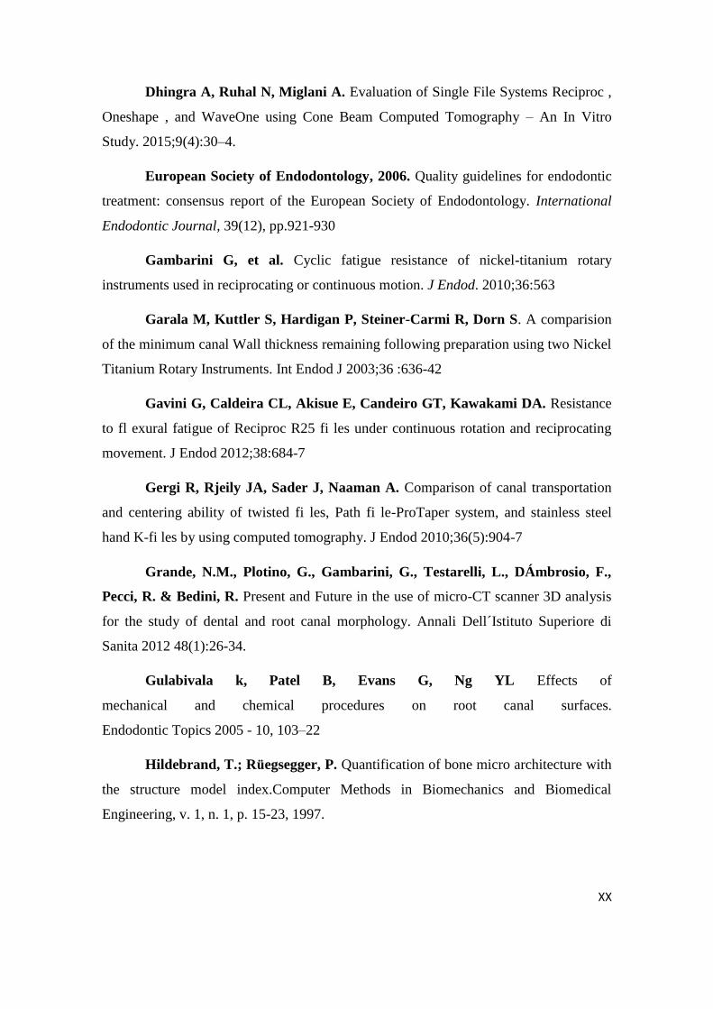
Dhingra A, Ruhal N, Miglani A. Evaluation of Single File Systems Reciproc ,
Oneshape , and WaveOne using Cone Beam Computed Tomography – An In Vitro
Study. 2015;9(4):30–4.
European Society of Endodontology, 2006. Quality guidelines for endodontic
treatment: consensus report of the European Society of Endodontology. International
Endodontic Journal, 39(12), pp.921-930
Gambarini G, et al. Cyclic fatigue resistance of nickel-titanium rotary
instruments used in reciprocating or continuous motion. J Endod. 2010;36:563
Garala M, Kuttler S, Hardigan P, Steiner-Carmi R, Dorn S. A comparision
of the minimum canal Wall thickness remaining following preparation using two Nickel
Titanium Rotary Instruments. Int Endod J 2003;36 :636-42
Gavini G, Caldeira CL, Akisue E, Candeiro GT, Kawakami DA. Resistance
to fl exural fatigue of Reciproc R25 fi les under continuous rotation and reciprocating
movement. J Endod 2012;38:684-7
Gergi R, Rjeily JA, Sader J, Naaman A. Comparison of canal transportation
and centering ability of twisted fi les, Path fi le-ProTaper system, and stainless steel
hand K-fi les by using computed tomography. J Endod 2010;36(5):904-7
Grande, N.M., Plotino, G., Gambarini, G., Testarelli, L., DÁmbrosio, F.,
Pecci, R. & Bedini, R. Present and Future in the use of micro-CT scanner 3D analysis
for the study of dental and root canal morphology. Annali Dell´Istituto Superiore di
Sanita 2012 48(1):26-34.
Gulabivala k, Patel B, Evans G, Ng YL Effects of
mechanical and chemical procedures on root canal surfaces.
Endodontic Topics 2005 - 10, 103–22
Hildebrand, T.; Ruegsegger, P. Quantification of bone micro architecture with
the structure model index.Computer Methods in Biomechanics and Biomedical
Engineering, v. 1, n. 1, p. 15-23, 1997.
Page 51
XXII
Hulsmann, M.; Peters, O. A.; Dummer, P. M. H. Mechanical preparation of
root canals: shaping goals, techniques and means. Endodontic Topics, v. 10, n. 1, p. 30-
76, 2005
Kim HC, Kwak SW, Cheung GS, Ko DH, Chung SM, Lee W. Cyclic fatigue
and torsional resistance of two new nickel-titanium instruments used in reciprocation
motion: Reciproc versus WaveOne. J Endod 2012;38:541-4.
Mehran AH, M AboEl-Fotouh. Comparison of Effects of ProTaper,
HeroShaper, and Gates Glidden Burs on Cervical Dentin Thickness and Root Canal
Volume
by using Multislice Computed Tomography. J Endod. 2008;34(10):1219-22
Nagy, c. D.; Bartha, K.; Bernath, M.; Verdes, E.; Szabo, J. The effect of root
canal morphology on canal shape following instrumentation using different
techniques. International Endodontic Journal, v. 30, n. 2, p. 133-140, 1997
Neelakantan P, Subbarao C, Subbarao CV Comparative evaluation of
modified canal staining and clearing technique, cone-beam computed tomography,
peripheral quantitative computed tomography, spiral computed tomography, and plain
and contrast medium-enhanced digital radiography in studying root canal morphology.
Journal of Endodontics (2010) 36, 1547–51
Nur BG, Ok E, Altunsoy M, Aglarci OS, Colak M. Evaluation of the root and
canal morphology of mandibular permanent molars in a south-eastern Turkish
population using cone-beam computed tomography. 2014;8(2):154–9
de Pablo OV, Estevez R, Peix Sanchez M, Heilborn C,
Cohenca N Root anatomy and canal configuration of the permanent mandibular first
molar: a systematic review. Journal of Endodontics (2010) 36, 1919–31
Peters OA. Current challenges and concepts in the preparation of root canal
systems: A review. J Endod 2004;30(8):559-67
Plotino, G., Grande, N.M., Pecci, R., Bedini, R., Pameijer, C.H. & Somma,
F. Three-dimensional imaging using microcomputed tomography for studying tooth
Page 53
XXIV
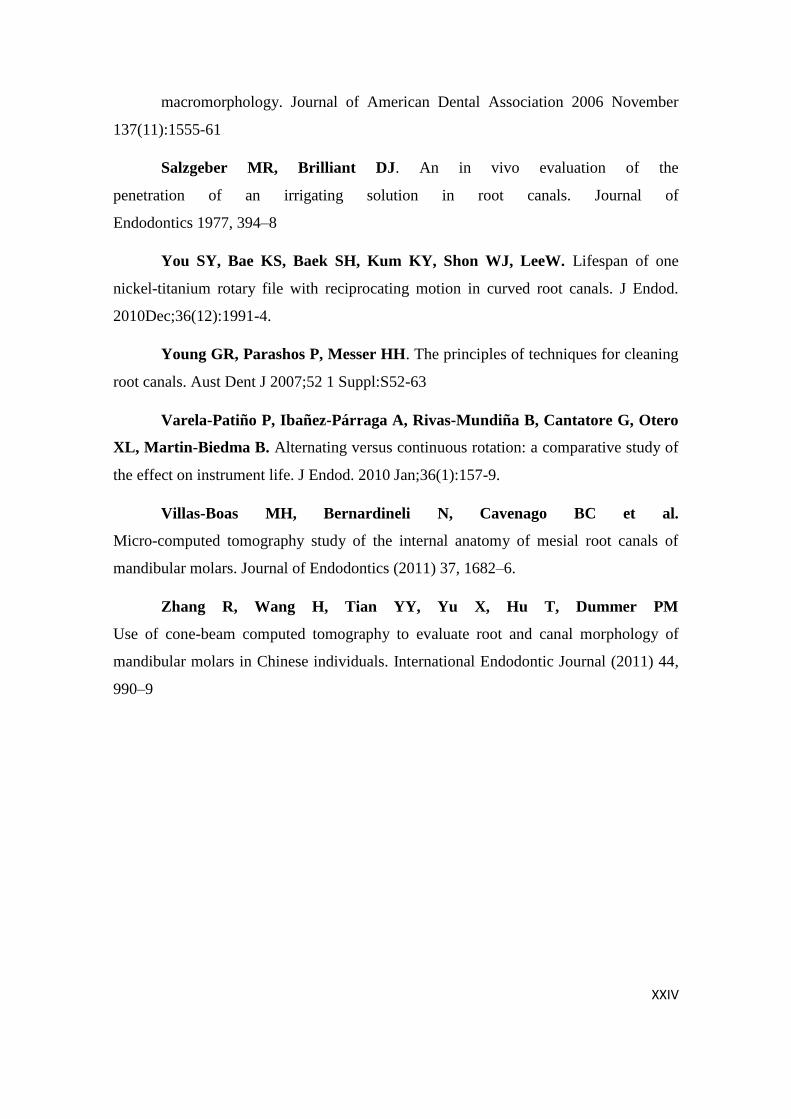
macromorphology. Journal of American Dental Association 2006 November
137(11):1555-61
Salzgeber MR, Brilliant DJ. An in vivo evaluation of the
penetration of an irrigating solution in root canals. Journal of
Endodontics 1977, 394–8
You SY, Bae KS, Baek SH, Kum KY, Shon WJ, LeeW. Lifespan of one
nickel-titanium rotary file with reciprocating motion in curved root canals. J Endod.
2010Dec;36(12):1991-4.
Young GR, Parashos P, Messer HH. The principles of techniques for cleaning
root canals. Aust Dent J 2007;52 1 Suppl:S52-63
Varela-Patiño P, Ibañez-Párraga A, Rivas-Mundiña B, Cantatore G, Otero
XL, Martin-Biedma B. Alternating versus continuous rotation: a comparative study of
the effect on instrument life. J Endod. 2010 Jan;36(1):157-9.
Villas-Boas MH, Bernardineli N, Cavenago BC et al.
Micro-computed tomography study of the internal anatomy of mesial root canals of
mandibular molars. Journal of Endodontics (2011) 37, 1682–6.
Zhang R, Wang H, Tian YY, Yu X, Hu T, Dummer PM
Use of cone-beam computed tomography to evaluate root and canal morphology of
mandibular molars in Chinese individuals. International Endodontic Journal (2011) 44,
990–9
Page 55
XXVI
APPENDIX
a) Abbreviations
- NiTi: Nickel-Titanium
- 2D: Bidimensional
- 3D: Tridimensional
- SMI: Structure Model Index
- CT: Computerized tomography
b) Symbols
- %: Percentage
- TM: Unregistered trademark
c) Units
- rpm: Rotations per minute
- mm: Millimeters
- N: Newton
- µm: Micrometer
- mL: Milliliter
- kV: Kilovolts
- µA: Micro amperes
- ms: Milliseconds
d) Figure Index
- Figure 1 – WaveOne GoldTM system
- Figure 2 – ReciprocTM system
- Figure 3 – ProTaper NextTM
- Figure 4 – Variability of canals system in mandibular first molar
- Figure 5 – Electric motor (Tecnika, Dentsply Maillefer, Schools Grant
Program)
- Figure 6 – Computerized Micro Tomography (IP Leiria)
Page 56
XXVII
e) Tables Index
- Table 1 – Morphometric data of the root canal before instrumentation
(mean ± standard deviation)
- Table 2 – Absolute change and percentage of root canal volume (mean ±
standard deviation)
- Table 3 – Absolute change and percentage of root canal SMI (mean ±
standard deviation)
- Table 4 – Absolute change and percentage of root canal surface (mean ±
standard deviation)
- Table 5 – Absolute change and percentage of root canal area (mean ±
standard deviation)
- Table 6 - Absolute change and percentage of root canal diameter (mean
± standard deviation)
f) Graphs Index
- Graph 1 – Volume box-plots
- Graph 2 – Surface box-plots
- Graph 3 – Area box-plots
- Graph 4 – Diameter box-plots
Page 57
XXVIII
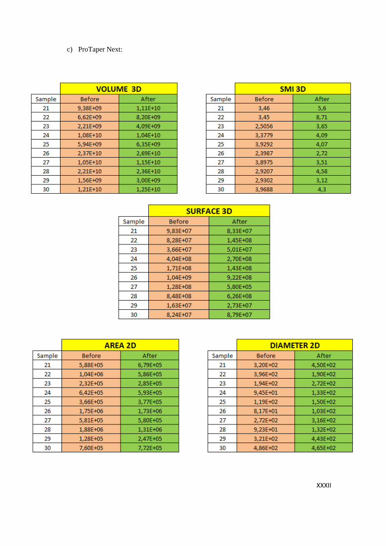
APPENDIX 2 - Tables of parameters observations (from µCT):
a) WaveOne Gold:
Page 61
XXXII
c) ProTaper Next:
Page 63
XXXIV
APPENDIX 3
Image example from micro CT (Sample nr. 3 - post instrumentation):