Palaeodiversity 2: 233–270; Stuttgart, 30.12.2009. 233 Comparative histology of sculptured dermal bones in basal tetrapods, and the implications for the soft tissue dermis FLORIAN WITZMANN Abstract The histology of the sculptured dermal bones of skull and pectoral girdle of 19 taxa of Palaeozoic and Meso- zoic basal tetrapods and of the porolepiform Laccognathus is investigated. The dermal bones consist generally of compact external and internal cortices that frame a cancellous or trabecular middle region. In Laccognathus, thin, unmineralized Sharpey’s fibers that are loosely arranged penetrate the external cortex between the odontodes. After the reduction of odontodes in finned stem-tetrapods, dermal sculpture developed via preferential growth of bone without the involvement of resorptive processes. In the stem-tetrapod Panderichthys, the Sharpey’s fibers are well-mineralized and indicate a tight connection to the overlying soft-tissue integument. The fibers are most numer- ous and most densely arranged in the bony ridges and tubercles, which constituted the main points of anchorage for the skin. The morphology and morphogenesis of the bony sculpture and its association with mineralized Sharpey’s fibers was retained during the fish-tetrapod transition and basically conserved in the different lineages of basal tetrapods including basal amniotes. The dermal bones of the stem-tetrapods Panderichthys, Acanthostega and Greererpeton are composed to a large degree of parallel-fibered bone, and a ‘fish-like’ character is the internal cortex that consists of isopedine. In crown-group tetrapods, the Sharpey’s fibers are generally much thicker and more densely arranged than in stem- tetrapods, and metaplastic bone can be demonstrated in addition to parallel-fibered bone in many taxa. These data suggest that the first crown-group tetrapods had attained a denser integument that might have provided a better resistance against water loss and mechanical damage during locomotion on land. In contrast to extant lissamphib- ians, the denser integument as well as ossified dermal scales and the comparatively large body size probably pre- cluded large-scale cutaneous respiration in most basal tetrapods. The middle region of dermal bones shows varying degrees of resorption and secondary growth among taxa. In heavily ossified forms such as Eryops or Mastodonsaurus, the weight of the skeleton helped them to stay under water, whereas skeletal lightening by reduced cortices and a highly porous middle region may have enhanced the agility and manoeuvrability during swimming. K e y w o r d s : Bone microstructure, fish-tetrapod transition, integument, Mesozoic, metaplasia, Palaeozoic, Sharpey’s fibers. Zusammenfassung In dieser Studie wird die Histologie der skulptierten Hautknochen des Schädels und des Schultergürtel von 19 Taxa basaler Tetrapoden aus dem Paläozoikum und Mesozoikum sowie des Porolepiformen Laccognathus unter- sucht. Die Hautknochen bestehen normalerweise aus kompakten äußeren und inneren Cortices, die eine spongiöse mittlere Region einschließen. Laccognathus besitzt dünne, nicht mineralisierte Sharpey’sche Fasern im externen Cortex, die locker zwischen den Odontoden angeordnet sind. Nach Reduktion der Odontoden bei fischartigen Stamm-Tetrapoden entstand die äußere Skulptur der Dermalknochen durch bevorzugtes Knochenwachstum ohne Anzeichen damit verbundener Knochenresorption. Bei dem Stamm-Tetrapoden Panderichthys sind die Sharpey’- schen Fasern gut mineralisiert und deuten auf eine feste Verbindung zwischen der Knochenoberfläche und der überlagernden Haut hin. Die Fasern sind am zahlreichsten und dichtesten in den knöchernen Tuberkeln und Leisten angeordnet, welche die Hauptverankerungspunkte der Haut darstellten. Morphologie und Morphogenese der Kno- chenskulptur und ihre Assoziation mit mineralisierten Sharpey’schen Fasern wurden während des Fisch-Tetrapoden Überganges beibehalten und in den verschiedenen Linien basaler Tetrapoden einschließlich basaler Amnioten be- wahrt. Die Hautknochen der Stamm-Tetrapoden Panderichthys, Acanthostega und Greererpeton bestehen zu einem großen Teil aus parallelfaserigem Knochen, und ein „fischartiges“ Merkmal ist das Vorhandensein von Isopedin im internen Cortex. Bei Kronengruppen-Tetrapoden sind die Sharpey’schen Fasern im allgemeinen deutlich dicker und dichter angeordnet als bei Stamm-Tetrapoden, und neben parallelfaserigem Knochen kann oft metaplastischer Kno- chen nachgewiesen werden. Diese Befunde deuten darauf hin, dass die ersten Kronengruppen-Tetrapoden eine derbe Haut besessen haben, die wahrscheinlich einen besseren Schutz gegen Wasserverlust und Verletzung beim Laufen über das Land bot. Im Gegensatz zu heutigen Lissamphibien verhinderten die derbere Haut sowie die häufig vorhandenen Knochenschuppen und das ungünstige Verhältnis von Körperoberfläche zu Volumen eine weit- reichende Hautatmung bei den meisten basalen Tetrapoden. Die mittlere Region der Hautknochen war von Taxon zu Taxon in unterschiedlichem Maße von Resorption und sekundärem Wachstum betroffen. Bei schwer verknöcherten Formen wie Eryops und Mastodonsaurus wirkte das Gewicht des Skeletts dem Auftrieb im Wasser entgegen, wohingegen eine Gewichtsreduktion durch dünnere Cor- tices und eine stark poröse mittlere Region die Wendigkeit beim Schwimmen sicherlich erhöht hat.
Comparative histology of sculptured dermal bones in basal tetrapods, and the implications for the soft tissue dermis
FLORIAN WITZMANN
A b s t r a c tThe histology of the sculptured dermal bones of skull and pectoral girdle of 19 taxa of Palaeozoic and Meso-
zoic basal tetrapods and of the porolepiform Laccognathus is investigated. The dermal bones consist generally of compact external and internal cortices that frame a cancellous or trabecular middle region. In Laccognathus, thin, unmineralized Sharpey’s fibers that are loosely arranged penetrate the external cortex between the odontodes. After the reduction of odontodes in finned stem-tetrapods, dermal sculpture developed via preferential growth of bone without the involvement of resorptive processes. In the stem-tetrapod Panderichthys, the Sharpey’s fibers are well-mineralized and indicate a tight connection to the overlying soft-tissue integument. The fibers are most numer-ous and most densely arranged in the bony ridges and tubercles, which constituted the main points of anchorage for the skin. The morphology and morphogenesis of the bony sculpture and its association with mineralized Sharpey’s fibers was retained during the fish-tetrapod transition and basically conserved in the different lineages of basal tetrapods including basal amniotes.
The dermal bones of the stem-tetrapods Panderichthys, Acanthostega and Greererpeton are composed to a large degree of parallel-fibered bone, and a ‘fish-like’ character is the internal cortex that consists of isopedine. In crown-group tetrapods, the Sharpey’s fibers are generally much thicker and more densely arranged than in stem-tetrapods, and metaplastic bone can be demonstrated in addition to parallel-fibered bone in many taxa. These data suggest that the first crown-group tetrapods had attained a denser integument that might have provided a better resistance against water loss and mechanical damage during locomotion on land. In contrast to extant lissamphib-ians, the denser integument as well as ossified dermal scales and the comparatively large body size probably pre-cluded large-scale cutaneous respiration in most basal tetrapods.
The middle region of dermal bones shows varying degrees of resorption and secondary growth among taxa. In heavily ossified forms such as Eryops or Mastodonsaurus, the weight of the skeleton helped them to stay under water, whereas skeletal lightening by reduced cortices and a highly porous middle region may have enhanced the agility and manoeuvrability during swimming.
K e y w o r d s : Bone microstructure, fish-tetrapod transition, integument, Mesozoic, metaplasia, Palaeozoic, Sharpey’s fibers.
Z u s a m m e n f a s s u n gIn dieser Studie wird die Histologie der skulptierten Hautknochen des Schädels und des Schultergürtel von 19
Taxa basaler Tetrapoden aus dem Paläozoikum und Mesozoikum sowie des Porolepiformen Laccognathus unter-sucht. Die Hautknochen bestehen normalerweise aus kompakten äußeren und inneren Cortices, die eine spongiöse mittlere Region einschließen. Laccognathus besitzt dünne, nicht mineralisierte Sharpey’sche Fasern im externen Cortex, die locker zwischen den Odontoden angeordnet sind. Nach Reduktion der Odontoden bei fischartigen Stamm-Tetrapoden entstand die äußere Skulptur der Dermalknochen durch bevorzugtes Knochenwachstum ohne Anzeichen damit verbundener Knochenresorption. Bei dem Stamm-Tetrapoden Panderichthys sind die Sharpey’- schen Fasern gut mineralisiert und deuten auf eine feste Verbindung zwischen der Knochenoberfläche und der überlagernden Haut hin. Die Fasern sind am zahlreichsten und dichtesten in den knöchernen Tuberkeln und Leisten angeordnet, welche die Hauptverankerungspunkte der Haut darstellten. Morphologie und Morphogenese der Kno-chenskulptur und ihre Assoziation mit mineralisierten Sharpey’schen Fasern wurden während des Fisch-Tetrapoden Überganges beibehalten und in den verschiedenen Linien basaler Tetrapoden einschließlich basaler Amnioten be-wahrt.
Die Hautknochen der Stamm-Tetrapoden Panderichthys, Acanthostega und Greererpeton bestehen zu einem großen Teil aus parallelfaserigem Knochen, und ein „fischartiges“ Merkmal ist das Vorhandensein von Isopedin im internen Cortex. Bei Kronengruppen-Tetrapoden sind die Sharpey’schen Fasern im allgemeinen deutlich dicker und dichter angeordnet als bei Stamm-Tetrapoden, und neben parallelfaserigem Knochen kann oft metaplastischer Kno-chen nachgewiesen werden. Diese Befunde deuten darauf hin, dass die ersten Kronengruppen-Tetrapoden eine derbe Haut besessen haben, die wahrscheinlich einen besseren Schutz gegen Wasserverlust und Verletzung beim Laufen über das Land bot. Im Gegensatz zu heutigen Lissamphibien verhinderten die derbere Haut sowie die häufig vorhandenen Knochenschuppen und das ungünstige Verhältnis von Körperoberfläche zu Volumen eine weit-reichende Hautatmung bei den meisten basalen Tetrapoden.
Die mittlere Region der Hautknochen war von Taxon zu Taxon in unterschiedlichem Maße von Resorption und sekundärem Wachstum betroffen. Bei schwer verknöcherten Formen wie Eryops und Mastodonsaurus wirkte das Gewicht des Skeletts dem Auftrieb im Wasser entgegen, wohingegen eine Gewichtsreduktion durch dünnere Cor-tices und eine stark poröse mittlere Region die Wendigkeit beim Schwimmen sicherlich erhöht hat.
234 PALAEODIVERSITY 2, 2009
1. Introduction
Dermal bones of the skull and the pectoral girdle de-velop within the integument, generally in the lower layer of the dermis (CASTANET et al. 2003). These ossifications are frequently penetrated by numerous canals that carried blood vessels and nerves (including the lateral line system of fishes and non-amniote basal tetrapods) to the external bone surface and into the directly overlying integument. The histomorphology of dermal bones as well as the struc-ture of their external surface are therefore well suited as osteological correlates of the integumentary structure in fossil vertebrates. In the different groups of fossil fishes and early vertebrates, the dermal bones of skull and pecto-ral girdle, the scales and armour plates have been subject to histological investigation for a long time, so that the
internal structure of these dermal ossifications are the most extensively studied among vertebrates (e. g., BYSTROW 1939, 1942, 1957; ØRVIG 1951, 1957, 1966, 1968, 1989; THOMSON 1977; GROSS 1930, 1957, 1973; BEMIS & NORTH-CUTT 1992; SMITH 1977).
In fossil tetrapods, the focus of histological investiga-tions lies most often on the long bones, including the ap-plication of skeletochronology as an important method to infer biological parameters like growth rate, individual age and mode of life, and a large amount of data has ac-cumulated over the last decades (e. g., DE RICQLÈS 1975a, b, 1976, 1977, 1981, 1993; CHINSAMY 1993; CHINSAMY-TURAN 2005; DAMIANI 2000; STEYER et al. 2004; SANDER & AN-DRÁSSY 2006; KLEIN & SANDER 2007, 2008; SANCHEZ et al. 2008). More recently, also the histology of dermal ossifi-cations of the trunk (i. e., osteoderms) in fossil amniotes
WITZMANN, HISTOLOGY OF SCULPTURED DERMAL BONES 235
has attracted increasing interest of palaeontologists and zoologists, e. g. in turtles (SCHEYER & SÁNCHEZ-VILLAGRA 2007; SCHEYER & ANQUETIN 2008), placodonts (SCHEYER 2007), dinosaurs (DE BUFFRÉNIL et al. 1986; DE RICQLÈS et al. 2001; SCHEYER & SANDER 2004; MAIN et al. 2005), xe-narthrans (HILL 2005, 2006), and basal tetrapods (WITZ-MANN & SOLER-GIJÓN 2008).
The histology of the dermal ossifications of skull and pectoral girdle in temnospondyls and other basal tetra-pods, in contrast, has received the attention of compara-tively few workers who investigated only a very limited range of taxa. Within the work on the Early Permian ‘branchiosaurs’ from the Döhlen Basin in Saxony, CRED-NER (1893, pl. 30, figs. 4–6; pl. 31, figs. 8–9) illustrated schematically the course of presumed blood vessels in dermal skull bones of the temnospondyl Onchiodon and presented a histological section of the vomer (CREDNER 1893, pl. 31, fig. 4).
SEITZ (1907) described the histology of a mandible fragment of ?Mastodonsaurus. He observed a compact outer region consisting of lamellar bone with simple vas-cular canals and primary osteons. More internally in the bone, SEITZ (1907) found secondary osteons (Haversian systems) and irregular caverns of a spongy region.
GROSS (1934) provided a short description of the der-mal skull bone histology of Mastodonsaurus, Metoposau-rus and Plagiosternum. He recognized that the dermal bones of these temnospondyls exhibit a diploë structure, i. e., a spongy middle region is framed by an external and an internal compact cortex, and designated the matrix of the cortical bone as zonal periosteal bone (zonarer Periost-knochen).
As GROSS (1934) noted, the internal cortex lacks the isopedine-like organization of many finned sarcoptery-gians. A detailed study of the histology of dermal skull bones in the stereospondyl Benthosuchus was published by BYSTROW (1935). He recognized horizontally aligned large canals in the middle, spongy region from which oblique canals branch off, traverse the external cortex and open to the external bone surface on the floor of the sculp-tural pits and furrows. BYSTROW (1935) described a fine network of capillaries additional to these large canals in the external region that open to the bone surface via small pores. He designated this network as ‘rete vasculosum’. In a later study, BYSTROW (1947) interpreted these capillaries as serving for cutaneous respiration and compared the vascularization of the dermal bones of Benthosuchus with those of the stereospondylomorphs Wetlugasaurus and Platyoposaurus, and the dvinosaur Dvinosaurus. In his studies, BYSTROW (1935, 1947) had focused mainly on the morphology and course of blood vessels within the bone, but he neither described the bone matrix proper nor the presence of extrinsic fibers.
In the first part of their comprehensive works on bone
histology, ENLOW & BROWN (1956) concentrated on the study of long bones of tetrapods, but also commented on the arrangement and orientation of vascular canals in der-mal bones of temnospondyls (Edops, Trimerorhachis, Eryops) and of the stem-amniote Seymouria. PEABODY (1961) investigated cyclical growth zones in sections of the dentaries of Early Permian ‘microsaurs’ and indetermi-nate ‘labyrinthodonts’ from Fort Sill, Oklahoma. On the basis of his findings, PEABODY (1961) discussed palaeocli-matic inferences like alternating wet and dry seasons for this locality.
In 1974, COLDIRON published his work on the possible function of dermal bone sculpture in temnospondyls and other basal tetrapods. He challenged BYSTROW’s assump-tion that the ‘rete vasculosum’ was functionally associated with cutaneous respiration since the capillaries describe an irregular pathway and thus an inefficient route of the blood to the bone surface and the skin. Based on split-line technique in dermal skull bones of Alligator and the orien-tation of the long-axis orientation in bone cell lacunae of dermal bones of Eryops, COLDIRON (1974) inferred the collagen-fiber orientation within the bone. He found the collagen fibers arranged parallel to one another in the in-ternal and middle region, but non-parallel and randomly oriented in the sculptured external region. Thus, COLDIRON (1974) concluded that dermal bone sculpture in basal tetra-pods and crocodilians is a strengthening adaptation by distributing the stress that acted on the dermal skull roof.
COSGRIFF & ZAWISKIE (1979) described a capillary net-work that opens to the sculptured surface in the dermal bones of the rhytidosteid temnospondyl Pneumatostega, although they did not prepare thin sections. Following BYSTROW (1947), they interpreted this vascularization as indication of cutaneous respiration.
DE RICQLÈS (1981), in his comprehensive work on long bones of fossil tetrapods, also commented on dermal skull bones of the temnospondyl Trematops and the nectridean Diplocaulus. In Trematops (DE RICQLÈS 1981, pl. 1, fig. 4), he described parallel-fibered bone with simple primary canals in the external cortex, and recognized lamellar-zonal bone in the external cortex of Diplocaulus (DE RIC-QLÈS 1981, pl. 2, fig. 3).
CASTANET et al. (2003) published a useful compilation of the present knowledge on bone histology of extant and extinct amphibians including stem-amniotes and early tet-rapods. They confirmed the diploë structure as described by GROSS (1934) and BYSTROW (1935, 1947) as basic pattern for most dermal bones. According to CASTANET et al. (2003), the external cortex consists of primary bone tissue with a lamellar structure and contains simple vascular ca-nals and primary osteons. Zones and annuli (and some-times lines of arrested growth) indicate that bone deposi-tion was cyclic. The middle spongy or cancellous region has undergone remodeling whose degree varies between
236 PALAEODIVERSITY 2, 2009
taxa. The internal region is lamellar and usually penetrat-ed by a small number of simple canals that run parallel to the internal bone surface. Sharpey’s fibers may cross this region at a steep angle.
In his analysis of placodont osteoderm histology, SCHEYER (2007) also referred to the internal structure of temnospondyl dermal bones and illustrated histological sections of Trimerorhachis, Mastodonsaurus and Ger-rothorax. He observed a diploë pattern with a generally high vascularization and secondary remodeling in the middle region. The external and internal cortices consist mainly of parallel-fibered bone with growth marks, and transitions to interwoven structural fibers (ISF) in Mast-odonsaurus exist. SCHEYER (2007) observed Sharpey’s fi-bers that penetrate the bone of the sculptural ridges in Mastodonsaurus.
The aim of the present paper is to close the gap in the literature about dermal bone histology in basal tetrapods, and to provide a basis for comparison with the histology of dermal bones plus the overlying integument in extant sculpture-bearing fishes and tetrapods, that will be car-ried out in a future paper (WITZMANN in progress). Consid-ered in the present study are dermal bones of the skull and the pectoral girdle, whereas the histology of osteoderms in temnospondyls is dealt with in a separate publication (WITZMANN & SOLER-GIJÓN 2008). Importance is especial-ly attached to the different types of bone tissues, the course of intrinsic bone fibers and the presence of extrin-sic fibers. A further focus will be the degree of vascular-ization and the type and morphology of the vascular ca-nals, and also the mode of growth of the dermal bones, especially of the external sculpture. The results will be taken to draw conclusions about the integument in which these bones were formed, and possible functional aspects of dermal bone sculpture in basal tetrapods will be dis-cussed.
I n s t i t u t i o n a l a b b r e v i a t i o n sCMNH Cleveland Museum of Natural History, Cleveland, Ohio (USA)MB Leibniz Institute for Research on Evolution and Biodi-
versity at the Humboldt University Berlin, Museum für Naturkunde (Germany)
MCZ Museum of Comparative Zoology, Harvard, Cam- bridge/Mass. (USA)
SMNS Staatliches Museum für Naturkunde Stuttgart (Ger- many)
UCMP University of California, Museum of Paleontology, Berkeley (USA)
UMZC University Museum of Zoology, Cambridge (UK)
A n a t o m i c a l a b b r e v i a t i o n scl bone cell lacunacr crevice (artefact)de dentineEC external cortex
en enamelER erosion roomfbs former bone surfacefl longitudinally cut bone fibersft transversely cut bone fibersGM growth marksIC internal cortexIL interstitial lamellaeISF interwoven structural fiber bundlesLB lamellar boneMR middle regionov opening of large vesselPB primary bonePFB parallel-fibered bonePO primary osteonPVC primary vascular canalRL resorption lineShF Sharpey’s fibersSB secondary boneSO secondary osteontr trabeculae
A c k n o w l e d g e m e n t sI am indebted to the following people who allowed me to
produce thin sections of dermal bones from collections under their care: JENNY CLACK (Cambridge), PAT HOLROYD and KEVIN PADIAN (Berkeley), OLIVER HAMPE, JOHANNES MÜLLER and JÜRGEN KRIWET (Berlin), MICHAEL RYAN (Cleveland), CHUCK SCHAFF (Harvard) and RAINER SCHOCH (Stuttgart). RODRIGO SOLER-GIJÓN (Berlin) is greatly acknowledged for many discussions about dermal bone histology and for drawing my attention to the con-nection between bone microstructure and the mode of life in aquatic animals. The helpful reviews of NICOLE KLEIN (Bonn), ANDREW MILNER (London) and TORSTEN SCHEYER (Zürich) im-proved the manuscript. HANS-PETER SCHULTZE (Lawrence) and RAINER SCHOCH (Stuttgart) gave many suggestions. I thank ANJA PIGOWSKE, HANS-RUDOLF KNÖFLER, HENRIK STÖHR (Berlin) and NORBERT ADORF (Stuttgart) for preparing the histological thin sections. This study was supported by the DFG.
2. Material and methods
Taxa whose dermal bones were investigated histologi-cally by thin sections and their inventory numbers are listed in Table 1. Altogether, 103 histological slides have been prepared from dermal bones of skull and pectoral girdle of 20 taxa of finned and limbed sarcopterygians. The dermal bone fragments were first embedded in syn-thetic resin (Paraloid B72, an ethyl-methacrylat-copoly-mere) and then cut vertically either parallel or transverse to the direction of the sculptural ridges. Thin-sections were prepared with a thickness of approximately 30–50 μm using the standard method of CHINSAMY & RAATH (1992). Additionally, one slide of Plagiosuchus pustuliferus (FRAAS, 1896) (MB.Hi.1705), one slide of Plagiosternum granulosum (FRAAS, 1889) (MB.Hi.1714), and six slides of dermal bones of Metoposaurus diagnosticus (VON MEYER, 1842) (MB.Hi.1718–1723) produced by WALTER GROSS in 1934 and stored in the Museum für Naturkunde Berlin,
WITZMANN, HISTOLOGY OF SCULPTURED DERMAL BONES 237
Tab. 1. Taxa investigated in this study with list of specimens and their inventory numbers.
Taxon Stratigraphy Locality Specimens/RemarksPorolepiform and stem-tetrapodsLaccognathus panderi GROSS, 1941 Middle Devonian Riga, Latvia MB.f.17666: several fragments of dermal skull
or pectoral girdle. 4 slidesPanderichthys rhombolepis (GROSS, 1930)
Late Devonian, Gauja beds Latvia MB.f.17548: several fragments of dermal skull
or pectoral girdle. 5 slides
Acanthostega gunnari JARVIK, 1952
Late Devonian, upper Famennian, Britta Dal Formation
Stensiö Bjerg, Green-land UMZC 150b: 1 fragment of cheek. 3 slides
Greererpeton burkemorani ROMER, 1969
upper Viséan/lower Namurian
Greer, West Virginia, USA
CMNH 11900: several fragments of dermal skull. 6 slides
Temnospondyls
Edops craigi ROMER, 1936 Early PermianTerrapin School, Archer County, Texas, USA
MCZ 1235: 3 fragments of dermal skull roof. 6 slides
Chenoprosopus milleri MEHL, 1913
Early Permian, Cutler Formation New Mexico, USA UCMP 41104: 5 pieces of dermal skull roof.
Pantylus cordatus COPE, 1881 Early Permian,Wichita Group Texas, USA UCMP 20296: fragment of dermal skull roof.
1 slideSeymouriamorpha
Seymouria baylorensis BROILI, 1904 Early Permian
Baylor County, Clear Fork, West Coffee Creek, Texas, USA
MCZ without number: 1 fragment of the lower jaw (?angular). 3 slides
EureptiliaLabidosaurus hamatus COPE, 1895
Early Permian,Clear Fork Group Texas, USA MCZ without number: 1 fragment of skull
table. 1 slide
238 PALAEODIVERSITY 2, 2009
were studied. The thin sections were examined by using a Leica DC 300 polarising stereosmicroscope with trans-mitted ordinary and polarised light. For the investigation of the bone structure by SEM, dermal bone fragments were first cut and polished, and the polished surface was then etched with 10 % HCL for 8–10 seconds.
All dermal bones sectioned in this study are assumed to belong to adults or, in the case of Sclerocephalus haeu-seri GOLDFUSS, 1847, Archegosaurus decheni GOLDFUSS, 1847 (based on skull length) and Acanthostega gunnari JARVIK, 1952 (based on bone thickness of the cheek), to subadults. The only small juvenile specimen in the sample belongs to Mastodonsaurus giganteus (JAEGER, 1828) and is listed and described separately from the adults of this species. For each taxon investigated here, the bone micro-structure and histology was consistent. Intraspecific vari-ability was only observed in Mastodonsaurus giganteus and Plagiosternum granulosum and affects the degree of vascularization of the bone (see description).
The terminology of FRANCILLON-VIEILLOT et al. (1990) and DE RICQLÈS et al. (1991) concerning bone histology will
be used throughout the text. Furthermore, I follow SCHEYER & ANQUETIN (2008) in their work on turtle shell bone histol-ogy in the use of the term ‘external’ and ‘internal’. An ap-propriate alternative designation is ‘superficial’ and ‘deep’, as used by HILL (2006) instead of ‘external’ and ‘internal’, respectively. The external sculptured surface of the dermal bone faces the body surface, whereas the internal surface is oriented to the visceral surface of the body. The term ‘interior’ corresponds to the inner or middle part of the bone. In general, the dermal bones show a diploë structure, i. e., a middle region that is cancellous or trabecular is mantled by compact external and internal cortices. Three-dimensionally interwoven bundles of collagen fibers with a well ordered fiber bundle arrangement showing no gen-eral isotropy under polarized light are designated here as ‘interwoven structural fibers’ (ISF) (SCHEYER & SANDER 2004; SCHEYER & SÁNCHEZ-VILLAGRA 2007; SCHEYER 2007; SCHEYER & ANQUETIN 2008). They are distinguished from woven or fibrous bone that shows general isotropy and the collagen fibers of which are irregularly and loosely ar-ranged (FRANCILLON-VIEILLOT et al. 1990; DE RICQLÈS et al.
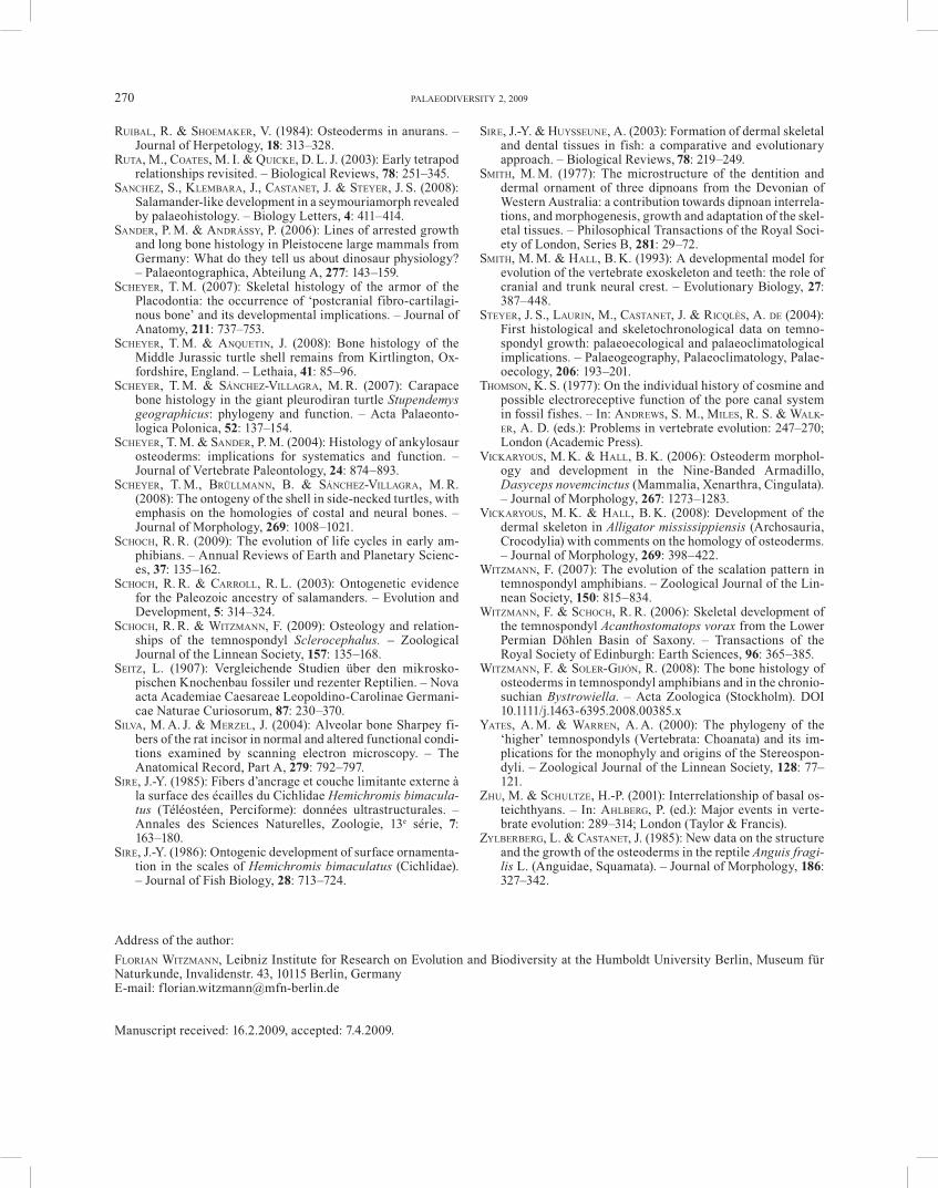
Fig. 1. Phylogenetic relationship of the taxa investigated in this study. After YATES & WARREN (2000), RUTA et al. (2003) and SCHOCH & WITZMANN (2009).
WITZMANN, HISTOLOGY OF SCULPTURED DERMAL BONES 239
1991). The sculptural tubercles and ridges on the external surface of the dermal bones are called ‘saddles’ and the grooves between them as ‘valleys’ in the thin sections. Canaliculi that are longer than the maximum diameter of the appertaining bone cell lacuna are designated as ‘long’, those that are approximately as long as the maximum di-ameter are ‘moderately long’, and those whose length is shorter than maximum diameter are termed ‘short’.
The interrelationships of basal tetrapods as found in the analyses of YATES & WARREN (2000), RUTA et al. (2003) and SCHOCH & WITZMANN (2009) are taken as the phyloge-netic framework of this study (Fig. 1). The term ‘crown-group tetrapod’ is used here in the sense of HENNIG (1966) as encompassing the last common ancestor of all living tetrapods and all its descendants, fossil and extant. The stem-group tetrapods define all fossil taxa that are more closely related to crown-group tetrapods than to the extant group that is most closely related to crown-group tetra-pods (either actinistians [e. g., ZHU & SCHULTZE 2001] or dipnoans [e. g., CLOUTIER & AHLBERG 1996]).
3. Description of histological thin sections
3.1. Outgroup finned sarcopterygian
3.1.1. Laccognathus panderi (Porolepiformes)
The external surface of the dermal skull bones of Lac-cognathus panderi GROSS, 1941 bears a sculpture of small, round to oval tubercles that give the bone a pustular ap-pearance. The sectioned bone has a thickness of approxi-mately 4.5 mm, and the ratio external cortex : middle re-gion : internal cortex is 1 : 1.5 : 0.6.
E x t e r n a l c o r t e x . As described by PANDER (1860), GROSS (1930) and ØRVIG (1957), the tubercles of the exter-nal bone surface represent odontodes (‘dermal teeth’) con-sisting of dentine with a cap of enamel (Fig. 2a–c), and several odontodes of earlier generations can be found ‘buried’ within the bone matrix of the external cortex and may be in the state of resorption. In some regions, the ex-ternal bone surface of an earlier growth phase can readily be traced as a sharp red-brownish line, extending from odontode to odontode. Sometimes this line is scalloped out to receive a vascular canal (Fig. 2a). Short, fine Sharp-ey’s fibers that measure approximately 2 μm in diameter cross the former external bone surface at approximately a right angle (Fig. 2a). They are regularly but not densely arranged, are hollow inside and filled by organic sub-stance, indicating that they were not or only poorly miner-alized in the living animal. The external cortex is com-posed mainly of fine parallel-fibered bone (Fig. 2b). Its fibers show regional changes in orientation: fibers that course approximately parallel to the surface of the section
appear bright or dark under polarized light, depending on the orientation of the slide, and have spindle-shaped bone cell lacunae that are bipolar, i. e., they possess short canali-culi at both tapering ends. Fibers that are cut approxi-mately perpendicularly remain dark under polarized light and the bone cell lacunae appear round. Vascularization is high and consists of anastomosing primary vascular ca-nals and primary osteons, that are often aligned in layers.
M i d d l e r e g i o n . The external cortex grades into the coarse cancellous and well vascularized middle region that possesses several primary and secondary osteons (Fig. 2c). In some areas, the bone is trabecular with large, irregularly shaped erosion rooms. The trabeculae are ir-regular in outline and lack lining of secondary bone. The primary bone matrix consists of parallel-fibered bone whose fibers show regionally different orientations, simi-lar to the external region. Near the transition to the inter-nal cortex, the bone matrix of the middle region contains several primary osteons aligned in rows.
I n t e r n a l c o r t e x . The internal cortex is separated from the middle region by a distinct transition. As out-lined by PANDER (1860) and GROSS (1930), the internal cortex of Laccognathus exhibits a plywood-structure that can be designated as isopedine (Fig. 2d). In polarized light, a regular pattern of horizontal bands is visible. Those bands in which the fibers are cut longitudinally appear bright or dark under polarized light (with elongate cell la-cunae), depending on the orientation of the slide, whereas those in which the fibers and bone cell lacunae are oblique-ly cut brighten up to a lesser extent. Bands with trans-versely cut fibers remain dark in polarized light (with round cell-lacunae) and exhibit a fine bright meshwork that delineates the single fiber bundles. Fine fibers, which are oriented approximately perpendicular to the internal bone surface, are discernable also in normal transmitted light. The internal cortex is avascular.
The dermal skull bones of Panderichthys rhombolepis (GROSS, 1930) bear basically a tubercular sculpture, and low sculptural ridges may connect the tubercles. The thickness of the sectioned bones amounts approximately 4 mm. The ratio external cortex : middle region : internal cortex is 1 : 0.8 : 0.4.
E x t e r n a l c o r t e x . The dermal bones of Pander-ichthys lack any dentine and enamel components, so that the sculptural tubercles and ridges are composed solely of bone tissue (Fig. 2e). Similar to odontodes, ‘buried’ tu-bercles of earlier generations are visible in the external region. As in Laccognathus, the bone matrix consists of
240 PALAEODIVERSITY 2, 2009
Fig. 2. a–d. Laccognathus panderi GROSS, 1941, MB.f.17666. Vertical sections of dermal bones of skull or pectoral girdle. a. External cortex in normal transmitted light; next to the odontode, the bone surface of an earlier growth stage with perpendicular, unmineral-ized Sharpey’s fibers is visible. b. External cortex with odontode in polarized light. c. External cortex with two odontodes and exter-nal part of middle region in normal transmitted light; primary and secondary osteons are arranged in rows. d. Internal cortex consist-ing of isopedine, polarized light. – e–f. Panderichthys rhombolepis (GROSS, 1930), MB.f.17548. Vertical sections of dermal bones of skull or pectoral girdle. e. External cortex in polarized light with sculptural valley and saddle, which consists solely of bone, the asterisk (*) indicates a ‘buried’ sculptural saddle of an earlier generation. f. Rows of primary osteons in a sculptural valley, normal transmitted light. – For abbreviations, see text.
WITZMANN, HISTOLOGY OF SCULPTURED DERMAL BONES 241
fine parallel-fibered bone whose fibers show locally dif-ferent orientations. Growth marks cannot be detected. Vascularization is moderate and consists of a succession of layers of primary osteons interior to the sculptural val-leys, and more interiorly, larger erosion cavities are visible (Fig. 2f). These layers of primary osteons are located in a parallel-fibered bone matrix with mostly transversely cut fibers (remain dark under polarized light) and round cell lacunae (Fig. 2e, f). Between these layers, layers of paral-lel-fibered bone whose fibers are cut longitudinally with spindular, bipolar bone cell lacunae are intercalated. This gives the bone the appearance of alternating dark and bright bands that wedge out at the lateral bases of the sculptural saddles under polarized light (Fig. 2e, on the left). In contrast to the valleys, the saddles are poorly vas-cularized by primary vascular canals, whereas more inte-riorly, several anastomosing primary vascular canals are present. Fine Sharpey’s fibers that extend far interiorly until to the middle region penetrate the sculptural saddles, both in the superficial saddles and in the ‘buried’ ones of earlier generations, but are absent in the valleys (Fig. 2e). The Sharpey’s fibers have a diameter of 5 to 6 μm. In po-larized light, the Sharpey’s fibers appear bright and dark, respectively, depending on the orientation of the slide. This indicates that they were well mineralized in the liv-ing animal.
M i d d l e r e g i o n . This region is coarse cancellous. In the more interior part, the region is trabecular with ir-regular, thin to moderately thick trabeculae that enclose large erosion cavities that are lined by secondary bone la-mellae (Fig. 3a). The primary matrix consists mostly of fine parallel-fibered bone. Several smaller secondary os-teons are present and some of them cut each other, but do not form Haversian tissue. The middle region is separated from the internal cortex by a distinct transition.
I n t e r n a l c o r t e x . The internal cortex consists of isopedine, similar to the internal cortex of Laccognathus, but appears less regular, since the bands are of different thickness and may fray out at their lateral ends (Fig. 3b). Fine fibers that are approximately perpendicular to the internal bone surface are visible in the dark bands. Scat-tered primary vascular canals run parallel or oblique to the internal bone surface.
3.2.2. Acanthostega gunnari
The investigated sections of Acanthostega gunnari JARVIK, 1952 were prepared through the cheek region (probably the squamosal, whose bone thickness varies between 1 mm and 3 mm) with polygonal sculpture. The ratio external cortex : middle region : internal cortex is 1 : 1.8 : 0.9.
E x t e r n a l c o r t e x . The bone matrix consists of
parallel-fibered bone that is mostly homogeneous, where-as in some regions, it appears coarse and less regular (Fig. 3c, d). As in the finned sarcopterygians desribed above, the bone fibers may change their orientation in different regions, and this is reflected by the shape of the bone cell lacunae. The bone is moderately to highly vascularized by primary vascular canals and primary osteons. Loosely ar-ranged Sharpey’s fibers with a diameter of 3 to 5 μm pen-etrate the sculptural saddles until to the middle region (Fig. 3c), but they can also be found in the valleys where they are less abundant. Growth marks are not visible in the external cortex.
M i d d l e r e g i o n . This region is coarse cancellous with primary osteons and several, partially large second-ary osteons (Fig. 3d). The bone matrix consists of fine parallel-fibered bone and of the lamellar bone of the sec-ondary osteons.
I n t e r n a l c o r t e x . Isolated primary vascular ca-nals may locally be present and run parallel to the internal bone surface. The bone matrix is composed of parallel-fi-bered bone that appears fine in most regions, but may also change its fiber orientation in irregular layers, so that the internal cortex resembles irregular isopedine in some re-gions (Fig. 3e). Fine fibers are visible that cross the inter-nal cortex approximately at a right angle.
3.2.3. Greererpeton burkemorani (Colosteidae)
Greererpeton burkemorani ROMER, 1969 has strongly sculptured dermal bones with high sculptural ridges. The skull fragment used for sectioning (?quadratojugal) has a sculpture of polygons and furrows of irregular outline, and its thickness ranges from 3.5 mm to more than 5 mm. The ratio external cortex : middle region : internal cortex is 1 : 1.4 : 1.3.
E x t e r n a l c o r t e x . As in the taxa described above, the bone matrix of the external cortex consists mainly of parallel-fibered bone the fibers of which show varying orientation in some regions (Fig. 3f), what is reflected in the shape of the bone cell lacunae (Fig. 4a). The intrinsic fibers of the parallel-fibered bone are mostly coarse and vary in extent and direction what gives the bone tissue often a less ordered appearance. Fine Sharpey’s fibers (Fig. 3f) that are well mineralized penetrate the external region at approximately a right angle to the surface and are more abundant in the sculptural saddles than in the val-leys. The Sharpey’s fibers have a diameter of 3 to 7 μm. The external region is well vascularized by partially anas-tomosing primary vascular canals (Fig. 4a) and scattered primary osteons. Growth marks cannot be observed.
M i d d l e r e g i o n . The external cortex grades into a fine to coarse cancellous region that is very well vascular-ized by numerous primary and secondary osteons whose
242 PALAEODIVERSITY 2, 2009
Fig. 3. a–b. Panderichthys rhombolepis (GROSS, 1930), MB.f.17548. Vertical sections of dermal bones of skull or pectoral girdle. a. Middle region in normal transmitted light with secondary osteons. b. Internal cortex consisting of isopedine, polarized light. – c–e. Acanthostega gunnari JARVIK, 1952, UMZC T 150b. Vertical section of the cheek, probably squamosal. c. Sculptural saddle of external cortex with Sharpey’s fibers and primary vascular canals, polarized light. d. Histological overview in polarized light, the external cortex is well vascularized, and secondary remodeling took place in the middle region. e. Internal region in polarized light, the bone fibers may show changing orientation in different layers, so that it has locally an isopedine-like appearance. – f. Greerer-peton burkemorani ROMER, 1969, CMNH 11900. Vertical sections of dermal skull bone (?quadratojugal). Low sculptural saddle of external cortex in polarized light; the parallel-fibered bone shows regional changes of its bone fiber orientation, and thin Sharpey’s fibers are present. – For abbreviations, see text.
WITZMANN, HISTOLOGY OF SCULPTURED DERMAL BONES 243
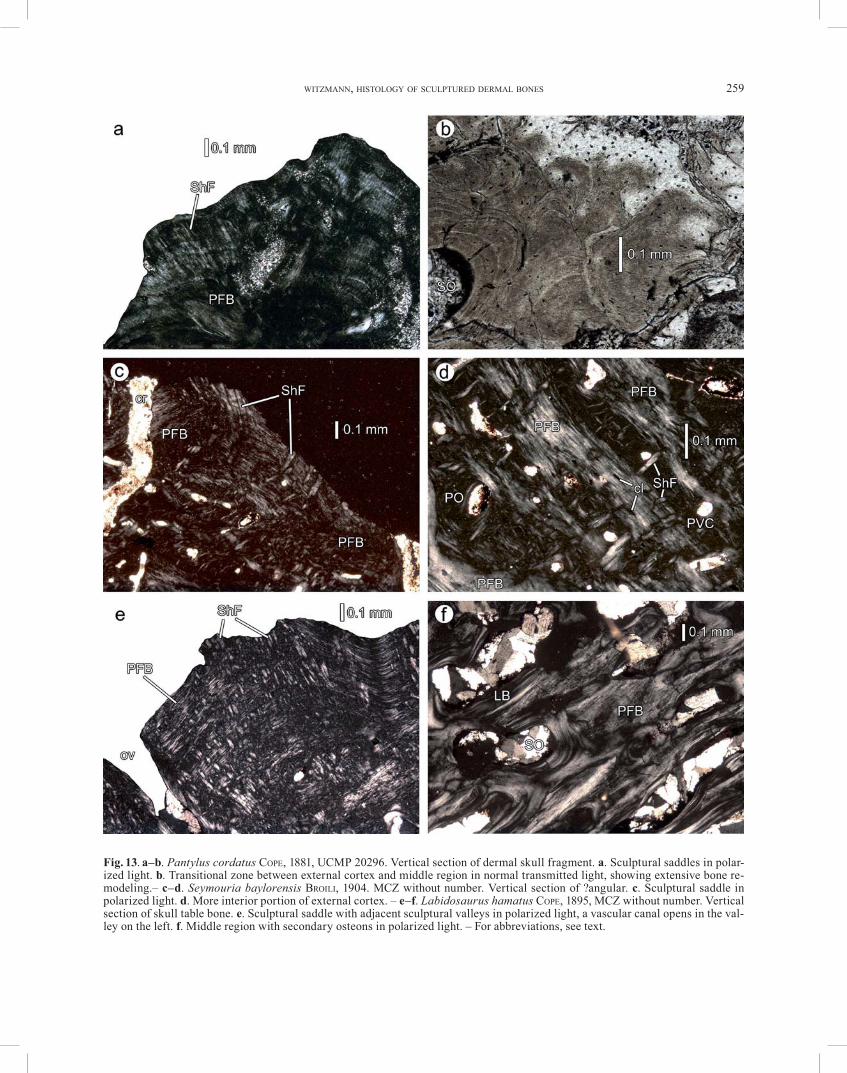
Fig. 4. a–b. Greererpeton burkemorani ROMER, 1969, CMNH 11900. Vertical sections of dermal skull bone (?quadratojugal). a. Low sculptural saddle of external cortex in normal transmitted light, the shape of the bone cell lacunae represents the direction of the bone fibers: on the left, the lacunae are spindular and indicate that the fibers are oriented parallel to the image plane, and on the right, the lacunae are round and indicate transverse section of the fibers. b. Internal cortex consisting of isopedine in polarized light. – c–f. Edops craigi ROMER, 1936, MCZ 1235. Vertical section of skull table bone. c. External cortex, sculptural saddle with Sharpey’s fibers, polarized light. d. External cortex showing growth marks and sparse vascularization, normal transmitted light. e. Middle region with primary matrix of fine, subparallel fibers and primary and secondary osteons, polarized light. f. Middle region with in-terwoven structural fibers as primary tissue, polarized light. – For abbreviations, see text.
244 PALAEODIVERSITY 2, 2009
Haversian canals may constitute large cavities, but true Haversian tissue cannot be observed. The interstitial, pri-mary bone matrix consists of fine parallel-fibered bone.
I n t e r n a l c o r t e x . The compact internal cortex consists of isopedine, and the shape of the bone cell lacu-nae reflects the orientation of the fibers. However, the discrete bands exhibit a broad variation in their thickness (Fig. 4b). Most parts of the internal cortex are avascular, but in the most interior part, isolated primary vascular canals and few secondary osteons are visible that are par-allel to the internal bone surface. Fine fibers that are ar-ranged perpendicular to the internal bone surface are vis-ible in the ‘dark layers’.
3.3. Crown-group tetrapods
3.3.1. Edops craigi (Temnospondyli, Edopoidea)
The bone fragments of Edops craigi ROMER, 1936 used for sectioning stem from the skull table and bear a rather irregular, polygonal sculpture of ridges and pits. Histo-logically, the middle region of the bone is much thickened with respect to the cortices. The sectioned bone fragments attain a thickness of more than 12 mm. The ratio external cortex : middle region : internal cortex is 1 : 3.6 : 0.6.
E x t e r n a l c o r t e x . In large areas, the external cortex consists of coarse parallel-fibered bone with spin-dular bone cell lacunae that have long, branching canali-culi. Distinct growth marks are present, and the bone can thus be designated as lamellar-zonal (Fig. 4c, d). Vascular-ization is in most parts low and consists of isolated pri-mary vascular canals and few primary osteons that may be aligned in single rows (Fig. 4d). Sculptural saddles of ear-lier generations are embedded within the cortex. Fan-shaped clusters of densely arranged Sharpey’s fibers ap-pear irregularly in the sculptural saddles, where they pen-etrate the bone tissue (Fig. 4c). In the sculptural valleys, the fibers are less numerous. The sometimes bifurcating Sharpey’s fibers measure around 21 μm in diameter and may extend to the middle region. In some areas more inte-rior within the cortex, primary interwoven structural fiber bundles (ISF) sensu SCHEYER & SÁNCHEZ-VILLAGRA (2007) are present and show a non-homogeneous distribution. Such interwoven structural fibers are more abundant in the middle region and are therefore described below. The outer parts of the external layer are in few places disturbed by areas of secondary bone remodeling with a distinct re-sorption line. These resorption structures at the bone sur-face are described in more detail for Eryops (see below). The external cortex is separated from the middle region by a distinct transition, with the occurrence of several small secondary osteons.
M i d d l e r e g i o n . This fine to coarse cancellous
region underwent extensive remodeling with numerous secondary osteons that constitute Haversian tissue in many areas. The interstitial primary bone consists in many areas of parallel-fibered bone. Rather fine primary fibers that are oriented subparallel, but may change their direction in succeeding layers, can frequently be observed (Fig. 4e). These layers can be arranged irregularly in a ‘flowmark’-like fashion. In some areas or layers, primary fibers cross each other at an angle of approximately 90° and constitute a three-dimensional network of interwoven structural fi-bers (Fig. 4f). Under polarized light, the birefringence patterns of the collagen fibers are well visible. Approxi-mately transversely cut fiber bundles have a globular ap-pearance. Focusing on different planes of the slide under the microscope shows that these structures do not repre-sent real globules like cell spaces, for instance, but fibers that extend perpendicularly or obliquely to the image plane. Between the fibers, bone cell lacunae of irregular outline with no or only stumpy canaliculi are present. These areas of fibers are remains of primary bone, which was remodelled and partially bounded by secondary bone. Near the internal cortex, primary osteons may be aligned regularly in layers.
I n t e r n a l c o r t e x . Only parts of the compact in-ternal cortex are preserved. As far as it can be discerned, it consists of coarse, less organized parallel-fibered bone that is avascular.
Bone fragments of the skull table of Chenoprosopus milleri MEHL, 1913 were sectioned that bear a sculpture of rounded polygons. The internal part of the middle region, and the complete internal cortex are eroded. The thickness of the sectioned bone fragments ranges from 3 mm to 6 mm.
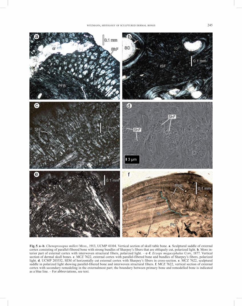
E x t e r n a l c o r t e x . The external cortex is mainly composed of coarse parallel-fibered bone that shows cy-clic growth marks (lamellar-zonal bone). The bone cells are variable in outline with varying length of their canali-culi, and are randomly distributed. In the external part of the sculptural saddles, densely arranged Sharpey’s fibers with a diameter of 8 to 17 μm are present (Fig. 5a). Some of the fibers may extend until to the external part of the middle region. The fibers may ramify into two or three branches. In the sculptural valleys, the Sharpey’s fibers are less numerous than in the saddles and may also be ab-sent in places. In the more interior part of the external cortex, the parallel-fibered bone shows transitions to a network of interwoven structural fibers that extend obliquely to the bone surface (Fig. 5b). The bone cell lacu-nae in this bone tissue are irregularly arranged and pos-
WITZMANN, HISTOLOGY OF SCULPTURED DERMAL BONES 245
Fig. 5. a–b. Chenoprosopus milleri MEHL, 1913, UCMP 41104. Vertical section of skull table bone. a. Sculptural saddle of external cortex consisting of parallel-fibered bone with strong bundles of Sharpey’s fibers that are obliquely cut, polarized light. b. More in-terior part of external cortex with interwoven structural fibers, polarized light. – c–f. Eryops megacephalus COPE, 1877. Vertical section of dermal skull bones. c. MCZ 7622, external cortex with parallel-fibered bone and bundles of Sharpey’s fibers, polarized light. d. UCMP 203332, SEM of horizontally cut external cortex with Sharpey’s fibers in cross-section. e. MCZ 7622, sculptural saddle in polarized light showing parallel-fibered bone and interwoven structural fibers. f. MCZ 7622, vertical section of external cortex with secondary remodeling in the externalmost part; the boundary between primary bone and remodelled bone is indicated as a blue line. – For abbreviations, see text.
246 PALAEODIVERSITY 2, 2009
sess no canaliculi. The external cortex is poorly vascular-ized with scattered primary vascular canals and isolated primary osteons. In one slide, a layer of primary osteons is visible interior to a sculptural valley. The external cortex grades into the middle region with the occurrence of scat-tered secondary osteons that increase in number and den-sity in a small transitional zone.
M i d d l e r e g i o n . This fine to coarse cancellous region is characterized by extensive remodeling of the bone. Numerous secondary osteons that may course in different directions are present that in parts build Haver-sian tissue. The interstitial primary bone consists of paral-lel-fibered bone that shows transitions to fine interwoven structural fibers in some regions.
The bone fragments of the dermal skull roof of Eryops megacephalus COPE, 1877 sectioned for this study bear pits of variable outline, and thus the sculpture appears rather irregular. The ratio external cortex : middle region : internal cortex is 1 : 3.3 : 0.6. The bone fragments used for sectioning attain a thickness of more than 14 mm.
E x t e r n a l c o r t e x . Densely arranged Sharpey’s fibers with a diameter of mostly 21 to 29 μm penetrate the external cortex until to the middle region (Fig. 5c, d). The Sharpey’s fibers may be numerous also in the valleys. Coarse, often less ordered parallel-fibered bone predomi-nates the bone matrix of the external cortex and has bone cell lacunae of variable shape that are arranged in rows and have branching, moderately long to long canaliculi. The bone matrix shows local islets of interwoven struc-tural fibers especially in the more interior parts of the cortex, but they may also be present individually in the more external parts (Fig. 5e). Conspicuous growth marks are present in the external cortex (Fig. 5f). Similar to Edops, the more superficial parts of the external cortex may be disturbed by areas of secondary bone remodeling (Fig. 5f). The remodelled bone lies discordantly on the primary bone, separated by a distinct resorption line. Whereas the adjacent primary bone is only sporadically pervaded by primary vascular canals and primary os-teons, the secondary bone is well vascularized. It consists of parallel-fibered bone and interwoven structural fibers and lacks the clear zonal pattern. In specimen MCZ 7622-2, the remodelled area was subsequently overgrown by ‘normal’ parallel-fibered bone.
M i d d l e r e g i o n . The external cortex is separated from the middle region by a short transition zone that con-tains scattered secondary osteons. The middle region is mostly fine to coarse cancellous and shows extensive re-modeling with Haversian tissue (Fig. 6a, b). Also subordi-nate areas are present in which large vascular spaces are
separated by irregularly arranged trabeculae that are rath-er thick and short. The interstitial primary bone consists of parallel-fibered bone and of the three-dimensional net-work of interwoven structural fibers (Fig. 6b–d), as de-scribed for Edops. The transversely cut fibers are visible as ‘globules’, and between the fibers, bone cell lacunae of irregular outline with no or short canaliculi are visible (Fig. 6c).
I n t e r n a l c o r t e x . This only fragmentarily pre-served region consists mostly of coarse parallel-fibered bone with a less ordered appearance. Transitions to islets of interwoven structural fibers may occur in some areas. Fine, oblique extrinsic fibers that are densely arranged and probably represent Sharpey’s fibers cross the internal cor-tex in MCZ 7622-2. The cortex is poorly vascularized by scattered primary osteons and primary vascular canals.
The bone fragments of the dermal skull roof of Ache-loma cumminsi COPE, 1882 used for sectioning possess rather regular, rounded sculptural pits on their external surface. The bone fragment investigated has a thickness of almost 9 mm, and the ratio external cortex : middle region : internal cortex is 1 : 6.1 : 1.2.
E x t e r n a l c o r t e x . The bone matrix is constituted by mostly homogeneous, fine parallel-fibered bone. Vas-cularization is poorly developed and consists of few pri-mary vascular canals and primary osteons. The sometimes bifurcating Sharpey’s fibers with a diameter of 15 to 31 μm are conspicuous. They are restricted to the sculptural sad-dles where they are moderately to densely arranged (Fig. 6e). Growth marks are present, but are indistinct and dif-ficult to follow. Bone cell lacunae are often oval in shape and possess branching canaliculi. They are more orderly arranged in the valleys than in the saddles, where they have frequently a rather round shape.
M i d d l e r e g i o n . The transition from the external and internal cortex to the trabecular middle region is abrupt. The middle region underwent strong resorption and remodeling and possesses numerous bone cavities of irregular shape and size, most of which are lined by sec-ondary lamellar bone. The rather short trabeculae are ir-regularly arranged. The primary bone matrix in the trabe-culae consists of homogeneous parallel-fibered bone.
I n t e r n a l c o r t e x . This region is made of avascu-lar, fine parallel-fibered bone, which is penetrated in some areas by thin fibers at an angle of approximately 60°. Bone cell lacunae are spindular, have long, branching canaliculi and are arranged in rows (Fig. 6f). They may form dark layers of densely arranged lacunae within the internal cortex.
WITZMANN, HISTOLOGY OF SCULPTURED DERMAL BONES 247
Fig. 6. a–d. Eryops megacephalus COPE, 1877, MCZ 7622. Vertical sections of dermal skull bones. a. Overview of middle region with Haversian tissue, normal transmitted light. b. Middle region with secondary osteons and interstitial primary bone tissue, normal transmitted light. c. Close up of interstitial primary bone tissue of middle region, showing ‘globular’ fibers in cross section, normal transmitted light. d. Middle region in polarized light, showing primary interwoven structural fibers. – e–f. Acheloma cumminsi COPE, 1882, MCZ 1490. Vertical section of skull roof fragment. e. Sculptural saddle of parallel-fibered bone with Sharpey’s fibers in polar-ized light. f. Internal cortex with bone cell lacunae, normal transmitted light. – For abbreviations, see text.
The postfrontal of a subadult specimen of Scleroceph-alus haeuseri GOLDFUSS, 1847 was sectioned. The bone has a maximum thickness of 4.2 mm, and the ratio external cortex : middle region : internal cortex amounts 1 : 4.2 : 1.1. Unfortunately, the bone was heavily altered by recrystal-lization, so that nothing can be said about the bone tissue itself or the intrinsic and extrinsic fibers. However, the slide shows the vascularization of the cortices, the bone’s microstructure and growth marks. The bone structure is best visible in reflected light (Fig. 7a, b). The external cortex is moderately to low vascularized by primary vas-cular canals (Fig. 7a). Bone cell lacunae are abundant, but their exact shape cannot be determined. Cyclical growth marks are conspicuous and follow the relief of the external bone sculpture. The middle region can be designated as trabecular with large erosion rooms that are lined by sec-ondary lamellar bone (Fig. 7a, b). The internal cortex is poorly vascularized by scattered primary vascular canals.
A prefrontal of a subadult specimen of Archegosaurus decheni GOLDFUSS, 1847 was sectioned. The external bone surface possesses a polygonal sculpture in the ossification centre and radiating ridges in the periphery. Unfortunate-ly, the external portion of the external cortex is eroded. This is rather common in Archegosaurus, since the speci-mens are preserved in siderite concretions (geodes) and divide into part and counterpart when the concretions are split. Because of this surface erosion, the ratio between the thickness of the cortices and the middle region cannot be calculated. The maximum thickness of the bone is ap-proximately 2.5 mm.
E x t e r n a l c o r t e x . The preserved parts of the ex-ternal cortex consist homogenously of parallel-fibered bone that is moderately to highly vascularized by primary canals, primary osteons (partially anastomosing) and, more interiorly, by secondary osteons (Fig. 7c). The latter mark the transition zone to the middle region. Bone cell lacunae are numerous, randomly arranged in the primary bone matrix and are of varying shape. The canaliculi are moderately long to long and branching. Sharpey’s fibers that penetrate the external bone surface are not visible (probably due to erosion of the external portion of the cor-tex). However, the prefrontal forms a rather thin, under-plating shelf for articulation with the lacrimal; in this re-gion, densely arranged, almost horizontally oriented Sharpey’s fibers are visible that connected the two adja-cent bones (Fig. 7d).
M i d d l e r e g i o n . The trabecular middle region experienced extensive erosion and remodeling. Large ero-sion rooms, some of them lined by secondary lamellar bone, are separated from each other by trabeculae of ir-regular outline and direction (Fig. 7c). Many of the trabe-culae are stout, others are slender and long. The interstitial bone matrix of the trabeculae may contain small primary or secondary osteons, which are sometimes cut. The pri-mary matrix of the middle region consists of parallel-fi-bered bone, and morphology and alignment of the bone cells corresponds to those of the external cortex.
I n t e r n a l c o r t e x . The internal cortex is separated from the trabecular middle region by a thin transition zone with small secondary osteons (Fig. 7c) and consists of rather coarse parallel-fibered bone. The matrix is pene-trated by scattered primary vascular canals that course in different directions. Bone cell lacunae are aligned in rows and have an elongate, flattened shape with long, branching canaliculi.
The bone fragment of Kupferzellia wildi SCHOCH, 1997 under study is derived from the skull table and probably represents a part of the supratemporal. Its dermal sculp-ture consists of rather regular, wide polygons. The thick-ness of the investigated bone amounts slightly more than 5 mm, and the ratio external cortex : middle region : inter-nal cortex is 1 : 1.7 : 0.5.
E x t e r n a l c o r t e x . The bone matrix consists mainly of coarse parallel-fibered bone with a rather ir-regular fibrous arrangement (Fig. 7e). In the sculptural saddles, the parallel-fibered bone is even less organized than in the valleys. The external cortex is moderately vas-cularized: more externally, only isolated, small primary vascular canals and primary osteons are visible, whereas more interiorly, a larger number of small primary canals and primary osteons (as well as few secondary osteons) are present and may anastomose. Sharpey’s fibers are re-stricted to the sculptural saddles and measure 5 to 16 μm in diameter. In the parallel-fibered bone of the sculptural valleys, the bone cell lacunae are mostly spindular with long canaliculi and are arranged in layers. In contrast, their shape is variable in the more coarse parallel-fibered bone of the saddles. Growth marks are present but diffi-cult to follow.
M i d d l e r e g i o n . This region is coarse cancellous with several secondary osteons of varying size, but Haver-sian tissue is not present (Fig. 7f). In the interior part, the Haversian canals may be quite large. The primary matrix consists of fine parallel-fibered bone with generally spin-dular cell lacunae. Local transitions to interwoven struc-
WITZMANN, HISTOLOGY OF SCULPTURED DERMAL BONES 249
Fig. 7. a–b. Sclerocephalus haeuseri GOLDFUSS, 1847, SMNS 90517. Vertical section of postfrontal, reflected light. a. External cortex with external portion of middle region. b. Close up of middle region with large erosion rooms lined by secondary bone. c–d. Archegosaurus decheni GOLDFUSS, 1847, MB.Am.152. Vertical section of prefrontal. c. Histological overview in normal trans-mitted light. d. Close up of shelf for articulation with the lacrimal with horizontally oriented Sharpey’s fibers in normal transmitted light. – e–f. Kupferzellia wildi, SCHOCH, 1997, SMNS 91247. Vertical section of ?supratemporal. e. External cortex with lateral part of sculptural saddle, polarized light. f. Middle region with secondary osteons, polarized light. – For abbreviations, see text.
250 PALAEODIVERSITY 2, 2009
tural fibers are present, in which most of the lacunae are round. Canaliculi are mostly short. More internally, the secondary osteons are smaller and aligned in two to four rows that are separated by avascular layers of parallel-fi-bered bone.
I n t e r n a l c o r t e x . Coarse parallel-fibered bone constitutes the bone matrix of the internal cortex, with lo-cally changing directions of its fibers. Most of the bone cell lacunae are spindular and have few canaliculi. Extrin-sic fibers are not visible. Vascularization is poor and con-sists of scattered primary vascular canals.
The dermal sculpture of Mastodonsaurus giganteus (JAEGER, 1828) is composed of regular, rather square pits on the skull table. The peripheral parts of the bones of the postorbital skull table consist of radiating ridges and fur-rows. Some sections for this study were cut through bones with polygonal sculpture, and others were made perpen-dicular or parallel to the radiating ridges. The ratio exter-nal cortex : middle region : internal cortex is 1 : 3 : 0.4. The bone thickness of the investigated specimens ranges from 8 mm to 17 mm. Additionally, the clavicular blade of a small juvenile specimen has been sectioned and is de-scribed separately below.
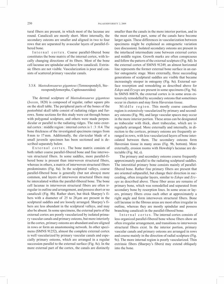
E x t e r n a l c o r t e x . The bone matrix consists of both rather coarse parallel-fibered bone and fine interwo-ven structural fibers. In some saddles, more parallel-fi-bered bone is present than interwoven structural fibers, whereas in others, a matrix of interwoven structural fibers predominates (Fig. 8a). In the sculptural valleys, coarse parallel-fibered bone is generally (but not always) more common, and layers of interwoven structural fibers may be intercalated within the parallel-fibered bone. The bone cell lacunae in interwoven structural fibers are often ir-regular in outline and arrangement, and possess short or no canaliculi (Fig. 8b). Rather short, but thick Sharpey’s fi-bers with a diameter of 25 to 28 μm are present in the sculptural saddles and are loosely arranged. Sharpey’s fi-bers are less abundant in the sculptural valleys, and may also be absent. In some specimens, the external parts of the external cortex are poorly vascularized by isolated prima-ry vascular canals and primary osteons, but more interiorly in the cortex, primary osteons are abundant and are aligned in rows or form an anastomosing network. In other speci-mens (SMNS 91252), almost the complete external cortex is well vascularized by primary vascular canals and espe-cially primary osteons, which are arranged in a regular succession parallel to the external surface (Fig. 8c). In the more external part of the cortex, the canals are distinctly
smaller than the canals in the more interior portion, and in the most external part, some of the canals have become larger again. These differences in vascularization between specimens might be explained as ontogenetic variation (see discussion). Isolated secondary osteons are present in the interlaced intermediate zone between external cortex and middle region. Growth marks are often conspicuous and follow the pattern of the external sculpture (Fig. 8d). In the external cortex of SMNS 91249, an almost horizontal line represents the former external bone surface in an ear-lier ontogenetic stage. More externally, three succeeding generations of sculptural saddles are visible that became increasingly steeper in ontogeny (Fig. 8e). External sur-face resorption and remodeling as described above for Edops and Eryops are present in some specimens (Fig. 9a). In SMNS 80878, the external cortex is in some areas ex-tensively remodelled by secondary osteons that sometimes occur in clusters and may form Haversian tissue.
M i d d l e r e g i o n . This mostly coarse cancellous region is extensively vascularized by primary and second-ary osteons (Fig. 9b), and large vascular spaces may occur in the more interior portion. These areas can be designated as trabecular with thick, short trabeculae, which are ir-regularly arranged. More externally and internally, in di-rection to the cortices, primary osteons are frequently ar-ranged in rows, with less vascularized layers of bone inter-calated between them. The secondary osteons form Haversian tissue in many areas (Fig. 9b, bottom). More externally, erosion rooms with Howship’s lacunae are de-tectable (Fig. 8d, e).
The primary and secondary osteons course frequently approximately parallel to the radiating sculptural saddles. The interstitial primary bone consists mainly of parallel-fibered bone. Rather fine primary fibers are present that are oriented subparallel, but change their direction in suc-ceeding, often irregular layers, similar to Edops and Ery-ops as described above. These fiber areas are remains of primary bone, which was remodelled and separated from secondary bone by resorption lines. In some areas or lay-ers, primary fibers cross each other at approximately a right angle and form interwoven structural fibers. Bone cell lacunae in the fibrous areas are most often irregular in outline, whereas they are mostly spindular and possess branching canaliculi in the parallel-fibered bone.
I n t e r n a l c o r t e x . The internal cortex consists of less organized parallel-fibered bone whose fibers show an often irregular arrangement, and transitions to interwoven structural fibers exist. In the interior portion, primary vascular canals and primary osteons are arranged in rows and course mainly in the direction of radial sculpture (Fig. 9c). The more internal region is poorly vascularized. Thin extrinsic fibers (Sharpey’s fibers) may extend obliquely into the bone.
WITZMANN, HISTOLOGY OF SCULPTURED DERMAL BONES 251
Fig. 8. Mastodonsaurus giganteus (JAEGER, 1828). Vertical sections of external cortex. – a. SMNS 91251. Slightly eroded sculptural saddle showing interwoven structural fibers. Polarized light. b. SMNS 91248. Bone cell lacunae in a matrix of interwoven struc-tural fibers. c. SMNS 91252. Sculptural saddle with numerous primary osteons that are often arranged in a regular succession paral-lel to the external surface. d. SMNS 91248. Sculptural saddle showing growth marks; the more interior portion is well vascularized. e. SMNS 91248. The former bone surfaces of three succeeding generations of a sculptural saddle are visible; the first is almost hori-zontally aligned. – For abbreviations, see text.
252 PALAEODIVERSITY 2, 2009
Fig. 9. a–e. Mastodonsaurus giganteus (JAEGER, 1828). a. SMNS 80878. Vertical section parallel to a sculptural ridge showing struc-tures of surface resorption. b. SMNS 91248. Middle region in normal transmitted light, vertical section. Zone of extensive remodel-ing and Haversian tissue. c. SMNS 91248. More interior part of internal region with primary osteons in rows. d–e. Juvenile specimen, SMNS 91255. Interclavicle, vertical section transverse to the sculptural ridges. – d. Histological overview, normal transmitted light. e. The fine to coarse cancellous middle region with a matrix of coarse parallel-fibered bone, polarized light. f. Metoposaurus diagnosticus (VON MEYER, 1842), MB.Hi.1719. Sculptural saddle of an unidentified dermal bone in normal transmit-ted light. – For abbreviations, see text.
WITZMANN, HISTOLOGY OF SCULPTURED DERMAL BONES 253
3.3.9. Clavicular blade of a small juvenile specimen of Mastodonsaurus giganteus
The section is aligned transversely to the radiating sculptural ridges of this approximately 2.1 mm thick bone. The cortices are so extensively vascularised that they re-semble closely the middle region. Therefore, it is no more appropriate to speak of a clear diploë structure, in contrast to the ontogenetically more advanced specimens. The de-termination of boundaries between the cortices and the middle region is thus rather arbitrary.
E x t e r n a l c o r t e x . The external cortex is highly vascularized by numerous, anastomosing primary vascu-lar canals and primary osteons (Fig. 9d). Also few second-ary osteons can be observed. There is no difference in the degree of vascularization and nature of the bone tissue between saddles and valleys. Growth marks are not visi-ble. The primary bone tissue consists of coarse, poorly organized parallel-fibered bone that may fray out in fibers with changing direction. Interwoven structural fibers and Sharpey’s fibers are not visible in the material under study. Most bone cell lacunae are randomly arranged, irregular in shape and possess few or no canaliculi.
M i d d l e r e g i o n . This fine to coarse cancellous region is distinguished from the cortices by the slightly larger diameter of many of its vascular canals, and the presence of several secondary osteons that may anasto-mose. Haversian tissue is not present. The rather small osteons are frequently arranged in layers (Fig. 9e). Few slightly larger erosion rooms without lining of lamellar bone are present. The interstitial primary tissue consists mainly of coarse, less organized parallel-fibered bone (Fig. 9e). In the primary bone matrix, the longitudinal axes of the spindular bone cell lacunae are directed parallel to the bone fibers. Canaliculi are hardly visible.
I n t e r n a l c o r t e x . The internal cortex is com-posed of coarse parallel-fibered bone. It is well vascular-ized by primary vascular canals and primary osteons that may anastomose (Fig. 9d). The bone cell lacunae are or-dered and spindular with few canaliculi.
It is not clear if the slides investigated here from Meto-posaurus diagnosticus (VON MEYER, 1842) are derived from the skull roof or the dermal pectoral girdle. The ratio external cortex : middle region : internal cortex is 1 : 1.5 : 0.3. However, the middle region has collapsed by the superimposed load of sediment in the specimens under study and was therefore originally probably somewhat thicker. The bone investigated attains a thickness of 9 mm.
E x t e r n a l c o r t e x . The more interior part of the cortex is well vascularized by numerous, partially anasto-mosing primary vascular canals of mostly a small diame-ter, whereas the number of canals decreases in direction to the external bone surface and the top of the saddles (Fig. 9f). The bone matrix is composed of homogeneous paral-lel-fibered bone and shows several distinct growth marks that allow to trace the growth of the rather steep sculptural saddles. Numerous bone cell lacunae are visible that ap-pear mostly irregular-elongate in shape. Poor preservation precludes recognition of canaliculi. In the sculptural sad-dles, the Sharpey’s fibers are arranged in clusters that are mostly present in the lateral parts of the saddles (Fig. 10a), where they are oriented interiorly and medially. In the sculptural valleys, the clusters are more densely arranged and extend deep into the cortex (Fig. 10a). The diameter of the fibers amounts 17 to 25 μm.
M i d d l e r e g i o n . The external cortex grades into the coarse cancellous middle region with numerous sec-ondary osteons that may form Haversian tissue. The pri-mary bone matrix is represented by fine, homogeneous parallel-fibered bone.
I n t e r n a l c o r t e x . The internal cortex is thin compared to the middle and external region and consists of homogeneous parallel-fibered bone that is largely avas-cular (Fig. 10b). It can be distinguished from the middle region by a distinct transition zone.
For the present study, a dermal bone fragment of Pla-giosuchus pustuliferus (FRAAS, 1896) (SMNS 82023), which derives probably from the skull roof and bears a sculpture of tubercles and low ridges between them, was sectioned. The bone thickness amounts 11 mm. In this fragment, the cortices are highly vascularized similar to the middle region, and thus a diploë structure is not visi-ble. The determination of external cortex, middle region, and internal cortex in the following is somewhat arbi-trarily and based on the higher density of vascular canals in the middle region and the presence of extrinsic fibers (Sharpey’s fibers) in the cortices. The vascularization is slightly less well developed in the external cortex of MB.Hi.1705 (maximum bone thickness 4 mm), a slide of an unidentified dermal bone of Plagiosuchus, than in that of SMNS 82023. Nevertheless, a clear diploë structure is not developed also in MB.Hi.1705.
E x t e r n a l c o r t e x . The complete external cortex is well vascularized by numerous, partially anastomosing primary vascular canals and primary osteons in SMNS 82023, whereas in MB.Hi.1705, the external portion of the cortex is nearly avascular. Densely arranged Sharpey’s
254 PALAEODIVERSITY 2, 2009
Fig. 10. a–b. Metoposaurus diagnosticus (VON MEYER, 1842), vertical sections through unidentified dermal bone. a. MB.Hi.1720. Sculptural saddle and external cortex in polarized light. b. MB.Hi.1721. Internal cortex in polarized light. – c–d. Plagiosuchus pus-tuliferus (FRAAS, 1896), SMNS 82023. Vertical section through fragment of dermal skull roof. c. Sculptural saddle with Sharpey’s fibers in polarized light. d. Middle region with primary vascular canals and erosion rooms, normal transmitted light. – e–f. Plagio-sternum granulosum (FRAAS, 1889), SMNS 91256. Vertical section of interclavicle. e. Sculptural saddle and valley with Sharpey’s fibers and external part of middle region, polarized light. f. External cortex with eroded surface, showing sculptural saddles of dif-ferent generations (indicated by arrows) and Sharpey’s fibers both in saddles and valleys, normal transmitted light. – For abbrevia-tions, see text.
WITZMANN, HISTOLOGY OF SCULPTURED DERMAL BONES 255
fibers penetrate the external region both in the sculptural saddles and valleys (Fig. 10c) and extend far interiorly within the cortex. The fibers have a diameter of mostly 22 to 26 μm and may ramify in two or three branches. The largest part of the bone tissue consists of coarse parallel-fibered bone with a more irregular fibrous arrangement. Spindle-shaped bone cell lacunae are present that possess branching canaliculi. In some areas, especially the sculp-tural saddles, also islets of interwoven structural fibers are discernable. Cyclic growth marks are not continuous and have an indistinct appearance in SMNS 82023, whereas they are more distinct in the external part of the external cortex in MB.Hi.1705.
M i d d l e r e g i o n . This coarse cancellous region is extensively vascularized by primary vascular canals and primary osteons, and isolated, medium-sized erosion cav-ities are present (Fig. 10d). Some of them show Howship’s lacunae, and secondary lamellar bone lining is absent in the SMNS specimen under study, whereas in MB.Hi.1705, scattered, small secondary osteons are discernable. The bone matrix consists in large parts of interwoven struc-tural fibers, and also parallel-fibered bone is present.
I n t e r n a l c o r t e x . The bone matrix consists most-ly of poorly organized parallel-fibered bone. Sharpey’s fibers are present that are shorter and distinctly thinner than those of the external cortex. Vascularization is well developed and consists of numerous primary vascular ca-nals that are often aligned in rows or may anastomose.
A fragment of an interclavicle of Plagiosternum gran-ulosum (FRAAS, 1889) (SMNS 91256) with polygonal sculpture was sectioned that has a maximum thickness of 11 mm. Additionally, the vertical section of an unidenti-fied, 7 mm thick dermal bone of Plagiosternum (MB.Hi.1714) was investigated. The ratio external cortex : mid-dle region : internal cortex is 1 : 2.9 : 0.8.
E x t e r n a l c o r t e x . The external cortex is com-posed of coarse, poorly ordered parallel-fibered bone, and islets of interwoven structural fibers can be found espe-cially in the more internal regions of the sculptural saddles and in the interior parts of the cortex (Fig. 10e). The bone cell lacunae in the parallel-fibered bone are of irregular shape with moderately long canaliculi. In the SMNS spec-imen, Sharpey’s fibers are densely arranged in both the sculptural saddles and in the valleys, comparable to the situation in Plagiosuchus. The diameter of the Sharpey’s fibers varies between 17 to 26 μm, and most of them ter-minate in approximately the external half of the external region. Zones and annuli are well visible and delineate sculptural saddles of earlier generations, in which Sharp-
ey’s fibers are visible (Fig. 10f). The external cortex of the SMNS specimen is moderately vascularized in its interior portion by primary vascular canals, whereas the external portion is largely avascular (Fig. 10f). In MB.Hi.1714, the complete cortex is well vascularized by anastomosing pri-mary canals whose diameters decrease in size in the more external portion of the cortex (Fig. 11a). The difference in the degree of vascularization between the Berlin and the Stuttgart specimen might be explained as ontogenetic variation (see discussion).
M i d d l e r e g i o n . A rather small transition zone between the external cortex and the middle region is char-acterised by scattered secondary osteons. The middle re-gion was extensively affected by remodeling. It is charac-terised by large erosion rooms that are separated by long, irregularly arranged trabeculae of varying thickness (Fig. 11b). Some erosion rooms are lined by secondary lamellar bone. The primary bone matrix within the trabeculae con-sists of well-ordered parallel-fibered bone with spindular bone cell lacunae.
I n t e r n a l c o r t e x . The bone tissue is composed of parallel-fibered bone with often irregularly arranged fi-bers, and transitions to interwoven structural fibers are locally present. Sharpey’s fibers that are distinctly thinner than those of the external region penetrate the internal cortex obliquely at an angle of about 60° and are densely arranged. Primary vascular canals that may anastomose are more numerous in the interior than in the internal part of the cortex. Cyclic growth marks are well visible (Fig. 11c).
The thin sections were prepared from bone fragments of the interclavicle and clavicle, ranging from 3.3 mm to 9 mm in thickness. The dermal sculpture is tubercular, and low sculptural ridges may connect the tubercles. The ratio external cortex : middle region : internal cortex is 1 : 1.9 : 0.9.
E x t e r n a l c o r t e x . The interior portion of the ex-ternal cortex is moderately to highly vascularized by pri-mary vascular canals and primary osteons that often branch and anastomose, whereas vascularization is low in the external portion of the cortex (Fig. 11d). The sculp-tural valleys and the lateral parts of the saddles consist mainly of fine to coarse parallel-fibered bone in which the bone cell lacunae are often spindular with moderately long to long canaliculi. The interior parts of the sculptural saddles, in contrast, show general isotropy under polar-ized light, and the bone cell lacunae are rather round and haven often long, branching canaliculi. The isotropy can probably be attributed to the superposition of the Sharp-
256 PALAEODIVERSITY 2, 2009
Fig. 11. a–c. Plagiosternum granulosum (FRAAS, 1889). a. MB.Hi.1714. Vertical section of unidentified dermal bone, sculptural saddle, normal transmitted light. b–c. SMNS 91256. Vertical section of interclavicle. b. Middle region showing trabecular structure with large erosion rooms. c. Internal cortex in normal transmitted light. – d–f. Gerrothorax sp. d. SMNS 91258. Vertical section of clavicle, histological overview of external cortex and middle region in normal transmitted light; the arrows indicate the bone surface of a former generation. e. SMNS 91257. Vertical section of clavicle in polarized light, showing the middle region with numerous primary osteons and larger erosion rooms. f. SMNS 83033. Vertical section of interclavicle, internal cortex in polarized light; note the oblique Sharpey’s fibers penetrating the bone. – For abbreviations, see text.
WITZMANN, HISTOLOGY OF SCULPTURED DERMAL BONES 257
ey’s fibers in the cores of the saddles. In some regions more interior in the cortex, the bone tissue may show tran-sitions to diagonally arranged interwoven structural fibers of irregular size. Rather thin Sharpey’s fibers with a diam-eter of 8 to 11 μm, that are not as densely arranged as in Plagiosuchus and Plagiosternum, are visible in the sculp-tural tubercles and are fewer in number in the valleys. Cyclic growth marks are present in the external cortex. In one interclavicle (SMNS 83033), a former generation of sculptural saddles is visible in the interiormost part of the external cortex (Fig. 11d). These old sculptural saddles are distinctly smaller and the valleys narrower than those of the actual external surface.
M i d d l e r e g i o n . The transition zone from the ex-ternal cortex to the middle region is rather thin and bears scattered secondary osteons. In most specimens, the mid-dle region varies from coarse cancellous to trabecular (Fig. 11e). In some areas, numerous primary osteons pre-dominate and may be arranged in rows, whereas in other areas, scattered, large erosion rooms prevail, that are sepa-rated by irregularly arranged trabeculae. In some parts, remodeling was so extensive that Haversian tissue was formed (Fig. 11f, on top). The primary tissue is parallel-fibered bone, but also a matrix of interwoven structural fibers that are diagonally arranged with respect to the bone surface occurs, whose sparse bone cell lacunae are mostly round and bear no or only short canaliculi.
I n t e r n a l c o r t e x . The internal cortex is com-posed of coarse, less ordered parallel-fibered bone. Rather loosely arranged Sharpey’s fibers of varying size extend into the internal region at an oblique angle (30–50°), some of them extending until to the middle region (Fig. 11f). Only very few primary vascular canals are visible. These are mostly situated more interiorly in the internal cortex and course parallel to the internal bone surface.
The sectioned bone fragments of Diplocaulus magni-cornis COPE, 1882, ranging from 7 mm to 14 mm in thick-ness, represent lateral parts of the conspicuous ‘horns’ of this nectridean, so that a sculptured compact cortex en-closes the middle region dorsally, laterally, and ventrally. Therefore, in the following, the cortex will be referred to as ‘external cortex’ only. The external cortex is much re-duced with respect to the extensive middle region, the ra-tio external cortex : middle region : external cortex is 1 : 11.1 : 1. The dermal sculpture is composed of rounded polygons.
E x t e r n a l c o r t e x . The externalmost portion of the rather thin cortex is for the largest part avascular with very few, small primary canals, whereas its more interior
part is well vascularized by primary canals and primary osteons. In a transitional region between cortex and mid-dle region, several small erosion rooms and primary and secondary osteons are present (Fig. 12a). The bone matrix of the cortex consists largely of coarse parallel-fibered bone (Fig. 12b), and Sharpey’s fibers are restricted to the sculptural saddles, where their arrangement is moderately dense. The fibers have a comparatively small diameter of 11 to 13 μm. The cores of many sculptural saddles show isotropy under polarized light; as outlined for Gerrotho-rax (see above), this might be attributed to superposition of the Sharpey’s fibers. In most specimens, interwoven structural fibers constitute the predominant matrix in the more interior part of the external cortex (Fig. 12c). In the parallel-fibered bone, the cell lacunae have often long, branching canaliculi and are most often spindular (in the valleys) and rounded (in the saddles). The cell lacunae within the interwoven structural fibers, in contrast, are irregular in shape and have often no or only stumpy canali-culi. Throughout the external cortex, growth marks are present that are continuous in saddles and valleys and fol-low their relief.
M i d d l e r e g i o n . The middle region of Diplocau-lus is highly porous and strongly affected by successive resorption and reconstruction of bone. Thin, long trabecu-lae that are mostly orientated in a dorsoventral and hori-zontal direction enclose rectangular shaped bone cavities of similar size (Fig. 12d). The ordered appearance of tra-beculae and cavities is unique among the basal tetrapods studied here. Most of the cavities have a thin covering of secondary lamellar bone (Fig. 12e). In the more external cavities, the lining of lamellar bone is frequently absent. The primary bone tissue consists in large parts of coarse parallel-fibered bone, but also areas with interwoven structural fibers do exist. Partially cut primary osteons can be discerned in the primary tissue of the trabeculae.
Specimen MCZ 2269-3 provides interesting data con-cerning the extensive resorption and remodeling of the middle region. Structures of resorption are visible both in the primary matrix and the secondary lamellae (Fig. 12f). The primary bone matrix exhibits cyclical growth marks, with the different zones being delimited by distinct growth lines. The secondary lamellae surround the zones of pri-mary bone matrix continuously, indicating their deposi-tion after the formation of the zones.
A small, 3 mm thick fragment of the dermal skull roof of Pantylus cordatus COPE, 1881 consisting of one sculp-tural saddle and one valley plus parts of the middle region was sectioned. Pantylus has a pronounced polygonal sculpture on its dermal skull roof. The internal cortex and
258 PALAEODIVERSITY 2, 2009
Fig. 12. Diplocaulus magnicornis COPE, 1882. Vertical sections of the ‘cheek’-region. – a. UCMP 203658. Histological overview of external cortex with middle region in normal transmitted light. b. UCMP 203505. Sculptural saddle (partially eroded) in polarized light showing fan-shaped Sharpey’s fibers and matrix of parallel-fibered bone. c. UCMP 203658. Interior part of external cortex (the more external part is eroded) with erosion rooms of the middle region. d. MCZ 2269. Overview of the trabecular middle region in normal transmitted light. e. UCMP 203658. Trabeculae of the middle region in normal transmitted light, resorptive structures are indicated by arrows. f. MCZ 2269. Trabeculae of middle region in normal transmitted light, the numbers 1–4 indicate the order of bone deposition, the arrow marks a resorptive structure. – For abbreviations, see text.
WITZMANN, HISTOLOGY OF SCULPTURED DERMAL BONES 259
Fig. 13. a–b. Pantylus cordatus COPE, 1881, UCMP 20296. Vertical section of dermal skull fragment. a. Sculptural saddles in polar-ized light. b. Transitional zone between external cortex and middle region in normal transmitted light, showing extensive bone re-modeling.– c–d. Seymouria baylorensis BROILI, 1904. MCZ without number. Vertical section of ?angular. c. Sculptural saddle in polarized light. d. More interior portion of external cortex. – e–f. Labidosaurus hamatus COPE, 1895, MCZ without number. Vertical section of skull table bone. e. Sculptural saddle with adjacent sculptural valleys in polarized light, a vascular canal opens in the val-ley on the left. f. Middle region with secondary osteons in polarized light. – For abbreviations, see text.
260 PALAEODIVERSITY 2, 2009
the more internal part of the middle region of this speci-men are eroded.
E x t e r n a l c o r t e x . Densely arranged Sharpey’s fibers penetrate the high, steep sculptural saddles (Fig. 13a). The diameter of the fibers varies between 13 and 21 μm. The primary bone matrix consists of parallel-fi-bered bone. The sculptural valley is diagenetically altered so that the original bone matrix cannot be discerned, and it cannot be stated if Sharpey’s fibers were present. The vascularization of the external cortex is low with scattered primary vascular canals and primary osteons. On the left side of the sculptural saddle, a conspicuous resorption line is visible, similar to the situation described for Edops, Eryops and Mastodonsaurus (see above). Here, surface erosion with formation of secondary bone including a sec-ondary osteon has taken place. The bone cell lacunae in the primary bone tissue are round throughout with no or short canaliculi. In the secondary bone tissue, the lacunae are larger, more numerous, and have longer canaliculi. Cyclic growth marks that follow the external sculpture are visible, but are absent in the remodelled area.
M i d d l e r e g i o n . The transitional zone between the external cortex and the middle region is still rather compact with scattered secondary osteons and several, ‘chaotic’ resorption lines, indicating that this region un-derwent extensive remodeling (Fig. 13b). In the middle region, the erosion cavities become larger and constitute extensive spaces, so that this region can be designated as trabecular. The cavities are bounded by thick, irregularly arranged trabeculae that may be covered by secondary lamellar bone. The primary bone matrix consists mostly of coarse, less ordered parallel-fibered bone.
3.3.16. Seymouria baylorensis (Seymouriamorpha)
From Seymouria baylorensis BROILI, 1904, a small der-mal bone fragment of the lower jaw, probably the angular, was studied. The 5.5 mm thick bone was sectioned per-pendicular to the sculptural ridges. The ratio external cor-tex : middle region : internal cortex is 1 : 2.7 : 0.9.
E x t e r n a l c o r t e x . The bone tissue comprises mostly poorly organized parallel-fibered bone (Fig. 13c). Bone cell lacunae appear spindular or round, depending on their orientation (see below), and have moderately long to long, branching canaliculi. In the interior portion of the cortex, layers of fine parallel-fibered bone are present, whose fibers are cut roughly longitudinally (with spindu-lar cell lacunae) and brighten up in polarized light. They alternate with those whose fibers are cut transversely (with round bone cell lacunae) and remain dark in polarized
light, so that the bone is reminiscent of an isopedine-like tissue (Fig. 13d). Scattered Sharpey’s fibers that may bi-furcate penetrate the sculptural saddles. They are of dif-ferent length and measure 21 to 32 μm in diameter. Com-pared e. g. to Eryops or Plagiosuchus (see above), they are rather loosely arranged. The Sharpey’s fibers are only sporadically present in the sculptural valleys. The interior portion of the cortex is moderately to highly vascularized by primary canals and primary osteons. In the external portion of the external cortex, however, vascularization is low and consists of isolated primary canals.
M i d d l e r e g i o n . This region is fine to coarse can-cellous and is characterized by scattered secondary os-teons of varying orientation. They may possess wide Hav-ersian canals. The primary bone matrix consists of homo-geneous parallel-fibered bone.
I n t e r n a l c o r t e x . The internal cortex is com-posed of parallel-fibered bone. It is well vasularized by different layers of primary vascular canals and primary osteons.
Many parareptiles and basal eureptiles possess dis-tinctly sculptured dermal bones, similar to those of tem-nospondyls (KISSEL et al. 2002; MODESTO et al. 2007; WITZ-MANN, unpublished data). From the captorhinid Labido-saurus hamatus COPE, 1895, a 5 mm thick bone fragment of the skull table with a reticular sculpture has been sec-tioned. The internal cortex is eroded in this specimen.
E x t e r n a l c o r t e x . This region is mainly com-posed of coarse parallel-fibered bone (Fig. 13e). The ma-jority of the well mineralized, long Sharpey’s fibers can be found in the sculptural ridges, whereas the sculptural val-leys are nearly devoid of them. The diameter of most Sharpey’s fibers varies between 16 and 29 μm. Vascular-ization is low and consists of scattered simple primary canals and primary osteons in the interior portion of the external cortex, whereas the external portion is almost avascular.
M i d d l e r e g i o n . The middle region is coarse can-cellous and is populated by numerous secondary osteons that course in different directions (Fig. 13f). Locally, Hav-ersian tissue is formed. Mostly fine, homogenous parallel-fibered bone constitutes the primary bone matrix. The bone cell lacunae are generally elongate. Canaliculi are not visible, probably due to poor preservation.
The histological features of the investigated taxa are summarized in Tables 2–4.
WITZMANN, HISTOLOGY OF SCULPTURED DERMAL BONES 261
4. Discussion
4.1. Growth of bony tubercles and ridges, and remodel-ing of the outer bone surface
Among extant tetrapods, growth of dermal bony tu-bercles and ridges has been studied in the osteoderms of squamates and in dermal skull bones and osteoderms of crocodiles. In squamates, the presence of pits and ridges on the external surface of osteoderms follows from both local resorption and growth of bone (ZYLBERBERG & CAS-TANET 1985; LEVRAT-CALVIAC & ZYLBERBERG 1986), where-as in crocodile dermal bones, DE BUFFRÉNIL (1982) stated that sculpture is mainly the result of local resorption. In contrast, VICKARYOUS & HALL (2008) found no evidence for morphogenesis of bone sculpture by resorption in Al-ligator mississippiensis and presumed that sculptural ridg-es develop by preferential bone growth. Concerning basal tetrapods, BYSTROW (1935, 1947) showed that the develop-ment of bone sculpture in the temnospondyls Benthosu-chus, Platyoposaurus and Dvinosaurus took place solely by growth of the bony ridges and tubercles, and resorptive processes were not involved.
The thin sections of the dermal bones of skull and pectoral girdle in the basal tetrapods investigated here corroborate BYSTROW’S findings and show that the dermal sculpture did not develop by local resorption of the bone surface, comparable to the pattern in basal tetrapod osteo-derms (WITZMANN & SOLER-GIJÓN 2008). The growth marks follow clearly the pattern of sculptural saddles and valleys. In general, the sculptural saddles in basal tetra-pods are characterized by a much greater number of Sharpey’s fibers than the valleys (see below). The pres-ence of these anchoring fibers can induce preferential growth of the mineralized tissue or bone around them (BOYDE 1972; SIRE 1985, 1986; MOWBRAY 2005), and this might have contributed to the formation of the bony tu-bercles and ridges that constitute the dermal sculpture in basal tetrapods (see also WITZMANN & SOLER-GIJÓN 2008). Nevertheless, resorptive structures are visible on the bone surface in the sculptural saddles in some taxa: the better vascularized secondary bone lies discordantly on the pri-mary bone and is separated from it by a distinct resorp-tion line that is often interiorly convex. Very similar cases of remodeling in the external cortex have been reported by SCHEYER & SÁNCHEZ-VILLAGRA (2007) in the shell bones of two turtle taxa, the extant Podocnemis erythrocephala (SPIX, 1824) and the fossil Taphrosphys sulcatus (LEIDY, 1856), where this phenomenon is interpreted as a reaction of bone to infection or trauma. The remodeling structures on the bone surface in the basal tetrapods investigated here might result from similar reasons and can be re-garded as unusual bone growth that did not contribute to the formation of dermal bone sculpture.
The fact that morphogenesis of bony ridges and tuber-cles took place by preferential growth in all investigated taxa of basal tetrapods strongly suggests that dermal sculpture evolved once in the finned stem-tetrapods after the reduction of the dermal components (odontodes) and was basically retained in all lineages of basal tetrapods including early amniotes (see below).
4.2. Vascularization of the external and internal cortex
BYSTROW (1935, 1947) found a network of capillaries in the external cortex of the stereospondyls Benthosuchus and Wetlugasaurus that he named the ‘rete vasculosum’. He suggested that this network carried blood capillaries into the overlying dermis. In the temnospondyls Platyo-posaurus and Dvinosaurus, and in the seymouriamorph Enosuchus, the same author found this network to be ab-sent and the cortex rather avascular. For BYSTROW (1947), this finding was of great palaeobiological significance. He called those forms that possess the ‘rete vasculosum’ the ‘hydrophilous labyrinthodonts’ and concluded that they were water-dwellers that breathed primarily through their wet skin; in contrast, those basal tetrapods in which this network is absent were considered as ‘xerophilous laby-rinthodonts’ that lived mainly terrestrially and relied pri-marily on lung-breathing rather than on cutaneous respi-ration, since the integument was interpreted to be less vascularized. BYSTROW (1947) regarded the temnospondyl Dvinosaurus as an exception since it lived aquatically and lacks the ‘rete vasculosum’. This discrepancy, however, was explained by the fact that Dvinosaurus possessed gills throughout its lifetime and therefore, cutaneous res-piration was not necessary for gas exchange (BYSTROW 1947).
The thin sections of basal tetrapod dermal bones in-vestigated here show that the external cortex varies in the density of vascularization (Tab. 2). In taxa such as the presumably semi-terrestrial Edops, Chenoprosopus and Eryops, and the rather terrestrial Acheloma, Pantylus and Labidosaurus, the external cortex is poorly vascularized or almost avascular. In most other taxa investigated, a moderate to high number of primary vascular canals is present in the more interior part of the cortex where they may anastomose and form a ‘rete vasculosum’ sensu BYSTROW (1935, 1947). However, there is a drop in vascu-larization towards the bone surface, and thus the more external portion of the cortex is populated by sparsely distributed primary canals, or the bone is even avascular. In some specimens of the aquatic Mastodonsaurus, the external cortex is throughout well vascularized by prima-ry canals and even more by primary osteons that are ar-ranged in layers and may anastomose. Towards the exter-nal surface of the bone, however, many of the canals get
262 PALAEODIVERSITY 2, 2009
Tab. 2. Summary of histological data concerning the external cortices of the dermal bones investigated. ** indicates that the Sharp-ey’s fibers are not mineralized. Abbreviations: int., interior; mod., moderate. For further abbreviations, see text.
Taxon Bone tissue Vascularization Sharpey’s fibers Growth marksØ in μm density
Porolepiform and stem-tetrapodsLaccognathus fine PFB, odontodes high 2** loose not visiblePanderichthys fine PFB moderate 5–6 mod. dense not visibleAcanthostega fine to coarse PFB mod. to high 3–5 loose not visibleGreererpeton coarse PFB mod. to high 3–7 loose not visibleTemnospondylsEdops coarse PFB > ISF low 21 mod. dense distinctChenoprosopus coarse PFB > ISF low 8–17 dense distinctEryops coarse PFB > ISF low 21–29 dense distinctAcheloma fine PFB low 15–31 dense weakSclerocephalus ? moderate to low ? ? distinctArchegosaurus fine PFB mod. to high (int.) ? ? not visibleKupferzellia coarse PFB > ISF moderate (int.) 5–16 loose weakMastodonsaurus coarse PFB, ISF mod. to high 25–28 loose distinctMastodonsaurus (juv.) coarse PFB high ? ? not visibleMetoposaurus fine PFB mod. to high (int.) 17–25 mod. dense distinctPlagiosuchus coarse PFB > ISF mod. to high 22–26 dense distinctPlagiosternum coarse PFB > ISF mod. to high (int.) 17–26 dense distinctGerrothorax fine /coarse PFB > ISF mod. to high (int.) 8–11 mod. dense distinctLepospondylsDiplocaulus coarse PFB, ISF high (int.) 11–13 mod. dense distinctPantylus fine PFB low 13–21 dense distinctSeymouriamorphaSeymouria coarse PFB mod. to high (int.) 21–32 loose not visibleEureptiliaLabidosaurus coarse PFB low 16–29 mod. dense not visible
distinctly smaller in their diameter. A similar pattern is also visible in the Berlin specimen of the aquatic Plagio-sternum (MB.Hi.1714). The fact that the primary vascular canals that may or may not form a network are mostly re-stricted to the more interior portion of the cortex renders a connection between cutaneous respiration and vascular-ization of the bone doubtful. Furthermore, even when the canals extend to the external portion of the cortex and to the bone surface as in the mentioned specimens of Mast-odonsaurus and Plagiosternum, they constitute a very ir-regular and thus less effective route for the blood to reach the soft-tissue integument, as correctly pointed out by COLDIRON (1974).
The primary vascular canals in the external cortex rather had the function to supply the bone during its growth. Their varying density, presence or absence might be explained with different growth rates in bone during ontogeny. In the compacta of long bones, the density of vascular canals is closely connected with the apposition rate of bone, i. e., a dense or sparse vascularization means a higher or lower growth rate, respectively (CHINSAMY 1993; STEYER et al. 2004; KLEIN & SANDER 2008, and refer-ences therein). The higher growth rate requires a dense vascularization since faster growing bone has higher en-ergy demands. With increasing age of the animals, vascu-
larization and growth rate of the long bone cortices de-creased, and the diameter of the vascular spaces becomes increasingly smaller due to their infilling with bone (KLEIN & SANDER 2008). Although these studies focused on growth rates in long bone compacta, a similar connection between vascularization and bone growth can also be as-sumed in dermal bones. The better vascularization of the more interior than the more external part of the external cortex in the dermal bones investigated here might sug-gest that the bone growth was faster in an earlier ontoge-netic stage and slowed down later. It is not possible that the vascular canals developed after the formation of the bone, since primary canals are incorporated into the bone dur-ing its deposition (FRANCILLON-VIEILLOT et al. 1990; CHIN-SAMY-TURAN 2005).
In this connection, it is interesting to regard the thin section of the clavicle of the small, juvenile Mastodonsau-rus. The external cortex is penetrated by an anastomosing network of numerous primary osteons. In contrast to more advanced specimens of the same taxon, neither the density nor the diameter of the vascular spaces decrease towards the bone surface, and the organization of the vascular net-work is irregular compared to the succession of primary osteons parallel to the external surface in the older speci-mens. The presence of numerous primary osteons through-
WITZMANN, HISTOLOGY OF SCULPTURED DERMAL BONES 263
out the external cortex of the small specimen suggests a rather rapid formation of the bone (MARGERIE et al. 2002). However, the bone matrix proper in this specimen consists of parallel-fibered bone that has an intermediate growth rate between the slow-growing lamellar and the fast-grow-ing woven or fibrous bone (FRANCILLON-VIEILLOT et al. 1990), so that the growth rate cannot be compared e. g. with that of fast growing archosaurs (see e. g. CHINSAMY-TURAN 2005). However, the parallel-fibered bone is rather poorly organized in this specimen, indicating a higher growth rate than in well ordered parallel-fibered bone.
In the thin sections investigated here, the vasculariza-tion pattern of the internal cortex is similar to the external one in that the more interior portion is mostly better vas-cularized by primary vascular canals and primary osteons, whereas the internal portion is poorly vascularized or avascular (Tab. 3). In the small Mastodonsaurus speci-men, the complete internal cortex is well vascularized, corresponding to the external cortex.
The secondary bone in the external cortex of Edops, Eryops, Pantylus and Mastodonsaurus that formed possi-bly after trauma or infection (see above) is much better vascularized than the adjacent primary bone. This could be explained by the enhanced physiological need for blood supply during bone repair.
4.3. Bone texture and implications for the mode of life
The middle region of the investigated dermal bones exhibits varying degrees of vascularization, resorption
and secondary bone growth (Tab. 4). Laccognathus and the stem-tetrapods investigated here have a cancellous middle region that may show large erosion spaces in some areas, with irregular and rather thick trabeculae. Second-ary remodeling is present, but Haversian tissue is not formed. Crown-group tetrapods like Edops, Chenoproso-pus, Eryops, Metoposaurus, Gerrothorax, Kupferzellia and Mastodonsaurus are heavily ossified, and the middle region possesses numerous secondary osteons (often forming Haversian tissue) and stout, thick trabeculae. In contrast, Plagiosuchus shows very little secondary re-modeling in its dermal bones, and a clear diploë structure is not present since the rather compact middle region is populated mostly by small primary vascular canals and primary osteons, similar to the osteoderms of this plagio-saurid (WITZMANN & SOLER-GIJÓN 2008). All these crown-group tetrapods lived primarily aquatically and were prob-ably not rapid and agile swimmers but rather ambush predators, and the weight of the skeleton would have helped them to dive and to stay under water. DE RICQLÈS & DE BUFFRÉNIL (2001) found osteosclerosis in the ribs of Mastodonsaurus and pachyostosis in the ribs of Gerrotho-rax, which fits into this interpretation. Only the postcra-nial skeleton of Eryops has adaptions for larger land ex-cursions (PAWLEY & WARREN 2006), although this animal was certainly still feeding in water (SCHOCH 2009) where it was lurking for prey.
Archegosaurus has more lightly built dermal skull bones since the middle region is trabecular with large erosive spaces. This gharial-like temnospondyl is inter-preted as an active swimmer searching for fishes in the
Tab. 3. Summary of histological data concerning the internal cortices of the dermal bones investigated. For abbreviations, see text.
Taxon Bone tissue Vascularization Extraneous fibersPorolepiform and stem-tetrapodsLaccognathus isopedine avascular fine, perpendicular to bone surfacePanderichthys isopedine low fine, perpendicular to bone surfaceAcanthostega PFB, isopedine low fine, perpendicular to bone surfaceGreererpeton isopedine low fine, perpendicular to bone surfaceTemnospondylsEdops coarse PFB avascular not visibleEryops coarse PFB > ISF low fine, oblique to bone surfaceAcheloma fine PFB avascular fine, oblique to bone surfaceSclerocephalus ? low ?Archegosaurus coarse PFB low not visibleKupferzellia coarse PFB low not visibleMastodonsaurus coarse PFB > ISF medium to low fine, oblique to bone surfaceMastodonsaurus (juv.) coarse PFB high not visibleMetoposaurus fine PFB avascular not visiblePlagiosuchus coarse PFB high fine, oblique to bone surfacePlagiosternum coarse PFB high (interior) fine, oblique to bone surfaceGerrothorax coarse PFB low coarse, oblique to bone surfaceSeymouriamorphaSeymouria fine PFB high not visible
264 PALAEODIVERSITY 2, 2009
open water (WITZMANN & SCHOCH 2006). The dermal bone of the subadult Sclerocephalus skull investigated here has a similar microstructure. At least the juvenile and sub-adult individuals of this taxon can be interpreted as rather agile predators, as indicated by their slender body outline, the long, deep swimming tail and the flexible gastral scales (WITZMANN 2007; SCHOCH & WITZMANN 2009).
The nectridean Diplocaulus shows a completely differ-ent microstructure of its dermal skull bones compared to the above mentioned temnospondyls. The cortices are much reduced and the extended middle region is com-posed of large erosive cavities that are framed by very thin, regularly arranged trabeculae. This highly porous structure is the result of extensive erosion and remodeling, and it can be designated as an ‘osteoporotic-like condition’ in the sense of DE RICQLÈS & DE BUFFRÉNIL (2001). The histological data of the dermal skull of Diplocaulus and the implication of skeletal lightening is in accordance with the palaeobiological hypothesis concerning the mode of life of this animal by CRUICKSHANK & SKEWS (1980). These authors showed on the basis of wind tunnel tests that the expanded posterolateral ‘horns’ of the flat, boomerang-shaped skull of Diplocaulus served as a hydrofoil that produced positive lift in slowly moving water of streams, that enabled this animal to rise rapidly of the substrate to-wards the prey in the water column. The reduced skeletal
mass observed in the Diplocaulus skull certainly enhanced the agility of the animal as well as its capability to acceler-ate quickly. However, to gain a more integrated picture, it would be interesting to investigate also the postcranial ele-ments of Diplocaulus (ribs, vertebrae, limb bones) con-cerning their microstructure in the future.
The new skull reconstruction of Plagiosternum by GASTOU (2007) shows that this plagiosaurid has a skull shape that is strikingly similar to that of Diplocaulus, and a corresponding function as a hydrofoil is well possible. This is supported by the fact that the dermal bones of Pla-giosternum investigated here are also lightly built with extensive erosion cavities in the middle region. Unfortu-nately, the postcranial anatomy of Plagiosternum is too poorly known to gain further information about its mode of life.
4.4. Implications of bone histology for the soft tissue dermis
4.4.1. Metaplastic bone
Metaplastic bone develops via direct transformation of pre-existing, dense connective tissue (HAINES & MOHUID-DIN 1968). Since a periost and thus osteoblasts are absent
Tab. 4. Summary of histological data concerning the middle region of the dermal bones investigated. Abbreviation: trab., trabecular. For further abbreviations, see text.
Taxon Primary tissue Vascularization Bone texture Haversian tissuePorolepiform and stem-tetrapodsLaccognathus fine PFB high (PO and SO) coarse cancellous (trab.) absentPanderichthys fine PFB high (PO and SO) coarse cancellous absentAcanthostega fine PFB high (PO and SO) coarse cancellous absentGreererpeton fine PFB high (PO and SO) fine to coarse cancellous absentTemnospondylsEdops PFB > ISF high (SO and PO) fine to coarse cancellous presentChenoprosopus PFB > ISF high (SO) fine to coarse cancellous presentEryops PFB > ISF high (SO) coarse cancellous (trab.) presentAcheloma fine PFB high (SO and ER) trabecular absentSclerocephalus ? high (SO and ER) trabecular absentArchegosaurus fine PFB high (SO) trabecular absentKupferzellia PFB > ISF high (SO) coarse cancellous absentMastodonsaurus PFB, ISF high (SO and PO) coarse cancellous (trab.) presentMastodonsaurus (juv.) coarse PFB high (SO and PO) fine to coarse cancellous absentMetoposaurus fine PFB high (SO) coarse cancellous presentPlagiosuchus ISF, PFB high (PVC, PO, ER) fine cancellous absentPlagiosternum fine PFB high (SO, ER) trabecular absentGerrothorax PFB, ISF high (PO, SO, ER) coarse cancellous to trab. presentLepospondylsDiplocaulus PFB, ISF high (SO, ER) trabecular absentPantylus fine PFB high (SO, ER) trabecular absentSeymouriamorphaSeymouria fine PFB moderate (SO, PO) fine to coarse cancellous absentEureptiliaLabidosaurus fine PFB high (SO) coarse cancellous present
WITZMANN, HISTOLOGY OF SCULPTURED DERMAL BONES 265
during metaplastic ossification (but not always, see SCHEY-ER et al. 2008), fibroblasts take over the role of osteoblastic cells (VICKARYOUS & HALL 2008). According to HAINES & MOHUIDDIN (1968), metaplastic bone is a dense fibrous tis-sue whose coarse fibers are interwoven, and the bone cell lacunae are arranged irregularly and have mostly short or stumpy canaliculi. In accordance, the interwoven struc-tural fibers described here in the dermal bones of skull and pectoral girdle of basal tetrapods can be interpreted as metaplastic in origin. However, none of the dermal bones investigated are composed completely of interwoven structural fibers. Interwoven structural fibers are found often as islets or larger areas in the more interior part of the external cortex, whereas the more external part con-sists often of parallel-fibered bone. As pointed out by MAIN et al. (2005), there exist many intermediate states between the periosteal bone of ‘normal intramebraneous bone’ and the metaplastic bone, and transitions from meta-plastic to periosteal tissue can be observed in the same section (e. g., interwoven structural fibers to parallel-fi-bered bone; see also GOODWIN & HORNER 2004, SCHEYER & SÁNCHEZ-VILLAGRA 2007 and WITZMANN & SOLER-GIJÓN 2008). Apart from the basal tetrapods described in this study, metaplastic tissue has been found within the pe-riosteal bone in the skull of pachycephalosaur dinosaurs (GOODWIN & HORNER 2004), in the ornamentation of abeli-saurid theropod skulls (HIERONYMUS & WITMER 2008), in dermal bones of the turtle shell (SCHEYER & SÁNCHEZ-VIL-LAGRA 2007) and in osteoderms of several tetrapod taxa such as chroniosuchians and temnospondyls (WITZMANN & SOLER-GIJÓN 2008), extant squamates (ZYLBERBERG & CASTANET 1985; LEVRAT-CALVIAC & ZYLBERBERG 1986), fos-sil and extant archosaurs (e. g., DE RICQLÈS et al. 2001; MAIN et al. 2005; SCHEYER & SANDER 2004; VICKARYOUS & HALL 2008), and extant anurans (RUIBAL & SHOEMAKER 1984). Whereas VICKARYOUS & HALL (2006) found no evi-dence of metaplasia in the dermal armour of the extant armadillo Dasypus, HILL (2006) demonstrated character-istics of metaplastic tissue in fossil and extant xenarthran osteoderms.
The prerequisite for metaplastic development of bone is a dense connective tissue, e. g. articulation facets, at-tachment sites of tendons and ligaments, or a dense dermis (HAINES & MOHUIDDIN 1968). Because metaplastic bone develops via direct transformation of preexisting connec-tive tissue, its occurrence in dermal bones of several basal crown-group tetrapods investigated here might indicate that their dermis was rather dense and composed of an interwoven network of strong collagenous fiber bundles. A mechanical advantage of metaplastic bone is a firm con-nection between bone and overlying soft-tissue, since the collagen fibers of the attached soft tissue are confluent with the collagen fibers within the metaplastic bone (HAINES & MOHUIDDIN 1968).
4.4.2. Sharpey’s fibers
The often closely packed, well-mineralized Sharpey’s fibers are numerous especially in the ridges and tubercles, and support the assumption of a rather dense integument in most investigated basal tetrapods. Sharpey’s fibers rep-resent pre-existing fibers of the dermis that became pro-gressively incorporated in the external cortex during its growth. A very similar pattern of Sharpey’s fibers has been reported in the shell bones of many turtles, in which dense connective tissue is tightly anchored to the sculp-tural projections of the dermal bones (SCHEYER & ANQUI-ETIN 2008). Also HILL (2006) described closely spaced, ossified Sharpey’s fibers that extend perpendicular to the external bone surface of xenarthran osteoderms and inter-weave with the collagen-fibers of the overlying dermis. Similarly, extraneous fibers firmly connect the scales of many teleosts (SIRE 1985, 1986) or the osteoderms of squa-mates (ZYLBERBERG & CASTANET 1985; LEVRAT-CALVIAC & ZYLBERBERG 1986) with the overlying integument. In these forms, the anchoring fibers extend uninterrupted from the bone into the dermis and continue until to the basement membrane of the epidermal-dermal boundary.
In general, the mineralization of Sharpey’s fibers themselves can vary considerably, and the degree of their mineralization is probably associated with their mechani-cal effectiveness (JONES & BOYDE 1974; SILVA & MERZEL 2004). The mineralized fibers in basal tetrapods, espe-cially when they have attained a large diameter, suggest a tight anchorage of the dermis to the external bone surface, particularly to the sculptural ridges and tubercles, which served as the main points of anchorage for the skin.
4.5. Dermal bone histology and the fish-tetrapod transition
4.5.1. The bone structure
Dermal sculpture that consists of bony tubercles and ridges is no acquisition of basal tetrapods, but developed already in their finned stem-forms after the reduction of the dental components (odontodes) in the dermal bones (see below). The basic morphology of the dermal sculp-ture, its morphogenesis by preferential growth, and its as-sociation with Sharpey’s fibers was retained during the fish-tetrapod transition and was basically conserved in the different lineages of basal tetrapods including basal am-niotes. In this respect, the dermal bones of basal tetrapods are highly conservative, and this applies also to the diploë-structure with compact external and internal cortices framing a cancellous to trabecular middle region that is affected by secondary remodeling to varying degrees.
The dermal bones of the limbed stem-tetrapods Acan-
266 PALAEODIVERSITY 2, 2009
thostega and Greererpeton are more ‘fish-like’ compared to those of the crown-group tetrapods in the possession of isopedine in the internal cortex, a tissue that has so far not been demonstrated in limbed tetrapods, and in the thinner Sharpey’s fibers within the external cortex. Similar to the finned sarcopterygians, the primary bone matrix of exter-nal cortex and middle region is mainly composed of paral-lel-fibered bone. However, limbed stem-tetrapods are more ‘tetrapod-like’ in that the bony tubercles and ridges (the ‘pit and ridge-sculpture’) are more pronounced and form a stronger relief.
In crown-group tetrapods, the sculpture is generally even more pronounced, what surely led to an increased consolidation of the bone-dermis contact, and also the Sharpey’s fiber morphology shows differences. In the po-rolepiform Laccognathus, the Sharpey’s fibers that pene-trated the bone surface between the odontodes were un-mineralized, whereas the fibers are well mineralized in the basal tetrapods investigated here. Interestingly, the Sharpey’s fibers are distinctly larger in diameter in most basal crown-group tetrapods than in Laccognathus and stem-tetrapods (Tab. 2), and often more densely arranged. Compared to the finned sarcopterygians and stem-tetra-pods, the degree of remodeling and the number and den-sity of secondary osteons are generally higher, with the exception of Plagiosuchus, in which secondary osteons are very scarce and thus gives a ‘juvenile’ impression. Furthermore, the cancellous to trabecular middle region occupies a proportionally larger space in most investigated basal crown-group tetrapods with respect to the compact cortices. The primary bone matrix in basal crown-group tetrapods consists to a large degree of parallel-fibered bone, and this holds true also for the internal cortex, where isopedine is completely reduced. However, also primary interwoven structural fibers can be recognized in the der-mal bones, in contrast to Laccognathus and stem-tetra-pods. The ability of the tetrapod dermis to form this meta-plastic tissue might have evolved somewhere before the split into the amphibian- (temnospondyl-) and amniote lineage in a stem-tetrapod more crownwards than Greer-erpeton. This is in accordance with the study of SIRE & HUYSSEUNE (2003), who found a ‘normal’ periosteal devel-opment without evidence of metaplasia in the postcranial armour plates of extant fishes.
4.5.2. The soft-tissue integument
If the interpretations based on the occurrence of meta-plastic tissue and Sharpey’s fiber morphology outlined in chapter 4.4. are correct, then many basal crown-group tetrapods had a more consolidated integument compared to Laccognathus and the stem-tetrapods investigated, that might be associated with their different modes of life. Like
finned sarcopterygians, Acanthostega and Greererpeton probably possessed internal gills (CLACK 2000; LEBEDEV & COATES 1995). They were primarily water dwellers that were nevertheless probably able to crawl on the shore, but not to walk effectively on land (CARROLL et al. 2005). In contrast, the earliest known temnospondyls of the Carbon-iferous are suggested to be capable of larger land excur-sions (MILNER & SEQUEIRA 1994; HOLMES et al. 1998). Therefore, a denser integument might have been required to reduce the extent of water loss in air and to withstand mechanical friction and abrasion during locomotion on land. The fact that the number and density of Sharpey’s fibers are generally reduced in the external cortex of Mast-odonsaurus could be explained with the larger amount of interwoven structural fibers in the external cortex of this taxon. As outlined by HAINES & MOHUIDDIN (1968), soft tissue is tightly anchored to metaplastic bone because the collagen fibers course uninterrupted between bone and soft-tissue. This can also be assumed for external cortex and overlying dermis of these capitosauroids, rendering a larger number of Sharpey’s fibers unnecessary. In con-trast, Metoposaurus has an external cortex that is solely composed of well-ordered parallel-fibered bone with no metaplastic tissue, and numerous clusters of densely ar-ranged Sharpey’s fibers connected the integument to the bone surface.
4.6. Odontodes and bony sculpture
4.6.1. Spatial arrangement of odontodes and bony ridges and tubercles
The presence of dental tissue (dentine and enamel/enameloid) on the external surface of dermal bones as ei-ther tubercles or ridges (odontodes; see description of Lac-cognathus above) can be regarded as plesiomorphic for osteichthyans and even for vertebrates in general (e. g., ØRVIG 1968, 1977; REIF 1982; SMITH & HALL 1993; JANVIER 1996). These dental components of the exoskeleton were reduced during the evolution of distinct vertebrate lineag-es, and also independently within actinopterygians and different groups of finned sarcopterygians. Also in tetra-pods and their immediate stem-forms from tristichopterids (including Eusthenopteron) crownwards, all non-oral den-tal elements were lost, and the tubercles and ridges on the dermal bone surface consist completely of bone (BYSTROW 1939; GROSS 1957; ØRVIG 1977; REIF 1982; JANVIER 1996).
In thin sections of odontode-bearing dermal bones of several ‘ostracoderms’, arthrodires, actinopterygians and finned sarcopterygians, different generations of odontodes are frequently visible within the external cortex, with the older generations being buried within the growing bone matrix (e. g., PANDER 1860; GROSS 1930; BYSTROW 1939;
WITZMANN, HISTOLOGY OF SCULPTURED DERMAL BONES 267
ØRVIG 1977; SMITH 1977) (see also Fig. 2c). A very similar pattern of superpositional growth is shown by the sculp-tural tubercles or ridges that consist completely of bone, e. g. in the basal tetrapods and their stem-forms investi-gated here (Figs. 2e, 11d). ‘Buried’ bony tubercles of ear-lier generations are generally readily visible in the bone matrix of the external cortex, closely resembling the spa-tial arrangement of successive generations of odontodes. A further similarity is that the odontodes/bony tubercles of successive generations are generally larger than those of preceding generations (e. g., BYSTROW 1935, 1939; ØRVIG 1977; this study). This similarity in arrangement, spatial distribution and outer morphology of the bony sculpture and the odontodes suggests that the bony tubercles and ridges have taken over parts of the functional role of the odontodes. This is supported by the histology of the der-mal tubercles e. g. in the Devonian fish-like sarcoptery-gian Holoptychius. BYSTROW (1939, fig. 10a, b) investigated thin sections of dermal skull bones of this porolepiform and found the sculptural tubercles to be composed of both bone and dentine, and noted the absence of enamel. He interpreted this as an evolutionary ‘transformation’ (Ver-wandlung) of dermal teeth (odontodes) into bony tubercles within ‘crossopterygians’ (BYSTROW 1939: 303; see also ØRVIG 1977).
4.6.2. Functional considerations and the reduction of odontodes
The dentinous tissue of the odontodes was formed dur-ing ontogeny in a dental papilla (consisting of mesenchy-mal soft tissue) in the dermis directly below the epider-mal-dermal junction, and enamel was produced by the adjoining dental organ (consisting of epidermis cells) in the basal part of the epidermis (ØRVIG 1977; REIF 1982). In contrast to odontodes, which arose always in the superfi-cial part of the dermis, the supporting, comparatively thick dermal bone developed in the middle and internal parts of the dermis (ØRVIG 1968). By the interactions of epidermal and ectomesenchymal tissue in the production of the odontodes, and their development above (enamel) and be-low (dentine and bone) the epidermal-dermal junction, the outer skeletal surface might have been well integrated with both dermis and epidermis via the odontodes. Fur-thermore, because of the superficial position of the odon-todes within the skin (the enamel cap might have extended beyond the soft-tissue integument, or it was covered solely by epidermis cells; see ØRVIG 1977, fig. 1), they stabilized the integument and served for mechanical protection (e. g. against abrasion) of the soft-tissue including the vessels and nerves that arose from the bone surface in the ‘val-leys’ between the odontodes.
The epidermal (and superficial dermal) portion of the
exoskeleton was lost when the odontodes were reduced and replaced by bony tubercles and ridges. As indicated by the occurrence of the mostly densely arranged Sharpey’s fi-bers, these bony sculptural elements laid deeper in the in-tegument than the odontodes and were covered by the dermis, and there are no indications that they extended until to the epidermis. REM studies reveal no imprints of epithelial cells as visible e. g. in the cosmine covering of dermal bones in fossil lungfishes (SMITH 1977; BEMIS & NORTHCUTT 1992). Analogous to certain fishes and squa-mates (see above), it is hypothesized here that the bundles of Sharpey’s fibers, that are found mainly and most closely packed in the bony tubercles and ridges, still maintained an association between the outer surface of the dermal bone and the superficial parts of the dermis and epidermal-der-mal junction, respectively. These extrinsic fibers led to a strengthening of the integument and its tight connection to the bone surface. Like the odontodes, the bony ridges and tubercles certainly also protected the numerous blood ves-sels mechanically that coursed in furrows or pits between them. The difference, however, is that the bony tubercles and ridges were embedded deeper within the dermis and did not extend to or beyond the epidermis (see above). This might be the reason why the skin became more dense and was more tightly bound to the bone surface by Sharpey’s fibers after the odontodes were reduced; this difference is well visible between the odontode-bearing Laccognathus with loosely arranged, unmineralized Sharpey’s fibers (Fig. 2a, c) and Panderichthys, that has a bony sculpture and stronger, mineralized extrinsic fibers (Fig. 2e).
5. Conclusions
The thick, well mineralized Sharpey’s fibers and the presence of metaplastic tissue suggest that the first crown-group tetrapods had attained a denser integument than their stem-forms. The more consolidated integument prob-ably provided a better resistance against water loss and mechanical damage such as abrasion during locomotion on land. Additional to this, there is also evidence that the epidermis in basal tetrapods was more similar to amniotes in being more strongly cornified and more complex com-pared to lissamphibians (MADDIN et al. 2007). These im-plications for the soft-tissue integument, the presence of well-ossified scales in the majority of Palaeozoic basal tetrapods (WITZMANN 2007) and the often large size of these animals strongly suggest that cutaneous respiration was not as substantial as in extant slamanders and anurans. It might have been comparably important in small-grow-ing, newt-like forms like branchiosaurids (BOY 1993) or the miniaturized lepospondyls (SCHOCH & CARROLL 2003). The thin, moist integument of extant amphibians, which is suitable to large-scale cutaneous gas exchange, can there-
268 PALAEODIVERSITY 2, 2009
fore probably be regarded as derived and is an adaptation to their small size, as already suggested by ROMER (1972). The ‘rete vasculosum’ did not serve for the purpose of cutaneous respiration as proposed by BYSTROW (1947), but rather for supply of the dermal bone during its growth.
6. References
BEMIS, W. E. & NORTHCUTT, R. G. (1992): Skin and blood vessels of the snout of the Australian Lungfish, Neoceratodus forst-eri, and their significance for interpreting the cosmine of Devonian lungfishes. – Acta Zoologica (Stockholm), 73: 115–139.
BOY, J. A. (1993): Synopsis of the tetrapods from the Rotliegend (Lower Permian) in the Saar-Nahe Basin (SW-Germany). – In: HEIDTKE, U. (ed.): New Research on Permo-Carbonifer-ous Faunas. – Pollichia-Buch, 29: 155–169.
BOYDE, A. (1972): Scanning electron microscopic studies of bone. – In: BOURNE, G. (ed.): The biochemistry and physiol-ogy of bone, Vol. 1: 259–309; New York (Academic Press).
BUFFRÉNIL, V. DE (1982): Morphogenesis of bone ornamentation in extant and extinct crocodilians. – Zoomorphologie, 99: 155–166.
BUFFRÉNIL, V. DE, FARLOW, J. O. & RICQLÈS, A. DE. (1986): Growth and function of Stegosaurus plates: evidence from bone histology. – Paleobiology, 12: 459–473.
BYSTROW, A. P. (1935): Morphologische Untersuchungen der Deckknochen des Schädels der Stegocephalen. 1. Mittei-lung. Schädel der Stegocephalen. – Acta Zoologica (Stock-holm), 16: 65–141.
BYSTROW, A. P. (1939): Zahnstruktur der Crossopterygier. – Acta Zoologica (Stockholm), 20: 283–338.
BYSTROW, A. P. (1942): Deckknochen und Zähne der Osteolepis und Dipterus. – Acta Zoologica (Stockholm), 23: 263–289.
BYSTROW, A. P. (1947): Hydrophilous and xerophilous laby-rinthodonts. – Acta Zoologica (Stockholm), 28: 137–164.
BYSTROW, A. P. (1957): The microstructure of dermal bones in arthrodires. – Acta Zoologica (Stockholm), 38: 239–275.
CARROLL, R. L., IRWIN, J. & GREEN, D. M. (2005): Thermal physi-ology and the origin of terrestriality in vertebrates. – Zoo-logical Journal of the Linnean Society, 143: 345–358.
CASTANET, J., FRANCILLON-VIEILLOT, H., RICQLÈS, A. DE & ZYLBER-BERG, L. (2003): The skeletal histology of the Amphibia. – In: HEATWOLE, H. & DAVIES, M. (eds.): Amphibian Biology, Vol. 5: Osteology: 1598–1683; Chipping Norton (Surrey Be-atty).
CHINSAMY, A. (1993): Image analysis and the physiological im-plications of the vascularisation of femora in archosaurs. – Modern Geology, 19: 101–108.
CHINSAMY, A. & RAATH, M. A. (1992): Preparation of fossil bone for histological examination. – Palaeontographica africana, 29: 39–44.
CHINSAMY-TURAN, A. (2005): The microstructure of dinosaur bone. Deciphering biology with fine-scale techniques. 195 pp.; Baltimore (Johns Hopkins University Press).
CLACK, J. A. (2000): The origin of tetrapods. – In: HEATWOLE, H. & CARROLL, R. L. (eds.): Amphibian Biology, Vol. 4: Palae-ontology: 979–1029; Chipping Norton (Surrey Beatty).
CLOUTIER, R. & AHLBERG, P. E. (1996): Morphology, characters, and the interrelationships of basal sarcopterygians. – In: STIASSNY, M. L. J., PARENTI, L. R. & JOHNSON, G. D. (eds.): Interrelationships of fishes: 445–480; San Diego (Academic Press).
COLDIRON, R. W. (1974): Possible functions of ornament in laby-
rinthodont amphibians. – Occasional Papers of the Museum of Natural History, University of Kansas, 33: 1–19.
COSGRIFF, J. W. & ZAWISKIE, J. M. (1979): A new species of the Rhytidosteidae from the Lystrosaurus zone and a review of the Rhytidosteoidea. – Palaeontologica africana, 22: 1–27.
CREDNER, H. (1893): Die Stegocephalen aus dem Rotliegenden des Plauen’schen Grundes bei Dresden. 10. Theil. – Zeit-schrift der Deutschen Geologischen Gesellschaft, 45: 639–704.
CRUICKSHANK, A. R. I. & SKEWS, B. W. (1980): The functional significance of nectridean tabular horns (Amphibia: Lepo-spondyli). – Proceedings of the Royal Society of London, Series B, 209: 513–537.
DAMIANI, R. J. (2000): Bone histology of some Australian Trias-sic temnospondyl amphibians: preliminary data. – Modern Geology, 24: 109–124.
ENLOW, D. H. & BROWN, S. O. (1956): A comparative histological study of fossil and recent bone tissues. Part I. – Texas Jour-nal of Science, 8: 405–443.
FRANCILLON-VIEILLOT, H., BUFFRÉNIL, V. DE, CASTANET, J., GER-AUDIE, J., MEUNIER, F. J., SIRE J.-Y., ZYLBERBERG, L. & RIC-QLÈS, A. DE (1990): Microstructure and mineralization of vertebrate skeletal tissues. – In: CARTER, J. G. (ed.): Biomin-eralization: Patterns and Evolutionary Trends: 471–530; New York (Van Nostrand Reinhold).
GASTOU, S. (2007): A revision of the plagiosaurid Plagiosternum granulosum FRAAS (Amphibia: Temnospondyli) from the Middle Triassic of Germany. Unpublished DES Report, Uni-versité Paris 6.
GOODWIN, M. B. & HORNER, J. R. (2004): Cranial histology of pachycephalosaurs (Ornithischia: Marginocephalia) reveals transitory structures inconsistent with head-butting behav-ior. – Paleobiology, 30: 252–267.
GROSS, W. (1930): Die Fische des mittleren Old Red Süd-Liv-lands. – Geologische und Paläontologische Abhandlungen, 22: 123–156.
GROSS, W. (1934): Die Typen des mikroskopischen Knochen-baues bei fossilen Stegocephalen und Reptilien. – Zeitschrift für Anatomie und Entwicklungsgeschichte, 103: 731–764.
GROSS, W. (1957): Mundzähne und Hautzähne der Acanthodier und Arthrodiren. – Palaeontographica, Abteilung A, 109: 1–40.
GROSS, W. (1973): Kleinschuppen, Flossenstacheln und Zähne von Fischen aus europäischen und nordamerikanischen bonebeds des Devons. – Palaeontographica, Abteilung A, 142: 51–155.
HAINES, R. W. & MOHUIDDIN, A. (1968): Metaplastic bone. – Journal of Anatomy, 103: 527–538.
HENNIG, W. (1966): Phylogenetic Systematics. 280 pp.; Cham-paign (University of Illinois Press).
HIERONYMUS, T. & WITMER, L. (2008): The facial skin of Majun-gasaurus crenatissimus (Abelisauridae: Saurischia): pro-nounced dermal metaplasia as the cause of rugosity in abeli-saurid skulls. – Journal of Vertebrate Paleontology, 28 (Sup-plement): 90A.
HILL, R. V. (2005): Integration of morphological data sets for phylogenetic analysis of Amniota: the importance of integu-mentary characters and increased taxonomic sampling. – Systematic Biology, 54: 1–18.
HILL, R. V. (2006): Comparative anatomy and histology of xen-arthran osteoderms. – Journal of Morphology, 267: 1441–1460.
HOLMES, R. R., CARROLL, R. L. & REISZ, R. R. (1998): The first articulated skeleton of Dendrerpeton acadianum (Temno-spondyli, Dendrerpetontidae) from the Lower Pennsylva-nian locality in Joggins, Nova Scotia, and a review of its re-
WITZMANN, HISTOLOGY OF SCULPTURED DERMAL BONES 269
lationships. – Journal of Vertebrate Paleontology, 18: 64–79.
JANVIER, P. (1996): Early Vertebrates. Reprint 2002. 393 pp.; Oxford (Clarendon Press).
JONES, S. J. & BOYDE, A. (1974): The organization and gross min-eralization patterns of the collagen fibers in Sharpey fiber bone. – Cell Tissue Research, 148: 83–96.
KISSEL, R. A., DILKES, D. W. & REISZ, R. R. (2002): Captorhinus magnus, a new captorhinid (Amniota: Eureptilia) from the Lower Permian of Oklahoma, with new evidence on the ho-mology of the astragalus. – Canadian Journal of Earth Sci-ences, 39: 1363–1372.
KLEIN, N. & SANDER, P. M. (2007): Bone histology and growth of the prosauropod dinosaur Plateosaurus engelhardti VON MEYER, 1837 from the Norian bonebeds of Trossingen (Ger-many) and Frick (Switzerland). – Special Papers in Palaeon-tology, 77: 169–206.
KLEIN, N. & SANDER, P. M. (2008): Ontogenetic stages in the long bone histology of sauropod dinosaurs. – Paleobiology, 34: 247–263.
LEBEDEV, O. A. & COATES, M. I. (1995): The postcranial skeleton of the Devonian tetrapod Tulerpeton curtum LEBEDEV. – Zoo-logical Journal of the Linnean Society, 114: 307–348.
LEVRAT-CALVIAC, V. & ZYLBERBERG, L. (1986): The structure of the osteoderms in the gekko: Tarentola mauritanica. – American Journal of Anatomy, 176: 437–446.
MADDIN, H. C., MUSAT-MARCU, S. & REISZ, R. R. (2007): Histo-logical microstructure of the claws of the African Clawed Frog, Xenopus laevis (Anura: Pipidae): implications for the evolution of claws in tetrapods. – Journal of Experimental Zoology, Part B, Molecular and Developmental Evolution, 308: 259–268.
MAIN, R. P., RICQLÈS, A. DE, HORNER, J. R. & PADIAN, K. (2005): The evolution and function of thyreophoran dinosaur scutes: implications for plate function in stegosaurs. – Paleobiology, 31: 291–314.
MARGERIE, E. DE, CUBO, J. & CASTANET, J. (2002): Bone typology and growth rate: testing and quantifying ‘Amprino’s rule’ in the mallard (Anas platyrhynchos). – Comptes rendus biolo-gies, 325: 221–230.
MILNER, A. R. & SEQUEIRA, S. E. K. (1994): The temnospondyl amphibians from the Viséan of East Kirkton, West Lothian, Scotland. – Transactions of the Royal Society of Edinburgh, Earth Sciences, 84: 331–361.
MODESTO, S., SCOTT, D. M., BERMAN, D. S., MÜLLER, J. & REISZ, R. (2007): The skull and the palaeoecological significance of Labidosaurus hamatus, a captorhinid reptile from the Lower Permian of Texas. – Zoological Journal of the Lin-nean Society, 149: 237–262.
MOWBRAY, K. (2005): Surface bone histology of the occipital bone in humans and chimpanzees. – The Anatomical Re-cord, Part B, 283: 14–22.
ØRVIG, T. (1951): Histologic studies of placoderms and fossil elasmobranchs. 1. The endoskeleton, with remarks on the hard tissues of lower vertebrates in general. – Arkiv för Zoologi, 2: 321–453.
ØRVIG, T. (1957): Remarks on the vertebrate fauna of the Lower Devonian of Escuminac Bay, P.Q., Canada, with special reference to the porolepiform crossopterygians. – Arkiv för Zoologi, 10: 367–426.
ØRVIG, T. (1966): Histologic studies of ostracoderms, placo-derms and fossil elasmobranchs. 2. On the dermal skeleton of two late Palaeozoic elasmobranchs. – Arkiv för Zoologi, 19: 1–39.
ØRVIG, T. (1968): The dermal skeleton; general considerations. – In: ØRVIG, T. (ed.): Current problems of lower vertebrate
phylogeny, Proceedings of the 4th Nobel Symposium: 373–397; Stockholm (Almquist & Wiksell).
ØRVIG, T. (1977): A survey of odontodes (‘dermal teeth’) from developmental, structural, functional, and phyletic points of view. – In: ANDREWS, S. M., MILES, R. S. & WALKER, A. D. (eds.): Problems in vertebrate evolution. – Linnean Society Symposium Series, 4: 53–75.
ØRVIG, T. (1989): Histologic studies of ostracoderms, placo - derms and fossil elasmobranchs. 6. Hard tissues of Ordovi-cian vertebrates. – Zoologica Scripta, 18: 427–446.
PANDER, C. H. (1860): Über die Saurodipterinen, Dendrodonten, Glyptolepiden und Cheirolepiden des devonischen Systems. 96 pp.; Petersburg (Kaiserliche Akademie der Wissen-schaften).
PAWLEY, K. & WARREN, A. A. (2006): The appendicular skeleton of Eryops megacephalus COPE, 1877 (Temnospondyli: Ery-opoidea) from the Lower Permian of North America. – Jour-nal of Paleontology, 80: 561–580.
PEABODY, F. (1961): Annual growth zones in living and fossil vertebrates. – Journal of Morphology, 108: 11–62.
REIF, W.-E. (1982): Evolution of dermal skeleton and dentition in vertebrates – the odontode regulation theory. – Evolutionary Biology, 15: 287–368.
RICQLÈS, A. DE (1975a): Quelques remarques paléohistologiques sur le problème de la néoténie chez les Stégocéphales. Problèmes Actuels de Paléontologie: Evolution des Verté-brés. – Colloque International du Centre National de Re-cherche Scientifique, 218: 351–363.
RICQLÈS, A. DE (1975b): Recherches paléohistologiques sur les os longs des tétrapodes. VII. Sur la classification, la significa-tion fonctionnelle et l’histoire des tissus osseux des tétra-podes. Première partie. – Annales de Paléontologie (Verté-brés), 61: 51–129.
RICQLÈS, A. DE (1976): Recherches paléohistologiques sur les os longs des tétrapodes. VII. Sur la classification, la significa-tion fonctionnelle et l’histoire des tissus osseux des tétra-podes. Deuxième partie. – Annales de Paléontologie (Verté-brés), 62: 71–126.
RICQLÈS, A. DE (1977): Recherches paléohistologiques sur les os longs des tétrapodes. VII. Sur la classification, la significa-tion fonctionnelle et l’histoire des tissus osseux des tétra-podes. Deuxième partie, suite. – Annales de Paléontologie (Vertébrés), 63: 33–56.
RICQLÈS, A. DE (1981): Recherches paléohistologiques sur les os longs des tétrapodes. VI. Stégocéphales. – Annales de Paléontologie (Vertébrés), 67: 141–160.
RICQLÈS, A. DE (1993): Some remarks on palaeohistology from a comparative evolutionary point of view. – In: GRUPE, G. & GARLAND, A. N. (eds.): Histology of ancient human bone: 37–77; Berlin (Springer).
RICQLÈS, A. DE & BUFFRÉNIL, V. DE (2001): Bone histology, het-erochronies and the return of tetrapods to life in water: where are we? – In: MAZIN, J.-M. & BUFFRÉNIL, V. DE (eds.): Secondary adaptation of tetrapods to life in water: 289–310; Munich (Dr. Friedrich Pfeil).
RICQLÈS, A. DE, MEUNIER, F. J., CASTANET, J. & FRANCILLON-VIEIL-LOT, H. (1991): Comparative microstructure of bone. – In: HALL, B. K. (ed.): Bone. Volume 3: Bone matrix and bone specific products: 1–78; Boca Raton (CRC Press).
RICQLÈS, A. DE, PEREDA SUPERBIOLA, X., GASPARINI, Z. & OLIVERO, E. (2001): Histology of dermal ossifications in an ankylosau-rian dinosaur from the Late Cretaceous of Antarctica. – Asociación Paleontológica Argentina, Publicación Especial, 7: 171–174.
ROMER, A. S. (1972): Skin breathing – primary or secondary? – Respiration Physiology, 14: 183–192.
270 PALAEODIVERSITY 2, 2009
RUIBAL, R. & SHOEMAKER, V. (1984): Osteoderms in anurans. – Journal of Herpetology, 18: 313–328.
RUTA, M., COATES, M. I. & QUICKE, D. L. J. (2003): Early tetrapod relationships revisited. – Biological Reviews, 78: 251–345.
SANCHEZ, S., KLEMBARA, J., CASTANET, J. & STEYER, J. S. (2008): Salamander-like development in a seymouriamorph revealed by palaeohistology. – Biology Letters, 4: 411–414.
SANDER, P. M. & ANDRÁSSY, P. (2006): Lines of arrested growth and long bone histology in Pleistocene large mammals from Germany: What do they tell us about dinosaur physiology? – Palaeontographica, Abteilung A, 277: 143–159.
SCHEYER, T. M. (2007): Skeletal histology of the armor of the Placodontia: the occurrence of ‘postcranial fibro-cartilagi-nous bone’ and its developmental implications. – Journal of Anatomy, 211: 737–753.
SCHEYER, T. M. & ANQUETIN, J. (2008): Bone histology of the Middle Jurassic turtle shell remains from Kirtlington, Ox-fordshire, England. – Lethaia, 41: 85–96.
SCHEYER, T. M. & SÁNCHEZ-VILLAGRA, M. R. (2007): Carapace bone histology in the giant pleurodiran turtle Stupendemys geographicus: phylogeny and function. – Acta Palaeonto-logica Polonica, 52: 137–154.
SCHEYER, T. M. & SANDER, P. M. (2004): Histology of ankylosaur osteoderms: implications for systematics and function. – Journal of Vertebrate Paleontology, 24: 874–893.
SCHEYER, T. M., BRÜLLMANN, B. & SÁNCHEZ-VILLAGRA, M. R. (2008): The ontogeny of the shell in side-necked turtles, with emphasis on the homologies of costal and neural bones. – Journal of Morphology, 269: 1008–1021.
SCHOCH, R. R. (2009): The evolution of life cycles in early am-phibians. – Annual Reviews of Earth and Planetary Scienc-es, 37: 135–162.
SCHOCH, R. R. & CARROLL, R. L. (2003): Ontogenetic evidence for the Paleozoic ancestry of salamanders. – Evolution and Development, 5: 314–324.
SCHOCH, R. R. & WITZMANN, F. (2009): Osteology and relation-ships of the temnospondyl Sclerocephalus. – Zoological Journal of the Linnean Society, 157: 135–168.
SEITZ, L. (1907): Vergleichende Studien über den mikrosko-pischen Knochenbau fossiler und rezenter Reptilien. – Nova acta Academiae Caesareae Leopoldino-Carolinae Germani-cae Naturae Curiosorum, 87: 230–370.
SILVA, M. A. J. & MERZEL, J. (2004): Alveolar bone Sharpey fi-bers of the rat incisor in normal and altered functional condi-tions examined by scanning electron microscopy. – The Anatomical Record, Part A, 279: 792–797.
SIRE, J.-Y. (1985): Fibers d’ancrage et couche limitante externe à la surface des écailles du Cichlidae Hemichromis bimacula-tus (Téléostéen, Perciforme): données ultrastructurales. – Annales des Sciences Naturelles, Zoologie, 13e série, 7: 163–180.
SIRE, J.-Y. (1986): Ontogenic development of surface ornamenta-tion in the scales of Hemichromis bimaculatus (Cichlidae). – Journal of Fish Biology, 28: 713–724.
SIRE, J.-Y. & HUYSSEUNE, A. (2003): Formation of dermal skeletal and dental tissues in fish: a comparative and evolutionary approach. – Biological Reviews, 78: 219–249.
SMITH, M. M. (1977): The microstructure of the dentition and dermal ornament of three dipnoans from the Devonian of Western Australia: a contribution towards dipnoan interrela-tions, and morphogenesis, growth and adaptation of the skel-etal tissues. – Philosophical Transactions of the Royal Soci-ety of London, Series B, 281: 29–72.
SMITH, M. M. & HALL, B. K. (1993): A developmental model for evolution of the vertebrate exoskeleton and teeth: the role of cranial and trunk neural crest. – Evolutionary Biology, 27: 387–448.
STEYER, J. S., LAURIN, M., CASTANET, J. & RICQLÈS, A. DE (2004): First histological and skeletochronological data on temno-spondyl growth: palaeoecological and palaeoclimatological implications. – Palaeogeography, Palaeoclimatology, Palae-oecology, 206: 193–201.
THOMSON, K. S. (1977): On the individual history of cosmine and possible electroreceptive function of the pore canal system in fossil fishes. – In: ANDREWS, S. M., MILES, R. S. & WALK-ER, A. D. (eds.): Problems in vertebrate evolution: 247–270; London (Academic Press).
VICKARYOUS, M. K. & HALL, B. K. (2006): Osteoderm morphol-ogy and development in the Nine-Banded Armadillo, Dasyceps novemcinctus (Mammalia, Xenarthra, Cingulata). – Journal of Morphology, 267: 1273–1283.
VICKARYOUS, M. K. & HALL, B. K. (2008): Development of the dermal skeleton in Alligator mississippiensis (Archosauria, Crocodylia) with comments on the homology of osteoderms. – Journal of Morphology, 269: 398–422.
WITZMANN, F. (2007): The evolution of the scalation pattern in temnospondyl amphibians. – Zoological Journal of the Lin-nean Society, 150: 815–834.
WITZMANN, F. & SCHOCH, R. R. (2006): Skeletal development of the temnospondyl Acanthostomatops vorax from the Lower Permian Döhlen Basin of Saxony. – Transactions of the Royal Society of Edinburgh: Earth Sciences, 96: 365–385.
WITZMANN, F. & SOLER-GIJÓN, R. (2008): The bone histology of osteoderms in temnospondyl amphibians and in the chronio-suchian Bystrowiella. – Acta Zoologica (Stockholm). DOI 10.1111/j.1463-6395.2008.00385.x
YATES, A. M. & WARREN, A. A. (2000): The phylogeny of the ‘higher’ temnospondyls (Vertebrata: Choanata) and its im-plications for the monophyly and origins of the Stereospon-dyli. – Zoological Journal of the Linnean Society, 128: 77–121.
ZHU, M. & SCHULTZE, H.-P. (2001): Interrelationship of basal os-teichthyans. – In: AHLBERG, P. (ed.): Major events in verte-brate evolution: 289–314; London (Taylor & Francis).
ZYLBERBERG, L. & CASTANET, J. (1985): New data on the structure and the growth of the osteoderms in the reptile Anguis fragi-lis L. (Anguidae, Squamata). – Journal of Morphology, 186: 327–342.
Address of the author:FLORIAN WITZMANN, Leibniz Institute for Research on Evolution and Biodiversity at the Humboldt University Berlin, Museum für Naturkunde, Invalidenstr. 43, 10115 Berlin, GermanyE-mail: [email protected]