Page 1
COMPARATIVE STUDY OF SEMISYNTHETIC DERIVATIVE OF NATAMYCINAND THE PARENT ANTIBIOTIC ON THE SPOILAGE OF
SHREDDED CHEDDAR CHEESE
ByEric C. Suloff
Thesis submitted to the faculty of the Virginia Polytechnic Institute and State University in partial fulfillment of the requirements for the degree of
Master of ScienceIn
Food Science and Technology
Approved:____________________________
Dr. Joseph E. Marcy, Chairman
_________________________ ______________________
Dr. Cameron R. Hackney Dr. Susan S. Sumner
2 December 1999Blacksburg, Virginia
Keywords: Pimaricin, Antibiotic, Antimycotic, Antifungal, Fungicide, Preservative, Fungi
Copyright 1999, Eric C. Suloff
Page 2
ii
COMPARATIVE STUDY OF SEMISYNTHETIC DERIVATIVE OF NATAMYCINAND THE PARENT ANTIBIOTIC ON THE SPOILAGE OF
SHREDDED CHEDDAR CHEESE
By
Eric C. Suloff
ABSTRACT
The polyene macrolide antibiotic natamycin (Antibiotic A-5283) is commonly used toretard the growth of surface molds on various cheese varieties. Natamycin is commonly appliedto the surface of cheese by dipping or spraying, using an aqueous dispersion containing 200 to300 ppm of the additive. The large molecular weight of natamycin, 666 g/mol, and conjugateddouble bond structure causes it to be extremely insoluble in water and most food grade solvents. The inability to apply natamycin in true solution creates void non-treated areas on the foodsurface. These non-treated areas promote the growth of fungal organisms.
A water soluble N-alkyl semisynthetic derivative of natamycin was synthesized by theMichael addition reaction of the parent with a N-substituted malemide. A comparative studyinvestigating the effectiveness of the semisynthetic derivative of natamycin and the parentantibiotic in suppressing mold growth on one month aged shredded Cheddar cheese modifiedatmosphere packaged (MAP) was performed. A 20 ppm natamycin treatment effectivelysuppressed visible mold growth (<104 CFU/g) in MAP samples for up to 30 days after opening. The 20 ppm semisynthetic derivative performed similarly to the 10 ppm natamycin treatment inretarding mold growth. Visible mold growth did not occur for these treatments in MAP samplesuntil 20 days after opening. Analysis of storage conditions revealed that an outgrowth of moldin shredded cheese occurred in MAP packages stored longer than 15 days. This bloom in moldgrowth was attributed to the degradation of natamycin and the semisynthetic derivativethroughout storage.
The stability and degradation of natamycin and the derivative were monitored throughoutthe study. Antibiotic concentration on the cheese surface was quantified by molecularabsorption spectrometry. Results from this study showed, heavily contaminated samples causedthe rate and loss of natamycin and the derivative to increase. Antibiotic concentration decreasedat a similar rate in MAP and open package conditions. Natamycin and derivative were found tohave similar degradation properties.
Page 3
iii
DEDICATION
This thesis is dedicated to my wife, Amy Lynn Suloff. I cannot express in words the love
and support Amy provided during this project. For these endearing qualities, and so many more
I want to thank her.
Page 4
iv
ACKNOWLEDGMENTS
I wish to express my sincere gratitude to my major advisor, Dr. Joseph E. Marcy, for his
suggestions, guidance, patience, understanding, and assistance which, without, my research
work would not have been possible.
I would like to extend further appreciation to my committee members, Dr. Cameron R.
Hackney and Dr. Susan Sumner, for their interest, advisement and input throughout my research
work.
Very special thanks to Dr. David G. Kingston and Dr. Prakash G. Jagtap for their
assistance with the synthesis of the natamycin derivative, to Tom Glass and Ann Campbell for
their help in mass spectroscopy and NMR analysis, to Laura Sammons for her advice and
guidance of fungal spoilage organisms, to Walter Hartman and John Chandler for their help
during the processing of cheese, to Brian Yaun, Lauren Knezovich and Wes Smittle for their
endless hours of support in the lab, and Harriet Williams for her constant support.
Additionally, I want to recognize the Nebraska Center for Mass Spectrometry for their
analysis of the natamycin derivative and the Wisconsin Milk Marketing Board for financially
supporting this research.
Page 5
v
CONTENTS
ABSTRACT . . . . . . . . . . . . . . . . . . . . . . . . . . . . . . . . . . . . . . . . . . . . . . . . . . . . . . . . . . . . . . . . ii
DEDICATION . . . . . . . . . . . . . . . . . . . . . . . . . . . . . . . . . . . . . . . . . . . . . . . . . . . . . . . . . . . . . iii
ACKNOWLEDGMENTS . . . . . . . . . . . . . . . . . . . . . . . . . . . . . . . . . . . . . . . . . . . . . . . . . . . . iv
CONTENTS . . . . . . . . . . . . . . . . . . . . . . . . . . . . . . . . . . . . . . . . . . . . . . . . . . . . . . . . . . . . . . . . v
CHAPTER 1: REVIEW OF LITERATURE . . . . . . . . . . . . . . . . . . . . . . . . . . . . . . . . . . . . . . . . 1Discovery and Origin . . . . . . . . . . . . . . . . . . . . . . . . . . . . . . . . . . . . . . . . . . . . . . . . . . . 1Physical and Chemical Properties . . . . . . . . . . . . . . . . . . . . . . . . . . . . . . . . . . . . . . . . . . 2
Solubility . . . . . . . . . . . . . . . . . . . . . . . . . . . . . . . . . . . . . . . . . . . . . . . . . . . . . . . 2Stability . . . . . . . . . . . . . . . . . . . . . . . . . . . . . . . . . . . . . . . . . . . . . . . . . . . . . . . . 4Toxicity . . . . . . . . . . . . . . . . . . . . . . . . . . . . . . . . . . . . . . . . . . . . . . . . . . . . . . . . 5
Derivatives of Polyene Macrolide Antibiotics . . . . . . . . . . . . . . . . . . . . . . . . . . . . . . . . 6Colloidal Dispersion Complexes . . . . . . . . . . . . . . . . . . . . . . . . . . . . . . . . . . . . . 6Semisynthetic Derivatives . . . . . . . . . . . . . . . . . . . . . . . . . . . . . . . . . . . . . . . . . . 7
Carboxyl Group Derivatives . . . . . . . . . . . . . . . . . . . . . . . . . . . . . . . . . . 8Amino Group Derivatives . . . . . . . . . . . . . . . . . . . . . . . . . . . . . . . . . . . 12
Mode of Antimicrobial Action . . . . . . . . . . . . . . . . . . . . . . . . . . . . . . . . . . . . . . . . . . . 14Resistance and Tolerance . . . . . . . . . . . . . . . . . . . . . . . . . . . . . . . . . . . . . . . . . . . . . . . 17Medical Applications . . . . . . . . . . . . . . . . . . . . . . . . . . . . . . . . . . . . . . . . . . . . . . . . . . 20Food Applications . . . . . . . . . . . . . . . . . . . . . . . . . . . . . . . . . . . . . . . . . . . . . . . . . . . . . 21Regulatory Status . . . . . . . . . . . . . . . . . . . . . . . . . . . . . . . . . . . . . . . . . . . . . . . . . . . . . 22Cheese Spoilage Organisms . . . . . . . . . . . . . . . . . . . . . . . . . . . . . . . . . . . . . . . . . . . . . 24Figures . . . . . . . . . . . . . . . . . . . . . . . . . . . . . . . . . . . . . . . . . . . . . . . . . . . . . . . . . . . . . 27
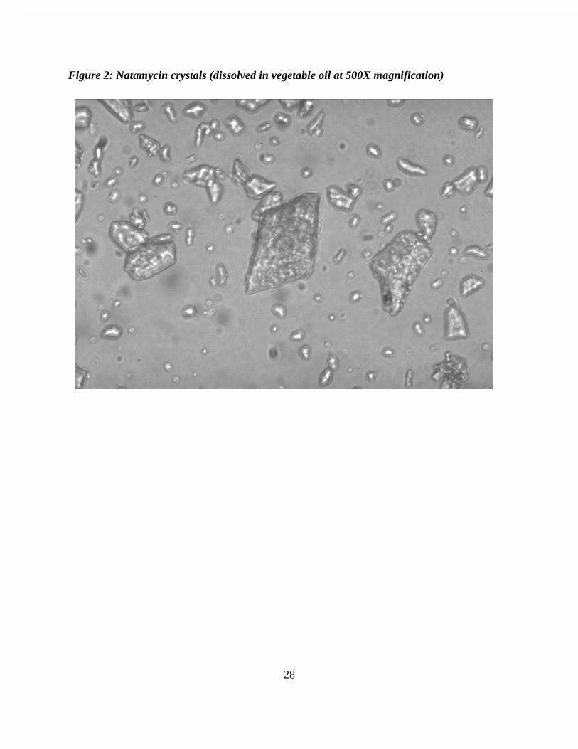
Figure 1: Chemical structure of natamycin . . . . . . . . . . . . . . . . . . . . . . . . . . . . 27Figure 2: Natamycin crystals (dissolved in vegetable oil at 500X magnification)
. . . . . . . . . . . . . . . . . . . . . . . . . . . . . . . . . . . . . . . . . . . . . . . . . . . . . . . 28References . . . . . . . . . . . . . . . . . . . . . . . . . . . . . . . . . . . . . . . . . . . . . . . . . . . . . . . . . . 29
CHAPTER 2: SYNTHESIS OF N-AMINOACYL DERIVATIVE OF NATAMYCIN . . . . . . 42Abstract . . . . . . . . . . . . . . . . . . . . . . . . . . . . . . . . . . . . . . . . . . . . . . . . . . . . . . . . . . . . 42Introduction . . . . . . . . . . . . . . . . . . . . . . . . . . . . . . . . . . . . . . . . . . . . . . . . . . . . . . . . . 42Materials and Methods . . . . . . . . . . . . . . . . . . . . . . . . . . . . . . . . . . . . . . . . . . . . . . . . . 44
Synthesis of Propylaminomalemide (PAM) Hydrochloride . . . . . . . . . . . . . . . 44Synthesis of Natamycin Derivative . . . . . . . . . . . . . . . . . . . . . . . . . . . . . . . . . . 45
Results and Discussion . . . . . . . . . . . . . . . . . . . . . . . . . . . . . . . . . . . . . . . . . . . . . . . . . 46Conclusions . . . . . . . . . . . . . . . . . . . . . . . . . . . . . . . . . . . . . . . . . . . . . . . . . . . . . . . . . 46Figures . . . . . . . . . . . . . . . . . . . . . . . . . . . . . . . . . . . . . . . . . . . . . . . . . . . . . . . . . . . . . 48
Page 6
vi
Figure 1: Major ions present in the positive ion FAB mass spectras for natamycin,PAM hydrochloride, and natamycin derivative . . . . . . . . . . . . . . . . . . . 48
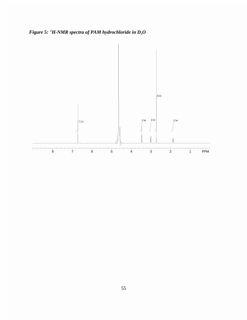
Figure 2: 1H NMR spectral data for PAM hydrochloride and natamycin derivative48References . . . . . . . . . . . . . . . . . . . . . . . . . . . . . . . . . . . . . . . . . . . . . . . . . . . . . . . . . . 49Appendix . . . . . . . . . . . . . . . . . . . . . . . . . . . . . . . . . . . . . . . . . . . . . . . . . . . . . . . . . . . 51
Figure 1: Reaction scheme for the preparation of propylaminomalemide (PAM)hydrochloride . . . . . . . . . . . . . . . . . . . . . . . . . . . . . . . . . . . . . . . . . . . . 51
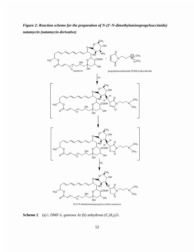
Figure 2: Reaction scheme for the preparation of N-(3'-N-dimethylaminopropylsuccimido) natamycin (natamycin derivative) . . 52
Figure 3: Low resolution FAB mass spectra of natamycin . . . . . . . . . . . . . . . . 53Figure 4: Low resolution FAB mass spectra of PAM hydrochloride . . . . . . 54Figure 5: 1H-NMR spectra of PAM hydrochloride in D2O . . . . . . . . . . . . . . . . 55Figure 6: Low resolution FAB mass spectra of N-(3'-N-dimethylaminopropylsuccimido) natamycin . . . . . . . . . . . . . . . . . . . . . . . . . . . 56Figure 7: High resolution FAB mass spectra of N-(3'-N-
dimethylaminopropylsuccimido) natamycin (natamycin derivative) . . 57Figure 8: 1H-NMR spectra of N-(3'-N-dimethylaminopropylsuccimido)
natamycin (natamycin derivative) in pyridine-d5 . . . . . . . . . . . . . . . . . 58
CHAPTER 3: MICROBIOLOGICAL CHALLENGE STUDY FOR SEMISYNTHETICDERIVATIVE OF NATAMYCIN AND PARENT ANTIBIOTIC . . . . . . . . . . . . . . . . 59Abstract . . . . . . . . . . . . . . . . . . . . . . . . . . . . . . . . . . . . . . . . . . . . . . . . . . . . . . . . . . . . 59Introduction . . . . . . . . . . . . . . . . . . . . . . . . . . . . . . . . . . . . . . . . . . . . . . . . . . . . . . . . . 59Experimental Design and Statistical Analysis . . . . . . . . . . . . . . . . . . . . . . . . . . . . . . . 61Materials and Methods . . . . . . . . . . . . . . . . . . . . . . . . . . . . . . . . . . . . . . . . . . . . . . . . . 61
Spore Stock Preparation . . . . . . . . . . . . . . . . . . . . . . . . . . . . . . . . . . . . . . . . . . 61Sample Production . . . . . . . . . . . . . . . . . . . . . . . . . . . . . . . . . . . . . . . . . . . . . . 62Microbiological Sampling . . . . . . . . . . . . . . . . . . . . . . . . . . . . . . . . . . . . . . . . . 63Package Integrity and Gas Sampling . . . . . . . . . . . . . . . . . . . . . . . . . . . . . . . . . 63
Results and Discussion . . . . . . . . . . . . . . . . . . . . . . . . . . . . . . . . . . . . . . . . . . . . . . . . . 64Package Integrity and Gas Analysis . . . . . . . . . . . . . . . . . . . . . . . . . . . . . . . . . 64Non-inoculated Treatment Analysis . . . . . . . . . . . . . . . . . . . . . . . . . . . . . . . . . 64Inoculated Treatment Analysis . . . . . . . . . . . . . . . . . . . . . . . . . . . . . . . . . . . . . 67Storage Condition Analysis . . . . . . . . . . . . . . . . . . . . . . . . . . . . . . . . . . . . . . . . 67
Conclusions . . . . . . . . . . . . . . . . . . . . . . . . . . . . . . . . . . . . . . . . . . . . . . . . . . . . . . . . . 67Figures . . . . . . . . . . . . . . . . . . . . . . . . . . . . . . . . . . . . . . . . . . . . . . . . . . . . . . . . . . . . . 69
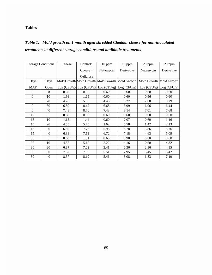
Figure 1: Mold growth on 1 month aged shredded Cheddar cheese at variousstorage conditions and antibiotic treatments . . . . . . . . . . . . . . . . . . . . . 69
References . . . . . . . . . . . . . . . . . . . . . . . . . . . . . . . . . . . . . . . . . . . . . . . . . . . . . . . . . . 70Appendix . . . . . . . . . . . . . . . . . . . . . . . . . . . . . . . . . . . . . . . . . . . . . . . . . . . . . . . . . . . 72
Figure 1: Application treatments for microbiological challenge study ofsemisynthetic derivative of natamycin and parent antibiotic . . . . . . . . 72
Page 7
vii
Figure 2: Storage treatments for microbiological challenge study of semisyntheticderivative of natamycin and parent antibiotic . . . . . . . . . . . . . . . . . . . . 72
Figure 3: Statistical model for microbiological challenge study of semisyntheticderivative of natamycin and parent antibiotic . . . . . . . . . . . . . . . . . . . . 73
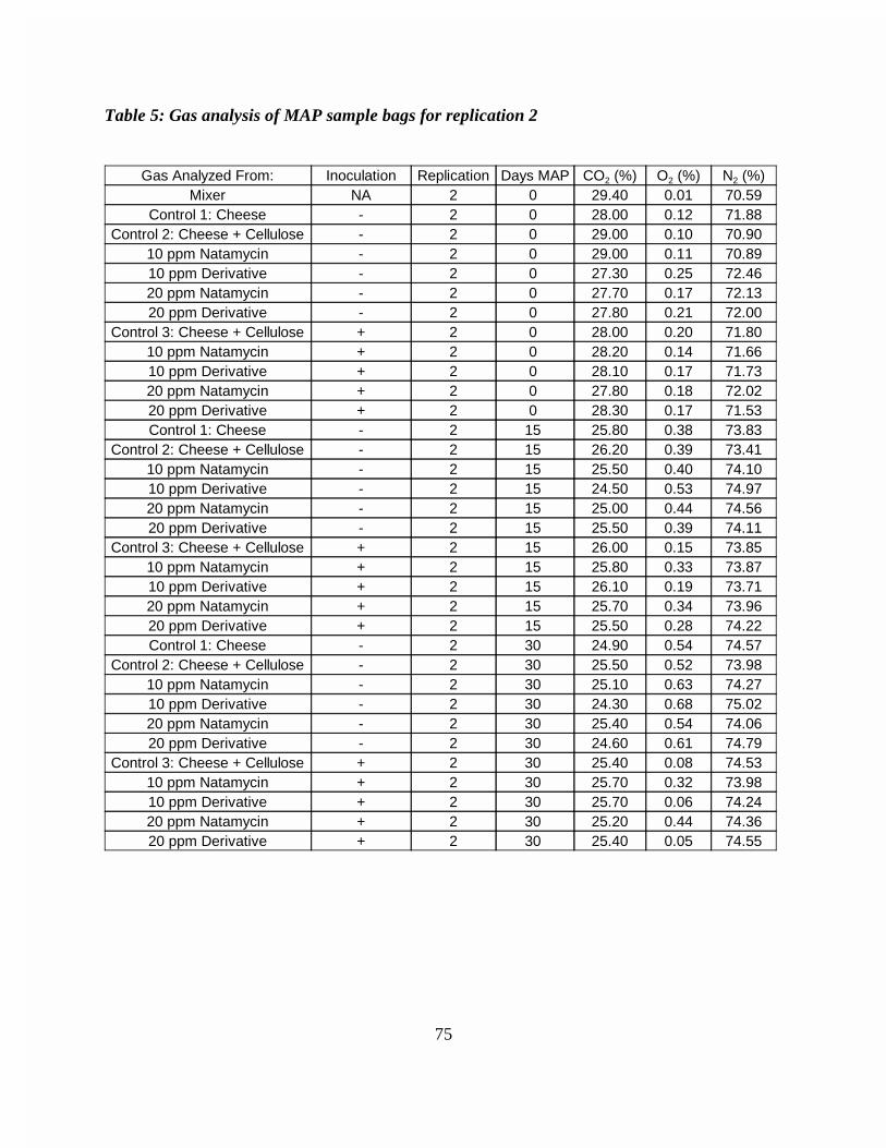
Figure 4: Gas analysis of MAP sample bags for replication 1 . . . . . . . . . . . . . . 74Figure 5: Gas analysis of MAP sample bags for replication 2 . . . . . . . . . . . . . . 75Figure 6: Gas analysis of MAP sample bags for replication 3 . . . . . . . . . . . . . . 76Figure 7: Mold growth on 1 month aged shredded Cheddar cheese for antibiotic
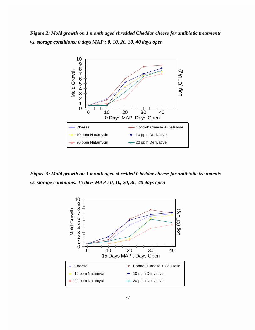
treatments vs. storage conditions: 0 days MAP : 0, 10, 20, 30, 40 daysopen . . . . . . . . . . . . . . . . . . . . . . . . . . . . . . . . . . . . . . . . . . . . . . . . . . . . 77
Figure 8: Mold growth on 1 month aged shredded Cheddar cheese for antibiotictreatments vs. storage conditions: 15 days MAP : 0, 10, 20, 30, 40 daysopen . . . . . . . . . . . . . . . . . . . . . . . . . . . . . . . . . . . . . . . . . . . . . . . . . . . . 77
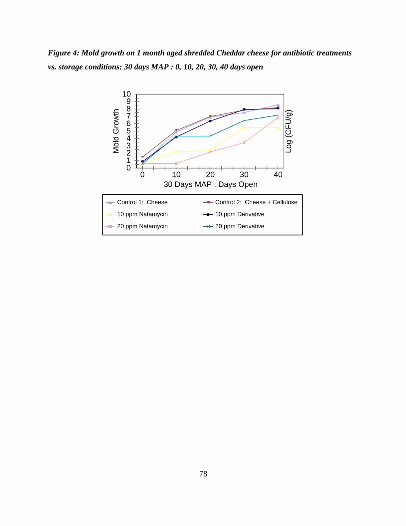
Figure 9: Mold growth on 1 month aged shredded Cheddar cheese for antibiotictreatments vs. storage conditions: 30 days MAP : 0, 10, 20, 30, 40 daysopen . . . . . . . . . . . . . . . . . . . . . . . . . . . . . . . . . . . . . . . . . . . . . . . . . . . . 78
Figure 10: Mold growth on 1 month ripened shredded Cheddar cheese inoculatedwith Penicillium roqueforti spores at various storage conditions andantibiotic treatments . . . . . . . . . . . . . . . . . . . . . . . . . . . . . . . . . . . . . . . 79
CHAPTER 4: STABILITY CHALLENGE STUDY FOR SEMISYNTHETIC DERIVATIVE OFNATAMYCIN AND PARENT ANTIBIOTIC . . . . . . . . . . . . . . . . . . . . . . . . . . . . . . . 80Abstract . . . . . . . . . . . . . . . . . . . . . . . . . . . . . . . . . . . . . . . . . . . . . . . . . . . . . . . . . . . . 80Introduction . . . . . . . . . . . . . . . . . . . . . . . . . . . . . . . . . . . . . . . . . . . . . . . . . . . . . . . . . 80Materials and Methods . . . . . . . . . . . . . . . . . . . . . . . . . . . . . . . . . . . . . . . . . . . . . . . . . 82
Spore Stock Preparation . . . . . . . . . . . . . . . . . . . . . . . . . . . . . . . . . . . . . . . . . . 82Sample Production . . . . . . . . . . . . . . . . . . . . . . . . . . . . . . . . . . . . . . . . . . . . . . 83Procedure for Determining Natamycin and Derivative Concentration on Cheese
. . . . . . . . . . . . . . . . . . . . . . . . . . . . . . . . . . . . . . . . . . . . . . . . . . . . . . . 83Results and Discussion . . . . . . . . . . . . . . . . . . . . . . . . . . . . . . . . . . . . . . . . . . . . . . . . . 84Conclusions . . . . . . . . . . . . . . . . . . . . . . . . . . . . . . . . . . . . . . . . . . . . . . . . . . . . . . . . . 86Figures . . . . . . . . . . . . . . . . . . . . . . . . . . . . . . . . . . . . . . . . . . . . . . . . . . . . . . . . . . . . . 88
Figure 1: Antibiotic concentration of 1 month aged shredded Cheddar cheese fornon-inoculated treatments vs. total days of storage . . . . . . . . . . . . . . . . 88
Figure 2: Antibiotic concentration of 1 month aged shredded Cheddar cheese forinoculated treatments vs. total days of storage . . . . . . . . . . . . . . . . . . . 88
Figure 5: Antibiotic concentration of 1 month aged shredded Cheddar cheese fornon-inoculated treatments at storage condition: 0 Days MAP : 0, 20 DaysOpen . . . . . . . . . . . . . . . . . . . . . . . . . . . . . . . . . . . . . . . . . . . . . . . . . . 89
Figure 6: Antibiotic concentration of 1 month aged shredded Cheddar cheese forinoculated treatments at storage conditions: 0 Days MAP : 0, 20 DaysOpen . . . . . . . . . . . . . . . . . . . . . . . . . . . . . . . . . . . . . . . . . . . . . . . . . . . 89
Page 8
viii
Figure 7: Antibiotic concentration of 1 month aged shredded Cheddar cheese fornon-inoculated treatments at storage condition: 15 Days MAP : 0, 20Days Open . . . . . . . . . . . . . . . . . . . . . . . . . . . . . . . . . . . . . . . . . . . . . . . 90
Figure 8: Antibiotic concentration of 1 month aged shredded Cheddar cheese forinoculated treatments at storage condition: 15 Days MAP : 0, 20 DaysOpen . . . . . . . . . . . . . . . . . . . . . . . . . . . . . . . . . . . . . . . . . . . . . . . . . . . 90
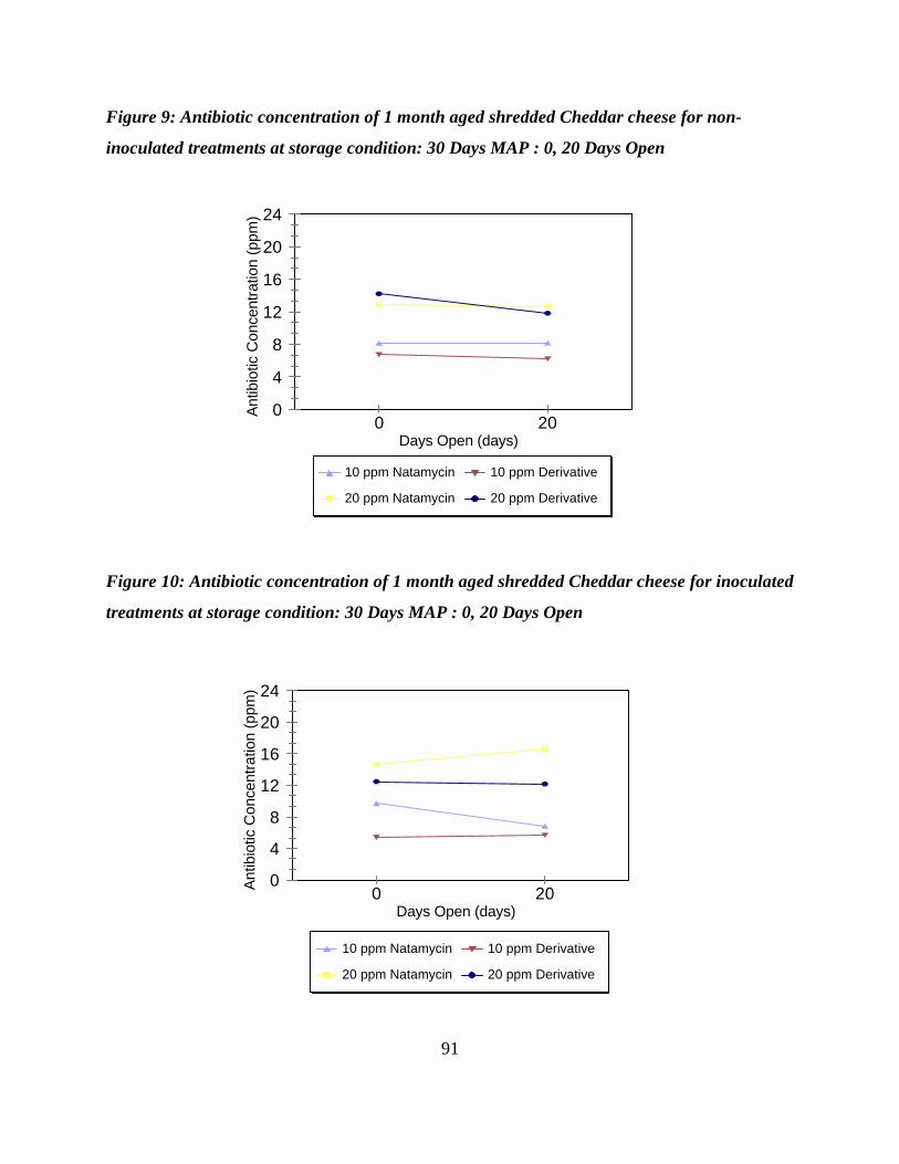
Figure 9: Antibiotic concentration of 1 month aged shredded Cheddar cheese fornon-inoculated treatments at storage condition: 30 Days MAP : 0, 20Days Open . . . . . . . . . . . . . . . . . . . . . . . . . . . . . . . . . . . . . . . . . . . . . . . 91
Figure 10: Antibiotic concentration of 1 month aged shredded Cheddar cheese forinoculated treatments at storage condition: 30 Days MAP : 0, 20 DaysOpen . . . . . . . . . . . . . . . . . . . . . . . . . . . . . . . . . . . . . . . . . . . . . . . . . . . 91
References . . . . . . . . . . . . . . . . . . . . . . . . . . . . . . . . . . . . . . . . . . . . . . . . . . . . . . . . . . 92
VITA . . . . . . . . . . . . . . . . . . . . . . . . . . . . . . . . . . . . . . . . . . . . . . . . . . . . . . . . . . . . . . . . . . . . 94
Page 10
1
CHAPTER 1: REVIEW OF LITERATURE
Discovery and Origin
In 1955, a Streptomyces strain was taken from a soil sample near the town of
Pietermaritzburg in Natal, a province of South Africa by Dutch scientists. The Streptomyces
strain was named Streptomyces natalensis (Raab, 1972; Thomas, 1976). A new, highly active
antibiotic was isolated from culture filtrates of Streptomyces natalensis and was named
pimaricin. Gist-brocades B.V. (Delft, The Netherlands) filed the patent application, “Pimaricin
and Process of Producing the Same,” on March 13, 1956 in the Netherlands (Struyk and
Waisvisz, 1960). Gist-brocades B.V. filed the U.S. patent application for pimaricin on March 7,
1957 (Struyk and Waisvisz, 1975).
The antibiotic tennecetin was isolated from a soil sample in Chattanooga, TN from a
Streptomyces strain in 1959. The strain was named Streptomyces chattanoogensis. Around the
same time, another antibiotic, A 5283, was isolated from Streptomyces gilvosporus. Tennecetin
and A 5283 were proved to have the identical chemical structure of pimaricin (Clark et al.,
1964). The production of pimaricin from Streptomyces gilvosporus was applied for patent in
the U.S. by the American Cyanamid Company on July 23, 1956 (American Cyanamid
Company, 1960).
In the late 1960's the World Health Organization (WHO) established a regulation
specifying that antibiotics produced from Streptomyces must carry names ending in “...mycin.”
Therefore, the antibiotic pimaricin changed its name to natamycin (Raab, 1972).
A review of literature shows that natamycin and pimaricin are interchangeably used to
describe this antibiotic. The food industry and most of the British Commonwealth use the name
natamycin to describe this substance, while most of the rest of the world and medical
community use the old name, pimaricin, more prevalently.
Page 11
2
Physical and Chemical Properties
Natamycin has an empirical formula of C33H47NO13 and a molar mass of 665.73 (Lück
and Jager, 1997). The International Union of Pure and Applied Chemistry (IUPAC) chemical
name for natamycin is 22-[(3-amino-3,6-dideoxy-�-D-mannopyranosyl)-oxy]-1,3,26-trihydroxy-
12-methyl-10-oxo-6,11,28-trioxatricyclo[22.3.1.05,7] octacosa-8,14,16,18,20-pentaene-25-
carboxylic acid. Chemical Abstract Service (CAS) registry number is 7681-93-8 (Brik, 1981).
The structural formula of natamycin is depicted in Figure 1.
The primary structure of natamycin is its large lactone ring of 25 C-atoms. The lactone
ring is linked to a mycosamine moiety, amino-sugar, by a glycosidic linkage, classifying
natamycin as a polyene macrolide antibiotic. In particular, natamycin is a tetraene antibiotic
because of its four conjugated double bonds. The mycosamine (3-amino-3,6-dideoxy-D-
mannose) of natamycin at the C15 position is a six-membered pyranose ring (Thomas, 1976).
Natamycin is a white, tasteless, and odorless powder. The antibiotic is crystalline in
structure and melts at a temperature of 180°C upon decomposition (Figure 2) (Oostendorp,
1981). Exposures in excess of one hour at temperatures above 120° C impair the antibiotic
activity of natamycin (Lück and Jager, 1997; Raab, 1972).
Solubility
Natamycin is extremely insoluble in water. Schaffner and Mechlinski (1972) report the
solubility of natamycin as 0.0520 mg/ml. In addition, natamycin is insoluble in higher alcohols,
ethers, esters, aromatic or aliphatic hydrocarbons, chlorinated hydrocarbons, ketones, dioxane,
cyclohexanol and various oils. Glacial acetic acid, methylpyrrolidone, dimethylformamide,
dimethylsulfoxide, glycerol, and propylene glycol are good solvents for natamycin (Raab, 1972).
The large molecular weight, 665.73 g/mol, and conjugated double bond structure
explains the poor solubility of natamycin in water and most alcohol solutions (Thomas, 1976).
Natamycin’s insolubility in most hydrocarbons is attributed to the three hydroxyl groups in the
lactone ring and the formation of a dipolar ion. Natamycin is an amino acid that contains both
Page 12
3
acidic and basic groups that undergo an intermolecular acid-base reaction. This reaction causes
natamycin to exist in the form of a dipolar ion, or zwitterion (McMurry, 1992).
The zwitterionic nature of natamycin allows it to react with acids or bases depending
upon the circumstance. In an aqueous acid solution, natamycin can accept a proton at the C12
carboxyl group and form a cation. Conversely, in an aqueous basic solution, natamycin loses a
proton from its amino group of the mycosamine moiety forming an anion. Amino acids that
possess this dual relationship with acids and bases are called amphoteric (McMurry, 1992).
The solubility and amphoteric nature of natamycin can be better understood by viewing
this antibiotic in two chains. The chain that contains the four double bonds is completely
hydrophobic, whereas the chain with the two hydroxyl groups has a hydrophillic and
hydrophobic face, rendering the chain amphipathic. The polar end of the amphipathic chain
contains the mycosamine and a carboxyl group. The other end is marginally non-polar with only
one hydroxyl group. Natamycin forms a cylindrical structure by aligning hydroxyl groups of its
amphipathic chain toward each other. The exterior of the cylinder is completely non-polar
(Thomas, 1976).
Thomas (1976) indicates another reason why many polyene antibiotics are extremely
insoluble and experience relatively high stability is the existence of a six-membered ketal ring
structure when the antibiotic is in a crystalline form. The ketal ring in natamycin is achieved by
joining carbon atoms 9 and 13.
Strong solvent-solute interactions explain the exceptional solubility of natamycin in
dimethylformamide and dimethylsulfoxide. Infrared studies by Mechlinski and Schaffner
(1972) indicate strong intermolecular hydrogen bonds involving hydroxyl, carboxyl, and amino
groups. These studies also suggest disruption of the zwitterion by solvent molecules. This
subjects the carboxyl and amino group to chemical reactions.
Natamycin’s solubility in methylpyrrolidone and other strong polar organic solvents is
not fully understood. It is believed that polyene antibiotic molecules form suspensions of
micelles rather then dissolving. Micellular suspensions may also cause polyene antibiotics to
exist in pure aqueous solutions without visible precipitation at concentrations of 50 )g/ml
(Oostendorp, 1981; Thomas, 1976).
Page 13
4
Stability
Natamycin is most stable in a crystalline powder form in the absence of heat and light.
No loss in biological activity occurs when stored at room temperature and protected from
ultraviolet light at 30 months. The stability of natamycin is lower in dry formulations of sodium
chloride, citric acid, and sucrose (Hoogerheide, 1961 and 1964, unpublished data).
Micellular suspensions of natamycin offer protection by shielding the labile sites of the
molecule (Thomas, 1976). Studies show no loss in biological activity for a 0.5% aqueous
suspension of natamycin with 0.1% polyoxyethylene stored in the dark at pH 6.5 for 24 months
(Hoogerheide, 1961 and 1964, unpublished data).
Ultraviolet light with wavelengths of 300 to 350 nm quickly inactivates natamycin
(Dekker and Ark, 1959). The tetraene structure of natamycin causes its instability in the
presence ultraviolet light. The photodynamic destruction of natamycin occurs by a unique
triplet-triplet transfer mechanism. Most light induced destructions occur as a result of oxidation
or by a free-radical mechanism. A triplet-triplet transfer occurs when a compound in an excited
triplet state collides with a compound in the singlet state at a lower energy state. The result is a
conversion of spin movement for the singlet state compound causing it to excite the molecule.
An experimental demonstration of this phenomenon was conducted for natamycin. Excited
riboflavin molecules in the triplet state bombarded natamycin in the 300 to 350 nm range
causing a triplet-triplet transfer. The triplet-triplet transfer caused the natamycin molecule to
quickly degrade the reactive tetraene structure (Posthuma and Berends, 1960; 1961).
Photodynamic destruction of natamycin caused by triplet-triplet transfer does not show
decreases in maxima peaks 279, 290, 304, and 318 nm in an ultraviolet absorption spectra
(Thomas, 1976).
Natamycin suspensions and solutions lose their biological activity by oxidation.
Oxidation is promoted by elevated temperatures, greater than 37°C, and in the presence of
chemical oxidants (Dekker and Ark, 1959; Barr, 1959). Chemical oxidants such as peroxides,
perchlorates, persulfates, permanganates, iodates, bromates, hypochlorites, sulfites, and
Page 14
5
anhydrides of organic acids cause the oxidative inactivation of natamycin. The ultraviolet
absorption spectra of natamycin following oxidative inactivation show marked decreases in
maxima peaks, 279, 290, 304, and 318 nm (Thomas, 1976).
Oxidation is amplified at pH levels below 5.0 and above 9.0 (Raab, 1972). In organic
acids, acid hydrolysis occurs resulting in the elimination of the mycosamine. Natamycin
changes its structure from a tetraene to a pentaene as a result (Thomas, 1976). Oxidative
inactivation may be prevented by the addition of chlorophyll, ascorbic acid, or a number of other
antioxidants (Dekker and Ark, 1959; Barr, 1959; Struyk et al., 1958).
The isoelectric point of natamycin is 6.5. Solutions of natamycin with pH values
between 5.0 and 9.0 are stable when stored in the dark (Raab, 1972). Natamycin undergoes
hydrogenation in the presence of a palladium catalyst. Single-stage hydrogenation occurs in an
alkaline medium, whereas a more complete hydrogenation is demonstrated in neutral or acidic
solutions (Raab, 1972).
“Physiochemically, the stability of natamycin depends greatly on the stability of the
tetraene moiety of the molecule. Readily occurring inactivation of natamycin (in solution) by
heat, by light, (ultraviolet light or in the presence of substances capable of transferring photo-
energy) or by oxidation always begins with the severing of these interconnected, unsaturated
double bonds” (Raab, 1972).
Toxicity
The toxicological effects of natamycin in this paper will be limited to oral doses. The
acute toxicity of natamycin has been determined for mice, rats, guinea pigs, and rabbits through
clinical studies with little agreement. The LD50 of natamycin after oral administration has been
determined to be 1.5 g/kg for mice and rats and 0.45 g/kg for guinea pigs (Struyk et al., 1958).
Levinskas et al. (1966) determined the LD50 as 2.7 - 4.7 g/kg and 1.4 g/kg for rats and rabbits,
respectively. The intake of natamycin did not cause any gross lesions on the animals that died.
It should be noted that irritation of the mucosa was seen in rabbits. Near lethal doses of
natamycin caused growth inhibition, diarrhea, and edema (Struyk et al., 1958).
Page 15
6
Levinskas et al. (1966) conducted chronic toxicity tests for natamycin in rats and dogs.
A two year study was performed for rats who were fed a 0.2% natamycin feed. This 1000 ppm
dosage of natamycin was well tolerated with only minor effects being observed. Observed
effects included, slight decrease in food intake and slight inhibition in growth. Lactation and
reproductions were normal for these animals over the two year test period. Results from chronic
toxicity tests for dogs were similar to those for rats. Feed containing 0.05% and 0.025%
natamycin, 500 and 250 ppm, were fed to dogs for over a two year period. Dogs subjected to
500 ppm dosages experience a slight increase in body weight. No observable effects occurred in
dogs administered 250 ppm dosages over the two year period. The acceptable daily intake
(ADI) has been established at 0.30 mg/kg by the WHO and Food and Agriculture Organization
of the United Nations (FAO). Approximately 0.002 mg/kg of natamycin is ingested by the
average cheese consumer. This consumption equals <1% of the established ADI (Oostendorp,
1981).
Derivatives of Polyene Macrolide Antibiotics
As previously mentioned, polyene macrolide antibiotics have characteristics such as
extreme insolubility in water, severe instability, and serious parenteral toxicity. Many attempts
have been made to overcome one or more of these problems by chemical modification of the
chemical structure. Extensive research in the past 25 years has been conducted in polyene
macrolide chemistry. Although some studies have been conducted on natamycin, the vast
majority of research on this family of antibiotics have been performed for amphotericin B
(Schaffner, 1987).
Colloidal Dispersion Complexes
The aqueous insolubility of polyene macrolide antibiotics is due to their intramolecular
aggregation. “Introduction of detergent-like molecules, such as bile acids and fatty alcohol’s
sulfonates, or of polar polymers, such as polyborates, produce reversible complexes with
Page 16
7
increased molecular dispersion in aqueous medium” (Schaffner, 1987).
Bartner et al. (1958) prepared a neutral solution of amphotericin B in an aqueous
solution of sodium desoxycholate. The preparation was lyophilized into a stable powder that
can be reconstituted with water or dextrose solution. It was discovered that soluble
amphotericin B prepared by the use of sodium desoxycholate does not enter into true solution
(molecular dispersion). Although aqueous solutions of this preparation appear clear, they
demonstrate the Tyndall effect, and exist in a fine colloidal dispersion of particles with
dimensions smaller than those of bacteria (Bartner et al., 1958). Amphotericin B sodium
desoxycholate complex (Fungizone®) is manufactured by Bristol-Meyers Squibb Company and
is the only polyene antibiotic complex approved by the Food and Drug Administration (FDA)
for intravenous use (Bristol-Meyers Squibb Company, 1999).
An amphotericin B borate complex has also been produced (Vandeputte, 1973). The
amphotericin B borate complex exhibits good water solubility, but only possess 80% the
antifungal activity and has a significantly higher acute parenteral toxicity than that of the
amphotericin B sodium desoxycholate complex (Schaffner, 1987).
Semisynthetic Derivatives
Chemical interactions with the zwitterionic carboxylate and the mycosamine of polyene
macrolide antibiotics have produced a variety of semisynthetic antifungal derivatives with
increased solubility as salts. Unlike colloidal dispersion complexes, these compound enter into
molecular dispersion. Chemical interaction with free carboxylic acid and amino base functional
groups is difficult. Water-soluble salts produced by direct interaction with strong acids or bases
exhibit marked instability due to extremes of pH in aqueous solutions. Adjustments of solutions
towards neutrality or the isoelectric pH of antibiotic results in precipitation due to colloidal
aggregation and micelle formation. At acidic pH, degradation occurs due to chromophore
isomerization and glycosidic hydrolysis. At alkaline pH, the macrocyclic lactone can be
saponified with alterations of molecular structure and conformation resulting in total loss of
biological activity. The conjugated polyene chromophore is also adversely affected by heat,
Page 17
8
oxidation, reduction, and ultraviolet radiation. Isomerization reactions, epoxide formation, or
uptake of hydrogen produce structural alterations, which also lead to reduced or total loss of
biological activity (Schaffner, 1987).
Carboxyl Group Derivatives
Mechlinski and Schaffner (1972) synthesized amphotericin B methyl ester.
Esterification was accomplished by bubbling diazomethane, prepared in tetrahydrofurane (THF),
in a mixture of DMSO and amphotericin B. Amphotericin methyl ester demonstrated an
equivalent biological activity to the parent antibiotic. Both compounds showed MIC values of
0.05-0.5 µg/ml for Saccharomyces cerevisiae, Aspergillus niger, and Candida albicans
(Mechlinski and Schaffner, 1972). Based on this study, it was concluded that esterification of
the carboxyl group has little effect on the activity of the antibiotic.
The methyl ester derivative of amphotericin B was basic and its solubility was only
marginally better than the parent. Schaffner and Mechlinski (1972) treated the methyl ester
derivative of amphotericin B with hydrochloric acid to form a water soluble salt of this
compound, amphotericin B methyl ester hydrochloride. This compound showed drastically
improved water solubility and exhibited full antifungal activity. The solubility (molecular
dispersion) of this compound was verified by UV spectroscopy. Amphotericin B methyl ester
hydrochloride enters into true solution, unlike Fungizone®. E.R. Squibb and Sons, Inc.
obtained the U.S. patent for amphotericin B methyl ester salts on August 9, 1977 (Sipos and
Keseleski, 1977).
The production of methyl ester hydrochloride derivatives of nystatin, natamycin,
mediocidin, candicidin, and trichomycin followed. All of these compounds exhibited good
solubility and antifungal activity. For example, natamycin methyl ester hydrochloride has a
solubility of 80.000 mg/ml in water at 25°C whereas the natamycin has a solubility of 0.052 mg/
ml for these same conditions (Schaffner and Mechlinski, 1972).
More comprehensive studies were performed examining the biological properties of
polyene macrolide ester salts (Bonner et al., 1972; Bruzzese et al., 1975). In vitro studies
Page 18
9
against S. cerevisiae and in vivo studies against C. albicans in mice confirmed previous findings
that polyene macrolide ester salts are at least as biologically active as their parent antibiotics and
in some cases even more active(Bonner et al., 1972). Bruzzese et al. (1975) findings agreed
with Bonner’s, but also observed that the lengthening of the aliphatic chain in the ester group
(methyl to butyl) produced a gradual decrease in activity. Bonner et al. (1972) also examined
the acute intraperitoneal toxicities of polyene macrolides and their methyl ester hydrochlorides
in mice. The methyl ester hydrochlorides were less toxic then their parent antibiotics for all
polyenes tested.
Subsequent research focused on improved methods for preparation of methyl ester
derivatives and extension of these methods to previously underivatized polyene antibiotics
(Bruzzese and Ferrari, 1974; Pandey et al., 1977). Researchers from around the world
challenged the efficacy and toxicity of amphotericin B methyl ester. Many in vitro, in vivo, and
toxicity studies were published for amphotericin B.
Bannatyne and Cheung (1977) compared the susceptibility of 465 clinical isolates of C.
albicans to amphotericin B and amphotericin B methyl ester. Results from the study showed
that amphotericin B and amphotericin B methyl ester had comparable activity against half of the
strains, but against the remainder of the strains the activity of amphotericin B methyl ester was
slightly lower than that of amphotericin B. In another study, the in vitro antifungal activity of
amphotericin B methyl ester was compared to the parent against a variety of pathogenic and
potentially pathogenic fungi (Howarth et al., 1975). Test organisms used in this study included:
Aspergillus fumigatus, A. niger, Blastomyces dermatitidis, C. albicans, C. guilliermondii, C.
pseudotropicalis, Cladosporium sp., Coccidioides immitis, Cryptococcus neoformans,
Histoplasma capsulatum, H. duboisii, Microsporum canis, M. gypseum, Muco pusillus,
Oidiodendron kalrai, Paracoccidioides brasiliensis, Phialophora compactum, P. dermatitidis,
P. verrucosa, Rhizopus arrhizus, Sepedonium sp., Sporothrix schenckii, Syncephalastrum,
Torulopsis glabrata, Trichophyton memtagrophytes, T. rubrum, and T. simii. Amphotericin B
methyl ester showed significant antifungal activity against most fungi, but was slightly less
active then amphotericin B. The yeast cells of most fungi were killed at concentrations of 1
µg/ml or less for either antibiotic. Filamentous forms of dimorphic fungi were more resistant
Page 19
10
requiring 10 to 50 µg/ml concentrations of these amphotericin B and amphotericin B methyl
ester to cause death to these organisms (Howarth et al., 1975).
In vivo studies of amphotericin B (Fungizone®) and amphotericin B methyl ester against
experimental histoplasmosis, blastomycosis, cryptococcosis, and candidosis in mice were
performed (Bonner et al., 1975). Mean effective doses (ED50) for amphotericin B methyl ester
were slightly higher for Histoplasma, Blastomyces, and Cryptococcus infections than those of
the parent compound. In contrast, the ED50 for Candida infections for amphotericin B were
slightly higher than that of amphotericin B methyl ester.
A similar in vivo study of amphotericin B (Fungizone®) and amphotericin B methyl
ester against C albicans, C. neoformans, and B. dermatitidis in mice was performed (Gadebusch
et al., 1976). Findings from these studies showed that amphotericin B methyl ester was
drastically less efficacious then amphotericin B. Researchers of this study proposed that
amphotericin B methyl ester has a lower avidity than amphotericin B for membrane-binding
sites, is inactivated to a greater extent at these sites, and is poorly distributed when compared to
the parent antibiotic.
In addition, the therapeutic efficacy of amphotericin B (Fungizone®) and amphotericin
B methyl ester delivered intraperitoneal or intravenous in mice infected with experimental
murine coccidioidomycosis while monitoring toxicity (Lawrence and Hoeprich, 1976).
Amphotericin B methyl ester was less effective than Fungizone® at low doses, but at higher
doses was more effective than the parent antibiotic. At higher doses over 30 days, Fungizone®
was lethal and/or nephrotoxic, whereas amphotericin B methyl ester was therapeutically
effective and nontoxic to the kidneys. Keim et al. (1976) performed toxicological studies of
amphotericin B methyl ester and amphotericin B in mice, rats, and dogs. It was found that the
acute and subacute toxicity of amphotericin B methyl ester was much less than for the parent
antibiotic. Amphotericin B methyl ester is 20 times less toxic than amphotericin B when
administered as a single intravenous dose in mice. Intraperitoneal studies in rats and dogs
showed the methyl ester derivative was one-forth and one-eighth as nephrotoxic as amphotericin
B.
Results from these studies indicated that amphotericin B methyl ester could be an
Page 20
11
improved therapeutic agent for the treatment of systemic mycoses when compared to that of
amphotericin B. Experimental in vitro and in vivo results showed that amphotericin B methyl
ester derivatives were somewhat less effective against fungi and yeasts, but there significantly
lower toxicity warranted their use. It was found that amphotericin B methyl ester could be safely
administered to mice at concentrations several times greater than concentrations of Fungizone®
that caused death (Lawrence and Hoeprich, 1976).
Monji et al. (1975) conducted an experiment to investigate the metabolic stability of
amphotericin B methyl ester. Intravenous and intraperitoneal administration of radioactive
amphotericin B 14C-methyl ester in mice revealed no de-esterification of the antibiotic
derivative. The most significant finding of this investigation was that there was no
accumulation of radioactivity in the kidneys after administration of radiolabelled amphotericin
B methyl ester. A similar study was performed for a non-human primate, Macaca mulatta
(Lawrence et al., 1980). Results agreed with the previous study, amphotericin B methyl ester
was metabolically stable and no de-esterification occurred.
Between 1975 and 1980, 50 patients suffering from mycoses caused by seven different
kinds of fungi were treated with amphotericin B methyl ester by intravenous and intrathecal
routes of administration. Doses administered were typically five times greater than those given
with Fungizone®. Nephrotoxicity with these treatments were uncommon, but other more
serious neurotoxic side-effects were evident. “In some of the patients receiving high doses for
prolonged periods of time, neuropsychiatric symptoms such as confusion, disorientation,
withdrawl, lethargy, depression, anorexia and more rarely stupor and coma became evident.
Electroencephalo abnormalities were also evident” (Schaffner, 1987). Ellis et al. (1982)
conducted histopathological examinations of the brains of patients who exhibited symptoms of
neurological toxicity and who died of their fungal infections. The autopsy study showed diffuse
leukoencephalopathy which consisted of proliferation of gemistocytic astrocytes, pallor of the
myelin and accumulation of macrophage. These conditions were found in the cerebrum and
cerebellar white matter (Ellis et al., 1982).
It was suggested (Hoeprich et al., 1982) and then later proved (Schaffner, 1987) that lots
of amphotericin B methyl ester used in the treatment of patients revealed a complex of polyene
Page 21
12
macrolide components. In addition to monomethyl ester amphotericin B, the complex consisted
of di-, tri-, tetra-, and penta-methylated amphotericins. Additional methylations occurred
exclusively on the free amino group and the hydroxyl groups of the mycosamine moiety. None
of the overmethylation involved the hydroxyl groups of the aglycone. It is believed that
intermolecular bonding prevented this occurrence.
Amphotericin B amides have been produced by reaction of amines with the free carboxyl
group of the polyene macrolide aglycone (Stefa�ska et al., 1980; Jarzebski et al., 1981). In vitro
studies of amphotericin B amides demonstrated that these compounds have similar activity to
that of the parent antibiotic, whereas some are virtually inactive. “The antifungal activity of the
amide derivative appear to depend on the lipophilicity of the amid substituent. No in vivo
chemotherapeutic data from animal studies have been performed” (Schaffner, 1987).
Grzybowska and Borowski (1990) have published a paper on the preparation of polyene
macrolide hydrazides, but no biological studies on these compounds have followed.
Amino Group Derivatives
Although most research of polyene macrolide semisynthetic derivatives has focused on
ester derivatives, the first derivatization of macrolide antibiotics occurred in 1961 by
successfully forming an N-alkyl derivative of natamycin (Schaffner and Borowski, 1961;
Lechevalier et al., 1961). It involved the treatment of amphotericin B with a large excess (50
moles) of acyl anhydride at room temperature for 1 hour. The yield of this procedure was 70-
75% and the purity of the compound was poor. Mechlinski and Schaffner (1972) later improved
this procedure by conducting the acylation with acetic anhydride in an improved reaction
medium for the antibiotic. The solution was a mixture of DMSO and methanol. It was found
that the N-acylation of amphotericin B drastically reduced this compounds biological activity.
The minimum inhibitory concentration (MIC) for N-acetyl amphotericin B, N-iodoacetyl
amphotericin B, N-propionyl amphotericin B, and N-succinyl amphotericin B was 1.0 - 10.0
µg/ml for test organisms S. cerevisiae, A. niger, and C. albicans compared to 0.05 - 0.5 µg/ml
for the parent antibiotic (Mechlinski and Schaffner, 1972).
Page 22
13
Based on research from 1972, the development of semisynthetic polyene antibiotics
derivatized at the free amino group did not occur. Researchers wrongfully hypothesized that an
intact amino group on polyenes was required for biological activity. It was not until the early
1980's when Schering-Plough Corp. ( Bloomfield, NJ) began developing N-aminoacyl
amphotericin B methyl ester derivatives that research in this area was revisited (Oblack et al.,
1981).
Schering-Plough Corp. developed N-D-ornithyl amphotericin B methyl ester in 1980.
Galgiani and VanWyck (1984) compared the efficacy of N-D-ornithyl amphotericin B methyl
ester and Fungizone® against C. albicans infection in rats. At low doses, 0.1 and 0.5 mg/kg, the
drugs were equally effective in preventing deaths. At doses of 2.0 mg/kg, mortality occurred in
rats treated with Fungizone®, whereas N-D-ornithyl amphotericin B methyl ester was effective
and non-toxic. Graybill and Kaster (1984) showed that Aspergillus was equally susceptible to
amphotericin B and N-D-ornithyl amphotericin B methyl ester. These findings were supported
by in vitro and in vivo tests in mice.
A comprehensive comparative in vitro and in vivo evaluation of N-D-ornithyl
amphotericin B methyl ester, amphotericin B methyl ester, and amphotericin B was performed
by researchers at Schering-Plough Corp. (Parmegiani et al., 1987). In vitro studies indicated
that N-D-ornithyl amphotericin B methyl ester was more active than the other two drugs against
Candida spp. and other fungi. In vivo, the dose response of N-D-ornithyl amphotericin B
methyl ester was similar to amphotericin B to produce a 10,000-fold reduction of C. albicans in
a mouse kidney infection and was much more effective then amphotericin B methyl ester. Acute
intravenous 50% lethal doses in mice revealed that N-D-ornithyl amphotericin B methyl ester
was one-ninth as toxic is amphotericin B, but twice as toxic as amphotericin B methyl ester.
Czerwi�ski et al. (1986) highlighted a general procedure for the preparation of N-
dimethylaminoacyl derivatives of polyene macrolide antibiotics by combining the native polyene
antibiotic in dimethylformamide (DMF) with an excess of N-hydroxysuccinimide ester in the
presence of triethylamine. Czerwi�ski et al. (1991) later published a more comprehensive paper
on the synthesis and biological properties of N-alkyl derivatives of amphotericin B. One
particular N-alkyl derivative of amphotericin, N-(3'-N-dimethylaminosuccimido) amphotericin
Page 23
14
B, was of interest. N-(3'-N-dimethylaminosuccimido) amphotericin B is achieved by the
Michael addition reaction of amphotericin B with the N-substituted malemide (N-(3'-N-
dimethylaminopropyl)malemide hydrochloride). MIC studies for N-(3'-N-
dimethylaminosuccimido) amphotericin B showed it to possess equivalent activity to
amphotericin B for test organisms S. cerevisiae, C. albicans, C. tropicalis, Geotrichum
candidum, Torulopsis candida, and Trichophyton nanum. N-(3'-N-dimethylaminosuccimido)
amphotericin B was less biologically active against C. arborea, C. mycoderma, and A. nidulans,
but more active against A. niger, Penicillium cytrinum, and Mucor mucedo (Czerwi�ski et al.,
1986). The uniqueness of N-(3'-N-dimethylaminosuccimido) amphotericin B is the presence of
an additional, basic nitrogen atom in the modified aminosugar moiety.
Mode of Antimicrobial Action
The mode of action of polyene antibiotics mimics the hemolytic activity of saponins
(Demel et al., 1965). Saponin, from an aqueous phase, penetrates and complexes with surface-
absorbed complexes, particularly cholesterol. Cholesterol is removed from the cell membrane by
a saponin solution. Extensive rearrangement of membrane lipids occurs and changes the state of
the membrane. The membrane changes from a free flowing lamellar state to a micellar state due
to the aggregation by the saponin-cholesterol complex. The micellar state resembles a hexagonal
lattice. The hexagonal lattice structure is more permeable than the original membrane due to its
pits and holes (Bangham and Horne, 1962; Lucy and Glauert, 1964).
Similarities between polyene antibiotics and saponins were realized after the effects of
nystatin, filipin, and amphotericin B were examined on Saccharomyces and Candida strains.
These studies indicated that losses of K+, NH4+, inorganic phosphate, carboxylic acids, sugar
phosphates, nucleotides, and protein occur from the action of polyene antibiotics. Harsch and
Lampen (1963) conducted a study examining the K+ transport system of S. cerevisiae after
exposure to the polyene antibiotic N-Acetylcandidin. At low levels, the binding of N-
Acetylcandidin to the membrane reduces the efficiency of the ion transport into and out of the
cell. The usual Na+-K+transport balance is adversely affected. This effect is reversed by the
Page 24
15
addition of K+and NH4+ and is known as sublysis.
N-Acetylcandidin, at high levels (>100 µg/ml), lysis the cell membrane by causing
hexagonal pits. Loss of NH4+ occurs and blocks the cells ability to accumulate K+ by depriving
it of a necessary energy source. The addition of K+and NH4+ did not cause this effect to be
reversible. Loss in efficiency and the ability to accumulate K+ by ion transport expedites cell
death (Harsch and Lampen, 1963).
A similar study investigating intracellular loss of potassium in C. albicans after
exposure to a number of polyene antibiotics was performed. Results from this study were less
conclusive. Some antibiotics performed identical to N-Acetylcandidin where others did not. A
few antibiotics caused irreversible membrane damage immediately at low concentrations.
(Zygmunt, 1966). These results suggested that all polyene antibiotics do not have a similar
mode of action.
Polyene macrolide antibiotics can be divided into two groups. Group 1 (46-63 carbon
atoms) include: N-Acetylcandidin, nystatin, candidin, candicidin, and amphotericins A and B).
These antibiotics have a specific effect on cation permeability at low concentrations. Their
effects on glycolysis can be completely or partially reversed under appropriate conditions
(Kinsky, 1967; Weissmann and Sessa, 1967). Group 2 (33-37 carbon atoms) include filipin,
etruscomycin, and natamycin). Antibiotics in group 2 cause more extensive cell membrane
damage than members of the group 1 because of their ability to interact with membrane
phospholipids. Antibiotic-phospholipid interaction induces increased surface pressure to the
sterol present in the membrane and causes reorientation of the cell membrane. Loss of vital cell
constituents and osmotic shock occurs to the cell (Demel et al., 1968; Morris and Hart, 1978).
The effects of group 2 polyene antibiotics cannot be reversed or prevented.
A relationship exists between the number of carbon atoms, size of macrolide ring, and
the destructive power of the antibiotics to the cell membrane. Polyene antibiotics with smaller
macrolide rings are more destructive to the cell membrane. No relationship for cell membrane
damage exists for number of conjugated double bonds or net charge (Kinsky, 1967). It is
important to note that relationships discussed are for relative degree of damage to the membrane
and not on relative potency of the antibiotics in inhibiting growth. Heptaenes inhibit growth at
Page 25
16
lower concentrations than tetraenes.
In 1965, it was discovered that the presence of sterol in the cell membrane is necessary
for polyene sensitivity by researchers at two institutions, independently, by similar methods.
Mycoplasma laidlawii strain A, unlike most mycoplasmas, does not require sterols. When
grown in the presence of cholesterol the organism incorporates the sterol into its membrane.
Weber and Kinsky (1965) discovered, “The polyene antibiotic, filipin, inhibited growth and
caused lysis of Mycoplasma laidlawii cells which had been cultured in the presence of
cholesterol. The antibiotic did not inhibit growth and did not promote lysis of the organism
when grown in the absence of cholesterol.” Similarly, Feingold (1965) observed these same
effects for Mycoplasma laidlawii when treated with amphotericin B.
The sterol hypothesis for the mode of action of polyene antibiotics was proven again by a
unique study on the effects of polyene antibiotics on growth and sterol-induction of oospore
formation by Pythium and Phytophthora species. Pythium and Phytophthora species,
destructive plant pathogenic fungi, require an exogenous source of sterol for reproduction by
sporangia or oospores (Hendrix and Lauder, 1966). Polyene antibiotics completely blocked
oospore formation for Pythium periplocum when cholesterol was incorporated.
The interaction of polyenes with sterols was quantified by measuring changes in surface
pressure occurring when polyenes were injected beneath monolayers of lipids or lipid mixtures
at varying initial surface pressures. Changes occurred because interactions altered the spatial
orientation of the lipid molecules with respect to the air-water interface. This research allowed
scientists to quantify and compare the relative activity of various polyenes with numerous sterols
(Hamilton-Miller, 1974).
Numerous in vitro studies were performed with polyene antibiotics on synthetic lecithin-
cholesterol bilayers to better understand the biological properties and selective membrane
toxicity of polyene antibiotics. Results from these studies lead to the following conclusions: (1)
polyene-induced permeability alteration in membrane systems is affected by the composition of
membrane phospholipid fatty acyl chains; (2) the selective toxicity of polyenes is related to the
distribution of double bonds in the sterol nucleus; (3) polyenes differ in membrane selectivity
(Zutphen et al., 1971; Norman et al., 1972a; Norman et al., 1972b; Hsuchen and Feingold,
Page 26
17
1973).
Brajtburg et al. (1990) also reviewed the molecular basis of sterol specificity of polyenes.
Spectrophotometry has demonstrated that polyene antibiotics bind more avidly to ergosterol than
to cholesterol and to ergosterol-containing membranes than to cholesterol-containing
membranes. Two theories have been presented to explain this phenomenon. One theory
suggests that polyene antibiotics and sterols, with the participation of H2O, form a “cage”
linking the sterol between the carboxyl and amino end of the molecule by hydrogen bonds.
Functional groups involved in the hydrogen bonds are the hydroxyl groups of the sterol and the
carboxyl group. The “cage” is completed by participation of the free amino group. Both
ergosterol and cholesterol are 3-�-hydroxy sterols, and it can be assumed that their reactions
with polyenes involving hydrogen bonds are equivalent (Brajtburg et al., 1990).
Another theory to help explain the sterol specificity involves the involvement of van der
Waals forces (non-specific forces). Ergosterol contains an double bond at C-22 on its alkyl side
chain whereas cholesterol does not contain this double bond. The double bond of ergosterol
limits the number of possible conformations this molecule can obtain. The predominant
confirmation of ergosterol is flat in shape. Cholesterol with no double bond is more flexible and
can exist in numerous different shapes, one being flat. The flat shape of the ergosterol molecule
may facilitate intermolecular contacts with the polyene macrolide. This theory helps better
explain the specificity of polyene antibiotics for ergosterol, whose conformational state favors
interaction (Brajtburg et al., 1990).
Chéron et al. (1988) conducted a quantitative structure-activity relationship study of
amphotericin B and sixteen semisynthetic derivatives obtained by modification at the carboxyl
and amino groups. Results from this study indicate: (1) the presence of positively charged
nitrogen atom is indispensable for biological activity and antibiotic-sterol interaction and (2) the
lack of free carboxyl group in the molecule favors the differentiation between cholesterol and
ergosterol containing cells.
Page 27
18
Resistance and Tolerance
There has never been a resistance problem with polyene antibiotics in practice since their
introduction in the 1950's. Laboratory experiments have shown that resistance to polyenes can
occur by sensitive organisms by compositional alteration of the cell membrane and resistance to
oxidative damage . However theoretically and experimentally achievable, resistance to polyene
antibiotics is of no clinical importance.
The sensitivity to natamycin of molds and yeasts isolated in cheese warehouses where
natamycin has been used for various periods was determined (De Boer and Stolk-Horsthuis,
1977). Strains of molds isolated in cheese warehouses showed no decrease in sensitivity to
natamycin after numerous transfers of the culture was subjected to sub-lethal doses of the
antibiotic. No differences were found in natamycin sensitivity between fungal populations for
warehouses utilizing natamycin for 10 years and ones that never introduced the antimycotic. A
similar study was performed in factories producing dry sausage. No differences were found in
natamycin sensitivity for mold and yeast isolates in factories using natamycin and ones that
never used the preservative (De Boer et al., 1979).
The Joint FAO/WHO Expert Committee on Food Additives (1976) addressed concerns
in using natamycin, a therapeutic agent, in food. Objections to the use of therapeutic antibiotics
in food presented to the committee were based on the following factors.
1. There is a rapid appearance of high levels of acquired resistance in
intestinal bacteria when antibiotics (especially tetracyclines or
streptomycin) are ingested. The levels may become so high, indeed, that
the greater concentrations used therapeutically cease to be effective.
2. Resistance to one antibiotic of a group (e.g. tetracyclines) is accompanied
by “cross-resistance” to others of that group.
3. There is a simultaneous transfer of resistance to antibiotics and drugs
unrelated to the one used to induce the resistance.
4. There is a ready transfer of resistance from harmless to pathogenic Gram-
negative bacteria, diminishing the therapeutic value of the antibiotic.
Page 28
19
The objections stated only occur minimally with natamycin or not at all. Natamycin has
no effect on bacteria and is not used to control them. Clinical resistance to natamycin for fungi
and yeasts has never occurred. Laboratory experiments have achieved organisms with minor
resistance to natamycin, but they are of no clinical importance. In addition, resistance among
fungi and yeasts were achieved by selection and not by induction. The same is true for related
polyenes.
Cross-resistance between polyene antibiotics is infrequent. In particular, natamycin-
resistant strains in vitro has never achieved cross-resistance with other polyene antibiotics.
Transferable resistance for yeasts and fungi can not occur because DNA cannot be transferred
across the cell-wall membrane barrier as it is in bacterial cells.
Studies investigating acquired-resistance to polyene antibiotics focused on C. albicans.
These studies indicated that polyene resistance is associated with altered sterol patterns (Athar
and Winner, 1971; Hamilton-Miller, 1972; Subden et al., 1977; Hitchcock et al., 1987). In vitro
studies showed the sterol metabolism of polyene-resistant mutant C. albicans was quantitatively
and qualitatively different then sensitive parent strains. It is believed that sterol patterns are
altered by changes in biosynthesis of lanosterol to ergosterol. Mutants lose the ability to
demethylate C-14 methyl groups by oxidation in the biosynthesis of ergosterol. A primitive
biogenetic precursor, 14-.-methyl sterol, is incorporated in place of ergosterol in the cell
membrane (Dekker and Gielink, 1979; Ziogas et al., 1983). All but one study, indicate a
decrease in ergosterol content for resistant C. albicans. Hamilton-Miller (1972) showed a two to
three time increase in ergosterol. In both cases, polyene-resistant mutants have protected their
cell membrane by substituting ergosterol with sterols that bind polyenes less effectively or by
adding sacrificial ergosterol for polyene binding. Resistance to polyene antibiotics may be due
to a single gene mutation. Woods (1971) showed this effect for nystatin resistance in yeasts and
it was later shown for natamycin resistance in A. nidulans (Dekker and Gielink, 1979).
Two related theories are offered to explain the mechanism of resistance for polyene
antibiotics. Pierce et al. (1978) proposes that sterol esters incorporated into the membrane
would increase resistance to polyenes. Sterol esters have a lower affinity for polyenes then do
Page 29
20
free sterols. Sokol-Anderson et al. (1988) believe that resistance of cells to oxidative damage
may be another factor affecting polyene antibiotic sensitivity. These theories are related to the
primitive biogenic precursor theory. Resistance to oxidation may cause fungi and yeasts to loss
the ability to demethylate C-14 methyl groups. Incorporation of methyl sterols into the
membrane will decrease the affinity for polyene antibiotics to bind to the cell membrane.
It should be noted that resistant fungi and yeast cultures showed decreased growth rate,
reduced production of germ tubes, slower production of chlamydospores, reduced suspension
stability, and reduced pathogenicity when compared to sensitive parent strains (Athar and
Winner, 1971). “ The quantitative and qualitative alterations in the sterols of the fungal
membrane responsible for reduced affinity to polyene antibiotics, seem at the same time to lower
the fitness of mutants which are resistant to these antibiotics” (Dekker and Gielink, 1979). If
mutation for resistance would occur in clinical conditions it is improbable or impossible that
these organisms would be able to establish appreciable numbers.
Medical Applications
Polyene antibiotics are widely used to treat superficial and systemic mycoses.
Amphotericin B, nystatin, and natamycin are the most useful clinically of all the polyene
antibiotics. Polyene antibiotics are ideal for treating fungal infections of the skin because of
their broad antifungal spectrum, high efficacy, good penetration, no metabolism, low
percutaneous absorption, and lack of systemic toxicity (Stock, 1981). Polyene antibiotics are
prescribed to treat superficial infections caused by Epidermophyton spp., Trichophyton spp.,
Microsporum spp., and C. albicans. Deep mycoses such as systemic candidiasis,
cryptococcosis, and systemic aspergillosis are also treated with polyene antibiotics (Purdue
Research Foundation, 1998).
Since 1980, more specific antibiotics for yeast and mold infections have been developed
and are being utilized. This is especially true for Candida infections of skin or mucous
membranes (oral, genital, and respiratory). Antiseptic and disinfectant preparations containing
Page 30
21
phenols, fatty acids, benzoic and salicylic acid, quaternary ammonium compounds,
hydroxychinolines, and triphenylmethandyes are used to treat superficial fungal infections
(Stock, 1981).
Intravenous administration of amphotericin B is the most effective agent for the
treatment of many systemic fungal infections (Keim et al., 1973). Administration of
amphotericin B intravenously is effective, but its use is limited to its poor solubility in most
vehicle solutions and because of its high relative nephrotoxicity. Coccidionidomycosis, the
fungal infection of Coccidioides immitis, more commonly known as meningitis, is a serious
chronic disease that requires amphotericin B for its treatment (Lawrence and Hoeprich, 1976).
No other therapeutic agent is available to treat this condition.
The only medical application of natamycin being used in the U.S. is for treatment of
fungal infections of the eye. Alcon Laboratories, Inc. (Fort Worth, TX) manufactures eye-drops
under the trade name, Natacyn A®. Natacyn A® is a preparation of a 5% solution of natamycin
that is used to treat fungal keratistis and allergy irritation of the eye (Alcon Laboratories, Inc.,
1999). Natamycin is the only FDA approved ocular antifungal agent in the U.S. (Primary Care
Optometry News, 1999).
A conversation with Dr. Carl P. Schaffner, Professor of Microbiology at Rutgers
University, notified me that their has been a resurgence in research concerning polyene
antibiotics and their derivatives (Schaffner, 1998). Dr. Schaffner and colleagues were
responsible for establishing the chemical structure of amphotericin B by X-ray crystallography
and was the first to experiment with semisynthetic derivatives of polyene antibiotics in the
1960's.
Research has been stimulated in this area due to increased incidence of systemic
mycoses, especially meningitis. Opportunistic pathogens, yeasts and molds, which are normally
non-problematic in healthy people, may become pathogenic in particular in patients with
impaired defense mechanisms. The immunocompromised population of the U.S. is increasing
every year by the growth in numbers of the elderly and people suffering from the HIV virus.
Treatment of systemic fungal infection by intravenous amphotericin B for AIDS patients is
common. Preliminary investigations has shown that this treatment has also made T-cells more
Page 31
22
resistant to the HIV virus. The use of polyene antibiotics to treat HIV infected individuals is
being studied. Dr. Schaffner also reported that the U.S. Army Corp. is investigating additional
applications of polyene antibiotics to treat numerous tropical diseases.
Food Applications
A good preservative agent satisfies the following requirements, (1) it is active against
spoilage organisms that can cause deterioration; (2) it must remain active under normal
conditions throughout the products shelf-life; (3) it is safe for human consumption; (4) it must
not increase the cost of the product appreciably; and (5) it must not affect the quality,
appearance, smell, color, or flavor of the product. Natamycin satisfies these general
requirements, but yet its usage as a food preservative is limited due to how it is regulated by the
FDA and the European Union (EU).
The potential uses of polyene antibiotics for the preservation of food was recognized
soon after their discovery. Klis et al. (1959) reported that a foliage spray of griseofulvin
controlled Botrytis cinera infections of lettuce and Alternaria solani infections of tomatoes. He
proposed to use polyene antibiotics to control the fungal spoilage of fresh fruits and vegetables
as well as dehydrated, smoked, and salted fish and meats. Amphotericin B and candicidin were
experimentally introduced into refrigerated apple juice to extend the shelf-life of this product
(Do and Salunkhe, 1964). Recently, the FDA discovered two juice manufacturers incorporating
natamycin in orange juice to extend the shelf-life of that product (Perry, 1998).
Although natamycin is useful in inhibiting fungi and yeast growth for fresh fruits and
vegetables, dried and salted meats, and beverages, its use for those product is illegal. The only
food application of natamycin allowed in the U.S. is for the treatment of block and sliced
cheeses. Typically, a 0.05 to 0.25% aqueous suspension of natamycin is applied to the surface
of cheese by spraying or dipping. Natamycin only penetrates the surface of cheese slightly,
concentrating on the surface, because of its insolubility in water (Luck and Jager, 1997). In
addition to the use of natamycin on cheeses, the EU also allows natamycin to be used as a
surface growth inhibitor for dried and cured sausages.
Page 32
23
Regulatory Status
The FDA classifies natamycin as a food additive permitted for direct addition to food for
human consumption (CFR, 1998a). The Code of Federal Regulations (CFR) specifies strict
usage and quality standards for natamycin. The CFR requires natamycin’s purity to be 97% ±
2% on an anhydrous basis, not contain more than 1 ppm arsenic, not contain more than 20 ppm
heavy metals (as Pb) (CFR, 1998a). Application and usage levels of natamycin according to the
CFR, “The additive may be applied to the surface of cuts and slices of cheese to inhibit mold
spoilage with the following limitations: (1) the additive may be applied by dipping or spraying
using an aqueous solution containing 200 to 300 ppm of the additive; (2) the additive may be
applied to the surface of those cuts and slices of cheese(s) listed in part 133 of this chapter only
if the cheese standards provide for the use of “safe and suitable” mold-inhibiting ingredients”
(CFR, 1998a). Cheese varieties in which a safe and suitable antimycotic agent(s) can be applied
to the cheese surface listed in part 133 of the CFR include: asiago fresh and asiago soft cheese,
asiago medium cheese, blue cheese, brick cheese, caciocavallo siciliano cheese, cheddar cheese,
edam cheese, gorgonzola cheese, gouda cheese, granular and stirred curd cheese, grated cheeses,
gruyere cheese, hard cheeses, monterey cheese and monterey jack cheese, mozzarella cheese and
scamorza cheese, muenster and munster cheese, parmesan and reggiano cheese, provolone
cheese, romano cheese, samsoe cheese, and swiss and emmentaler cheese (CFR, 1998b).
Natamycin is regulated by the EU differently then how it is regulated by the FDA. The
European Parliament and Council Directive (95/2/EC) of February 20, 1995 on food additives
other than colors and sweeteners allows natamycin to be used as a surface treatment to prevent
mold growth on hard, semi-hard and semi-soft cheeses, and dried, cure sausages. Maximum
level allowed on the food surface is 1 mg/dm2 (Jukes, 1998). The EU has designated a
numbering system for all food additives. Natamycin’s designation is E235. The numbering
system is being adapted for international use by the Codex Alimentarius Commission who are
developing an International Numbering System (INS).
The EU specifies the maximal level of natamycin on the cheese surface, 1 mg/dm2.
Page 33
24
Conversely, the FDA only regulates the concentration at which natamycin may be applied, 200
to 300 ppm aqueous solution. The maximum level of natamycin on the cheese surface in the
U.S. is not regulated. This loophole in the regulation promotes processors to use higher levels of
natamycin than would be achieved by one application of a 200 to 300 ppm aqueous solution.
The CFR also specifies the way in which natamycin is applied, dipping or spraying in an
aqueous solution. The insoluble nature of natamycin in water makes application difficult.
Constant clogging of spray nozzles and errors in preparing natamycin aqueous solutions results
in inappropriate application. On December 1, 1998 the FDA amended the food additive
regulations to provide for the safe use of a dry form of natamycin as an antimycotic in cheeses
(FR, 1998). The action was in response to a petition filed by Protein Technologies International,
Inc. (St. Louis, MO).
Requirements for certification are required for natamycin for medical application. The
CFR dictates standards of identity, strength, quality and purity for a natamycin ophthalmic
suspension, the only specified application (CFR, 1998c). Part 436 of the CFR details tests and
methods of assay of antibiotic and antibiotic-containing drugs.
Cheese Spoilage Organisms
Bullerman (1981) reported that 82% of mold isolates on refrigerated Cheddar cheese
belongs to the genus Penicillium, 7% Aspergillus, 1% Fusarium, 1% Alternaria, and the
remaining 9% were distributed over several other genera. In another study, the most frequent
spoilage molds on refrigerated cheeses included; Penicillium 61%, Aspergillus 12%,
Cladosporium 9%, and Phycomycetes 13% (Jarvis, 1983). Jarvis (1983) reported the most
common Penicillium species isolated in cheese samples were P. roqueforti, P. griseofulvum, P.
chrysogenum, P. expansum, P. cyclopium, P. crustosum, P. citrinum, P. aurantiogriseum, P.
brevi-compactum,, and P. viridicatum.
The most comprehensive study of cheese spoilage organisms to date was performed by F.
Lund and colleagues in 1994. In this study, 371 fungal isolates were identified in hard, semi-
hard, and semi-soft cheeses from Denmark, France, Greece, UK, and other countries.
Page 34
25
Penicillium sp. accounted for 91% of the fungal isolates (Lund et al., 1995). Most of the
isolates (88%) found on the cheese belonged to the following species; P. commune, P.
nalgiovense, P. verrucosum, P. solitum, P. roqueforti, Aspergillus versicolor, P. crustosum, P.
atramentosum, P. chrysogenum, and P. echinulatum (Lund et al., 1995).
Lund et. al. (1995) reported that P. commune, P. verrucosum, P. solitum, P. roqueforti,
and P. nalgiovense accounted for most of the mold isolates in hard (e.g. Cheddar) vacuum
packaged cheese. Inadequately vacuum-packed cheese showed a prevalence of P. roqueforti
(Lund et al., 1996). Contamination of cheese by P. roqueforti and P. commune can be
attributed to their prevalence in cheese warehouses and ripening rooms. Penicillium roqueforti
and P. commune are ripening molds commonly used to promote specific flavor and color
characteristics for several cheese varieties.
The production of mycotoxins by cheese spoilage organisms is a food safety concern.
Ochratoxin is a mycotoxin produced by species in the genera Aspergillus and Penicillium, two
common cheese spoilage organisms. The primary and most toxic form of ochratoxin is
ochratoxin A (OA). Ochratoxin is commonly produced by P. verrucosum and A. ochraceous.
OA production has been reported by A. ostianus, A. quercins (A. melleus), and A. sulphureus (A.
fresenii) (Marquardt and Frohlich, 1992). The production of ochratoxin by Penicillium species
is a subject of much controversy. The following Penicillium species are a few of those that have
been reported in literature as producing ochratoxin: P. commune, P. chrysogenum, P.
cyclopium, P. palitans, P. purpurescens, P. variable, P. verrucosum and P. viridicatum
(Pohlmeier and Bullerman, 1978; Bullerman, 1980; Northolt et al., 1979; Marquardt and
Frohlich, 1990). However, Pitt (1987) proved that P. viridicatum does not produce ochratoxin
A as was widely reported. Penicillium verrucosum was the only species found to reliably
produce OA in the subgenus Penicillium. This study found 48 of 84 isolates of P. verrucosum
were capable of producing OA, with some isolates additionally able to produce citrinin. It is
believed that earlier reports of OA production were due to misidentification of the fungi
especially in the case of P. viridicatum (Pitt, 1987; Pitt and Hocking 1997).
The natural occurrence of OA in cheese has not been extensively surveyed, but instead
most research has focused on isolating fungi contaminating cheeses and identifying their
Page 35
26
capability of producing mycotoxins in culturing media. In a 1976 study of moldy cheese
trimmings, Bullerman found one cheese sample positive for the presence of OA. In a study by
Pohlmeier and Bullerman (1978), a Cheddar cheese isolate (Penicillium sp.) was able to produce
OA on Cheddar at 5, 12 and 25°C. Migration of the mycotoxin in the cheese was seen up to 7
mm from the fungus. A survey of Cheddar cheeses by Bullerman and Olivigni (1974) found
82.2% of fungal isolates were Penicillium followed by 6.6% Aspergillus. Of the Penicillium
species the researchers found 29.2% to be toxic to chicken embryos, with two isolates producing
OA. Penicillium has been found to be the most common spoilage causing fungi in a variety of
cheeses (Bullerman, 1976; Bullerman, 1981). Bullerman (1980) found 86% of all domestic
cheese and 80% of all imported cheeses to contain Penicillium as the major mycoflora. A.
ochraceous was isolated from both domestic and imported cheeses at 0.8% and 0.2%,
respectively. This study also found 4.7% of domestic cheese and 2.1% of imported cheese
isolates were capable of producing OA on culture media. In a study of visibly molded cheeses in
The Netherlands by Northolt et al. (1980), P. verrucosum was frequently isolated and A.
ochraceous was found in one sample. Sinha and Ranjan (1991) isolated A. ochraceous as the
dominant spoilage organism in molded Bhutanese cheese. They additionally tested the cheese
for mycotoxins and found five of nineteen to contain OA at levels ranging from 42 mg/kg to 116
mg/kg. Hocking and Faedo (1992) isolated P. verrucosum as one of the causes of thread mold
spoilage in Cheddar cheese vacuum packaged for aging. Lund et al. (1995) found P.
verrucosum to be a frequent contaminant of a variety of cheeses from different countries. All P.
verrucosum isolates were found to produce OA on culture media in this study.
Natamycin has been shown to delay mold growth on Gouda and Cheddar cheese during
aging (Lück and Cheeseman,1978). Shahani et al. (1977) studied the effect of natamycin on A.
parasiticus and P. patulum, producers of aflatoxin and patulin. The study showed that
natamycin was effective in inhibiting growth on blocks of Cheddar and Parmesan cheese. The
cheeses were tested for patulin and aflatoxin, but none was detected. Shahani et al. (1973)
found that natamycin was able to retard growth of A. flavus, A. ochraceous, P. cyclopium and P.
patulum on a variety of foods. This study found that at 1.0 ppm of natamycin inhibited
mycotoxin production from 25 to 97%.
Page 36
27
O
O
H3C
OO
OH
COOH
OH
1 3 5 7
OH
O
9 11
13
1517192123
25 26
27
O
HONH2
OH
CH3
1'3'
5'
6'
Figures
Figure 1: Chemical structure of natamycin
Page 37
28
Figure 2: Natamycin crystals (dissolved in vegetable oil at 500X magnification)
Page 38
29
References
Alcon Laboratories, Inc. 26 February 1999. “Naphcon A”. http://www.alconlabs.com/us.
American Cyanamid Company. 1960. New antifungal substance, pimaricin, and method of
producing same. British Patent 846,933.
Athar, M.A. and Winner, H.I. 1971. The development of resistance by Candida species to
polyene antibiotics in vitro. J. Med. Microbiol. 4:505-517.
Bangham, A.D. and Horne, R.W. 1962. Action of saponin on biological cell membranes. Nature.
196:952-955.
Bannatyne, R.M. and Cheung, R. 1977. Comparative susceptibility of Candida albicans to
amphotericin B and amphotericin B methyl ester. Antimicrob. Ag. Chemother. 12:449-450.
Barr, F.S. 1959. Tennecetin, a new antifungal antibiotic. Toxicological studies. Antib. And
Chemother. 9:406-410
Bartner, E., Zinnes, H., Moe, R.A. and Kulesza, J.S.. 1958. Studies on a new solubilized
preparation of amphotericin B. In Antibiotics Annual 1957-1958. Medical Encyclopedia Inc.,
New York. 53-58.
Bonner, D.P., Mechlinski, W., and Schaffner, C.P. 1972. Polyene macrolide derivatives. III.
Biological properties of polyene macrolide ester salts. J. Antibiotics. 25:261-262.
Bonner, D.P., Tewari, R.P., Solotorovsky, M., Mechlinski, W, and Schaffner, C.P. 1975.
Comparative chemotherapeutic activity of amphotericin B and amphotericin B methyl ester.
Antimicrob. Ag. Chemother. 7:724-729
Page 39
30
Brajtburg, J., Powderly, W.G., Kobayashi, G.S., and Medoff, G. 1990. Amphotericin B: Current
understanding of mechanisms of action. Antimicrob. Ag. Chemother. 34:183-188.
Brik, H. 1981. Natamycin. Ch, In Analytical Profiles of Drug Substances, Vol. 10. K. Florey
(Ed.), p. 513-561. Academic Press, Inc. New York.
Bristol-Meyers Squibb Company. 6 March 1999. “Fungizone (Amphotericin B for injection)
intravenous”. http://www.bms.com/products/index.html.
Bruzzese, T. and Ferrari, R. 1974. A new polyenic antibiotic. British Patent 1,359,473.
Bruzzese, T., Cambieri, M., and Recusani, F. 1975. Synthesis and biological properties of alkyl
esters of polyene antibiotics. J. Pharm. Sci. 64:462-463.
Bullerman, L.B. and Olivigni, F.J. 1974. Mycotoxin producing-potential of molds isolated from
Cheddar cheese. J. Food Sci. 39:1166-1168.
Bullerman, L.B. 1976. Toxigenic potential of molds isolated from moldy cheese trimmings. In
Abstracts of Papers and Presented at the Sixty-third Annual Meeting of IAMFES. J. of Milk and
Food Technol. 9:705.
Bullerman, L.B. 1980. Incidence of mycotoxic molds in domestic and imported cheeses. J. Food
Safety. 2:47-58.
Bullerman, L.B. 1981. Public health significance of molds and mycotoxins in fermented dairy
products. J. Dairy Sci. 64:2439-2452.
CFR. 1998a. Code of Federal Regulations. Title 21. Part 172. U.S. Government Printing Office,
Washington, DC.
Page 40
31
CFR 1998b. Code of Federal Regulations. Title 21. Part 133. U.S. Government Printing Office,
Washington, DC.
CFR. 1998c. Code of Federal Regulations. Title 21. Part 449. U.S. Government Printing Office,
Washington, DC.
Chéron, M., Cybulska, B., Mazerski, J., Grzybowska, J., Czerwi�ski, A., and Borowski, E.
1988. Quantitative structure-activity relationships in amphotericin B derivatives. Biochem.
Pharmac. 37:827-836.
Clark, W.L., Shirk, R.J. and Kline, E.F. 1964. Pimaricin, a new food fungistat. Fourth
International Symposium on Food Microbiology, Göteborg, Sweden, June 1-5.
Czerwi�ski, A., Grzybowska, J., Borowski, E. 1986. N-Dimethylaminoacyl derivatives of
polyene macrolide antibiotics. J. Antibiotics. 39:1025-1027.
Czerwi�ski, A., König, A., Zieniawa, T., Sowi�ski, P., Sinnwell, V., Milewski, S., and
Borowski, E. 1991. New N-alkyl derivatives of amphotericin B synthesis and biological
properties. J. Antibiotics. 44:979-984.
De Boer, E. and Stolk-Horsthuis, M. 1977. Sensitivity to natamycin (pimaricin) of fungi isolated
in cheese warehouses. J. Food Prot. 40:533-536.
De Boer, E., Labots, H., Stolk-Horsthuis, M.., and Visser, J.N. 1979. Sensitivity to natamycin of
fungi in factories producing dry sausage. Fleischwirtsch. 59:1868-1869.
Dekker, J. and Ark, P.A. 1959. Protection of the antibiotic pimaricin from oxidation and
ultraviolet light by chlorophyllin and other compounds. Antib. And Chemother. 9:327-332.
Page 41
32
Dekker, J. and Gielink, A.J. 1979. Acquired resistance to pimaricin in Cladosporium
cucumerinum and Fusarium oxysporum f.sp. narcissi associated with decreased virulence. Neth.
J. Pl. Path. 85:67-73.
Demel, R.A., Van Deenen, L.L.M. 1965. Penetration of lipid monolayers by polyene antibiotics.
J. Biol. Chem. 240:2749-2753.
Demel, R.A., Crombag, F.J.L., Van Deenen, L.L.M., and Kinsky, S.C. 1968. Interaction of
polyene antibiotics with single and mixed lipid monomolecular layers. Biochim. Biophys. Acta.
150:1-14.
Do, J.Y. and Salunkhe, D.K. 1964. Effects of chemical treatments on refrigerated apple juice.
Food Technol. 18:182-184.
Egmond, H.P. van. 1989. Mycotoxigenic fungal contaminants of cheese and other dairy
products. Ch. 7, In Mycotoxins in Dairy Products. H.P. Van Egmond (Ed.), Elsevier Applied
Science, New York.
Eliot, S.C., Vuillemard, J.C., and Emond, J.P. 1998. Stability of shredded Mozzarella cheese
under modified atmospheres. J. Food Sci. 63:1075-1080.
Ellis, W.G., Sobel, R.A., and Nielsen, S.D. 1982. Leukoencephalopathy in patients treated with
amphotericin B methyl ester. J. Infect. Dis. 146:125-137.
Feingold, D.S. 1965. The action of amphotericin B on Mycoplasma laidlawii. Biochem.
Biophys. Res. Commun. 19:261-267.
FR 1998. Federal Register. Vol. 63. p. 66,014. U.S. Government Printing Office, Washington,
DC.
Page 42
33
Galgiani, J.N. and VanWyck, D.B. 1984. Ornithyl amphotericin methyl ester treatment of
experimental Candidiasis in rats. Antimicrob. Ag. Chemother. 26:108-109.
Gadebusch, H.H., Pansy, F., Klepner, C., and Schwind, R. 1976. Amphotericin B and
amphotericin B methyl ester ascorbate. I. Chemotherapeutic activity against Candida albicans,
Cryptococcus neoformans, and Blastomyces dermatitidis in mice. J. Infect. Dis. 134:423-427.
Graybill, J.R. and Kaster, S.B. 1984. Experimental murine Aspergillus. Comparison of
amphotericin B and a new polyene antifungal drug, SCH 28191. Am. Rev. Respir. Dis.
129:292-295.
Grzybowska, J. and Borowski, B. 1990. Hydrazides - A novel type of derivatives of polyene
macrolide antifungal antibiotics. J. Antibiotics. 43:907-908.
Hamilton-Miller, J.M.T. 1972. Sterols from polyene-resistant mutants of Candida albicans. J.
Gen. Microbiol. 73:201-203.
Hamilton-Miller, J.M.T. 1974. Fungal sterols and the mode of action of the polyene antibiotics.
Adv. Appl. Microbiol. 17:109-134.
Harsch, M. and Lampen, J.O. 1963. Modification of K+ transport in yeast by the polyene
antifungal antibiotic N-acetylcandidin. Biochem. Pharm. 12:875-883.
Hitchcock, C.A., Barrett-Bee, K.J., and Russell, N.J.1987. The lipid composition and
permeability to azole of an azole- and polyene-resistant mutant of Candida albicans. J. Med.
Vet. Mycology. 25:29-37.
Hendrix, J.W. and Lauder, D.K. 1966. Effect of polyene antibiotics on growth and sterol-
induction of oospore formation by Pythium periplocum. J. Gen. Microbiol. 44:115-120.
Page 43
34
Hocking, A.D. and Faedo, M. 1992. Fungi causing thread mould spoilage of vacuum packaged
Cheddar cheese during maturation. International J. of Food Microbiol. 16:123-130.
Hoeprich, P.D. 1982. Editorial: Amphotericin B methyl ester and leukoencephalopathy: The
other side of the coin. J. Infect. Dis. 146:173-176.
Hoogerheide, J.C. 1961 and 1964. Unpublished Data, Quoted by Raab 1972.
Howarth, W.R.,Tewari, R.P., and Solotorovsk, M.. 1975. Comparative in vitro antifungal
activity of amphotericin B and amphotericin B methyl ester. Antimicrob. Ag. Chemother. 7:58-
63.
Hsuchen, C. and Feingold, D.S. 1973. Selective membrane toxicity of the polyene antibiotics:
Studies on lecithin membrane models (liposomes). Antimicrob. Ag. Chemother. 4:309-315.
Jarvis, B. 1983. Mould and mycotoxins in mouldy cheese. Microbiologie, Aliments, Nutrition.
1:187-191.
Jarzebski, A., Falkowski, L., and Borowski, E. 1982. Synthesis and structure-activity
relationships of amides of amphotericin B. J. Antibiotics. 35:220-229.
Joint FAO/WHO Expert Committee on Food Additives. 1976. Evaluation of certain food
additives. World Health Organization Technical Report Series, No. 599. 7-32.
Jukes, D. 12 November 1998. Personal Communication. The University of Reading, United
Kingdom.
Page 44
35
Keim, G.R., Poutsiaka, J.W., Kirpan, J., Keysser, J.H. 1973. Amphotericin B methyl ester
hydrochloride and amphotericin B: Comparative acute toxicity. Science. 179:584-585.
Keim, G.R., Sibley, P.L., Yoon, Y.H., Kulesza, J.S., Zaidi, I.H., Miller, M.M., and Poutsiaka,
J.W. 1976. Comparative toxicological studies of amphotericin B methyl ester and amphotericin
B in mice, rats, and dogs. Antimicrob. Ag. Chemother. 10:687-690.
Kinsky, S.C. 1967. Polyene antibiotics. Ch, In Antibiotics - Mechanisms of Action, Vol. 1 D.
Gottlieb and P.D. Shaw (Ed.), p. 122-141. Springler-Verlag Inc. New York.
Klis, J.B., Witter, L.D., and Ordal, Z.J.. 1959. The effect of several antifungal antibiotics on the
growth of common food spoilage fungi. Food Technol. 13:124-127.
Lawrence, R.M. and Hoeprich, P.D. 1976. Comparison of amphotericin B and amphotericin B
methyl ester: Efficacy in murine coccidioidomycosis and toxicity. J. Infect. Dis. 133:168-174.
Lawrence, R.M., Hoeprich, P.D., Jagdis, F.A., Monji, N., Huston, A.C., and Schaffner, C.P.
1980. Distribution of doubly radiolabelled amphotericin B methyl ester and amphotericin B in
the non-human primate, Macaca mulatta. J. Antimicrob. Chemother. 6:241-249.
Lechevalier, H., Borowski, E., Lampen, J.O. and Schaffner, C.P. 1961. Water-soluble N-acetyl
derivatives of heptaene macrolide antifungal antibiotics: Microbiological studies. Antibiot. And
Chemother. 11:640-647.
Levinskas, G.J., Ribelin, W.E., and Shaffer, C.B. 1966. Acute and chronic toxicity of pimaricin.
Toxicol. Appl. Pharmacol. 8:97-109.
Lück, E. and Cheeseman, C.E. 1978. Mould growth on cheese as influenced by pimaricin or
sorbate treatments. South African J. of Dairy Technol. 10:143-146.
Page 45
36
Lück, E. and Jager, M. 1997. Antimicrobial Food Additives, 2nd ed. Springer-Verlag, New York,
p. 214-218.
Lucy, J.A. and Glauer, A.M.. 1964. Structure and assembly of macromolecular lipid complexes
composed of globular micelles. J. Mol. Biol. 8:727-748.
Lund, F., Filtenborg, O., and Frisvad, J.C. 1995. Associated mycoflora of cheese. Food
Microbiol. 12:173-180.
MacPherson, D.T., Corbett, D.F., Costello, B.C., Driver, M.J., Greenlees, A.R., MacLachlan,
W.S., Shanks, C.T., and Taylor, A.W. 1993. Adventures in polyene macrolide chemistry: The
derivatisation of amphotericin B. Ch 14, In Recent Advances in the Chemistry of Anti-infective
Agents. P.H. Bentley and R. Ponsford (Ed.), p. 205-222. The Royal Society of Chemistry,
Cambridge, England.
Marquardt, R.R. and Frohlich, A.A. 1990. Ochratoxin A: an important western Canadian storage
mycotoxin. Can. J. Phys. And Pharm. 68:991-999.
Marquardt, R.R. and Frohlich, A.A. 1992. A review of recent advances in understanding
ochratoxicosis. J. Animal Sci. 70:3968-3988.
McMurry, J. 1992. Organic Chemistry, 3rd ed., Brooks/Cole Publishing Co., Pacific Grove,
California. 1041.
Mechlinski, W. and Schaffner, C.P. 1972. Polyene macrolide derivatives. I. N-acylation and
esterification reactions with amphotericin B. J. Antibiotics. 25:256-258.
Page 46
37
Michalska-Trenkner, E. 1970. Induced resistance of Candida albicans strains to sodium salt of
N-succinyl perimycin. Chemotherapy. 15:19-25.
Monji, N., Bonner, D.P., Hashimoto, Y., and Schaffner, C.P. 1975. Studies on the absorption,
distribution, and excretion of radioactivity after intravenous and intraperitoneal administration
of 14C-methyl ester of amphotericin B. J. Antibiot. 28:317-324.
Morris, H.A. and Hart, P.A. 1978. Pimaricin - What is it?. Cult. Dairy Prod. Journ. 13:22-24.
Norman, A.W., Demel, R.A., De Kruyff, B., and Van Deenen, L.L.M. 1972a. Studies on the
biological properties of polyene antibiotics. J. Biol. Chem. 247:1918-1929.
Norman, A.W., Demel, R.A., De Kruyff, B., Geurts Van Kessel, W.S.M., and Van Deenen,
L.L.M. 1972b. Studies on the biological properties of polyene antibiotics: Comparison of other
polyenes with filipin in their ability to interact specifically with sterol. Biochim. Biophys. Acta.
290:1-14.
Northolt, M.D., Egmond, H.P. van, and Paulsch, W.E. 1979. Ochratoxin A production by some
fungal species in relation to water activity and temperature. J. Food Prot. 42:485-490.
Northolt, M.D., Egmond, H.P. van, Soentoro, P., and Deijll, E. 1980. Fungal growth and the
presence of sterigmatocystin in hard cheese. J. Assoc. Off. Anal. Chem. 63:115-119.
Oblack, D.L., Hewitt, W.L., and Martin, W.J. 1981. Comparative in vitro susceptibility of yeasts
to amphotericin B and three methyl ester derivatives. Antimicrob. Ag. Chemother. 19:106-109.
Oostendorp, J.G. 1981. Natamycin. Antonie van Leeuwenhoek. 47:170-171.
Page 47
38
Pandey, R.C. and Rinehart, Jr., K.L. 1977. Polyene antibiotics. IX. An improved method for the
preparation of methyl esters of polyene antibiotics. J. Antibiotics. 30:158-162.
Parmegiani, R.M., Loebenberg, D., Antonacci, B., Yarosh-Tomaine, T., Scupp, R., Wright, J.J.,
Chu, P.J.S., and Miller, G.H. 1987. Comparative in vitro and in vivo evaluation of N-D-ornithyl
amphotericin B methyl ester, amphotericin B methyl ester, and amphotericin B. Antimicrob. Ag.
Chemother. 31:1756-1760.
Perry, T. 6 November 1998. “Fruit juice fraud and deception”. http://www.uvm.edu/~tmperry/.
Pierce, A.M., Pierce, H.D., Unrau, A.M., and Oehlschlager, A.C. 1978. Lipid composition and
polyene antibiotic resistance of Candida albicans mutants. Can. J. Biochem. 56:135-142.
Pitt, J.I. 1987. Penicillium viridicatum, Penicillium verrucosum, and production of ochratoxin
A. Appl. Environ. Microbiol. 53:266-269.
Pitt, J.I. and Hocking, A.D. 1997. Fungi and Food Spoilage, 2nd Ed. Blackie Academic and
Professional, New York.
Pohlmeier, M.M. and Bullerman, L.B. 1978. Ochratoxin production by a Penicillium species
isolated from cheese. In: Abstracts of papers presented at the sixty-fifth annual meeting of
IAMFES. J. Food Prot. 41:828-833.
Posthuma, J. and Berends, W. 1960. Triplet-triplet transfer as a mechanism of a photodynamic
reaction. Biochim. Biophys. Acta. 41:538-541.
Posthuma, J. and Berends, W. 1961. Energy transfer in aqueous solutions. Biochim. Biophys.
Acta. 51:390-392.
Page 48
39
Primary Care Optometry News. 26 February 1999. “Infection-causing organism dictates
microbial keratitis treatment”. http://www.slackinc.com/eye/pcon/199603/kera.htm.
Purdue Research Foundation. 23 April 1998. “Antifungal agents”.
http://www.vet.purdue.edu/depts/bms/courses/bms514/chmrx/antifung.htm.
Raab, W.P. 1972. Natamycin (Pimaricin) Its Properties and Possibilities in Medicine. Georg
Thieme Publishers Stuttgart, London.
Schaffner, C.P. and Borowski, E. 1961. Biologically active N-acyl derivative of polyene
macrolide antifungal antibiotics. Antibiot. And Chemother. 11:724-732
Schaffner, C.P. and Mechlinski, W. 1972. Polyene macrolide derivatives. II. Physical-chemical
properties of polyene macrolide esters and their water soluble salts. J. Antibiotics. 25:259-260.
Schaffner, C.P. 1987. Amphotericin derivatives. Ch, In Recent Trends in the Discovery,
Development and Evaluation of Antifungal Agents, Proceedings of an International
Telesymposium, May 1987. R.A. Fromtling, (Chair), 595-618. J.R. Prous Science Publishers,
Barcelona, Spain.
Schaffner, C.P. 19 May 1998. Personal Communication. Waksman Institute of Microbiology,
Rutgers - The State University of New Jersey, Piscataway, New Jersey.
Shahani, K.M., Bullerman, L.B., Barnhart, H.M., and Hartung, T.E. 1973. Effect of an
antifungal, pimaricin, upon the retardation of food spoilage and incidence of toxins. Abstracts in
Int. Congress for Bacteriology. 41.
Sinha, A.K. and Ranjan, K.S. 1991. A report of mycotoxin contamination in Bhutanese cheese.
J. Food Sci. and Technol. 28:398-399.
Page 49
40
Sipos, F. and Keseleski, A.J. 1977. Amphotericin B methyl ester salts. U.S. Patent 4,041,232.
Sokol-Anderson, M., Sligh, J.E., Elberg, S., Brajtburg, J., Kobayashi, G.S., and Medoff, G.
1988. Role of cell defense against oxidative damage in the resistance of Candida albicans to the
killing effect of amphotericin B. Antimicrob. Ag. Chemother. 32:702-705.
Stefa�ska, B., Bylec, E., and Borowski, E. 1980. The synthesis of amides of polyene macrolide
antibiotics. J. Antibiotics. 33:103-104.
Stock, R. 1981. How effective are antimycotic drugs?. Phar. Int. October 1981:232-236.
Struyk, A.P. and Waisvisz, J.M. 1960. Pimaricin and process of producing the same. British
Patent 844,289.
Struyk, A.P. and Waisvisz, J.M. 1975. Pimaricin and process of producing the same. U.S. Patent
3,892,850.
Struyk, A.P., Hoette, I., Drost, G., Waisvisz, J.M, Eek, T. van, and Hoogerheide, J.C. 1958.
Pimaricin, a new antifungal antibiotic. Antibiot. Ann. 878-885.
Subden, R.E., Safe, L., Morris, D.C., Brown, R.G., and Safe, S. 1977. Eburicaol, lichesterol,
ergosterol, and obtusifoliol from polyene antibiotic-resistant mutants of Candida albicans. Can.
J. Microbiol. 23:751-754.
Thomas, A.H. 1976. Analysis and assay of polyene antifungal antibiotics. The Analyst. 101:321-
339.
Vandeputte, J. 1973. Borate complex of polyene macrolide antibiotics. U.S. Patent 3,740,424.
Page 50
41
Webber, M.M. and Kinsky, S.C. 1965. Effect of cholesterol on the sensitivity of Mycoplasma
laidlawii to the polyene antibiotic filipin. J. Bacteriol. 89:306-312.
Weissmann, G. and Sessa, G. 1967. The action of polyene antibiotics on phospholipid-
cholesterol structures. J. Biol. Chem. 242:616-625.
Woods, R.A. 1971. Nystatin resistant mutants of yeast: Alterations in sterol content. J. Bact.
108:69-73.
Ziogas, B.N., Sisler, H.D., and Lusby, W.R. 1983. Sterol content and other characteristics of
pimaricin-resistant mutants of Aspergillus nidulans. Pestic. Biochem. Physiol. 20:320-329.
Zutphen, H.V., Demel, R.A., Norman, A.W., and Van Deenen, L.L.M. 1971. The action of
polyene antibiotics on lipid bilayer membranes in the presence of several cations and anions.
Biochim. Biophys. Acta. 241:310-330.
Zygmunt, W.A. 1966. Intracellular loss of potassium in Candida albicans after exposure to
polyene antifungal antibiotics. Appl. Microbiol. 14:953-956.
Page 51
42
CHAPTER 2: SYNTHESIS OF N-AMINOACYL DERIVATIVE OF NATAMYCIN
Abstract
The polyene macrolide antibiotic natamycin (Antibiotic A-5283) is commonly used to
prevent the growth of surface molds on different cheese varieties. Natamycin is commonly
applied to the surface of cheese by dipping or spraying, using an aqueous solution containing
200 to 300 ppm of the additive (CFR, 1998a). The large molecular weight of natamycin, 666
g/mol, and conjugated double bond structure causes it to be extremely insoluble in water and
most food grade solvents. The inability to apply natamycin in true solution creates void non-
treated areas on the food surface. These non-treated areas promote the growth of fungal
organisms. Altering the chemical structure of natamycin to make it more water soluble could
increase its effectiveness in retarding the growth of surface mold. A water soluble N-alkyl
semisynthetic derivative of natamycin was synthesized by the Michael addition reaction of the
parent with an N-substituted malemide. The use of a water soluble natamycin semisynthetic
derivative as a preservation agent may drastically increase the shelf-life of shredded cheese.
Introduction
The effectiveness of natamycin to prevent or retard surface mold growth is limited by its
ability to be evenly distributed on the food surface and its stability in the food system. In its
native form, natamycin exists as an extremely insoluble crystal (Oostendorp, 1981). Each
crystal imparts a zone of inhibition which successfully prevents or retards the growth of mycotic
organisms. In an ideal situation, natamycin crystals will be distributed on a food surface in a
manner in which all the zones of inhibition overlap. Unfortunately, this ideal situation can not
be achieved because of natamycin’s physical properties.
The insolubility of natamycin is particularly problematic for cheese producers because of
the manner in which natamycin is applied to the surface of cheese. For instance, a 200 to 300
ppm solution of natamycin is typically sprayed onto shredded cheese during tumbling (CFR,
Page 52
43
1998). Because of natamycin’s relative insolubility in water, less than 5 ppm of natamycin
actually enters into true solution and is applied in a proficient manner (Schaffner and
Mechlinski, 1972).
Natamycin’s solubility can be improved by chemical modification. Medical research
shows that chemical modification of the free carboxyl and amino functional groups of polyene
macrolide antibiotics have produced compounds with increased selective toxicity and solubility
in water. Czerwi�ski et al. (1991) prepared an N-alkyl derivative of amphotericin B (AmB) by
reacting the parent antibiotic with an N-substituted malemide. Synthesis of the semisynthetic
derivative followed the Michael addition reaction. Minimum inhibitory concentration (MIC)
studies for this new antibiotic showed that it was more active against Aspergillus niger,
Penicillium cytrinum, and Mucor mucedo. Since 82% of molds on refrigerated cheese belongs
to the genus Penicillium and 7% to the genus Aspergillus a similar semisynthetic derivative may
extend the shelf-life of cheeses (Bullerman, 1981).
In addition to the selective nature of the AmB derivative, it is believed that resistance
and tolerance of fungal organisms to this or any other polyene macrolide would be of no clinical
importance. After nearly 40 years of clinical use, reports of fungi resistant to AmB are
infrequent (Brajtburg et al., 1990). In one experiment, strains of molds isolated in cheese
warehouses showed no decrease in sensitivity to natamycin after numerous transfers of the
culture was subjected to sub-lethal doses of the antibiotic. No differences were found in
natamycin sensitivity between fungal populations for warehouses utilizing natamycin for 10
years and warehouses that never used the antimycotic (De Boer and Stolk-Horsthuis, 1977).
It is also hypothesized that the acute and chronic toxicity of water soluble polyene
macrolide antibiotics is lower than that of their parents (Lawrence and Hoeprich, 1976). Lower
toxicity would be expected for water soluble compounds because their increased water solubility
would prevent their accumulation in various tissues, e.g., the kidneys (Schaffner, 1987).
The uniqueness of the AmB derivative is the presence of an additional, basic nitrogen
atom in the modified aminosugar moiety (Czerwi�ski et al., 1991). The nitrogen atom in this
derivative is protonable to ensure the formation of a hydrogen bond with the oxygen atom of a
sterol hydroxyl group. The destructive mode of action of polyene macrolides is their ability to
Page 53
44
form antibiotic-sterol complexes (Hamilton-Miller, 1974). These compounds disrupt cell
membrane permeability causing the loss of inorganic ions and metabolites. Loss of these
constituents cause death to the cell.
Chéron et al. (1988) conducted a quantitative structure-activity relationship study for
polyene macrolide antibiotics which revealed information on polyene macrolide antibiotics
sterol specificity. Results from the study showed that the lack of free carboxyl group in the
molecules favors the differentiation between cholesterol and ergosterol containing cells. The
procedure Czerwi�ski et al. (1991) used to prepare their AmB derivative ensured the protonation
of the free carboxyl group allowing for increased sterol specificity of ergosterol. The formation
of antibiotic-ergosterol complexes are much more destructive to cells than other, particularly
cholesterol, complexes.
The objective of this research was to synthesize a semisynthetic derivative with increased
water solubility and stability. Presently, the dairy industry has difficulty predicting the shelf-life
of high surface area processed cheese. Berry (1999) states that the greatest obstacle shredded
cheese manufacturers face is product return due to mold growth. Natamycin’s ability to retard
mold growth on processed cheese is unpredictable due to its limited solubility. The
effectiveness of natamycin to prevent mold growth on cheese would be more predictable if its
application was more uniform. A more soluble natamycin derivative would allow better
application to the surface of cheese and may retard mold growth in a more efficient manner.
Materials and Methods
Synthesis of Propylaminomalemide (PAM) Hydrochloride
N-(3-dimethylaminopropyl)maleimide hydrochloride was synthesized according to
known procedure (Brantley, 1984). For this experiment, 3-dimethylaminopropylamine (10.2 g,
100 mmol) was placed in a 100 ml flask with mechanical stirring. Maleic anhydride (9.8 g, 100
mmol) was added in portions and the temperature was maintained at 0-5°C by the addition of
ice. When most of the anhydride was dissolved, the mixture was allowed to warm until solution
Page 54
45
was complete. Water was removed with a rotary film evaporator (Schott Duran Evapotec™,
West Germany). Acetic anhydride (16 ml) was slowly added to the viscous syrup. Flask was
placed in an ice bath to maintain temperature of solution below 60°C. Solution was allowed to
stand for 12 hours.
Ethanol (25 ml) was added and the mixture was evaporated with a rotary film evaporator
to remove excess acetic anhydride. The residue was dissolved in anhydrous ethanol (25 ml) and
gaseous hydrogen chloride was passed into the mixture until it was strongly acidic (pH < 3).
Excess anhydrous ethyl ether was added to the solution to precipitate the product. Ether was
removed and anhydrous ethanol (250 ml) was added. Solution was warmed to 50°C to fully
dissolve precipitate in ethanol. Mixture was cooled to room temperature, undisturbed, and
allowed to precipitate. Solution was filtered through porous filter paper (Whatman™ Filter
Paper #41, Whatman, Inc., Clifton, NJ) in a Buchner funnel and collected in an amber sample
jar. Product was dried in a vacuum oven (Fisher Isotemp® Vacuum Oven Model 281, Fisher
Scientific, Pittsburgh, PA) at 35°C overnight.
Synthesis of Natamycin Derivative
Synthesis was modeled after a similar procedure for AmB (Czerwi�ski et al., 1991).
Natamycin was obtained in a 2.5% sterile water suspension (Aldrich, Milwaukee, WI).
Suspension was freeze dried at 25°C using a laboratory freeze dryer (Virtis, Gardiner, NY).
Synthesis of natamycin derivative proceeded by the Michael addition reaction. Natamycin (4 g,
6 mmol) was dissolved in dimethylformamide (100 ml) and triethylamine (1 ml, 0.14 mmol).
PAM hydrochloride (6.56 g, 36 mmol) and a few drops of dimethylaminopyridine (0.1 ml) were
added slowly to the mixture. The mixture was stirred and subjected to a constant overpressure
of argon for 12 hours. Reaction was protected from light.
Anhydrous ethyl ether (250 ml) was added to the mixture to precipitate the product.
Ether was removed and product was dried in vacuum oven at 35°C overnight. Crude product
was then purified by column chromatography (silica (SiO2); chloroform (CHCl3) - methanol
(MeOH), 13:7). Thin layer chromatography was performed to monitor purification. Solvent
Page 55
46
was removed from product in a rotary film evaporator and dried in a vacuum oven at 35°C
overnight.
Results and Discussion
The structure confirmation of natamycin and PAM hydrochloride was obtained before
synthesis of derivative (Figures 1 and 2). The low resolution fast atom bombardment (FAB)
mass spectra of natamycin demonstrated a well represented parent ion. Major ions represented
in spectra included 666 (M+H)+, 648 (M+ H-H2O)+, 504 (M+H-aminosugar)+. The parent ion of
PAM hydrochloride, 182 (M+H)+, was seen in the low resolution FAB mass spectra of PAM
hydrochloride. NMR analysis confirmed the mass spectra results for PAM hydrochloride. The1H-NMR spectra of PAM hydrochloride showed, in addition to other signals, / = 2.72 ppm (s)
(6H), representing the N-methyl groups, and / = 6.72 ppm (s) (2 H) representing the vinyl
protons of the malemide ring. The structure confirmation of the natamycin derivative, N-
(3'-N-dimethylaminopropylsuccimido) natamycin, was confirmed by high and low resolution
mass spectroscopy and NMR analysis (Figures 1 and 2). Major ions represented in the low
resolution FAB mass spectra for the derivative included 848 (M+H)+, 870 (M+Na)+, and 686
(M+H-aminosugar)+. The high resolution mass spectra showed the presence of the parent ion,
848 (M+H)+. The 1H-NMR spectra of N-alkyl natamycin showed the signal, / = 2.35 ppm (s)
(6H), representing the N-methyl groups, but did not show any signals indicating vinyl protons of
the malemide ring. These results suggest that no free PAM hydrochloride was present and the
proposed N-alkyl natamycin compound was intact.
Conclusions
Previous researchers synthesized active water soluble AmB derivatives inclusive with a
protonable nitrogen atom by the Michael addition reaction (Czerwi�ski et al., 1991). This route
can also be used to synthesize natamycin derivatives with several modifications. The addition of
dimethylaminopyridine allowed reaction to proceed more quickly and acted as a catalyst.
Page 56
47
Purification of crude product was enhanced by using column chromatography (SiO2; CHCl3 -
MeOH, 13:7) and showed better results then counter-current system.
Natamycin derivatives may have widespread use in the food industry as preservative
agents against surface mold growth. Currently, natamycin is the only antibiotic approved for the
direct addition to food for human consumption by the FDA. The selective nature, low relative
toxicity, and ability to remain active against potential resistant organisms makes natamycin an
ideal food preservative. The limiting factors for natamycin is its ability to be evenly distributed
on a food surface and stability in the food system. These limiting factors can be overcome by
chemical modification. Additional research must be performed to better understand how these
chemical modifications will effect the efficacy, selectivity, stability, and toxicity of these
antibiotic derivatives.
In addition, processing parameters must be analyzed and optimized for natamycin and
natamycin derivatives. The development of tailor made solvent systems for specific antibiotics
warrants investigation. Solvent systems incorporated with food grade additives that preserve the
stability of natamycin and promotes further solubility is another approach to improve the
effectiveness of natamycin and like preservatives. Also, molecular studies for natamycin and
semisynthetic derivatives are needed to better understand the antimycotic action taken place at
the antibiotic-food interface.
Page 57
48
Tables
Table 1: Major ions present in the positive ion fast atom bombardment mass spectras for
natamycin, propylaminomalemide hydrochloride, and natamycin derivative
Compound Formula MW Major Ions (m/z)Natamycin C33 H47O13N 665 666 (M+H)+, 648 (M+H-H2O)+,
504 (M+H-aminosugar)+
PAM
Hydrochloride
C9H15O2N2 184 182 (M-2H)+
Natamycin
Derivative
C42H61O15N3 847 848 (M+H)+, 870 (M+Na)+,
686 (M+H-aminosugar)+
Table 2: 1H NMR spectral data for propylaminomalemide hydrochloride and natamycin
derivative
Compound Formula / (ppm) AffiliationPAM
Hydrochloride
C9H15O2N2 2.72 (6H) N-methyl groups
6.72 (2H) vinyl protons of the malemide ring
Natamycin
Derivative
C42H61O15N3 2.35 (6H) N-methyl groups
Page 58
49
References
Berry, D. 1999. Natamycin for shredded cheese. Dairy Foods. 100:45.
Brajtburg, J., Powderly, W.G., Kobayashi, G.S., and Medoff, G. 1990. Amphotericin B: Current
Understanding of Mechanisms of Action. Antimicrob. Ag. Chemother. 34:183-188.
Brantley, R.K., Haeffner-Gormley, L., and Wetlaufer, D.B. 1984. Preparation of a positively
charged malemide and its application to the high-performance liquid chromatographic
separation of the tryptic peptides of lysozyme. J. Chromatogr. 295:220-225.
Bullerman, L.B. 1981. Public health significance of molds and mycotoxins in fermented dairy
products. J. Dairy Sci. 64:2439-2452.
CFR, 1998. Code of Federal Regulations. Title 21. Part 172. U.S. Government Printing Office,
Washington, DC.
Chéron, M., Cybulska, B., Mazerski, J., Grzybowska, J., Czerwi�ski, A., and Borowski, E.
1988. Quantitative structure-activity relationships in amphotericin B derivatives. Biochem.
Pharmac. 37:827-836.
Czerwi�ski, A., König, A., Zieniawa, T., Sowi�ski, P., Sinnwell, V., Milewski, S., and
Borowski, E. 1991. New N-alkyl derivatives of amphotericin B synthesis and biological
properties. J. Antibiotics. 44:979-984.
De Boer, E. and Stolk-Horsthuis, M. 1977. Sensitivity to natamycin (pimaricin) of fungi isolated
in cheese warehouses. J. Food Prot. 40:533-536.
Page 59
50
FR 1998. Federal Register. Vol. 63. p. 66,014. U.S. Government Printing Office, Washington,
DC.
Hamilton-Miller, J.M.T. 1974. Fungal sterols and the mode of action of the polyene antibiotics.
Adv. Appl. Microbiol. 17:109-134.
Lawrence, R.M. and Hoeprich, P.D. 1976. Comparison of amphotericin B and amphotericin B
methyl ester: Efficacy in murine coccidioidomycosis and toxicity. J. Infect. Dis. 133:168-174.
Oostendorp, J.G. 1981. Natamycin. Antonie van Leeuwenhoek. 47:170-171.
Schaffner, C.P. and Mechlinski, W. 1972. Polyene macrolide derivatives. II. Physical-chemical
properties of polyene macrolide esters and their water soluble salts. J. Antibiotics. 25:259-260.
Schaffner, C.P. 1987. Amphotericin derivatives. Ch., In Recent Trends in the Discovery,
Development and Evaluation of Antifungal Agents, Proceedings of an International
Telesymposium, May 1987. Fromtling, R.A. (Chair), 595-618. J.R. Prous Science Publishers,
Barcelona, Spain.
Page 60
51
H3C
N
H3C
H2C
CH2
H2C
NH2 +
3-dimethylaminopropylamine maleic anhydride
O
O
O
H3CN
H3C
N+
O
O
H
H3CN
H3C
NH
O
OHO
O
O
H3C
H3C
H3CN
H3C
NH
O
O
H3C
O
N
O
O
propylaminomaleimide (PAM) acetic acid
O
C
H3C OH
acetic acid
O
C
H3C OH
+
acetic anhydride
H
O-
+N
H3C
H3C H
Cl-
+
(a)
(b)
(c)
(c)
(d)
Appendix
Figure 1: Reaction scheme for the preparation of propylaminomalemide (PAM)
hydrochloride (Brantley, 1984).
Scheme 1 (a) solid H2O (b) H2O removed in vacuo (c) i. CH3CH2OH; ii. CH3CH2OH
removed in vacuo (d) i. anhydrous CH3CH2OH; ii. gaseous HCl iii. anhydrous (C2H5)2O.
Page 61
52
O
O
H3C
OO
OHCOOH
OHOH
O
O
HO NH2
OH
CH3
O
O
H3C
O O
OHCOOH
OHOH
O
O
HO
OH
CH3
N
N+
N
CH3
CH3
propylaminomaleimide (PAM) hydrochloride
Cl-
N
O
O
N+CH3
CH3H
H
H
O
O
H3C
O O
OHCOOH
OHOH
O
O
HO
OH
CH3
-CN
N+
O
N
CH3
CH3
OH
H
O
O-
O
O
H3C
O O
OHCOOH
OHOH
O
O
HO
OH
CH3
N
O
N
CH3
CH3
ONH
natamycin
+
N-(3'-N-dimethylaminopropylsuccimido) natamycin
(a)
(b)
Figure 2: Reaction scheme for the preparation of N-(3'-N-dimethylaminopropylsuccimido)
natamycin (natamycin derivative)
Scheme 2 (a) i. DMF ii. gaseous Ar (b) anhydrous (C2H5)2O.
Page 62
53
Figure 3: Low resolution FAB mass spectra of natamycin
m/z
Page 63
54
Figure 4: Low resolution FAB mass spectra of PAM hydrochloride
m/z
Page 64
55
1.75 1.98 2.04
6.00
1.94
8 7 6 5 4 3 2 1 PPM
Figure 5: 1H-NMR spectra of PAM hydrochloride in D2O
Page 65
56
Figure 6: Low resolution FAB mass spectra of N-(3'-N-dimethylaminopropylsuccimido)
natamycin (natamycin derivative)
Page 66
57
Figure 7: High resolution FAB mass spectra of N-(3'-N-dimethylaminopropylsuccimido)
natamycin (natamycin derivative)
Page 67
58
1.44
7.98
1.00 0.97 0.08
0.84 0.99
2.67
1.15 0.47 0.61
4.85 5.51
6.12
2.63
9.29
7 6 5 4 3 2 1 PPM
Figure 8: 1H-NMR spectra of N-(3'-N-dimethylaminopropylsuccimido) natamycin
(natamycin derivative) in pyridine-d5
Page 68
59
CHAPTER 3: MICROBIOLOGICAL CHALLENGE STUDY FOR SEMISYNTHETIC
DERIVATIVE OF NATAMYCIN AND PARENT ANTIBIOTIC
Abstract
A comparative study investigating the effectiveness of a semisynthetic derivative of
natamycin and the parent antibiotic in suppressing mold growth on one month aged shredded
Cheddar cheese modified atmosphere packaged (MAP) was performed. A 20 ppm natamycin
treatment effectively suppressed visible mold growth (<104 CFU/g) for 30 days after opening in
MAP samples. The 20 ppm semisynthetic derivative performed similarly to the 10 ppm
natamycin treatment in retarding mold growth. Visible mold growth did not occur for these
treatments until 20 days after opening MAP samples. Analysis of storage conditions revealed
that an outgrowth of mold in shredded cheese occurs when MAP packages are stored longer than
15 days prior to opening. This bloom in mold growth is attributed to the degradation of
natamycin and the semisynthetic derivative throughout storage.
Introduction
Natamycin is the only approved antimycotic agent that can be used to prevent the growth
of mold on cheese. The use of sorbates will effectively retard the growth of mold on cheese, but
common cheese spoilage organisms such as Aspergillus and Penicillium degrade sorbate
through decarboxylation (Sofos and Busta, 1981; Marth et al., 1966). The degradation of
sorbate is accompanied by the formation of 1,3-pentadiene, a volatile compound with an
unpleasent hydrocarbon-like odor (Lück and Jager, 1997). Therefore, the dairy industry must
rely on one preservative agent to prevent or retard the growth of mold on processed cheese. One
of the greatest obstacles shredded cheese manufacturers face is product return due to mold
growth (Berry, 1999). Mold growth on shredded cheese usually occurs because of the
ineffective application of natamycin.
The effectiveness of natamycin to prevent or retard surface mold growth is limited by its
Page 69
60
ability to be evenly distributed on the food surface and its stability in the food system. In its
native form, natamycin exists as an extremely insoluble crystal (Oostendorp, 1981). Each
crystal imparts a zone of inhibition which successfully prevents or retards the growth of mycotic
organisms. In an ideal situation, natamycin crystals will be distributed on a food surface in a
manner in which all the zones of inhibition overlap. Unfortunately, this ideal situation can not
be achieved because of natamycin’s physical properties.
Natamycin is commonly applied to the surface of cheese by dipping or spraying, using a
dispersion containing 200 to 300 ppm of the additive (CFR, 1998). Because of natamycin’s
relative insolubility in water, less than 5 ppm of natamycin actually enters into true solution and
is applied in a proficient manner (Schaffner and Mechlinski, 1972).
Cheese processors recognize the ineffectiveness of one application of natamycin in
preventing mold growth on shredded cheese and therefore, subject shreds to more than one
application of a 200 to 300 ppm aqueous solution of natamycin. This practice is legal since the
FDA only regulates the concentration at which natamycin may be applied, 200 to 300 ppm
aqueous solution. The maximum level of natamycin on the cheese surface in the U.S. is not
regulated.
On December 1, 1998 the U.S. FDA amended the food additive regulations to provide
for the safe use of a dry form of natamycin as an antimycotic in cheese (FR, 1999). The action
was in response to a petition filed by Protein Technologies International, Inc., St. Louis,
Missouri. The effectiveness of natamycin in preventing mold growth on cheese when applied in
a dry form is not understood. In fact, preliminary findings reveal that the addition of 1% water /
cheese (w/w) enhances natamycin’s effectiveness (Suloff et al., 1999, unpublished data).
The objective of this study was to gain insight on the antimycotic action of natamycin
and a natamycin semisynthetic derivative of natamycin for typical usage and storage conditions
for shredded cheese. Antimycotic concentrations of 0, 10, and 20 ppm on the cheese surface
were examined. Concentrations are consistent with industry standards. Modified atmosphere
packaging (MAP) and typical storage conditions were simulated. In addition, half of the
samples of cheese were inoculated with Penicillium roqueforti spores.
The addition of P. roqueforti spores simulated heavily contaminated cheese. Initially, it
Page 70
61
was believed that inoculation of cheese with mold spores was needed in order to see differences
between high concentrations of natamycin and derivative. Penicillium roqueforti was selected
as the inoculation organisms because of its prevalence as a spoilage organism in the dairy
industry. Bullerman (1981) reported that 82% of molds on refrigerated cheese belongs to the
genus Penicillium. Lund et al. (1995) reported that P. commune, P. verrucosum, P. solitum, P.
roqueforti, and P. nalgiovense accounted for most of the mold isolates in hard (e.g. Cheddar)
vacuum packaged cheese. Inadequately vaccum-packed cheese showed a prevalence of P.
roqueforti (Lund et al., 1996). Therefore, Penicillium roquefortii Thom (ATCC #52322)
isolated from a Cheddar cheese substrate in Australia was selected as the inoculum organism
(ATCC, Manassas, VA).
Experimental Design and Statistical Analysis
A split-plot design was employed. There were 11 application treatments with three
variables, antimycotic agent, inoculation, and antimycotic concentration. Nested within these
11 application treatments were 7 storage treatments. The experiment was repeated three times in
a completely randomized block design. LSD analysis adjusted by Tukey-Kramer procedure for
treatment means was performed. A 95% confidence interval was established to detect statistical
differences. Statistical analysis was performed using SAS® version 7.0 software, (SAS Institute,
Inc., Cary, NC).
Materials and Methods
Spore Stock Preparation
Penicillium roquefortii Thom (ATCC #52322) (ATCC, Manassas, VA) was cultured in
Malt Extract Broth (MEB) (Difco, Detroit, MI) and then plated on Malt Extract Agar (MEA)
(Difco, Detroit, MI). Conidia and ascospores were harvested by washing each plate with 10 ml
of sterile potassium phosphate : magnesium chloride diluent (PMC) + 1% Tween 80 diluent and
Page 71
62
gently rubbing surface with a sterile glass spreader (Nielsen et al. 1988). Suspensions were
pipetted into sterile centrifuge tubes. Washing procedure was repeated two more times for a
total of three washes per plate. Samples were vortexed for 2 minutes and decanted through
sterile glass wool into a sterile 250 ml flask. After thorough mixing, 25 ml aliquots of spore
suspension were transferred into sterile centrifuge tubes. Tubes were centrifuged for 10 minutes
on low speed without braking (Sorvall® Inc., RC-5B centrifuge (Newtown, CT). Supernatant
was removed and 10 ml of MEB was added to the centrifuge tubes. Mixture was then
transferred to a 250 ml sterile flask. After thorough mixing, 1 ml spore suspensions were placed
in 2 ml cryogenic tubes and immersed in liquid nitrogen for 24 hours. Cryogenic tubes were
stored in cryogenic freezer at -80°C prior to lyophilization (Forma Scientific, Inc., Model 5479
freezer, Marjetta, OH).
Spores were freeze dried in laboratory freeze dryer at 25°C for 24 hours to remove all
water (Virtis, Gardiner, New York). The concentration of spores per gram was determined by
enumerating on Dichloran Rose Bengal (DRB) Agar (Oxoid Unipath, Ogensburg, NY)
supplemented with 0.1% chloramphenicol (Aldrich, Milwaukee, WI) (Ellis et al. 1993). A spore
stock cellulose powder was made by adding the appropriate amount of freeze dried spores to
common-type cellulose.
Sample Production
One month aged Cheddar cheese (Alto Dairy, Black Creek, WI) was shredded by a
electric shredder (Paxton Corp., Shelton, CT) in 2 kg batches. A 2% w/w cellulose mixture (40
g) (Gist-brocades, Menomonee Falls, WI) was added to each of the 11 application treatments
and tumbled for 2 minutes in a 110 L fabricated automated tumbler. The cellulose mixture in
five of the application treatments was spiked with approximately 2 X 10⁵ P. roqueforti spores.
The addition of these spores targeted a final concentration of 102 CFU/g for the shredded cheese.
During tumbling, a total of 1% (w/w) water (20.4 g) was sprayed into the tumbler and
onto the cheese. Incorporated into the 1% water spray was the appropriate concentrations of
Page 72
63
natamycin (Aldrich, Milwaukee, WI) or derivative. No antimycotic was added to the 1% water
for 0 ppm treatments, but for 10 and 20 ppm treatments, the 1% water addition included 0.1 and
0.2% antimycotic agent.
From the 2 kg batch, 66 g samples were placed in packaging bags (Cryovac Corp.,
Duncan, SC). Bags were MAP using a proportional gas blender (Smith Equipment, Model 299-
037F, Watertown, SD) and vacuum packaging machine (Koch, Model X-200, Kansas City,
MO). Gas mixture was comprised of 75% nitrogen and 25% carbon dioxide. Each package was
examined for leakage by a fabricated chambered gas leak detector. Samples were stored at 4°C
in a portable walk-in refrigerator (W.H. Porter Inc. Model 1200, Holland, MI). Three
replications of this experiment were conducted over a three day period.
Microbiological Sampling
Packages were opened after 0, 15, and 30 days of MAP storage. Opened packages were
sampled after 0, 10, 20, 30, and 40 days. For each sampling bag, 33 g of shredded cheese was
weighed and added to Fisher Scientific filtered stomacher bags (Pittsburgh, PA). A sterile 99 ml
sodium citrate diluent (2% w/v) was added to the stomacher bag and the sample was stomached
(Seward Medical, Model 400, London, England) for 1 minute. Each sample was plated in
duplicate on DRBC agar. DRBC plates were incubated at 22°C for 5 days (Eliot et al. 1998).
Mold colonies were counted calculated, and recorded.
Package Integrity and Gas Sampling
During sample production, each package was examined for leakage by a fabricated
chambered gas leak detector. Nitrogen and carbon dioxide concentrations in packages were
checked immediately after packaging and again after 15 and 30 days of MAP storage by a
oxygen/carbon dioxide analyzer (Illinois Instruments, Inc., Model 6600, Melrose Park, IL).
Page 73
64
Results and Discussion
Package Integrity and Gas Analysis
Gas analysis of MAP sample bags for replication 1, 2, and 3 showed no appreciable
decrease in carbon dioxide or nitrogen. Oxygen concentration for bags tested rarely exceeded
1%. The oxygen concentration for the 99 sample bags tested demonstrated a 95% confidence
level of 0.36% ± 0.049%. These results indicate that the occurrence of mold growth in shredded
cheese samples was not caused by leaker packages or improper MAP conditions.
Non-inoculated Treatment Analysis
Each sample bag was visually inspected for mold growth prior to enumeration. Results
from this visual inspection revealed that sparsely spotted mold growth occurred in most samples
when mold counts reached 104 CFU/g. For discussion purposes, it will be assumed that the
product is unacceptable to the consumer when mold counts exceed 104 CFU/g.
Initial mold counts for non-inoculated treatments stored at 0 days MAP : 0 days open, 15
days MAP : 0 days open, and 30 days MAP : 0 days open were comparatively and statistically
the same (Figure 1). MAP conditions effectively suppressed mold growth to less than 101
CFU/g for all non-inoculated treatments except cheese + cellulose (control) stored at 30 days
MAP : 0 days open (Figure 1). Control samples had an average mold count of 101 CFU/g.
No statistical difference was shown between treatments stored at 0 days MAP : 10 days
open and 15 days MAP : 10 days open. Mold counts for treatments stored at 0 days MAP : 10
days open ranged from nearly 102 to less than 101 CFU/g with control and cheese samples being
the most heavily contaminated (Figure 1). Treatments stored at 15 days MAP : 10 days open
had mold counts from slightly greater than 102 to less than 101 CFU/g with 10 ppm and 20 ppm
natamycin treatments showing the least growth (Figure 1).
The 20 ppm natamycin treatment stored at 30 days MAP : 10 days open was statistically
different from the control, cheese, 10 ppm and 20 ppm derivative treatments. The 20 ppm
Page 74
65
natamycin treatment was not however statistically different from the 10 ppm natamycin
treatment. Mold growth for 20 ppm natamycin treatment stored at 30 days MAP : 10 days open
was less than 101 CFU/g (Figure 1). The 10 ppm natamycin treatment exhibited a mold count of
102 while the control and derivative treatments had mold counts from 104 to 105 CCU/g (Figure
1).
Statistical difference was shown between the 20 ppm natamycin treatment and control
and 10 ppm derivative treatments stored at 0 days MAP : 20 days open. Mold counts for the 20
ppm natamycin and 20 ppm derivative treatments were 102 and 103 CFU/g, respectively (Figure
1). Cheese, control, 10 ppm natamycin, and 10 ppm derivative showed heavy mold growth with
counts ranging from 104 to 105 CFU/g (Figure 1).
The 10 ppm and 20 ppm natamycin treatments and the 20 ppm derivative treatment
stored at 15 days MAP : 20 days open performed similarly and were not statistically different.
The control and the 10 ppm derivative treatment were statistically different from both the 10
ppm natamycin, 20 ppm natamycin, and 20 ppm derivative treatments. Mold counts for 10 ppm
natamycin, 20 ppm natamycin and 20 ppm derivative treatments were 101, 101, and 102 CFU/g,
respectively (Figure 1). Cheese, control, and 10 ppm derivative treatments were heavily
contaminated with mold counts exceeding 104 CFU/g (Figure 1).
The 10 ppm and 20 ppm natamycin treatments stored at 30 days MAP : 20 days open
performed similarly in suppressing mold growth to 103 CFU/g (Figure 1). These treatments were
statistically different from cheese, control, and 10 ppm derivative treatments. Although not
statistically different from the 10 ppm and 20 ppm natamycin treatments, the 20 ppm derivative
treatment was heavily contaminated with a mold count exceeding 104 CFU/g (Figure 1).
All treatments stored at 0 days MAP : 30 days open were heavily contaminated with
mold counts exceeding 106 CFU/g (Figure 1). No statistical difference was shown between any
of the treatments. The 20 ppm natamycin treatment was the most effective treatment in
retarding mold growth for storage treatments: 15 days MAP : 30 days open and 30 days MAP :
30 days open. The 20 ppm natamycin treatment retarded mold growth to 103 CFU/g for both
storage conditions (Figure 1). All other treatments stored under those conditions had mold
counts exceeding 105 CFU/g (Figure 1). Even though the 20 ppm natamycin treatment was
Page 75
66
comparatively different from all other treatments for these storage conditions, statistical
differences were only found between the 20 ppm treatments and the two control treatments.
For storage treatments, 0 days MAP : 40 days open, 15 days MAP : 40 days open, and 30
days MAP : 40 days open there were no statistical differences between any of the treatments.
All samples were heavily contaminated with mold counts exceeding 104 CFU/g (Figure 1).
Statistical differences of mold counts for treatments stored under the same conditions
were not indicative of practical microbiological concerns concerning spoilage in many instances.
LSD analysis adjusted by the Tukey-Kramer procedure for treatment means was performed as
part of the statistical analysis. The Tukey-Kramer procedure is one of the most conservative
procedures in statistics for calculating confidence intervals. Therefore, it is possible that this
procedure would indicate no statistical difference between treatments when one may exist in a
practical sense. The transit time required for samples to exceed mold counts of 104 CFU/g is a
better benchmark to examine the effectiveness of each treatment.
Statistical analysis of main treatment effects revealed several important conclusions. The
10 ppm natamycin, 20 ppm natamycin, and 20 ppm derivative treatments were statistically
different from control. These differences show that these treatments were effective in retarding
mold growth in shredded Cheddar cheese. No statistical difference was found between control
and the 10 ppm derivative treatment. The 10 ppm derivative treatment was ineffective.
No statistical difference occurred between 10 ppm and 20 ppm natamycin. Also, no
statistical difference was found between 10 ppm natamycin and 20 ppm derivative. These
treatments can be used interchangeable with one another with no noticeable change in mold
growth. Although no statistical difference was found between 10 ppm and 20 ppm natamycin
treatments, there is a statistical difference between 20 ppm natamycin and 20 ppm derivative.
Inoculated Treatment Analysis
The inoculated portion of this experiment did not reveal any meaningful results. No
statistical differences were found between inoculated control and antibiotic treatments. Mold
counts exceeded 104 CFU/g for all treatments after 10 days open.
Page 76
67
Storage Condition Analysis
Statistical analysis of main storage condition effects were performed in a similar manner
to main treatment effects. No statistical differences in mold counts were found between 0 days
MAP, 15 days MAP, and 30 days MAP for packages at 0 days open. Statistical differences were
found between 0 days MAP and 30 days MAP as well as between 15 days MAP and 30 days
MAP at 10 days open. No statistical difference was found between 0 days MAP and 15 days
MAP for 10 day open packages. These results indicate that mold growth is much more rapid
after packages have been stored in MA conditions longer than 15 days.
Results from 20 day open packages confirm this statement. No statistical difference was
found between 0 day MAP and 15 day MAP packages after 20 days open, but statistical
differences were found between samples stored at 0 day MAP and 30 day MAP as well as 15
day MAP and 30 day MAP after 20 days open. No statistical differences were found between
storage conditions after 30 days open. Forty day open samples are not meaningful to discuss.
Conclusions
Natamycin was more effective than the derivative in retarding mold growth in one month
aged shredded Cheddar cheese at every concentration and storage condition. The application of
20 ppm natamycin effectively retarded mold growth below 104 CFU/g for samples stored at 15
and 30 days MAP and opened for as long as 30 days. Visual mold growth in most samples
occurred after mold counts exceeded 104 CFU/g. Although the 20 ppm natamycin treatment was
not statistically different from the 10 ppm natamycin treatment, it was different in preventing
visible mold growth on the shreds. The 20 ppm natamycin treatment was the most effective
treatment in preventing mold growth.
The 10 ppm natamycin and 20 ppm derivative performed similarly. No statistical or
comparative differences were found between these two treatments. Mold counts did not exceed
104 CFU/g for samples opened as long as 20 days for all MAP storage conditions. Cheese,
Page 77
68
control, and the 10 ppm derivative treatment showed visible mold growth in most samples at 20
days open and were noticeably ineffective at suppressing mold growth.
Unfortunately, the inoculated portion of the study did not reveal any meaningful results.
No differences were found for any of the antibiotic treatments. Analysis of storage conditions
revealed an important observation about MAP storage. The outgrowth of mold in shredded
Cheddar cheese stored in MAP conditions longer than 15 days is much greater than samples
stored under those conditions less than 15 days. This bloom in mold growth may occur as a
result of the degradation of natamycin and the derivative throughout storage.
The development of semisynthetic derivatives of natamycin should continue. Even
though the semisynthetic derivative tested in this study was less effective in preventing mold
growth in processed shredded Cheddar cheese it still maintained a substantial degree of efficacy.
In addition, the derivative may have processing advantages due to its increased water solubility.
The chance of spray nozzles becoming clogged during application is less likely with the
derivative then natamycin.
Page 78
69
Tables
Table 1: Mold growth on 1 month aged shredded Cheddar cheese for non-inoculated
treatments at different storage conditions and antibiotic treatments
Storage Conditions Cheese Control:
Cheese +
Cellulose
10 ppm
Natamycin
10 ppm
Derivative
20 ppm
Natamycin
20 ppm
Derivative
Days
MAP
Days
Open
Mold Growth
Log (CFU/g)
Mold Growth
Log (CFU/g)
Mold Growth
Log (CFU/g)
Mold Growth
Log (CFU/g)
Mold Growth
Log (CFU/g)
Mold Growth
Log (CFU/g)0 0 0.60 0.60 0.60 0.60 0.60 0.60 0 10 1.98 1.69 0.60 0.60 0.96 0.60 0 20 4.26 5.98 4.45 5.27 2.00 3.29 0 30 6.80 8.42 6.68 6.99 6.06 6.44 0 40 7.48 8.70 7.43 8.14 7.01 7.68 15 0 0.60 0.60 0.60 0.60 0.60 0.60 15 10 1.15 1.44 0.60 2.07 0.60 1.16 15 20 4.55 5.75 1.62 5.58 1.42 2.13 15 30 6.50 7.75 5.95 6.78 3.86 5.76 15 40 6.89 7.12 6.72 7.18 4.63 5.09 30 0 0.60 1.51 0.60 0.90 0.60 0.60 30 10 4.87 5.10 2.22 4.16 0.60 4.32 30 20 6.87 7.02 2.41 6.36 2.16 4.35 30 30 7.52 7.89 5.51 7.95 3.45 6.42 30 40 8.57 8.19 5.46 8.08 6.83 7.19
Page 79
70
References
Berry, D. 1999. Natamycin for shredded cheese. Dairy Foods. 100:45.
CFR, 1998. Code of Federal Regulations. Title 21. Part 172. Government Printing Office,
Washington, DC.
Eliot, S.C., Vuillemard, J.C., and Emond, J.P. 1998. Stability of shredded Mozzarella cheese
under modified atmospheres. J. Food Sci. 63:1075-1080.
Ellis, W.O., Smith, J.P., Simpson, B.K., and Ramaswamy, H. 1993. Effect of inoculum level on
aflatoxin production by Aspergillus flavus under modified atmosphere packaging (MAP)
conditions. Food Microbiol. 10:9-12.
Finol, M.L., Marth, E.H., and Lindsay, R.C. 1982. Depletion of sorbate from different media
during growth of Penicillium species. J. Food Protect. 45:398-404.
FR, 1998. Federal Register. Vol. 63. p. 66,014. Government Printing Office, Washington, DC.
Lück, E. and Jager, M. 1997. Antimicrobial Food Additives, 2nd ed. Springer-Verlag, New York,
p. 152-167.
Marth, E.H., Capp, C.M., Hasenzahl, L., Jackson, H.W., and Hussong, R.V. 1966. Degradation
of potassium sorbate by Penicillium species. J. Dairy Sci. 49:1197-1205.
Nielsen, R.V., Beuchat, L.R., and Frisvad, J.C. 1988. Growth of and fumitremorgin production
by Neosartorya fischeri as affected by temperature, light, and water activity. Appl. Environ.
Microbiol. 54:1504-1510.
Page 80
71
Oostendorp, J.G. 1981. Natamycin. Antonie van Leeuwenhoek. 47:170-171.
Schaffner, C.P. and Mechlinski, W. 1972. Polyene macrolide derivatives. II. Physical-chemical
properties of polyene macrolide esters and their water soluble salts. J. Antibiotics. 25:259-260.
Sofos, J.N. and Busta, F.F. 1981. Antimicrobial activity of sorbate. J. Food Protect. 44:614-622.
Suloff, E.C., Marcy, J.E. and Yaun, B. 1999. Unpublished data. Dept. of Food Science and
Technology, Virginia Polytechnic Institute and State University, Blacksburg, VA.
Page 81
72
Appendix
Table 1: Application treatments for microbiological challenge study of semisynthetic
derivative of natamycin and parent antibiotic
Application Treatment (1) Inoculation (2) Antibiotic
ConcentrationNone Negative 0 ppm
Common-type cellulose Negative 0 ppm Common-type cellulose + natamycin Negative 10 ppmCommon-type cellulose + derivative Negative 10 ppmCommon-type cellulose + natamycin Negative 20 ppmCommon-type cellulose + derivative Negative 20 ppm
Common-type cellulose Positive 0 ppmCommon-type cellulose + natamycin Positive 10 ppmCommon-type cellulose + derivative Positive 10 ppmCommon-type cellulose + natamycin Positive 20 ppmCommon-type cellulose + derivative Positive 20 ppm
(1) Applications with common-type cellulose contain 2% (w/w) cellulose mixture(2) Inoculation with Penicillium roqueforti spores
Table 2: Storage treatments for microbiological challenge study of semisynthetic derivative
of natamycin and parent antibiotic
Storage Treatment0 Days MAP / 0 Days Opened0 Days MAP / 10 Days Opened0 Days MAP / 20 Days Opened0 Days MAP / 30 Days Opened0 Days MAP / 40 Days Opened 15 Days MAP / 0 Days Opened
15 Days MAP / 10 Days Opened15 Days MAP / 20 Days Opened15 Days MAP / 30 Days Opened15 Days MAP / 40 Days Opened30 Days MAP / 0 Days Opened
30 Days MAP / 10 Days Opened30 Days MAP / 20 Days Opened 30 Days MAP / 30 Days Opened30 Days MAP / 40 Days Opened
Page 82
73
Figure 1: Statistical model for microbiological challenge study of semisynthetic derivative of
natamycin and parent antibiotic
Yijk = m + Ri + Aj + 0Aij + Bk + (AB)jk + 0B
ijk
i = replication
j = factor A
k = factor B
where ) = overall mean
Ri = replicate effect
Aj = effect of jth level of factor A
Bk = effect of kth level of factor B
(AB)jk = interaction
0A
ij = error A
0B
ijk = error B
Page 83
74
Table 4: Gas analysis of MAP sample bags for replication 1
Gas Analyzed From: Inoculation Replication Days MAP CO2 (%) O2 (%) N2 (%)Mixer NA 1 0 30.00 0.05 69.95 Mixer NA 1 0 29.90 0.05 70.05
Control 1: Cheese - 1 0 28.80 0.21 70.99 Control 2: Cheese + Cellulose - 1 0 28.30 0.37 71.33
10 ppm Natamycin - 1 0 26.30 0.20 73.50 10 ppm Derivative - 1 0 28.90 0.10 71.00 20 ppm Natamycin - 1 0 27.10 0.21 72.69 20 ppm Derivative - 1 0 27.00 0.16 72.84
Control 3: Cheese + Cellulose + 1 0 27.60 0.13 72.27 10 ppm Natamycin + 1 0 27.50 0.17 72.34 10 ppm Derivative + 1 0 27.80 0.22 71.98 20 ppm Natamycin + 1 0 27.70 0.14 72.16 20 ppm Derivative + 1 0 28.20 0.36 71.44 Control 1: Cheese - 1 15 26.40 0.41 73.19
Control 2: Cheese + Cellulose - 1 15 24.80 0.55 74.66 10 ppm Natamycin - 1 15 24.80 0.50 74.70 10 ppm Derivative - 1 15 25.20 0.55 74.25 20 ppm Natamycin - 1 15 25.70 0.47 73.83 20 ppm Derivative - 1 15 24.90 0.48 74.62
Control 3: Cheese + Cellulose + 1 15 25.00 0.43 74.57 10 ppm Natamycin + 1 15 25.40 0.41 74.19 10 ppm Derivative + 1 15 25.40 0.46 74.14 20 ppm Natamycin + 1 15 25.10 0.46 74.44 20 ppm Derivative + 1 15 25.10 0.50 74.40 Control 1: Cheese - 1 30 24.80 0.71 74.49
Control 2: Cheese + Cellulose - 1 30 23.00 0.86 76.15 10 ppm Natamycin - 1 30 23.70 0.77 75.54 10 ppm Derivative - 1 30 24.50 0.72 74.78 20 ppm Natamycin - 1 30 24.70 0.67 74.63 20 ppm Derivative - 1 30 23.70 0.69 75.61
Control 3: Cheese + Cellulose + 1 30 24.70 0.08 75.23 10 ppm Natamycin + 1 30 24.60 0.61 74.79 10 ppm Derivative + 1 30 24.80 0.09 75.11 20 ppm Natamycin + 1 30 24.40 0.66 74.94 20 ppm Derivative + 1 30 25.00 0.22 74.78
Page 84
75
Table 5: Gas analysis of MAP sample bags for replication 2
Gas Analyzed From: Inoculation Replication Days MAP CO2 (%) O2 (%) N2 (%)Mixer NA 2 0 29.40 0.01 70.59
Control 1: Cheese - 2 0 28.00 0.12 71.88Control 2: Cheese + Cellulose - 2 0 29.00 0.10 70.90
10 ppm Natamycin - 2 0 29.00 0.11 70.8910 ppm Derivative - 2 0 27.30 0.25 72.4620 ppm Natamycin - 2 0 27.70 0.17 72.1320 ppm Derivative - 2 0 27.80 0.21 72.00
Control 3: Cheese + Cellulose + 2 0 28.00 0.20 71.8010 ppm Natamycin + 2 0 28.20 0.14 71.6610 ppm Derivative + 2 0 28.10 0.17 71.7320 ppm Natamycin + 2 0 27.80 0.18 72.0220 ppm Derivative + 2 0 28.30 0.17 71.53Control 1: Cheese - 2 15 25.80 0.38 73.83
Control 2: Cheese + Cellulose - 2 15 26.20 0.39 73.4110 ppm Natamycin - 2 15 25.50 0.40 74.1010 ppm Derivative - 2 15 24.50 0.53 74.9720 ppm Natamycin - 2 15 25.00 0.44 74.5620 ppm Derivative - 2 15 25.50 0.39 74.11
Control 3: Cheese + Cellulose + 2 15 26.00 0.15 73.8510 ppm Natamycin + 2 15 25.80 0.33 73.8710 ppm Derivative + 2 15 26.10 0.19 73.7120 ppm Natamycin + 2 15 25.70 0.34 73.9620 ppm Derivative + 2 15 25.50 0.28 74.22Control 1: Cheese - 2 30 24.90 0.54 74.57
Control 2: Cheese + Cellulose - 2 30 25.50 0.52 73.9810 ppm Natamycin - 2 30 25.10 0.63 74.2710 ppm Derivative - 2 30 24.30 0.68 75.0220 ppm Natamycin - 2 30 25.40 0.54 74.0620 ppm Derivative - 2 30 24.60 0.61 74.79
Control 3: Cheese + Cellulose + 2 30 25.40 0.08 74.5310 ppm Natamycin + 2 30 25.70 0.32 73.9810 ppm Derivative + 2 30 25.70 0.06 74.2420 ppm Natamycin + 2 30 25.20 0.44 74.3620 ppm Derivative + 2 30 25.40 0.05 74.55
Page 85
76
Table 6: Gas analysis of MAP sample bags for replication 3
Gas Analyzed From: Inoculation Replication Days MAP CO2 (%) O2 (%) N2 (%)Mixer NA 3 0 29.70 0.03 70.27
Control 1: Cheese - 3 0 28.40 0.18 71.42Control 2: Cheese + Cellulose - 3 0 28.30 0.10 71.60
10 ppm Natamycin - 3 0 28.10 0.10 71.8010 ppm Derivative - 3 0 28.10 0.13 71.7720 ppm Natamycin - 3 0 28.10 0.16 71.7420 ppm Derivative - 3 0 28.20 0.14 71.66
Control 3: Cheese + Cellulose + 3 0 28.20 0.11 71.6910 ppm Natamycin + 3 0 28.10 0.08 71.8210 ppm Derivative + 3 0 28.40 0.07 71.5320 ppm Natamycin + 3 0 28.50 0.09 71.4120 ppm Derivative + 3 0 28.60 0.07 71.34Control 1: Cheese - 3 15 25.90 0.43 73.67
Control 2: Cheese + Cellulose - 3 15 26.20 0.45 73.3510 ppm Natamycin - 3 15 26.40 0.41 73.1910 ppm Derivative - 3 15 26.10 1.22 72.6820 ppm Natamycin - 3 15 26.60 0.37 73.0320 ppm Derivative - 3 15 26.20 1.20 72.60
Control 3: Cheese + Cellulose + 3 15 26.40 1.07 72.5310 ppm Natamycin + 3 15 27.40 0.37 72.2310 ppm Derivative + 3 15 26.70 0.30 73.0020 ppm Natamycin + 3 15 26.90 0.36 72.7420 ppm Derivative + 3 15 26.40 0.32 73.28Control 1: Cheese - 3 30 25.90 0.59 73.52
Control 2: Cheese + Cellulose - 3 30 24.40 0.74 74.8610 ppm Natamycin - 3 30 25.90 0.54 73.5610 ppm Derivative - 3 30 25.50 0.56 73.9420 ppm Natamycin - 3 30 25.30 0.61 74.0920 ppm Derivative - 3 30 26.60 0.45 72.95
Control 3: Cheese + Cellulose + 3 30 27.00 0.05 72.9510 ppm Natamycin + 3 30 27.30 0.23 72.4710 ppm Derivative + 3 30 25.70 0.05 74.2520 ppm Natamycin + 3 30 26.20 0.51 73.2920 ppm Derivative + 3 30 26.90 0.11 72.99
Page 86
77
0 1 2 3 4 5 6 7 8 9
10
Mol
d G
row
th
Log
(CF
U/g
)
0 10 20 30 400 Days MAP: Days Open
Cheese Control: Cheese + Cellulose
10 ppm Natamycin 10 ppm Derivative
20 ppm Natamycin 20 ppm Derivative
0 1 2 3 4 5 6 7 8 9
10
Mol
d G
row
th
Log
(CF
U/g
)
0 10 20 30 4015 Days MAP : Days Open
Cheese Control: Cheese + Cellulose
10 ppm Natamycin 10 ppm Derivative
20 ppm Natamycin 20 ppm Derivative
Figure 2: Mold growth on 1 month aged shredded Cheddar cheese for antibiotic treatments
vs. storage conditions: 0 days MAP : 0, 10, 20, 30, 40 days open
Figure 3: Mold growth on 1 month aged shredded Cheddar cheese for antibiotic treatments
vs. storage conditions: 15 days MAP : 0, 10, 20, 30, 40 days open
Page 87
78
0 1 2 3 4 5 6 7 8 9
10
Mol
d G
row
th
Log
(CF
U/g
)
0 10 20 30 4030 Days MAP : Days Open
Control 1: Cheese Control 2: Cheese + Cellulose
10 ppm Natamycin 10 ppm Derivative
20 ppm Natamycin 20 ppm Derivative
Figure 4: Mold growth on 1 month aged shredded Cheddar cheese for antibiotic treatments
vs. storage conditions: 30 days MAP : 0, 10, 20, 30, 40 days open
Page 88
79
Table 7: Mold growth on 1 month ripened shredded Cheddar cheese inoculated with
Penicillium roqueforti spores at various storage conditions and antibiotic treatments
Storage Conditions Control:
Inoculated
Cheese +
Cellulose
10 ppm
Natamycin
10 ppm
Derivative
20 ppm
Natamycin
20 ppm
Derivative
Days
MAP
Days Open Mold Growth
Log (CFU/g)
Mold Growth
Log (CFU/g)
Mold Growth
Log (CFU/g)
Mold Growth
Log (CFU/g)
Mold Growth
Log (CFU/g)0 0 3.05 3.05 3.11 3.05 3.07 0 10 4.31 3.66 3.86 3.19 3.89 0 20 5.95 5.82 6.02 5.29 6.20 0 30 6.74 6.74 6.39 6.35 6.66 0 40 6.78 7.36 6.72 7.08 6.67
15 0 2.82 2.84 2.91 2.81 2.80 15 10 5.29 4.28 5.24 3.63 5.06 15 20 6.24 6.09 6.37 5.11 6.24 15 30 6.50 6.49 7.11 6.32 6.79 15 40 7.52 7.09 7.83 7.36 7.23 30 0 3.20 3.77 3.44 3.22 3.68 30 10 5.90 5.88 6.35 4.63 5.97 30 20 7.35 7.24 7.58 6.07 7.15 30 30 8.14 7.84 7.63 7.13 7.73 30 40 8.23 7.90 8.11 7.53 7.84
Page 89
80
CHAPTER 4: STABILITY CHALLENGE STUDY FOR SEMISYNTHETIC
DERIVATIVE OF NATAMYCIN AND PARENT ANTIBIOTIC
Abstract
A comparative study investigating the stability of a semisynthetic derivative of
natamycin and the parent antibiotic on one month aged shredded Cheddar cheese MAP
packaged was performed. Antibiotic concentration on the cheese was quantified by molecular
absorption spectroscopy (IDF, 1992). Heavily contaminated samples caused the rate and loss of
natamycin and the derivative to increase. Antibiotic concentration decreased at a nearly
identical rate in MAP and open package conditions. Natamycin and derivative have similar
degradation properties.
Introduction
The effectiveness of natamycin to prevent or retard mold growth is limited to its ability
to be evenly distributed on the food surface and its stability in the food system. In its native
form, natamycin exists as an extremely insoluble crystal (Oostendorp, 1981). Each crystal
imparts a zone of inhibition which successfully prevents or retards the growth of mycotic
organisms. It is not however known whether the crystalline state of natamycin is necessary for
activity. Physical and chemical conditions which effect natamycin’s stability are well
documented, but how these conditions effect natamycin in a food system have not been
previously studied. This study investigated the stability of natamycin and a semisynthetic
derivative of natamycin on one month aged shredded Cheddar cheese modified atmosphere
packaged (MAP).
Exposure to ultraviolet light and chemical oxidation are the two most important
degradation factors for natamycin. Ultraviolet light with wavelengths of 300 to 350 nm quickly
inactivates natamycin (Dekker and Ark, 1959). The tetraene structure of natamycin causes its
instability in the presence ultraviolet light. The photodynamic destruction of natamycin occurs
Page 90
81
by a unique triplet-triplet transfer mechanism. Most light induced destructions occur as a result
of oxidation or by a free-radical mechanism. A triplet-triplet transfer occurs when a compound
in an excited triplet state collides with a compound in the singlet state at a lower energy state.
The result is a conversion of spin movement for the singlet state compound causing it to excite
the molecule. An experimental demonstration of this phenomenon was conducted for
natamycin. Excited riboflavin molecules in the triplet state bombarded natamycin in the 300 to
350 nm range causing a triplet-triplet transfer. The triplet-triplet transfer caused the natamycin
molecule to quickly degrade the reactive tetraene structure (Posthuma and Berends 1960 and
1961). Photodynamic destruction of natamycin caused by triplet-triplet transfer does not show
decreases in maxima peaks 279, 290, 304, and 318 nm in an ultraviolet absorption spectra
(Thomas 1976).
Natamycin suspensions and solutions lose their biological activity by oxidation.
Oxidation is promoted by elevated temperatures, greater than 37°C, and in the presence of
chemical oxidants (Dekker and Ark 1959 and Barr 1959). Chemical oxidants such as peroxides,
perchlorates, persulfates, permanganates, iodates, bromates, hypochlorites, sulfites, and
anhydrides of organic acids cause the oxidative inactivation of natamycin. The ultraviolet
absorption spectra of natamycin following oxidative inactivation show marked decreases in
maxima peaks, 279, 290, 304, and 318 nm (Thomas 1976).
Oxidation is amplified at pH levels below 5.0 and above 9.0 (Raab 1972). In organic
acids, acid hydrolysis occurs resulting in the elimination of the mycosamine. Natamycin
changes its structure from a tetraene to a pentaene as a result (Thomas 1976). Oxidative
inactivation may be prevented by the addition of chlorophyll, ascorbic acid, or a number of other
antioxidants (Dekker and Ark 1959 and Barr 1959).
The stability of natamycin and derivative on one month aged shredded Cheddar cheese
was investigated. Antibiotic treatments of 10 and 20 ppm on the cheese surface were examined.
Concentrations are consistent with industry standards. MAP and typical storage conditions were
simulated. In addition, half of the samples of cheese were inoculated with Penicillium
roqueforti spores.
The addition of P. roqueforti spores simulated heavily contaminated cheese. The
Page 91
82
purpose of inoculating cheese with mold spores was to see how natamycin and derivative
concentrations are affected during spoilage. Penicillium roqueforti was selected as the
inoculation organism because of its prevalence as a spoilage organism in the dairy industry.
Bullerman (1981) reported that 82% of molds on refrigerated cheese belongs to the genus
Penicillium. The specific Penicillium strain selected, Penicillium roquefortii Thom, was
isolated from a Cheddar cheese substrate in Australia (ATCC, Manassas, VA).
The objective of this study was to gain insight on the stability and degradation of
natamycin and a natamycin semisynthetic derivative during typical storage conditions for
shredded cheese. The effects of antibiotic application, MAP, open package conditions, and
mold growth on natamycin and the derivative’s stability were examined.
Materials and Methods
Spore Stock Preparation
Penicillium roquefortii Thom (ATCC #52322) (ATCC, Manassas, VA) was cultured in
Malt Extract Broth (MEB) (Difco, Detroit, MI) and then plated on Malt Extract Agar (MEA)
(Difco, Detroit, MI). Conidia and ascospores were harvested by washing each plate with 10 ml
of sterile potassium phosphate : magnesium chloride diluent (PMC) + 1% Tween 80 diluent and
gently rubbing surface with a sterile glass spreader (Nielsen et al. 1988). Suspensions were
pipetted into sterile centrifuge tubes. Washing procedure was repeated two more times for a
total of three washes per plate. Samples were vortexed for 2 minutes and decanted through
sterile glass wool into a sterile 250 ml flask. After thorough mixing, 25 ml aliquots of spore
suspension were transferred into sterile centrifuge tubes. Tubes were centrifuged for 10 minutes
on low speed without braking (Sorvall® Inc., RC-5B centrifuge (Newtown, CT). Supernatant
was removed and 10 ml of MEB was added to the centrifuge tubes. Mixture was then
transferred to a 250 ml sterile flask. After thorough mixing, 1 ml spore suspensions were placed
in 2 ml cryogenic tubes and immersed in liquid nitrogen for 24 hours. Cryogenic tubes were
stored in cryogenic freezer at -80°C prior to lyophilization (Forma Scientific, Inc., Model 5479
Page 92
83
freezer, Marjetta, OH).
Spores were freeze dried in laboratory freeze dryer at 25°C for 24 hours to remove all
water (Virtis, Gardiner, New York). The concentration of spores per gram was determined by
enumerating on Dichloran Rose Bengal (DRB) Agar (Oxoid Unipath, Ogensburg, NY)
supplemented with 0.1% chloramphenicol (Aldrich, Milwaukee, WI) (Ellis et al. 1993). A spore
stock cellulose powder was made by adding the appropriate amount of freeze dried spores to
common-type cellulose.
Sample Production
One month aged Cheddar cheese (Alto Dairy, Black Creek, WI) was shredded by a
electric shredder (Paxton Corp., Shelton, CT) in 2 kg batches. A 2% w/w cellulose mixture (40
g) (Gist-brocades, Menomonee Falls, WI) was added to each of the 11 application treatments
and tumbled for 2 minutes in a 110 L fabricated automated tumbler. The cellulose mixture in
five of the application treatments was spiked with approximately 2 X 10⁵ P. roqueforti spores.
The addition of these spores targeted a final concentration of 102 CFU/g for the shredded cheese.
During tumbling, a total of 1% (w/w) water (20.4 g) was sprayed into the tumbler and
onto the cheese. Incorporated into the 1% water spray was the appropriate concentrations of
natamycin (Aldrich, Milwaukee, WI) and derivative. No antimycotic was added to the 1% water
for 0 ppm treatments, but for 10 and 20 ppm treatments, the 1% water addition included 0.1 and
0.2% antimycotic agent.
From the 2 kg batch, 66 g samples were placed in packaging bags (Cryovac Corp.,
Duncan, SC). Bags were MAP using a proportional gas blender (Smith Equipment, Model 299-
037F, Watertown, SD) and vacuum packaging machine (Koch, Model X-200, Kansas City,
MO). Gas mixture was comprised of 75% nitrogen and 25% carbon dioxide. Each package was
examined for leakage by a fabricated chambered gas leak detector. Samples were stored at 4°C
in a portable walk-in refrigerator (W.H. Porter Inc. Model 1200, Holland, MI). Three
replications of this experiment were conducted over a three day period.
Page 93
84
Procedure for Determining Natamycin and Derivative Concentration on Cheese
Packages were opened after 0, 15, and 30 days MAP storage. Opened packages were
sampled after 0 and 20 days. Natamycin and derivative concentration of shredded cheese
samples was determined using a spectrometric method presented by De Ruig (1987) and
outlined in IDF 140A:1992 with several modifications (IDF, 1992). The sample size was
increased to 15 g from 5 g . Proportional increases in extraction liquids, methanol and water,
were adjusted accordingly. A
15 g control sample incorporated with 2% cellulose by weight, but no preservative agents was
used to establish the spectrometric baseline
Results and Discussion
Antibiotic treatments targeted 10 and 20 ppm of natamycin and the derivative on the
cheese surface. Initial concentrations, measured at 0 days MAP : 0 days open, deviated from
target concentrations by an average of 6 and 11% for natamycin and derivative. Antibiotic
concentration for non-inoculated treatments decreased linearly during storage independent of
MAP or open conditions (Figure 1). No trend could be established for inoculated treatments
(Figure 2). The 10 ppm natamycin non-inoculated treatment showed a loss of 19% after 30
days of MAP storage (Figure 3). The loss was 15% for the 10 ppm natamycin inoculated
treatment. (Figure 3). Examining the same conditions for the derivative showed that its
degradation was much greater. The 10 ppm derivative treatments for non-inoculated and
inoculated samples showed losses of 25 and 47% after 30 days of MAP storage (Figure 3).
Analysis of the 20 ppm concentrations showed that degradation of the derivative was less than
that of natamycin. The 20 ppm natamycin treatments for non-inoculated and inoculated samples
had identical losses of 33% after 30 days of MAP storage (Figure 3). For the same storage
conditions, the 20 ppm derivative treatment for non-inoculated and inoculated samples showed
losses of 25 and 17%.
Results indicate that the derivative showed a greater percent loss at a concentration of 10
Page 94
85
ppm, but a lower percent loss at a concentration of 20 ppm. Degradation for the 20 ppm
derivative treatment may have been slowed as a result of its solubility. Although natamycin is
most stable in a crystalline powder form, micellular suspensions of natamycin offer protection
by shielding the labile sites of the molecule (Thomas, 1976). Assuming that the derivative
would behave similarly to natamycin, the increased solubility of the derivative would provide
additional protection. This increased protection may have not been noticeable until antibiotic
concentrations reached 20 ppm.
The degradation of natamycin is nearly complete after it has been opened for 20 days.
Percent loss values for natamycin and the derivative were mostly negative and varied when
comparing antibiotic loss for open conditions between MAP samples. No degradation pattern
was established when comparing antibiotic concentration for storage conditions: 0 days MAP :
20 days open, 15 days MAP : 20 days open, and 30 days MAP : 20 days open (Figure 3).
A much more informative comparison of antibiotic concentration was seen when the
percent loss of natamycin and the derivative were measured after 20 days open for each MAP
storage condition. A negative linear trend was observed when examining these results. Non-
inoculated treatments opened for 20 days and stored at 0 days MAP showed losses of 24, 33, 24,
and 27% for 10 ppm natamycin, 10 ppm derivative, 20 ppm natamycin, and 20 ppm derivative
treatments (Figure 4). Antibiotic losses were greater for inoculated treatments stored at the
same conditions. The 10 ppm natamycin, 10 ppm derivative, 20 ppm natamycin, and 20 ppm
derivative inoculated treatments showed antibiotic losses of 48, 46, 25, and 36% (Figure 4).
The rate of antibiotic degradation for all antibiotic concentrations was almost identical for non-
inoculated treatments (Figure 5). The same was true for inoculated samples (Figures 6)
MAP samples stored for 15 days and opened for 20 days had lower percent loss values
then MAP samples stored for 0 days and opened for 20 days. This confirms the observation that
antibiotic degradation occurs during MAP storage. The 10 ppm natamycin, 10 ppm derivative,
20 ppm natamycin, and 20 ppm derivative non-inoculated treatments had antibiotic losses of 18,
15, 22, and 27% (Figure 4). Inoculated treatments for these same antibiotic concentrations had
antibiotic losses of 21, 27, 13, and 5% (Figure 4). The rate of antibiotic degradation for all
antibiotic concentrations was again similar for non-inoculated and inoculated samples (Figures 7
Page 95
86
and 10).
Percent loss values further decreased for MAP samples stored for 30 days. The 10 ppm
natamycin, 10 ppm derivative, 20 ppm natamycin, and 20 ppm derivative non-inoculated
treatments had antibiotic losses of 0, 8, and 1% (Figure 4). Inoculated treatments for these same
antibiotic concentrations had antibiotic losses of 27, -5, -13, and 2% (Figure 4). The rate of
antibiotic degradation had a linear trend for the non-inoculated samples, but inoculated samples
showed no degradation trend.
The rate of antibiotic degradation was affected by the degree of spoilage. Samples
heavily contaminated with mold showed a faster rate and greater loss of natamycin and
derivative during MAP and open storage conditions. This result was anticipated. The
outgrowth of mold in packages may cause stress on the tetraene moiety of the antibiotics. In
addition, mold growth may amplify oxidative degradation. Some metabolic byproducts of
fungal organisms are chemical oxidants which severe the interconnected, unsaturated double
bonds of the antibiotics (Raab, 1972).
The stability of natamycin and the derivative was compromised somewhat greater during
opened conditions then MAP conditions. Extensive degradation of both antibiotics did however
occur during MAP storage. Antibiotic concentrations were nearly identical for non-inoculated
samples stored at 0 days MAP : 20 days open and 30 days MAP : 0 days open. The rate of
degradation slowed tremendously for MAP samples stored longer than 15 days. These results
indicate that regardless of initial concentration, antibiotic agents reach a equilibrium within the
MAP package after 15 days. Little antibiotic degradation occurs after this equilibrium is
established. The stability of natamycin and the derivative were similar. Review of Figures 1, 2,
5, 6, 7, and 8 confirm this statement.
Conclusions
The rate and loss of natamycin and derivative is increased for heavily contaminated
samples. The outgrowth of mold may cause stress on the tetraene moiety of the antibiotics and
cause the severing of interconnected, unsaturated double bonds. The severing of these bonds
Page 96
87
renders natamycin and the derivative inactive. Similar degradation can occur in the presence of
chemical oxidants. Oxidative degradation may have occurred in the presence of chemical
oxidants produced by the metabolic byproducts of fungal organisms.
Natamycin and derivative degradation occurred nearly identical in MAP and open
conditions. This finding suggests that increased oxygen concentrations do not amplify antibiotic
degradation. It also suggests that a MA of 75% nitrogen and 25% carbon dioxide is just as
destructive to natamycin and natamycin synthetics as opened conditions.
Degradation caused by ultraviolet light was not discussed in this paper. The inactivation
of natamycin by ultraviolet light does not show decreases in maxima peaks 279, 290, 304, and
318 nm and therefore would not be detected by analytical tests utilized in this study. It is
hypothesized that the photodynamic destruction of the derivative is similar to one discussed for
natamycin.
Page 97
88
0
4
8
12
16
20
Ant
ibio
tic C
once
ntra
tion
(ppm
)
0 15 20 30 35 50Total Storage (days)
10 ppm Natamycin 10 ppm Derivative
20 ppm Natamycin 20 ppm Derivative
0
4
8
12
16
20
24
Ant
ibio
tic C
once
ntra
tion
(ppm
)
0 15 20 30 35 50Total Storage (days)
10 ppm Natamycin 10 ppm Derivative
20 ppm Natamycin 20 ppm Derivative
Figures
Figure 1: Antibiotic concentration of 1 month aged shredded Cheddar cheese for non-
inoculated treatments vs. total days of storage
Figure 2: Antibiotic concentration of 1 month aged shredded Cheddar cheese for inoculated
treatments vs. total days of storage
Page 98
89
0
4
8
12
16
20
24
Ant
ibio
tic C
once
ntra
tion
(ppm
)
0 20Days Open (days)
10 ppm Natamycin 10 ppm Derivative
20 ppm Natamycin 20 ppm Derivative
0
4
8
12
16
20
24
Ant
ibio
tic C
once
ntra
tion
(ppm
)
0 20Days Open (days)
10 ppm Natamycin 10 ppm Derivative
20 ppm Natamycin 20 ppm Derivative
Figure 5: Antibiotic concentration of 1 month aged shredded Cheddar cheese for non-
inoculated treatments at storage condition: 0 Days MAP : 0, 20 Days Open
Figure 6: Antibiotic concentration of 1 month aged shredded Cheddar cheese for inoculated
treatments at storage conditions: 0 Days MAP : 0, 20 Days Open
Page 99
90
0
4
8
12
16
20
24
Ant
ibio
tic C
once
ntra
tion
(ppm
)
0 20Days Open (days)
10 ppm Natamycin 10 ppm Derivative
20 ppm Natamycin 20 ppm Derivative
0
4
8
12
16
20
24
Ant
ibio
tic C
once
ntra
tion
(ppm
)
0 20Days Open (days)
10 ppm Natamycin 10 ppm Derivative
20 ppm Natamycin 20 ppm Derivative
Figure 7: Antibiotic concentration of 1 month aged shredded Cheddar cheese for non-
inoculated treatments at storage condition: 15 Days MAP : 0, 20 Days Open
Figure 8: Antibiotic concentration of 1 month aged shredded Cheddar cheese for inoculated
treatments at storage condition: 15 Days MAP : 0, 20 Days Open
Page 100
91
0
4
8
12
16
20
24
Ant
ibio
tic C
once
ntra
tion
(ppm
)
0 20Days Open (days)
10 ppm Natamycin 10 ppm Derivative
20 ppm Natamycin 20 ppm Derivative
0
4
8
12
16
20
24
Ant
ibio
tic C
once
ntra
tion
(ppm
)
0 20Days Open (days)
10 ppm Natamycin 10 ppm Derivative
20 ppm Natamycin 20 ppm Derivative
Figure 9: Antibiotic concentration of 1 month aged shredded Cheddar cheese for non-
inoculated treatments at storage condition: 30 Days MAP : 0, 20 Days Open
Figure 10: Antibiotic concentration of 1 month aged shredded Cheddar cheese for inoculated
treatments at storage condition: 30 Days MAP : 0, 20 Days Open
Page 101
92
References
Barr, F.S. 1959. Tennecetin, a new antifungal antibiotic. Toxicological Studies. Antib. And
Chemother. 9:406-410.
Bullerman, L.B. 1981. Public health significance of molds and mycotoxins in fermented dairy
products. J. Dairy Sci. 64:2439-2452.
Dekker, J. and Ark, P.A. 1959. Protection of the antibiotic pimaricin from oxidation and
ultraviolet light by chlorophyllin and other compounds. Antib. And Chemother. 9:327-332
Ellis, W.O., Smith, J.P., Simpson, B.K., and Ramaswamy, H. 1993. Effect of inoculum level on
aflatoxin production by Aspergillus flavus under modified atmosphere packaging (MAP)
conditions. Food Microbiol. 10:9-12.
IDF. 1992. International Dairy Federation Standard 140A:1992 - Cheese and cheese rind -
determination of natamycin content - method by molecular absorption spectrometry and by high
performance liquid chromatography.
Oostendorp, J.G. 1981. Natamycin. Antonie van Leeuwenhoek. 47:170-171.
Nielsen, R.V., Beuchat, L.R., and Frisvad, J.C. 1988. Growth of and fumitremorgin production
by Neosartorya fischeri as affected by temperature, light, and water activity. Appl. Environ.
Microbiol. 54:1504-1510.
Posthuma, J. and Berends, W. 1960. Triplet-triplet transfer as a mechanism of a photodynamic
reaction. Biochim. Biophys. Acta. 41:538-541.
Page 102
93
Posthuma, J. and Berends, W. 1961. Energy transfer in aqueous solutions. Biochim. Biophys.
Acta. 51:390-392.
Ruig, W.G. de. 1987. Determination of natamycin in cheese and cheese rind: Interlaboratory
collaborative study. J. Assoc. Off. Anal. Chem. 70:949-955.
Thomas, A.H. 1976. Analysis and assay of polyene antifungal antibiotics. The Analyst. 101:321-
339.
Page 103
94
VITA
The author, Eric C. Suloff, was born on January 17, 1975 in Reading, Pennsylvania. He
is the son of Charles and Christine Suloff who reside in Allentown, Pennsylvania. Eric attended
Parkland High School in Orefield, Pennsylvania and received his diploma in June of 1993. In
May 1997, he received his B.S. degree in Food Science from the Pennsylvania State University,
in State College, Pennsylvania.
In August of 1997, he entered Virginia Polytechnic Institute and State University to
begin graduate study in pursuit of a Master of Science degree in Food Science and Technology.
Eric married his high school sweetheart and longtime friend, Amy Lynn, in July 1998. Eric and
Amy reside in Salem, Virginia and are anxiously awaiting the arrival of their first child at the
time of this writing.