Cite this article: Lee VH, Conners JJ, John S, Busl KM, Song SY, et al. (2016) Comparison of ABC/2 with the Simplified ABC/2 Formula in Calculating Intra-cerebral Hemorrhage Volume. J Neurol Disord Stroke 4(1): 1111. 1/3
*Corresponding authorVivien H. Lee, Department of Neurological Sciences, Section of Cerebrovascular disease, Rush University Medical Center, 1725 W. Harrison St #1121 Chicago, IL 60612, Tel: 312-942-4500; Fax: 312-563-2206; Email:
Comparison of ABC/2 with the Simplified ABC/2 Formula in Calculating Intracerebral Hemorrhage VolumeVivien H. Lee1*, James J. Conners1, Sayona John2, Katharina M. Busl2, Sarah Y. Song1, Shawna Cutting1, Laurel Cherian1, Sebastian Pollandt2, Torrey Boland2, Diana Goodman2, George Lopez2, Thomas P. Bleck2, and Rajeev Garg2
1Department of Neurological Sciences, Rush University Medical Center, Chicago2Department of Neurological Sciences, Rush University Medical Center, Chicago
Abstract
Background: Bedside ABC/2 methods of measuring intracerebral hemorrhage (ICH) volumes are valuable clinical and research tools. The two most common methods of bedside calculation are the ABC/2 formula and the sABC/2 formula.
Methods: We retrospectively reviewed 142 consecutive spontaneous ICH patients admitted to our institution from November 1, 2009 to October 1, 2010. ICH volume was calculated using both ABC/2 and sABC/2 methods. Based on the ICH volume calculated by the ABC/2 formula, the patients were divided into 3 groups: small (< 30cc), medium (30-60cc) and large (> 60cc).
Results: Among 142 patients with ICH, the location was deep in 68 (47.8%), lobar in 52 (36.6%), and infratentorial in 22 (15.4%). The mean volume of ICH using ABC/2 was 17.5 cc (range, 0.19 to 90.3) and using sABC/2 was 30.4 cc (range, 0.26 to 171). The mean difference in volumes between ABC/2 and sABC/2 was 12.8 cc (SD + 17.6). The mean percentage difference in volumes between ABC/2 and sABC/2 was 76.9% (SD + 39.7). The differences between the volume by sABC/2 and ABC/2 increased with larger ICH volumes, with the equation of the regression line as follows: sABC2 = 0.2959659 + 1.7190626 ABC2 (p < 0.0001).
Conclusion: sABC/2 and ABC2 methods do not correlate, and the differences between the methods increase with larger ICH volumes. Clinicians should be aware of the limitations of bedside ICH volume measurement tools.
INTRODUCTIONThe ABC/2 formula has been validated as a reliable and quick
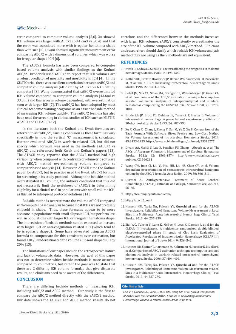
bedside method of measuring intracerebral hemorrhage (ICH) volume on CT. Kwak first estimated ICH volume on CT using the formula for the volume of an ellipsoid and estimating π to be 3, thus yielding the ABC/2 formula [Kwak] (Figure 1) [1]. Kothari added a weighting scale to the value of C (C= the number of approximate cuts weighted by % area of ICH seen per cut (< 25% = 0 slices, 25-50% = 0.5 slices, and > 75% = 1 slice) *(referred to as ABC/2 formula) [2] (Figure 1). This is in contrast to the Kwak formula which simply uses the total number of slices the hemorrhage is seen for its vertical measurement (Figure 1). We sought to directly compare the Kothari ABC/2 formula with the Kwak sABC/2.
PATIENTS AND METHODS With approval from the Institutional Review Board, we
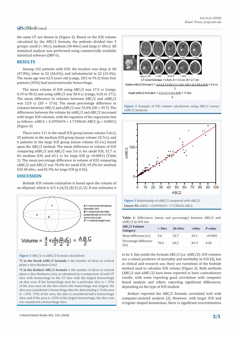
retrospectively reviewed 142 spontaneous ICH patients admitted to our institution from November 1, 2009 to October 1, 2010 with CT brain available for review. We excluded patients with SAH, traumatic ICH, multifocal ICH, hemorrhage into tumors, or isolated intraventricular hemorrhage (IVH). CT brain images were independently reviewed by board-certified vascular neurologist or neurointensivist for ICH volume measurements. The ABC/2 method (Figure 1) was used based upon prior published description using weighted percentage for C measurement [2]. The sABC/2 method was based upon prior published description (Figure 1) [1,3]. ICH volume was calculated using both ABC/2 and sABC/2 methods and a sample of the calculations used on
CentralBringing Excellence in Open Access
Lee et al. (2016)Email:
J Neurol Disord Stroke 4(1): 1111 (2016) 2/3
the same CT are shown in (Figure 2). Based on the ICH volume calculated by the ABC/2 formula, the patients divided into 3 groups: small (< 30cc), medium (30-60cc) and large (> 60cc). All statistical analysis was performed using commercially available statistical software (JMP 6).
RESULTSAmong 142 patients with ICH, the location was deep in 68
(47.8%), lobar in 52 (36.6%), and infratentorial in 22 (15.4%). The mean age was 62.5 years old (range, 28.5 to 93.3) Sixty four patients (45%) had intraventricular hemorrhage.
The mean volume of ICH using ABC/2 was 17.5 cc (range, 0.19 to 90.3) and using sABC/2 was 30.4 cc (range, 0.26 to 171). The mean difference in volumes between ABC/2 and sABC/2 was 12.8 cc (SD + 17.6). The mean percentage difference in volumes between ABC/2 and sABC/2 was 76.9% (SD + 39.7). The differences between the volume by sABC/2 and ABC/2 increased with larger ICH volumes, with the equation of the regression line as follows: sABC2 = 0.2959659 + 1.7190626 ABC2 (p < 0.0001) (Figure 3).
There were 111 in the small ICH group (mean volume 5.6cc), 25 patients in the medium ICH group (mean volume 32.7cc), and 6 patients in the large ICH group (mean volume 65.1cc) based upon the ABC/2 method. The mean difference in volume of ICH comparing sABC/2 and ABC/2 was 5.6 cc for small ICH, 32.7 cc for medium ICH, and 65.1 cc for large ICH (p <0.0001) (Table 1). The mean percentage difference in volume of ICH comparing sABC/2 and ABC/2 was 78.4% for small ICH, 69.2% for medium ICH 30-60cc, and 81.9% for large ICH (p 0.56).
DISCUSSIONBedside ICH volume estimation is based upon the volume of
an ellipsoid, which is 4/3 π (A/2) (B/2) (C/2). If one estimates π
to be 3, this yields the formula ABC/2 (i.e. sABC/2). ICH volumes are a robust predictor of mortality and morbidity in ICH [4], but in clinical and research use, there are variations of the bedside method used to calculate ICH volume (Figure 2). Both methods (ABC/2 and sABC/2) have been reported to have contradictory results, with some reporting good correlation with computer based analysis and others reporting significant differences, depending on the type of ICH studied.
Kothari reported the ABC/2 formula correlated well with computer-assisted analysis [2]. However, with larger ICH and irregular shaped hematomas, there is significant overestimation
Figure 1 ABC/2 vs sABC/2 formula calculations*C in the Kwak sABC/2 formula = the number of slices in vertical plane x slice thickness (cm)*C in the Kothari ABC/2 formula = the number of slices in vertical plane x slice thickness (cm), as calculated by a comparison of each CT slice with hemorrhage to the CT slice with the largest hemorrhage on that scan. If the hemorrhage area for a particular slice is > 75% of the area seen on the slice where the hemorrhage was largest, the slice was considered 1 hemorrhage slice for determining C. If the area is ~25% -75% of the area, the slice is considered half a hemorrhage slice; and if the area is <25% of the largest hemorrhage, the slice was not considered a hemorrhage slice.
Figure 2 Example of ICH volume calculations using ABC/2 versus sABC/2 methods.
Figure 3 Relationship of sABC/2 compared with ABC/2.Linear Fit sABC2 = 0.2959659 + 1.7190626 ABC2.
Table 1: Differences (mean and percentage) between ABC/2 and sABC/2 by ICH size.ABC/2 Volume Category < 30cc 30-60cc >60cc P value
error compared to computer volume analysis [5,6]. Xu showed ICH volume was larger with ABC/2 (58.4 cm3 vs 50.4) and that the error was associated more with irregular hematoma shape than with size [5]. Divani showed significant measurement error comparing ABC/2 with 3 dimensional volumes, which was worse for irregular shaped ICH [6].
The sABC/2 formula has also been compared to computer based volume analysis with similar findings as the Kothari ABC/2. Broderick used sABC/2 to report that ICH volumes are a robust predictor of mortality and morbidity in ICH [4]. In the GUSTO trial, there was excellent correlation between sABC/2 and computer volume analysis (68.7 cm3 by sABC/2 vs 63.3 cm3 by computer) [3]. Wang demonstrated that sABC/2 overestimated ICH volume compared to computer volume analysis (43.6ml vs 33.8ml) and this error is volume dependent, with overestimation seen with larger ICH [7]. The sABC/2 has been adopted by most clinical academic training programs as an easier bedside method of measuring ICH volume quickly. The sABC/2 formula has also been used for screening in clinical studies of ICH such as MISTIE, ATACH and CLEAR [8-12].
In the literature both the Kothari and Kwak formulas are referred to as “ABC/2”, causing confusion as these formulas vary specifically in how the vertical “C” measurement is calculated. Huttner evaluated ABC/2 in warfarin-related ICH, but did not specify which formula was used in the methods (sABC/2 vs ABC/2) and referenced both Kwak and Kothari’s papers [13]. The ATACH study reported that the ABC/2 method had high variability when compared with centralized volumetric software with ABC/2 method overestimating volume compared to computer based analysis [14]. However, ATACH cited the Kothari paper for ABC/2, but in practice used the Kwak sABC/2 formula for screening in its study protocol. Although the bedside method overestimated ICH volume, the authors concluded that this did not necessarily limit the usefulness of sABC/2 in determining eligibility for a clinical trial in populations with small volume ICH, as this led to infrequent protocol violations [14].
Bedside methods overestimate the volume of ICH compared with computer based analysis because most ICHs are not precisely ellipsoid in shape. Thus, these formulas appear to be more accurate in populations with small ellipsoid ICH, but perform less well in populations with larger ICH or irregular hematoma shape. The imprecision of bedside methods can be expected to increase with larger ICH or anti-coagulation related ICH (which tend to be irregularly shaped). Some have advocated using an ABC/3 formula to compensate for this consistent over-estimation, but found ABC/3 underestimated the volume ellipsoid shaped ICH by 20% [13].
The limitations of our paper include the retrospective nature and lack of volumetric data. However, the goal of this paper was not to determine which beside methods is more accurate compared to volumetrics, but rather the goal was to state that there are 2 differing ICH volume formulas that give disparate results, and clinicians need to be aware of the differences.
CONCLUSIONThere are differing bedside methods of measuring ICH,
including sABC/2 and ABC2 method. Our study is the first to compare the ABC/2 method directly with the sABC/2 method. Our data shows the sABC/2 and ABC2 method results do not
correlate, and the differences between the methods increases with larger ICH volumes. sABC/2 consistently overestimates the size of the ICH volume compared with ABC/2 method. Clinicians and researchers should clarify which bedside ICH volume analysis method they are using as the 2 methods are not equivalent.
REFERENCES1. Kwak R, Kadoya S, Suzuki T. Factors affecting the prognosis in thalamic
hemorrhage. Stroke. 1983; 14: 493–500.
2. Kothari RU, Brott T, Broderick JP, Barsan WG, Sauerbeck LR, Zuccarello M, et al. The ABCs of measuring intracerebral hemorrhage volumes. Stroke. 1996; 27: 1304–1305.
3. Gebel JM, Sila CA, Sloan MA, Granger CB, Weisenberger JP, Green CL, et al. Comparison of the ABC/2 estimation technique to computer-assisted volumetric analysis of intraparenchymal and subdural hematomas complicating the GUSTO-1 trial. Stroke 1998; 29: 1799-1801.
4. Broderick JP, Brott TG, Duldner JE, Tomsick T, Huster G. Volume of intracerebral hemorrhage. A powerful and easy-to-use predictor of 30-day mortality. Stroke. 1993; 24: 987–993.
5. Xu X, Chen X, Zhang J, Zheng Y, Sun G, Yu X, Xu B. Comparison of the Tada Formula With Software Slicer: Precise and Low-Cost Method for Volume Assessment of Intracerebral Hematoma Stroke. 2014; 45:3433-3435. http://www.ncbi.nlm.nih.gov/pubmed/25316277
6. Divani AA, Majidi S, Luo X, Souslian FG, Zhang J, Abosch A, et al. The ABCs of Accurate Volumetric Measurement of Cerebral Hematoma. Stroke. 2011; 42: 1569-1574. http://www.ncbi.nlm.nih.gov/pubmed/21566231
7. Wang CW, Juan CJ, Liu YJ, Hsu HH, Liu HS, Chen CY, et al. Volume dependent overestimation of spontaneous intracerebral hematoma volume by the ABC/2 formula. Acta Radiol. 2009; 50: 306–311.
8. Qureshi AI. Antihypertensive Treatment of Acute Cerebral Hemorrhage (ATACH): rationale and design. Neurocrit Care. 2007; 6: 56–66.
9. http://braininjuryoutcomes.com/
10. http://atach2.com/
11. Hussein HM, Tariq NA, Palesch YY, Qureshi AI and for the ATACH Investigators. Reliability of Hematoma Volume Measurement at Local Sites in a Multicenter Acute Intracerebral Hemorrhage Clinical Trial. Stroke. 2013; 44: 237-239.
12. Ziai WC, Tuhrim S, Lane K, McBee N, Lees K, Dawson J, et al for the CLEAR III Investigators. A multicenter, randomized, double-blinded, placebo-controlled phase III study of Clot Lysis Evaluation of Accelerated Resolution of Intraventricular Hemorrhage (CLEAR III). International Journal of Stroke 2014; 9: 536–542.
13. Huttner HB, Steiner T, Hartmann M, Köhrmann M, Juettler E, Mueller S, et al. Comparison of ABC/2 estimation technique to computer-assisted planimetric analysis in warfarin-related intracerebral parenchymal hem orrhage. Stroke. 2006; 37: 404–408.
14. Hussein HM, Tariq NA, Palesch YY, Qureshi AI and for the ATACH Investigators. Reliability of Hematoma Volume Measurement at Local Sites in a Multicenter Acute Intracerebral Hemorrhage Clinical Trial. Stroke. 2013; 44:237-239.
Lee VH, Conners JJ, John S, Busl KM, Song SY, et al. (2016) Comparison of ABC/2 with the Simplified ABC/2 Formula in Calculating Intracerebral Hemorrhage Volume. J Neurol Disord Stroke 4(1): 1111.


![a c:] 5 ooÐ L B 10.5 1 - Microsoft Word Abc Abc Abc Abc Abc Abc Abc Abc Abc Abc Abc Abc 1 - Microsoft Word Abc Abc Abc 505 7ï—L Mic SmartArt 1 - Microsoft Word Aa MS B 10.5 (Ctrl+L)](https://static.documents.pub/doc/80x56/5b180d777f8b9a19258b6a1e/a-c-5-ood-l-b-105-1-microsoft-word-abc-abc-abc-abc-abc-abc-abc-abc-abc-abc.jpg)


