Abstract. The type species of the genus Acteocina, Acteon wethenlli Lea, 1833, is synonymized with Volvaria canaliculata Say, 1826, type species of the genus Utriculastra; because Utriculastra and Cylichnel- la are synonymous, Acteocina is the senior synonym. The systematics of A. canaliculata and A. atrata spec. nov. are examined, and a neotype is designated for A. canaliculata. Descriptions are presented of shell, radular, and gizzard plate morphologies; geographic and bathymetric distributions are re-evalu- ated based on museum collections. Florid-i-a-n specimens of A. canaliculata have planktotrophic devel- opment, hatching in 4 days and settling in 24 days; A. atrata has capsular metamorphic development, hatching in 9 days as benthic juveniles. A preliminary review of Acteocina candei (d'Orbigny, 1841) is given, and a lectotype is designated. Acteocina candei distinctly differs conchologically from A. canali- culata, is resurrected from synonymy, and is considered a valid species.
INTRODUCTION
Acteocina canaliculata (Say, 1826) and A. candei (d'Orbi- gny, 1841) are small, cephalaspid gastropods common throughout much of the western Atlantic. Intraspecific variability of shell and radular characters has obscured interspecific differences, making them consistently diffi- cult to distinguish. Publications reporting either species may actually have considered both species simultaneously or confused the two.
Acteocina canaliculata and A. candei have been assigned to many genera, most notably Utriculus Brown, 1844, Tornatina A. Adams, 1850, Relusa Brown, 1827, and Ac- teocina Gray, 1847. Acteocina has had the most frequent usage in recent years. However, MARCUS (1956:41), MARCUS (1977:2), and CERNOHORSKY (1978:83) advocat- ed restriction of the genus Acteocina to fossil forms because (1) the type species, A. welherilli (Lea, 1833), was de- scribed as a fossil whose internal anatomy will never be known, and (2) knowledge of internal anatomy is neces- sary to define cephalaspid genera according to modern standards. Thus, they concluded that fossil species of this group cannot be reliably allocated to Recent genera. Fol- lowing this reasoning, MARCUS (1977) allocated A. cana- liculata and A. candei (which she synonymized with A. canaliculata) to Utriculastra Thiele, 1925. RUDMAN (1978)
incorrectly placed A. canaliculata (and thus A. candei) in the genus Tornatina, apparently unaware of MARCUS'
(1977) work restricting Tornatina on the basis of radular and gizzard plate morphologies, and because he incor- rectly believed that the genus Cylichnella Gabb, 1873, lacked jaws (see MARCUS, 1956:39; GOSLINER, 1979:88). GOSLINER (1979) determined that Utriculastra was a ju- nior synonym of Cylichnella based on similar arrange- ments of the reproductive system. Therefore, according to the more recent literature, A. canaliculata and A. candei should be placed in the genus Cylichnella.
The familial placement of Acteocina (as Cylichnella, etc.) has likewise been varied; it has most recently been placed (MARCUS, 1977; RUDMAN, 1978; GOSLINER, 1980) in the Scaphandridae G. O. Sars, 1878. This family was consid- ered (ABBOTT, 1974; CERNOHORSKY, 1978), apparently on a conchological basis alone, to be a junior synonym of Cylichnidae H. & A. Adams, 1854 (not of A. Adams, 1850, as stated by ABBOTT, 1974; not of Rudman, 1978). Although RUDMAN (1978:105) incorrectly reproposed Cy- lichnidae as a new family, he restricted the family to the genus Cylichna on the basis of anatomy. Therefore, ac- cording to Rudman, Cylichnidae and Scaphandridae are both valid and distinct families. GOSLINER (1980) refuted RUDMAN'S (1978) distinction between Cylichnidae and
P. S. Mikkelsen & P. M. Mikkelsen, 1984 Page 165
Scaphandridae, considering them synonymous. However, Gosliner chose Scaphandridae as the senior synonym, be- cause he incorrectly considered Cylichnidae of Rudman, 1978, rather than of H. & A. Adams, 1854. Cylichnidae H. & A. Adams, 1854, is the proper senior synonym and would be the correct family for Acteocina following GOSLINER'S (1980) synonymy. Following RUDMAN (1978), Scaphandridae is correct. The family Acteocinidae Pils- bry, 1921, may be valid as well (see CERNOHORSKY, 1978).
In 1962, WELLS & WELLS attempted to distinguish "Retusa" canaliculata from Acteocina candei. The diag- nostic characters used were: radular characters (shape and number of denticles for lateral and rachidian teeth), pro- toconch appearance (degree of protrusion and number of whorls), shell shape (spire height, basal shape, convexity of the whorls, and apertural shape), habitat, and type of larval development. Wells & Wells found direct-devel- oping larvae in field-collected egg masses of "R." canali- culata, and assumed planktonic development in A. candei from the appearance of the protoconchs of the adults. However, FRANZ (1971a, b) found planktotrophic devel- opment in eggs deposited in the laboratory by adults of A. canaliculata collected from Connecticut. Franz explained this discrepancy by suggesting (1) that Wells & Wells, using field-collected egg masses from North Carolina, had unknowingly reared some other cephalaspid, (2) that poe- cilogony (multiple patterns of development within a species) exists for A. canaliculata, or (3) that cryptic species were present. GOSLINER'S (1979) observations from the estuarine Pictou Harbor, Nova Scotia, also determined planktotrophic development for A. canaliculata.
MARCUS (1977:14) examined additional specimens of "Utriculastra" candei and UU," canaliculata and deter- mined that their shell and radular characters were highly variable and overlapping. Although Marcus noted differ- ent morphologies in the gizzard plates of each species, these differences were attributed to varying degrees of des- iccation, and the plates were considered "identical." Pos- sibly due to WELLS & WELLS' (1962) questionable devel- opmental observations, MARCUS (1977) failed to address Wells & Wells' criterion of developmental type as a dis- tinguishing character between the two species. Based on her observations, and on identical morphologies of the male reproductive structures, Marcus synonymized "£/." candei with "U." canaliculata. This synonymy had been suggested many years earlier by DALL (1889:45).
Fifteen shells and 5 radulae of "Utriculastra" canalicu- lata were figured by MARCUS (1977), but no attempt was made to document further the degree of variability. MIK- KELSEN & MIKKELSEN (1982) closely examined the shell and radular variability of "U." canaliculata (as defined by MARCUS, 1977) and indicated that "U." candei is an im- mature form of "£/." canaliculata, thus supporting the syn- onymy of Marcus. MIKKELSEN & MIKKELSEN (1983) not- ed the presence of two types of larval development in the "single species," "Cylichnella" canaliculata, and suggested that "C." candei be resurrected as valid.
In the present study, embryonic, larval, and postlarval morphological characters, and examination of museum collections are utilized to:
(1) show that Wells & Wells' "Retusa canaliculata" is actually an undescribed species, living sympatrically with Acteocina canaliculata in eastern Florida;
(2) determine the correct generic placement for A. ca- naliculata and A. candei;
(3) examine in detail the postlarval intraspecific vari- ation of the shell, radula, and gizzard plates of A. cana- liculata and the new species;
(4) describe the larval development of A. canaliculata as well as that of the new species; and
(5) partially characterize Acteocina candei, in contrast with A. canaliculata and the new species.
MATERIALS AND METHODS
Collections
The principal study site was the 195 km-long Indian River lagoon, along the central east coast of Florida (Fig- ure 7D). Salinity generally ranged from 18 to 36 ppt, although extremes of 8 and 42 ppt were recorded during the study. Live snails were sieved, using 0.5 mm-mesh screens, from bare or vegetated subtidal sand or mud sub- strates. Dried or wet-preserved specimens from various museums and private collections were utilized to redeter- mine geographic and bathymetric distributions. Fossil type specimens were examined for synonymies; however, be- cause additional fossil material was not thoroughly stud- ied, detailed paleontological distributions are not given. Cited repositories and other sources are as follows:
ANSP—Academy of Natural Sciences of Philadelphia, Philadelphia, PA
BM(NH)—British Museum (Natural History), London CAS—California Academy of Sciences, San Francisco, CA ChM—Charleston Museum, Charleston, SC D. Franz Collection—Department of Biology, Brooklyn
College of the City University of New York, Brooklyn, NY
HMNS—Houston Museum of Natural Science, Hous- ton, TX
IRCZM—Indian River Coastal Zone Museum, Harbor Branch Foundation, Ft. Pierce, FL
MACN—Museo Argentine de Ciencias Naturales, Bue- nos Aires, Argentina
MCZ—Museum of Comparative Zoology, Harvard Uni- versity, Cambridge, MA
MORG—Museo Oceanografico, Rio Grande, Brazil PRI—Paleontological Research Institution, Ithaca, NY ROM1Z—Royal Ontario Museum, Toronto, Ontario,
Canada R. Van Dolah Collection—South Carolina Marine Re-
sources Research Institute, Charleston, SC UNC-IMS—Institute of Marine Sciences, University of
North Carolina, Morehead City, NC
Page 166 The Veliger, Vol. 27, No. 2
USNM—National Museum of Natural History, Wash- ington, DC.
In synonymies, a dagger (t) preceding a species name indicates fossil type specimens. In "Material examined" sections, an "L" indicates that at least some of the speci- mens in the lot were live-collected and contained soft parts; an "E" indicates that all specimens were empty shells.
Either original figures or type specimens were exam- ined of all western Atlantic /Weonna cana/icuWa-like forms, fossil and Recent, described to date. Types were examined if figures even remotely resembled the species discussed herein. Two exceptions were CyWina uir^nica Conrad, 1868, and Tbrno/ma cy/mdrico Emmons, 1858, for which no types could be located to clarify the ambig- uous original figures.
Postlarval Observations
Specimens with intact protoconchs, collected from var- ious localities throughout the study area, were chosen for statistical analyses. These encompassed a wide range of shell lengths, but excluded specimens not retained by the 0.5 mm-mesh collecting sieve. Each shell was illustrated using a stereomicroscope and camera lucida providing permanent records of shell length, shell width, spire height and angle (Figure 1 A), and percent of protoconch protru- sion (Figure ID), Drawings were essential because par- tial destruction of the shells was necessary to remove the retracted animals.
Percent of protoconch protrusion was determined (Fig- ures 1E-G) utilizing the circle, (x — h)' + (y — k)' = r=, approximated by the periphery of the protoconch, where (h, k) are the coordinates of the center, (x, y) are the coordinates of any point on the circle, and r is the radius. With the ordinate axis drawn through the center of the circle, and the abscissa through the lateral suture points of the protoconch with the first postnuclear whorl, the y-co- ordinate (h) of the center equals zero, and the equation reduces to Equation I: x* + (y — k)* = r'. Measurement of the protoconch's exposed height (b) and width at the suture (2a) yields 3 points on the circle: (a, 0), (—a, 0), and (0, b). For point (a, 0) or (—a, 0), y = 0, and Equa- tion I becomes Equation II: a' + k' = r*. For point (0, b), x = 0, and Equation I becomes Equation III: b* — k' = r^ or Equation IV: b — k = r. Subtracting Equation II from Equation IH yields Equation V. k = (b* — a')/2b. Using a and b obtained from actual protoconch measure- ments, Equation V can be solved for k, which is used in Equation IV to determine the radius: r = b — (b — a)/ 2b. The diameter of the circle (2r) and the exposed height (b) are then used to determine the percentage (P) of pro- toconch protrusion: P = b/2r x 100%.
Radulae and gizzard plates were extracted by dissolving the surrounding soft tissue in a solution of 10% sodium hydroxide at 20°C (LiNDBERG, 1977). In addition to sug- gestions by TURNER (1960) and SoLEM (1972), handling
of small radulae was facilitated by use of a cat's vibrissa mounted on the tip of a probe. Gizzard plates were ob- served and subsequently stored in 70% ethanol, or dried. Radulae were simultaneously stained and permanently mounted on glass slides, using Turtox CMC-9AF tow- viscosity stain-mountant tinted with acid fuchsin (Masters Chemical Co., Inc., Des Plaines, IL). Due to the ex- tremely small size of the radula, this one-step operation eliminated the loss of many radulae. Each radula was mounted so that at least some of the lateral teeth were oriented as in Figure 1B. All slide-mounted radulae were illustrated using a compound microscope and camera lu- cida. These drawings provided the width, angle, and num- ber of denticles for lateral teeth (Figure IB), plus the width and number of denticles for rachidian teeth (Figure 1C). Radulae used for scanning electron microscopy were cleaned by sonication, following the method of SOLEM (1972). Radulae and gizzard plates were air dried and scanned using a Zeiss Novascan-30.
Shell terminology is after SMITH (1967a 758-760) and KNIGHT (1952:7-9); radular terminology is after BERTSCH (1977). Providing regressions of various characters follows the initiative of BERTSCH (1976).
Larval Development
Adults were collected during each month of the year from at least one of several locations (Figure 7D). Adults from a single site were left together for about 24 h in a finger bow] with seawater and sand substrate from the collection site. Individuals were then isolated in compart- mented plastic trays, each containing filtered seawater and a thin layer of sand. Each compartment was checked daily, for up to 14 days, and egg masses were removed to com- partmented plastic trays containing seawater only. Adults and developing egg masses were maintained at ambient laboratory conditions of 22-25°C with variable lighting, or under incubation at 22°C with a daily light cycle of 12 h light/12 hdark.
Planktotrophic larvae were reared using larval culture sieves placed in 300-mL beakers. Each larval sieve con- sisted of a 4.5-cm section of acrylic tubing, 7.6 cm in diameter, closed at the base by a 33 fim-mesh nitex screen. Larvae from a single egg mass were reared in the same sieve. Survivorship was maximized by adding antibiotics (5 mg/mL streptomycin sulfate plus 5 mg/mL penicillin- G) to 0.5 ^m-filtered, 36-ppt seawater (SwiTZER-DuNLAP & HADF1ELD, 1981:207). Cetyl alcohol Hakes were floated on the water to prevent larvae from being trapped by the surface tension (HURST, 1967). Veligers were fed to excess with .Skp/wmopfero sp., a lOftm-diameter, unicellular green alga. Water and food supply were changed every other day.
Direct-developed hatchlings and metamorphosed planktic larvae were transferred to 300-mL beakers of filtered seawater. Amorphous organic material, collected
P. S. Mikkelsen & P. M. Mikkelsen, 1984 Page 167
LENGTH
WIDTH Q AT SUTURE
'-* LENGTH
^^ k.
EXPOSED HEIGHT
F
2
JO«
{•'.Oh r>^ M\ ]
t b
Xa,0) \
AT *~-*._
^
—2
fiH
i b M
k.o) \ *^ ___>
Figure 1
Shell and radular parameters. A. Shell in apertural view. B. Lateral radular tooth. C. Rachidian radular tooth. D. Apical view of protoconch. E, F, and G. Diagrammatic protoconchs of increasing degree of protrusion, showing points and distances used in calculating percent protrusion.
from sedimentary detritus at the collection site or from a laboratory running-seawater system, provided food for the
juveniles. After this material was suspended by swirling, the larger particles were allowed to settle and the finer suspended material was decanted off. It was this fine or- ganic material which was added in small quantities to the
beakers containing the juveniles, to form a thin bottom layer of food. Juveniles were visually located and trans- ferred to new seawater and food once a week.
Larval shells were prepared for SEM either by pres- ervation in 80% ethanol or by soaking in a dilute solution of household bleach to remove the soft tissues. The latter
Page 168 The Veliger, Vol. 27, No. 2
method was considered least desirable because opercula were invariably lost. The larval shells were then dried, mounted, and scanned as described above for the radulae
The holotype of Acteon uietherilli Lea, 1833 (type species of the genus Acleocina) is extremely worn, and lacks its protoconch. The type locality of A. uietherilli is Deal, New Jersey, from deposits of Miocene age (RICHARDS, 1968). L. D. Campbell (personal communication, 1983) indicat- ed that the type locality may be as young as Pleistocene.
In all discernible conchological characters, Acteoana
luetherilli agrees with the Recent species Voluaria canali- citlata Say, 1826, type species of the genus Utriculastra.
This synonymy was first suggested by OLSSON & HAR- BISON (1953) and reiterated by OLSSON & MCGINTY
(1958), OLSSON (1964), and CAMPBELL el al. (1975). The genus Acleocina is therefore applicable to Recent forms (contrary to MARCUS, 1956:41). The anatomical charac- teristics of A. canaliculala may now be assumed to be ap- plicable to the genus. In addition, the genus Utriculastra
becomes a junior synonym of Acleocina. Utriculastra and Cylichnella are presently synonymous,
in accordance with the anatomical studies of GOSLINER
(1979). Although GOSLINER (1979) additionally included Tornastra Marcus, 1977, in this synonymy, we prefer to omit it here because of the distinctive gizzard plate and radular anatomy of Bulla eximia Baird, 1863, the type species of Tornastra (see MARCUS, 1977). DALL (1908) listed eight additional synonyms of Cylichnella, but they are either incorrect or unconfirmed herein.
Our experience has determined that Acteocina-hke
species can be distinguished conchologically. In the case of Acleocina, we have conchologically matched its fossil type specimen with a Recent species, and have subse- quently defined the genus using the internal characteris- tics of Recent specimens. This action does not differ from the conchological matching of dead-collected Recent type specimens (of prosobranch and other mollusks) with living individuals, which are then used to redescribe the species, complete with internal anatomy. Although this procedure is followed frequently in malacology, it has been consid- ered inappropriate for cephalaspids (MARCUS, 1956;
MARCUS, 1977). In our opinion, it must be considered acceptable (for example, in the case of A. candei).
Until further studies at the species level are complete for Acleocina and other closely related genera, resolution of the familial placement of Acleocina is not possible.
Other material: Prince Edward Island: 34L, MCZ 38870.— Maine: Isle au Haut(?): 10E, MCZ ex.14531 (in part). — Massachusetts: Duxbury: 76L, USNM 358256.—New Bedford: 24L, USNM 57310.—Rhode Island: Westerly: 200 +L, USNM 358281.—Connecti- cut: Noank (Beebe Cove), 20L, D. Franz Collection.— New Jersey: Little Egg Harbor: 8E, D. Franz Collec- tion.— Maryland: Point No Point: 100+L, USNM 379507.—North Carolina: Cape Hatteras: 3L, D. Franz Collection—off Cape Lookout: 500+ L, USNM 523583.—Neuse River: 51 L, UNC-IMS 9859.1-.51.— South Carolina: Charleston Harbor: 3E, ChM 30.183.11.—Georgia: Jekyll Island: 12E, UNC-IMS 8074.1-12.—Florida: St. Augustine: 18E, USNM 358292 (in part). —Cape Canaveral: 4E, ChM 43.28.4734.—Merritt Island (Pleistocene fossils): 6E, IRCZM 65:2000.—Turnbul) Creek (vouchers): 14L,
Figure 2
Acteoana canaliculala. A. Neotype, 3.80 mm, ANSP A9721A. B. SAY'S (1832) original figure. C. Neotype, crawling animal. D. Map of Charleston, SC, area showing Folly River collection site for the neotype. E. Folly River site at high tide.
Acteocina canaliculata was originally described from specimens taken from the coast of South Carolina. The type locality is believed to be in the vicinity of Charleston, where the collector, Mr. Stephen Elliott, lived (MAZYCK,
1913) and probably did the majority of his collecting. SAY
(1826:211) briefly described the species: "Shell whitish, immaculate, cylindric, with very minute obsolete wrinkles; spire convex, very little elevated, mammillated at the tip; volutions above five, with their shoulder very obtusely grooved; labrum with the edge arcuated; labium over- spread with a calcareous lamina, and with a single oblique fold or small tooth near the base." Say also stated that "the arcuated form of the edge of the labrum is only per- ceived when the part is viewed in profile." Although a figure did not accompany the original description, the species was figured later (as Bullina canaliculata in "American Conchology" (SAY, 1832:pl. 39) and is repro- duced herein (Figure 2B).
Type material
A series of 25 specimens of Acteocina canaliculata la- beled as syntypes was procured from the Academy of Nat- ural Sciences of Philadelphia (ANSP 57312). However, these are probably not type specimens because: (1) the specimen label reads "71 canaliculata," indicating the ge- nus Tornatina, (2) the cited locality is "Georgia," and (3) "J. S. Phillips" is recorded as donor of the lot. Thus, the status of ANSP 57312 as a type lot is questionable.
A search (by ANSP personnel) of the remaining lots of Acteocina canaliculata and related species in the ANSP collections failed to yield the original type material. His- torical information obtained from Virginia O. Maes (per- sonal communication, 1982) indicates that the types were probably destroyed. According to her, Say removed his
molluscan collections from the ANSP to New Harmony, Indiana in 1825 where much of his material was subse-
quently lost in a fire. Long after Say's death in 1884, his wife returned whatever was salvaged of Say's collections to Dr. H. A. Pilsbry at the ANSP. Unfortunately, no records exist of the species involved in these transactions, and we conclude that the types of Volvana canaliculata were probably destroyed in the New Harmony fire. To the best of our knowledge, a neotype has not been desig- nated.
Lacking any type specimens, one must rely entirely upon the original description by Say and his subsequent illus- tration. Unfortunately, the description and figures could apply to any of several species of Acteocina. However, a survey of South Carolina material in museum collections (USNM, ANSP, MCZ, ChM) and from Dr. L. Camp- bell (University of South Carolina at Spartanburg) in- cluded only one species living in shallow, estuarine waters (equivalent to SAY'S [1826] "coast of South Carolina").
This species closely approximates Say's original illustra- tion. In addition, the majority of specimens labeled as A. canaliculata in early museum collections were also of this
same form. We take this as sufficient evidence to consider the species in South Carolina estuaries to be Say's Volvana canaliculata.
Following the determination of Say's Volvana canali- culata, designation of a neotype was necessary to avoid future confusion. On December 3, 1982, specimens of Ac- teocina canaliculata were collected by the authors from soft, subtidal mud, below zones of Spartina and oysters in the Folly River, an estuarine channel just south of Charleston Harbor, Charleston County, South Carolina (Figures 2D- E). The site was located adjacent to the State Route 171 bridge over the Folly River. Salinity ranged (on an incom- ing tide) from 27 to 30 ppt during collection of the spec- imens. The neotype and specimens collected with it were distributed to various repositories (see Material examined, above).
A redescription of Acteocina canaliculata follows, based primarily on our observations of Indian River specimens, but consistent with material from South Carolina.
Diagnosis
Teleoconch thick-walled, cylindrical to pyriform. Shoulder rounded; subsutural sculptural band indistinct. Spire height variable, but usually less than 20% of total shell length. Protoconch distinctly tapered toward its or- igin, showing strongly coiled sutures in lateral view. Lat-
Figure 3
Acteocina canaliculata, adults. A. Shell, from Indian River lagoon, in apertural view, 4.58 mm. B. Gizzard plates, view of grinding surfaces. C. Radula with lateral teeth reflected to expose rachidians. D. Lateral teeth, showing wing-like expansion with denticles. E, F, and G. Protoconch: E. "Apical" view. F. "Posterior" view. G. "Umbilical" view. Scales: B = 100 nm; C = 10 ^m; D = 5 Mm; E, F, and G = 50 /xm.
Page 172 The Veliger, Vol. 27, No. 2
eral radular teeth with wing-like expansion bearing one row of denticles. Unpaired gizzard plate T-shaped. Tis- sues of gizzard, pallial caecum, and Hancock's organs light
orange to pink in live material.
Distribution
Prince Edward Island, Canada; Maine (dead shells only); Massachusetts to peninsular Florida and entire Gulf coast to Texas; intertidal to 40 m.
Description
Shell characters
The orthostrophic, dextral teleoconch of adult Acteocina
canaliculala is smooth and cylindrical, and has up to three whorls (Figure 3A). Large specimens tend toward a pyr- iform shape, i.e., with greater shell width at the posterior end. The aperture is narrow posteriorly and flares ante- riorly; it extends from 80 to 94% of the shell length. The parietal area bears a slight callus, ending in a columella with a single fold. The shell walls are porcelaneous white and thick, approximately 175 fim at the midpoint of the body whorl of adults. The shoulder is somewhat keeled in young to immature individuals, becoming more round- ed with maturity, with an indistinct, impressed, subsu- tural sculptural band. Indian River specimens attained a shell length and width of 5.0 mm and 2.3 mm respectively; the specimens varied in spire angle from 76 to 142 degrees and in spire height from 6 to 20%. The periostracum is thin and transparent, unless environmentally stained, in which case it is often spirally banded.
The sutures of the smooth, hyperstrophic, sinistral pro- toconch (Figures 3E, G) are strongly curved in both "lat- eral" views (i.e., from the larval shell's umbilical or apical aspect), appearing slightly umbilicate in "umbilical" view. In its "posterior" view (Figure 3F), the protoconch tapers toward its distal end, or origin. The percent of protoconch protrusion varied from 25 to 74%.
Correlation coefficients (r) were calculated for shell length versus the four other characters measured (Table 1). Shell length versus shell width showed the strongest r-value, while percent of protoconch protrusion, percent spire height, and spire angle yielded very low coefficients. Percent spire height versus spire angle showed a fairly strong negative coefficient.
Radular characters
The radular formula of Acteocina canaliculala is 1-R-l (Figure 3C), with 11-19 rows in specimens 2 mm or more in length. The rachidian teeth ranged in width from 17 to 34 jim and are centrally notched, with each rounded half bearing 4-11 sharply pointed denticles. The lateral
teeth (Figure 3D) are sickle-shaped and unicuspid, with the cusp bearing a wing-like expansion supporting one row of several denticles. A blunt, basal tubercle is present for articulation with adjoining lateral teeth. Ontogenetic
increases were noted in the lateral teeth in number of denticles from 6 to 18, in width from 41.6 to 71,9 /zm, and in angle from 79 to 104 degrees. Variation in number of lateral tooth denticles within a single radula was also noted (Figure 3C). Shortly after metamorphosis, lateral teeth of juveniles were about 15 (im in length, and smooth; faint traces of denticles were evident on some lateral teeth at 7-9 days post-metamorphosis.
Correlation coefficients (r) for shell length versus rad- ular characters (Table 1) were generally low, with the exception of lateral and rachidian tooth widths. Lateral tooth width versus lateral tooth angle (r = 0.06) and shell length versus lateral tooth angle (r = 0.01) were particu- larly low.
Other features
The extended, living animal of Acteocina canaliculala is of typical cephalaspid form (Figure 2C). The foot and mantle are translucent white in color, with scattered opaque white dots. Although the shell walls are thick and
porcelaneous, they are translucent enough to allow limited observation of the internal anatomy. The gizzard, pallial caecum, and Hancock's organs are light orange to pink in color.
The calcified portion of the gizzard of Acteocina cana- liculala consists of three plates (Figure 3B): a "pair" of non-identical, but similarly elongated, plates opposing a larger "unpaired" plate. The unpaired plate is most dor- sal in the crawling animal and is distinctly T-shaped, regardless of the method of extraction, preparation, or degree of desiccation.
The reproductive system did not differ in gross arrange- ment from that described by MARCUS (1977) or GOSLINER
(1979).
Oviposition
Oviposition in the laboratory occurred at all hours ex- cept those between 1800 and 2400. Egg masses were usu- ally deposited within the first few days after collection of the adults. Spawning occurred monthly in the field and in the laboratory, giving no indication of reproductive sea- sonally in the Indian River Lagoon animals.
Egg mass
The egg mass of Acteocina canaliculala (Figure 4A) cor- responds to "type C" of HURST'S (1967) opisthobranch egg mass forms. It is gelatinous and ovoid, ranging in maximum diameter from 1.9 to 6.4 mm (mean = 3.7 mm). It is firmly anchored at the sediment surface by a mucous thread, up to 60 mm long, which may bifurcate one or more times. Fresh egg masses were usually coated with sand grains, although most fell off within the first few hours after deposition. Uncleaved egg diameters ranged from 63.2 to 85.4 /im (mean = 77.5 ^m). The number of eggs per mass ranged from 189 to 1293 (mean = 631 eggs/
GO
Table 1
Statistical analysis of characteristics examined in Acteocina canaliculata and A. atrata spec, nov.; r, correlation coefficient; y = mx + b, regression line equation; n, number of cases; **, not applicable.
IT X vs. Character y
Acleocina canaliculata Acteocina atrata
Characi r y = mx + b n r y = mx + b n
Shell length vs. Shell width 0.93 y = 0.445x + 0.163 56 0.98 y = 0.490x + 0.0534 49
Shell length VS. Percent spire height 0.09 y = 0.358x + 13.6 56 0.27 y = 1.30x + 8.16 49
Shell length vs. Spire angle -0.23 y = -4.05x + 114 56 -0.57 y = -11.6x + 153 49
Shell length vs. Percent protoconch protrusion 0.10 y = 1.31x + 52.1 56 0.20 y = 2.03x + 49.0 49
Shell length vs. Radular rows 0.20 y = 0.506x + 12.7 56 0.52 y = 0.975x + 12.2 49
Shell length vs. Lateral tooth denticles 0.48 y = 1.83x + 5.08 56 0.18 y = 0.638x + 17.2 49
Shell length vs. Rachidian tooth denticles 0.46 y = 0.775x + 3.82 56 0.52 y = 0.797x + 4.46 49
Shell length VS. Lateral tooth width 0.85 y = 8.79x + 25.5 56 092 y = 13.5x + 18.4 49
Shell length vs. Rachidian tooth width 0.76 y = 3.91x + 11.1 56 0.79 y = 3.83x + 15.5 49
Shell length VS. Lateral tooth angle 0.01 y = -0.07x + 92.6 56 0.54 y = 4.33x + 92.9 49
Lateral tooth width vs. Lateral tooth angle 0.06 y = 0.047x + 89.8 56 0.60 y = 0.326x + 86.6 49
Lateral tooth width VS. Lateral tooth denticles 0.52 y = 0.190x + 0.79 56 0.60 y = 0.0526x + 15.9 49
Lateral tooth width VS. Rachidian tooth width 0.76 y = 0.374x + 3.70 56 0.82 y = 0.270x + 11.2 49
Rachidian tooth width vs. Rachidian tooth denticles 0.55 y = 0.185x + 1.96 56 0.58 y = 0.18lx + 2.02 49
Percent spire height vs. Spire angle -0.77 y = -3.54x + 153 56 -082 y = -3.49x + 156 49
Shell length vs. Number of eggs 0.90 y = 393x - 801 32 0.55 y = 38.2x - 56.6 54
Shell length VS. Egg mass diameter 0.88 y = 1.42x - 1.46 32 0.41 y = 0.534x + 1.42 54
Shell length vs. Egg tube diameter ** ** ** 0.42 y = 92.3x + 256 26
Shell length VS. Egg diameter 0.72 y = 10.4x + 39.9 7 0.22 y = 5.19x + 131 15
Shell length VS. Egg capsule length -0.10 y = -2.46x + 163 15 0.02 y = 0.91x + 317 21
Shell length vs. Egg capsule width -0.50 y = -6.36x + 150 15 -0.30 y = -8.39x + 294 21
Egg capsule . rngth vs. Egg capsule width 0.56 y = 0.302x + 83.7 15 0.60 y = 0.373x + 143 21
XT 2-
n m CD 3
\0 oo
CD
Page 174 The Veliger, Vol. 27, No. 2
mass). Each egg was encased in an oval capsule, about 155 x 130 Mm (range: 141-174 x 121-139 nm); capsules were interconnected by chalazal material. No internal transparent tubes, nurse eggs, or auxiliary yolk-like ma- terial were present. Statistical analyses showed high cor- relation coefficients (r) for maternal shell length versus egg diameter, egg mass diameter, and number of eggs per mass
(Table 1).
Larval development
Acleocina canaliculata exhibits planktotrophic develop- ment. Pre-hatching development was rapid and consistent between egg masses. The first two cleavages, to 2 cells at 2.5 h and to 4 cells at 3.5 h, are total, spiral and equal, after which they are unequal and form distinct animal and vegetal poles. The 8-celled stage appears at 4.5 h followed by the id-celled stage at 5 h into development. Further development proceeds rapidly to a multicelled or blastula stage, and by 23 h to a heart-shaped gastrula with its slit-like blastopore clearly visible.
The trochophore stage forms, and it begins to spin on its lateral axis (effectively doing "backwards somer- saults") at about 28.5 h into development. The mouth, metapodial rudiment, and prototroch bearing fine cilia are clearly visible at this stage, and the vegetal pole appears as an undifferentiated spherical mass. Two anal cells ap- pear on the lower right surface of the visceral hump.
The third day of development is characterized by the
appearance of the rapidly spinning veliger stage (Figure 4B). By then, the larva has switched to an antero-posterior axis of rotation. A well-developed velum with long cilia, a subvelum, two otocysts, a paucispiral operculum be- neath a pointed metapodium, a ciliated median metapo- dial band, and a spherical embryonic shell approximately 85 urn in diameter are present. As with other opistho- branchs, torsion does not occur as a larval process, except in the migrating anal cells, and the viscera differentiate in their post-torsional positions. On the 4th day of devel- opment, the veliger (Figure 4C) has a large, well-formed right eye. Its shell averages 140 nm long, and is nearly symmetrical, being only slightly skewed to the larval left. A colorless, larval kidney is prominent adjacent to the anus, and cilia are visible within the lumen of the midgut. Hatching can be mechanically stimulated by handling or rupturing the egg mass at about 90 h, but if not induced, will occur by 100-110 h after deposition. Hatching is rapid and nearly synchronous, being completed (i.e., the entire mass emptied) within 10 to 20 min.
Post-hatching development depended greatly on the de-
gree of feeding by individual larvae. A large proportion (77.4%) of the hatched veligers in our most successful culture did not metamorphose; most of these were appar- ently unable to feed, as evidenced by their colorless diges- tive systems and little or no growth. The remaining in- dividuals (22.6%) began feeding immediately, as evidenced by the bright green coloration of their guts, and developed rapidly. Acquisition of juvenile characteristics after hatch- ing was a function of the degree of growth, expressed as shell length, rather than age of the specimen. The degree of development among the individuals in a culture was also highly variable.
When larvae were not fed, many individuals survived for two weeks although no shell growth was observed. Although the left eye appeared in some individuals after four days, it remained smaller than the first-formed right eye. The left digestive diverticulum noticeably decreased in size during this period. All starved larvae died by the third week after hatching.
In feeding individuals, the left eye appeared, and equaled the right in size, by the fourth post-hatching day at 163 um shell length. By 270 (im shell length, the propodium began to swell, the right side of the mantle opened, and the larval heart began pulsating.
Metamorphosis at 300 fim shell length (Figures 5A-C) began after 14 to 20 days, independent of any special substrate. In newly metamorphosed individuals, both lar- val and adult hearts beat actively, and the nearly umbil- icate aspect of the shell was visible from the larval right (Figure 5A). Immediately after metamorphosis, shell growth accelerated at the larval right, beginning the change in direction of coiling. Shells of juveniles approximately
one week post-metamorphosis are shown in Figures 5D and E. Attempts at rearing A. canaliculata juveniles past this stage of development were unsuccessful.
Remarks
Specimens of Acleocina canaliculata show a strong re- semblance to those of Retusa oblusa (Montagu, 1803), es- pecially in New England where the two species overlap in distribution. Living or live-collected material of R. ob- tusa may be distinguished from A. canaliculata by the pres- ence of a much thinner shell and by the absence of eyes and a radula. In the chalky condition often found in dead shells, however, the two species are more similar in ap- pearance, and one must rely upon conchological charac- ters alone. The bulbous protoconch of R. obtusa, indicating direct development, is the most reliable feature in these
Figure 4
Acleocina canaliculata, larval development. A. Egg mass. B and C. Larval stages showing appearance of larvae and corresponding larval shells. B. Early veliger in egg capsule, three days post-deposition. C. One-eyed veliger at hatching, four days post-deposition. Scales: B and C = 20 /*m.
P. S. Mikkelsen & P. M. Mikkelsen, 1984 Page 175
Page 176 The Veliger, Vol. 27, No. 2
cases. /ZgfuJd oAfuio also usually exhibits an umbilical chink and a weaker or absent columellar fold.
The maximum shell length of 9 mm for Ac(«o«na ca- %a/:cw&ifo, given by MARCUS (1977:14) and others, is in error, being attributable to #%/b cono/icu/o&z d'Orbigny, 1841, a probable synonym of 7brHaK//o 6u//afo Kiener, 1834.
Although current literature (ABBOTT, 1974) character- izes Xc(gocina ccno/;cu/a(o as a shallow-water, estuarine species, this viewpoint was derived from the incorrect re- sults of WELLS & WELLS (1962). Our examination of live- collected museum specimens determined wider ecological and bathymetric ranges—from estuarine to oceanic, and from interlidal to a depth of 40 m,
Xckocma c/wztwignj» Richards, 1947, is herein syn- onymized with A cana/:c«Wa for the first time, based on examination of type material. TbnWino coiz/acAryma Guppy, 1867, is herein removed from synonymy with A wef/wnf/i (=A cano/ic%/akz), as stated by DALL (1890) and PiLSBRY (1921). The neotype of 7. cowWirynio (USNM 369322), designated by WOODRING (1928), has a proto- conch indicating direct development and spiral striae over the entire teleoconch; it does not agree in morphology with any of the species discussed herein.
In his synonymy of 5u//a (Tornafwa) M/ffAfrg/Zi [sic], DE GREGORIO (1890) listed "=?(1887. Tbrno/zna crarr:- jbbco/a Conr. Meyer . . .)." This refers to ZM/o crojj^Aca Conrad, 1847, and to MEYER's (1887) listing and figure of that species. Both Meyer's figure and the lectotype (ANSP 13412) and paralectotypes (ANSP 13413) of a. croj-MjOAca exhibit cylindrical shells with keeled shoulders around low to nearly involute spires. This is very different from A wefAenM; therefore, we also remove 5. cr&M:j&/w%z from synonymy with A uW/#n//:.
FRANZ's (1971b) observations on larval development of Ackocwa coWicubZo from Connecticut compare well with our observations from eastern Florida. Franz noted ovi- position only at night, while Florida specimens oviposited from 2400 to 1800; he also noted a smaller range (250- 700) in the number of eggs per mass. Franz found sand grains adhering to the newly deposited egg mass; we ob- served this also, but found that most of the sand grains fell off within a few hours after deposition. Floridian X. cana/icu/okz egg capsules were generally smaller, as was the diameter of the uncleaved ovum. The larval shell, at hatching, was also slightly smaller in Floridian specimens. Shell length at metamorphosis in Florida was identical to that found in Connecticut; however, metamorphosis oc- curred after 14 to 20 days post-hatching in Florida, twice as long as in Connecticut.
Other material: A/or(A Cerodna.- off Cape Haiteras: 14E, USNM 322831.—off Cape Lookout: 4L, UNC-IMS 9278.1-4.—.SWA Coro/wa 5L, R Van Dolah Collec- tion.—Georgia, off Savannah: 7E, UNC-IMS 7544.1- 7.--fbndb. off St. Augustine: IE, IRCZM 65:1943.— off Miami: 2E, USNM 358309 (in pan).—'Tortugas': 26L, USNM 358304 (in pan).—orTSanibel Island: 4E. MCZ 245063—off Calhoun County: 7E, ANSP 83825.—d/cAama. off Mobile: 4E, USNM 323745 — r«w. of Galveston: 1L, HMNS 8133; 3E, HMNS 9266.—Creojgrjnfdbf. Northwest Cuba: 76L, USNM 358210 (in part).—Jamaica: 11E, USNM 442626.— Haiti: 2E, USNM 383220 (in pan).—/.*.»«- /Inkfkf. British Virgin Islands: 6L, ANSP 338601—Antigua. 23E, USNM 500363 (in part).—Barbados: 17E, USNM 500360 (in part).—Grenada: 24E, ANSP 296954.—Bonaire: 7E, ANSP 351033.—Cmdrd ,4mgr- ica Yucatan: 6E, USNM 323195—Belize: 1E, ANSP 20656—Limon Bay, Panama: IE, USNM 760350 — Sou/A Xmfnca. Cabo Orange, Brazil: 13E, MORG 21.811—Fernando de Noronha, Brazil: 12E, MORG 20.608—Sao Paulo, Brazil: 31L, MORG 21.743 — Uruguay: 8E, MORG 20.070—San Antonio, Argen- tina: 17E, MORG 17.917.
Original description
D'ORBIGNY (1841:128-129) originally described shells of 2)u//a cand« as "oval, oblong, thick, slightly narrowing
Figure 5
Xckocina canaA'cu&zkz, post-larval development. A, B, and C Shell at metamorphosis. D and E. Shells at approx- imately one week post-metamorphosis. Scale — 50 41m.
Page 178 The Veliger, Vol. 27, No. 2
P. S. Mikkelsen & P. M. Mikkelsen, 1984 Page 177
P. S. Mikkelsen & P. M. Mikkelsen, 1984 Page 179
•
anteriorly, smooth, bright, marked slightly by a few lines of growth. Spire prominent, conical, canaliculate on the suture, aperture narrow, ending in a point posteriorly,
very enlarged anteriorly, and supplied, on the columella, with a ridge resembling a tooth. Color uniformly white." d'Orbigny also described the general appearance of the hyperstrophic protoconch. No distinction was made be- tween B. candei and the earlier described Volvaria cana- liculata.
Type material
The type material of Bulla candei (Figures 6A, C) con- sists of three specimens (3.1, 2.5, and 2.1 mm lengths) glued to a strip of black paper. All three have intact pro- toconchs and appear to have been dead-collected. A fourth specimen originally part of the lot was found to be missing in 1982 by BM(NH) personnel, but is believed to have been the smallest of the four based on the size of its impression in the glue. We herein designate the largest specimen of the remaining three as lectotype (Figure 6A), because it most closely approximates d'Orbigny's illustra- tion (1842:pl. IV bis, figs. 1-4; reproduced herein, Figure 6D) as well as the dimensions (3 mm long by 2 mm wide) given in the original description (D'ORBIGNY, 1841). The two smaller specimens are herein designated as paralec-
totypes. The type locality was given as the "Antilles."
Diagnosis
Shell, radula and gizzard plates as in Acteocina canali-
culata, except: subsutural sculptural band strongly im- pressed with distinct axial ribbing; spire height usually greater than 20% of total shell length; shell shape more uniformly cylindrical.
Distribution
Cape Hatteras to peninsular Florida and entire Gulf coast to Texas; throughout the Caribbean, including the Greater and Lesser Antilles, coasts of Central America and South America to San Antonio, Argentina; recorded
from 3 to 42 m.
Remarks
Acleocina candei is primarily an offshore species. From the appearance of its protoconch, it is probably planktic- developing. The adult shell is extremely similar to that of the planktic-developing, congener A. canahculata. We are
unable to distinguish the two species on the basis of shell
thickness, radula, gizzard plates, or gross reproductive anatomy. The only consistent diagnostic character for A. candei is the presence of a strongly impressed, and usually ribbed, subsutural band of sculpture (Figure 6F), appar- ently what D'ORBIGNY (1841) meant by "canaliculate on the suture." This subsutural band of sculpture is as reli- able a conchological character to separate A. candei from A. canaliculata as are the involute spire and double colu- mellar fold used to distinguish A. bidenlata (D'ORBIGNY,
1841) from either A. candei or A. canaliculata (all three species have extremely similar radulae and gizzard plates). Acleocina candei also usually exhibits a more uniformly cylindrical (i.e., less pyriform) shell shape, greater spire height, and a more highly protruded protoconch.
Acteocina candei and A. canaliculata were synonymized by MARCUS (1977). According to her illustrations, MARCUS
(1977:figs. 39, 42a), also included a third species (de- scribed as new below). Whether both A. canaliculata and A. candei were actually present in Marcus' material can- not be reliably determined from her text or illustrations. The subsutural sculptural band below the suture in A. candei is distinct, consistent, and comprises a good specific character in our opinion. This is perhaps the same feature noted by D'ORBIGNY (1841) as "canaliculate on the su- ture," and by DALL (1922:96) as "channel at the suture." However, Dall probably considered specimens of both A.
candei and A. canaliculata in observing that the "channel" varied from "clear-cut and sharp" ("A. candei) to "ob- solete" (=A. canaliculata). Because of this distinct sculp- tural feature, we resurrect A. candei from synonymy, giv- ing it full specific status. Until living specimens of A. candei can be further studied, the relationship between it and A. canaliculata is uncertain. Therefore, only a prelim- inarily revised diagnosis and distribution for A. candei are given herein; detailed description awaits more complete study.
Based on examination of their type material, the fol- lowing fossil species are herein synonymized with Acteo-
cina candei: Acteocina anelaspira Woodring, 1928 (synon- ymy previously suggested by WOODRING [1970]); Acteocina canaliculata vaughani Gardner, 1948; and Acleocina elach-
Acleocina candei. A. Lectotype, 3.28 mm, BM(NH) 1854.10.4.17 (in part). B. Shell from offshore of Indian River region, 2.89 mm. C. Syntypes, BM(NH) 1854.10.4.17, glued to a strip of black paper; lectotype is at extreme left. D. D'ORBIGNY'S (1842) original illustration. E. Same as B, "apical" view of protoconch. F. Same as B, posterolateral view of shoulder, showing sculptured subsutural band. Scales: E = 50 pm; F = 100 (im.
Page 180 The Veliger, Vol. 27, No. 2
Ulriculaslra canaliculata: MIKKELSEN & MIKKELSEN, 1982: 38 (in part).
Holotype: (Figures 7 A, B), 3.72 mm, USNM 838029. Paratypes: 25 each to USNM 838030, ANSP A 10112,
MCZ 294223, IRCZM 65:1996, BM(NH) 1983098, UNG-IMS 9860.1-.25, HMNS 13002, CAS 035945, and MORG 22.549.
Type locality: Indian River lagoon, Indian River County, Florida; in shallow, subtidal mud east of Round Island, on the eastern side of the lagoon (Figures 7C, D), 27°33.53'N latitude, 80°09.91'W longitude. Salinity at the time of collection was 17 ppt.
Other material: Florida: St. Augustine: 10E, USNM 358292 (in part).—Turnbull Creek (vouchers): 11L, USNM 836098; 3L, IRCZM 65:2001.—Haulover Canal area (vouchers): 10L, ROM1Z B2463.—Merritt Island (Pleistocene fossils): 8E, USNM 371722; 8E, IRCZM 65:1999; 8E, PR I 30017a-h.—Round Island (vouch- ers): 24L, ROM1Z B2461.—Lantana: 61E, ANSP 357893.—Miami area: 193E, USNM 270719 (in part).—Sanibel Island: 70E, MCZ 282080-b.—Sara- sota: 7E, USNM 36019— Clearwater area: 2L, ANSP 357892.—Tampa Bay: 21E, ANSP 356485.—Cedar Keys: 13L, USNM 36020 (in part).—off Ft. Walton: 3E, MCZ 282779.—Bahamas: Great Abaco: 2E, ANSP 299498.
Diagnosis
Teleoconch thin-walled, cylindrical. Shoulder keeled, with a distinct subsutural, sculptural band. Protoconch globose, showing nearly straight suture in lateral view. Lateral radular teeth with one row of denticles directly on inner carinate edge of cusp. Unpaired gizzard plate heart-shaped. Tissues of gizzard, pallial caecum, and
Hancock's organs black.
Etymology
The specific name alrata, from the Latin alralus, means "clothed in black" and refers to the black pigment present on various tissues of the living animal.
Distribution
Both sides of peninsular Florida; Bahamas (one record of dead shells from Great Abaco); less than 2 m. Uncon-
firmed literature record from Pamlico Sound, North Car- olina (WELLS & WELLS, 1962).
Description
Shell characters
The heterostrophic shell of Acteocina alrata (Figure 8A) consists of a smooth, hyperstrophic, sinistral protoconch, followed by a cylindrical, orthostrophic, dextral teleoconch with up to three whorls in adults. The teleoconch is off- white, smooth, translucent, and more thinly shelled than that of A. canaliculata, with the shell wall approximately 112 jum thick at the midpoint of the adult body whorl. The shell is angulate or keeled at the shoulder, and shows a prominent subsutural sculptural band. The aperture extends from 78 to 97% of the shell length, and flares widely at the rounded to truncate anterior end. In Indian River material, the shells reached a maximum length and
width of 5.4 mm and 2.7 mm respectively, and varied in spire angle from 80 to 160 degrees and in spire height from 3 to 22%. Large shells (3 mm and over) showed a strong tendency for the last whorl to be set at an abnor- mally high level on the body whorl, thus decreasing the expected final spire height and increasing the expected final spire angle. Periostracum as in A. canaliculata. The protoconch (Figures 8E-G), when viewed "laterally" (either from the larval shell's umbilical or apical aspect)
exhibits an only slightly curved suture. From its posterior view (Figure 8F), the protoconch is globose and does not taper. The percent of protoconch protrusion varied from 27 to 70%.
Correlation coefficients (r) for shell characters (Table 1) followed a pattern similar to those for Acleocina cana- liculata.
Radular characters
The radular formula of Acleocina atrata is 1-R-l, with 14-19 rows in specimens 2 mm or more in length. The rachidian teeth are centrally notched, with each rounded half bearing 5-11 sharply pointed denticles. The width of the rachidian teeth ranged from 24 to 39 /xm. The lateral teeth (Figures 8C, D) are distinct from A. canaliculata, being sickle-shaped, unicuspid, and bearing one row of finer denticles directly on the cusp's inner carinate edge. A blunt basal tubercle is present for articulation with adjoining lateral teeth. The lateral teeth varied ontoge- netically in number of denticles from 14 to 25, in width
from 46.8 to 97.8 (ira, and in angle from 98 to 125 degrees.
Figure 7
Acleocina atrata, spec. nov. A. Holotype, 3.72 mm, USNM 838029. B. Same as A, crawling animal. C. Round Island cove, the type locality of A. atrata. D. Map of the central east coast of Florida, showing primary collection sites (dots); detailed insert shows type locality (star), east of Round Island and north of the Indian River County/ St. Lucie County line.
P. S. Mikkelsen & P. M. Mikkelsen, 1984 Page 181
•
D TURNBUL CREEK
HAULOVER
INDIAN RIVER
BANANA RIVER
COCOA
PINEDA
-28° 00'
30'
81,00'
SEBASTIAN INLET
ROUND ISLAND
ATLANTIC OCEAN
FT. PIERCE INLET
ST. LUCIE INLET
80°00'
Page 182 The Veliger, Vol. 27, No. 2
P. S. Mikkelsen & P. M. Mikkelsen, 1984 Page 183
Variation within a single radula was also noted, especially in number of lateral tooth denticles. Hatchlings possessed radulae with 4-5 denticles per lateral tooth, increasing to 6 denticles in 14-20 days. These denticles are of the same form as those in the adult specimens, located directly on the inner carinate edge of the cusp; no wing-like expan- sion supporting the denticles, as is present in A. canali-
culala, was seen at any stage of development. Correlation coefficients (r) for shell length versus rad-
ular characters (Table 1) were generally low, with the exception of lateral and rachidian tooth widths, as in Ac- teocina canaliculala. However, lateral tooth width versus lateral tooth angle (r = 0.60) was an order of magnitude higher than in A. canaliculala; shell length versus lateral tooth angle was also notably higher.
Other features
The foot and mantle coloration in living Acleocina atra- ta is similar to A. canaliculala. The thinly walled adult shell of A. atrata allows visual observation of a portion of the internal anatomy, enhanced in this species by the pres- ence of black pigment (absent in A. canaliculala) in the tissue over the gizzard and the pallial caecum (Figure 7B). Black pigment is also visible in the lateral groove between the cephalic shield and the propodium, in the vicinity of Hancock's organs.
The gizzard plates of Acleocina alrata are similar to those of A. canaliculala, except that the unpaired plate is distinctly heart-shaped (regardless of method of extrac- tion, preparation, or degree of desiccation).
Gross reproductive anatomy was examined and found not to differ from that described by MARCUS (1977) and GoSLINER (1979) for Acleocina canaliculala. Preliminary observations by T. M. Gosliner (personal communication, 1983) determined differences in penial morphology be- tween A. atrata and A. canaliculala.
Oviposition
Oviposition occurred in a time frame identical to that of Acleocina canaliculala. Spawning occurred monthly in the field and in the laboratory, giving no indication of
reproductive seasonality in the Indian River lagoon.
Egg mass
The egg mass of Acleocina atrata (Figures 9A, A') is very similar in general shape and size to that of A. cana- liculala. It is also gelatinous, ovoid to spherical (from 2.3
to 5.0 mm in maximum diameter, mean = 3.5 mm), and is anchored at the sediment surface by a mucous thread. When freshly deposited, the egg masses were always heavily coated with sand grains, which persisted for most of the developmental period, but most of which dropped off during the last 1-2 days before hatching. The un- cleavcd egg diameter ranged from 132.3 to 175.9 nm (mean = 151.6 jum), and each mass contained from 23 to 148 eggs (mean = 91.1 eggs/mass). Each egg was enclosed in an oval capsule about 320 X 262 }±m (range: 268-347 jum x 235-288 Mm). The capsules were interconnected by chalazal material, and were also loosely packed into trans- parent tubes, 427.0-807.7 jira in diameter (mean = 609.4 (im), irregularly coiled within the egg mass. There were no nurse eggs or auxiliary yolk-like material.
In contrast to Acleocina canaliculala, statistical analyses showed low correlation coefficients (r) for shell length ver- sus egg diameter, mass diameter, and number of eggs/ mass (Table 1).
Larval development
Acteocina atrata exhibits capsular metamorphic devel- opment (as defined by BONAR, 1978). Although the time between developmental stages varied among egg masses, development within an individual mass was well synchro- nized. Early cleavages resulted in a 2-celled stage at 3 h, 4 cells at 4 h, 8 cells at 5 h, and 16 cells at 6 h. Further development proceeded rapidly to a multicelled or blastula stage, and then to the heart-shaped gastrula by 20 to 25 h into development.
During the second 24-h period, the trochophore stage appears and begins to spin on its antero-posterior axis, 40-45 h into development. The mouth, metapodial rudi- ment, and prototroch bearing fine cilia are clearly visible at this stage; the vegetal pole appears as an undifferen- tiated spherical mass. Two anal cells are present on the lower right surface of the visceral hump.
The third day after deposition is characterized by a rapidly spinning, early veliger stage (Figures 9B, B')- A cone-shaped shell, 177 jim in length, has formed with its apex at the veliger's left. The velum is well formed, with a strong median elevation. The anal cells have migrated
to a position below the lower right edge of the velum. A median ridge is evident on the rounded metapodium, and the operculum is visible at the edge of the foot. The viscera have begun to separate within the visceral hump.
By the fourth day, continued growth has modified the conical aspect of the larval shell, seemingly decreasing its
Figure 8
Acleocina atrata, spec, nov., adults. A. Shell from Indian River lagoon in apertural view, 4.18 mm. B. Gizzard plates, view of grinding surfaces. C. Outer edge of lateral radular teeth showing basal tubercle. D. Inner edge of lateral teeth showing denticles. E, F, and G. Protoconch: E. "Apical" view. F. "Posterior" view. G. "Umbilical" view. Scales: B = 100 jim; C and D = 5 pm; E, F, and G = 50 ^m.
Page 184 The Veliger, Vol. 27, No. 2
P. S. Mikkelsen & P. M. Mikkelsen, 1984 Page 185
length to 165 fim. The direction of coiling from this point onward is decidedly skewed to the larval right. Spinning has slowed from the previous day, but continues steadily. The pro podium has begun to expand. The digestive or- gans are clearly differentiated, as is a large, and now fully functional, retractor muscle on the larval left. The tor- sional process is represented solely by the migration of the
anal cells, as in Acteocina canaliculaia. On day five, motion has slowed drastically although the
larvae (Figures 9C, C) continue to turn, now frequently changing direction of rotation. The velar lobes are still fully extended, with the single row of fine preoral cilia erect and beating. No postoral band, or subvelum, has developed nor will develop in this species. The propodium
has continued to develop and the median metapodial band is now a strong keel. A pair of otocysts has appeared in the base of the foot. A small right eye is visible.
The continued decrease of spinning movement and the appearance of the left eye, initially smaller than the right, occur on day six. As the larva stops rotation by day seven, the larval heart (at the center of the larva, just dorsal to the foregut) begins to pump. A mantle opening appears to the right of the cephalic area; at its edges beat cilia longer than those of the velum. Also as rotation ceases, the velum becomes reduced to form the cephalic shield of the juvenile snail, with its row of fine cilia still visible. The propodium is well inflated and nearly equal in size
to the metapodium. The larva of eight days, with a shell length of 260 jum,
nearly fills its egg capsule, and spends much time fully retracted. A radula of four rows is present, with about four denticles on each lateral tooth.The osphradium and the two-chambered adult heart have formed in the mantle, appressed to the interior right side of the shell. As the continuous crawling movements that precede hatching be- gin on day nine, 200-210 h after deposition, the adult heart begins to beat. The egg capsules appear thin and weakened by this time, and the larvae break through them to crawl freely within the egg tubes. Nearly all of the larvae will have hatched from their capsules before breaks in the tubes and mass are located and benthic life can begin. At hatching (Figures 10A-D), shell length is 300 /urn, the larval retractor muscle and operculum are still present, and there is no evidence of gizzard plates. In lateral view (Figure 10D), the short and only slightly curved suture line of the larval shell is distinct.
Immediately after hatching, shell growth becomes ac- celerated at the larval right, beginning the change in di- rection of coiling. The teleoconch first appears within the
apertural edge of the protoconch, creating a distinct su- ture. At the larval left, the leading edge of the teleoconch is still internal four days after hatching (Figures 10E, F). Starved hatchlings of similar age showed no trace of te- leoconch growth. The operculum is lost during the second post-hatching week, and the black pigment characteristic
of Acteocina alrata appears during the sixth week, at 0.75 mm shell length.
During the first 2-3 weeks after hatching, when the transition from sinistral to dextral coiling takes place, the increase in shell length is slow, gaining only about 0.01 mm/week. When this transitional phase of growth is com- pleted, shell length increases at a mean rate of 0.11 mm/ week under laboratory conditions; this rate of growth con- tinues until a shell length of about 2.5 mm has been at- tained, approximately 23 weeks after hatching. At this time, increase in shell length again slows considerably.
Remarks
There is a possibility that Acteocina atrala is not prop-
erly placed in the genus Acteocina, because of distinct dif- ferences in the lateral radular teeth, specifically the ab- sence of the denticle-bearing "wing." In this regard, the lateral teeth of A. atrala more closely approximate those of the types of Tornaslra and Paracteocina Minichev, 1966 (see MARCUS, 1977), than they do those of the type of Acteocina. Therefore, generic placement of A. atrata in Acteocina may be tentative, pending generic revision of the
group. Fossil specimens of Acteocina atrata have been recently
discovered at a canal excavation site on Merritt Island, Brevard County, Florida. The Pleistocene fossiliferous layer was approximately 1.8 m under the surface. The specimens were found with roughly equal numbers of A. canaliculaia. The venerid bivalve Paraslarle triquetra (Con- rad, 1846) was also present in great numbers, paralleling Recent conditions in the Indian River lagoon.
The development of Acteocina atrala is similar to that of Relusa obtusa, the only other cephalaspid with well- documented capsular metamorphic development (see SMITH, 1967a, b; BONAR, 1978). However, there are no- table differences {A. alrata versus R. obtusa): a smaller egg diameter (151.6 versus 245 nm) and a greater number of eggs per mass (means of 91.1 versus 33 eggs, and max- ima of 148 versus 46). In A. atrala, the entire develop- mental progression toward hatching is greatly accelerated at all stages. Hatching in A. atrala occurs 3.2 times faster: 9 days versus 29 in R. obtusa. Several anatomical differ-
Figure 9
Acteocina atrata, spec, nov., larval development. A. Egg mass with sand coating. A'. Same as A, enlarged slightly, with sand removed. B and C. Larval stages showing appearance of larvae corresponding to larval shells. B. Early veliger in egg capsule, three days post-deposition. B'. Same as B, shell only. C. One-eyed veligers in capsules, five days post-deposition. C. Same as C, shells only. Scales: B and B' = 1 5 ^m; C and C = 50 ^m.
Page 186 The Veliger, Vol. 27, No. 2
P. S. Mikkelsen & P. M. Mikkelsen, 1984 Page 187
•-
Table 2
Distinguishing characteristics of Acteocina canaliculala, A. atrata spec, nov., and A. candei.
Spherical early embryonic shel' Pointed foot Subvelum present Long velar cilia
Pyriform Rounded shoulder Thick-walled (175 tim)
Tapered Curved suture
Pink-pigmented T-shaped unpaired plate
Denticles on wing of cusp Few denticles: 6-18 in adults
Sand-covered Tubes present Few (<150) large eggs
Capsular metamorphic Hatch on day 9, as benthic juveniles
Spins on antero-posterior axis
Cone-shaped early embryonic shell Rounded foot Subvelum absent Short velar cilia
Cylindrical Keeled shoulder Thin-walled (112 |im)
Globular Straight suture
Black-pigmented Heart-shaped unpaired plate
Denticles directly on cusp edge Many finer denticles: 14-25 in adults
Planktic (?)
Cylindrical Sculptured shoulder Thick-walled
Tapered Curved suture
Not pigmented (?) T-shaped unpaired plate
Denticles on wing of cusp Few denticles
ences are also apparent. The initial appearance of the shell in the R. obtusa veliger is rounded and globular (SMITH, 1967b), not distinctly cone-shaped as in A. atrata.
The larval kidney adjacent to the anus and the ciliated mid-velar groove of R. oblusa are both absent from A, atrata larvae. Also, the right larval retractor muscle of R.
obtusa, composed of various muscle fibers to the right pos- terior interior of the shell, was not noted in A. atrata, although numerous small bundles attached to various dor- sal and posterior locations were present. In addition, the prominent eyes, presence of a radula, pulsating larval heart, and median metapodial keel of A. atrata were not noted
by SMITH (1967b) for R. obtusa. Although we once considered the possibility of poecilog-
ony for Acteocina canaliculala, the consistent differences in egg, larval, and adult characters (Tables 2, 3) indicate the definite presence of two distinct species. Preliminary elec- trophoretic examination (unpublished data, M. J. Hara- sewych, January 1983) of A. canaliculala and A. atrata has shown strong and consistent differences in their ester- ase systems, with strong tendencies in other enzyme sys-
tems as well.
DISCUSSION
Because most lots of Acteocina canaliculala in early mu- seum collections are now known to have been correctly identified, the recent taxonomic confusion between A. ca- naliculala and A. candei apparently stems from WELLS & WELLS (1962). Although their conception of A. candei was correct, their "Relusa canaliculala" was in fact A. atrata; the true A. canaliculala seems to have been excluded en- tirely. FRANZ (I97lb:18l) was correct in his identification as well as in his belief that Wells & Wells' observations of direct development in A. canaliculala were for "some other cephalaspid." Our own earlier work reflects the taxonomic problems initiated by WELLS & WELLS (1962) and augmented by MARCUS (1977), who considered Ac-
leocina canaliculala, A. candei, and A. atrata, all as a single species. While considering Marcus' synonymy valid, we (MIKKELSEN & MIKKELSEN, 1982) determined "A. can-
dei" (actually A. canaliculala) to be a juvenile form of "A. canaliculata" (actually A. atrata). This incorrect result is now known to have been influenced by the coincidental lack at that time of large specimens of A. atrata. After
Figure 10
Acteocina alrala, spec, nov., post-larval development. A, C, and D. Shell at hatching. B. Appearance of hatchling corresponding to shell in A. E and F. Shells at four days post-hatching, showing unequal growth of teleoconch. Scales: A-D = 50 ^m (marker at B); E and F = 25 #m.
Page 188 The Veliger, Vol. 27, No. 2
Table 3
Results of /-tests to determine distinctness of the means of character distributions between Acteoana canaliculata and A. atrala spec. nov. (* indicates characters useful in dis-
denticles 0.0000 Egg mass diam- Rachidian tooth eter 0 2929
denticles 0.0010 Lateral tooth
width 0.0000 Rachidian tooth
width 0.0000 Lateral tooth
angle 0.0000 'Number of eggs 0.0000 *Egg capsule
length 0.0000 *Egg capsule
width 0.0000 *Egg diameter 0.0000
larval development studies had convinced us of the pres- ence of two distinct species (while still confusing the names), we suggested the resurrection of A. candei as a valid species (MIKKELSEN & MIKKELSEN, 1983). It was not until type material was examined that we determined the distinct characteristics for these species, and the correct identifications of our specimens.
Our search for previous names for Acteocina atrala in- cluded investigation of Bulla pusilla PfeifTer, 1840. Per- sonnel of the British Museum (Natural History) and the Museum fur Naturkunde der Humboldt-Universitat zu Berlin agreed that Pfeiffer's types were probably de- stroyed in the Stettin museum during World War II. This opinion was also held by DANCE (1966:197, 285). The Humboldt museum located a lot of 33 specimens of "Bulla pusilla" from Cuba in R. W. Dunker's collection, which were presumably obtained from PfeifTer. This lot included 32 specimens of Acteocina candei and one specimen of a species of Acteon. However, in view of the uncertain status of this lot, the lack of definite type material, and the am- biguity of the original description, it is unknown what PfeifTer intended to be called Bulla pusilla and the name must be considered a nomen dubium.
Analysis of Acteocina canaliculata and A. atrala from the Indian River lagoon showed overlap of all shell and radular meristic and morphometric characters (Figures 11, 12). Results of /-tests (Table 3) showed some signifi- cantly distinct means of the character distributions; how-
SHELL LENGTH
MM: r i i 1 1 1 I6
1 1 1
m^m?mmkmg$
I I5 , ,2 .3 ,4
1 1 1 1 1 1 1 1 1 II 1 1 l i
SHELL WIDTH
MM i i r
» • ' * : :*l
i«, ,
mmmmmmmmw I", , ,1.5 ,2.0 l l 1 i i i i I 1 l i i
SPIRE HEIGHT
PERCENT: i1 i 1 ,25
I--... T 4
BsamwammsBa 1 I21 i5 i i9 i i13 i ,"
SPIRE ANGLE
DEGREES:
1 : 1. •
70
i«m^R^^^^#
I • ISO I i i90 i i"° i ii3°
PROTOCONCH PROTRUSION
PERCENT: .20 1 ,
i mWsdasB mmiM
mggsmggEmm ,70 ,30 , ,40 , ,50 , ,60 ,
Figure 11
Variation in shell characters of Acteocina canaliculata (stippled; n = 56) and A. atrata (cross-hatched; n = 49). Ranges (horizontal solid line), means (vertical mid-point of pattern), and ± SD (pattern) are plotted.
P. S. Mikkelsen & P. M. Mikkelsen, 1984 Page 189
r.,..,.:.:::::::.:.::.:..:t. :::::..• I RADULAR
ROWS NUMBER: |11
HHH!Hl r9 I I13 1 I15 I I17 1
LATERAL
i i
L#8mm#p&g m TOOTH DENTICLES,
NUMBER: |° , GSS#^####|
, I10, 1 , I14 , 1 , I18 • 1 , 1",
RACHIDIAN E ...:....:•:,., t'.-,:.:,:,,:'.... . i
TOOTH DENTICLES.
NUMBER: |4 , 1^^^^^^^^^
i I , I6 , 1 , I8 , 1 , I10 ,
LATERAL
i i
I .I . ,„„| TOOTH WIDTH
/UM: ,40, , E&^^R^^^B^^B^^^
,50 ,60 ,70 ,80 ,90 l 1 l i l i 1 l i i i I "l i i l I" l l l l 1 1 i i
RACHIDIAN TOOTH WIDTH
/JM: |15 , , i r ^^^^^^^
' . I20. . . . I" 30, , , , I3', , ,
LATERAL i T | TOOTH ANGLE
DEGREES: ,75 , ,
^^^m^^ i i , , ,»*, . . , I95, , , , I*K, , , l'«. , ,
Figure 12
Variation in radular characters of Acteocina canaliculata (stippled; n = 56) and A. atrata (cross-hatched; n = 49). Symbols as in Figure 11.
MASS DIAMETER
MM:
rm:L, '..l:..:/:,.,:..:::/,,-: 1 n=32
, . . •• n*54
1 1 1
tmmmmm . . I2 3 4 , I . I Is •
TUBE DIAMETER
/UM:
Ino tub** Drawn! in C canaliculate)
mamimmmimmwwiM n=26
1 1 i400 "° «», . . . r0, i i
CAPSULE LENGTH
/UM
n=i5 JIM-
• i . i,5°, , , , i200, . , . I250I , i rayj^r ,3O0 1 1 1 • I
CAPSULE WIDTH
/UM
JE ,300 1 1 1
n=i5
n=26
i 1
^^1 ,150 ,200 ,250
1 l 1 1 1 1 1 1 1 1 1 1 1 1 1 1 1
EGG DIAMETER
,UM:
n=7 mmmmi
^m%& n=i5
I I r30 , i80 , 1100 , i,2° , i140 ,160
EGGS PER MASS
NUMBER:
1 •... t JJ;32
r° i20° , i400 i i600 , iao° i ,1000 n=54
.1200
Figure 13
Variation in eggs and egg mass characteristics of Acteocina canaliculata (stippled) and A. atrata (cross-hatched). Symbols as in Figure 11.
Page 190 The Veiiger, Vol. 27, No. 2
ever, because of the overlap of the ranges, these characters cannot be reliably used in specimen identification. Con- sistent subjective differences do exist in the sculpture of the shoulder, the shape of the protoconch, the presence or absence of black pigment on the animal, the placement of lateral tooth denticles, and the shape of the unpaired giz- zard plates. However, distinct meristic and morphometric differences exist in the characteristics of the eggs and egg capsules (Figure 13, Table 3).
The distinguishing characters for /Weocina cana/icwWa, X. ofrafo, and X. azndk; are summarized in Table 2. The most useful of these in the routine sorting of samples are the shape of the protoconch, features of the shoulder, and the presence or absence of black pigment on portions of the animal. Caution is advised, however, in the frequent cases where the protoconch is either worn or absent, or where the tissue has been stained or preserved. Also, po- tentially misleading black coloration may occur in the digestive gland of individuals of X cana/icuWa that have been feeding on a blackish food source. This coloration, however, is restricted to the digestive gland and does not affect the areas of the gizzard, pallial caecum, or Han- cock's organs.
ACKNOWLEDGMENTS
A large portion of the time required to conduct this work was generously allowed by Dr. Robert W. Virnstein and Mr. John E. Miller, both of Harbor Branch Foundation, Inc. (HBF), and by Dr. Robert H. Gore of Smithsonian Institution's Marine Station at Link Port (SIFP). Their comments concerning the study are also very much ap- preciated. Ms. Patricia Linley (HBF), Drs. Kerry B. Clark (Florida Institute of Technology), David R. Franz (Brooklyn College), Richard Houbrick (USNM), Ter- rence M. Gosliner (USNM), Eileen Jokinen (University of Connecticut), Eveline du Bois-Reymond Marcus (Uni- versity of Sao Paulo, Brazil), R. Tucker Abbott (Ameri- can Malacologists, Inc.), and David R. Lindberg (Mu- seum of Paleontology, University of California, Berkeley) suggested pertinent references and/or reviewed various drafts of the manuscript. We thank Julie Piraino (SIFP) for instructions concerning SEM preparation and for op- eration of the scanning electron microscope, Kristen Metzger (HBF), Carol Browder (HBF), Dr. Joseph Rosewater (USNM) and Diane Bohmhauer (USNM) for providing literature, Tom Smoyer (HBF) for photogra- phy, M. G. Harasewych for electrophoresis, and the HBF Division of Marine Science secretarial staff for typing various drafts of the manuscript. We also gratefully ac- knowledge the instruction and time donated by John E. Miller (HBF), whose experience and methodology in handling minute echinoderm ossicles was of great value in the manipulation of small radulae. Katie Nail (HBF), Chris Donohoe (HBF), and Kenneth Severin (SIFP) as- sisted with computer analyses.
The following generously loaned specimens under their care for the various aspects of this study: Dr. Joseph Rose- water (USNM), Dr. John Taylor and Ms. Kathie Way (BM(NH)), Dr. Robert Robertson and Ms. Mary A. Garback (ANSP), Dr. Kenneth J. Boss, Dr. Ruth D. Turner, and Ms. Carey Westermann (MCZ), Dr. Albert E. Sanders (ChM), Dr. Thomas Pulley and Ms. Con- stance Boone (HMNS), Prof. Dr. Rudolf Kilias (Mu- seum fur Naturkunde der Humboldt-Universitat zu Ber- lin), Dr. Robert Van Dolah (South Carolina Marine Resources Research Inst., Charleston, SC), Dr. Lyle D. Campbell (Univ. of South Carolina at Spartanburg), Hugh J. Porter (UNC-IMS), Dr. David R. Franz (Brooklyn College, NY), Dr. E. de C. Rios (MORG), Dr. Juan L. Botto (MACN), and Raymond Grizzle (Water Resources Department, Brevard Co., FL). Mr. & Mrs. William O. Boger of Merritt Island, Florida, discovered the fossil specimens of Xckocma a(ro*a and brought them to our attention. Collecting assistance was also provided by K. D. Cairns, M. A. Capone, K. D. Clark, C Curran, and B. Fry. A special thanks goes to Mr. and Mrs. Richard E. Petit for their assistance, advice, and hospitality.
This is Contribution Number 296 of the Harbor Branch Foundation, Inc., and Contribution Number 74 of Smith- sonian Institution's Marine Station at Link Port.
LITERATURE CITED
ABBOTT, R. T. 1974. American seashells. 2nd cd. Van Nos- trand Reinhold: New York. 663 pp., 24 pis.
ADAMS, A. 1850. Monograph of the family Bullidae. Pp. 553- 608, pis. 120-125. /n. G. B. Sowerby (ed.), Thesaurus con- chyliorum, vol. II. London.
ADAMS, H. & A. ADAMS. 1854. The genera of Recent Mol- luscs; arranged according to their organization, vol. II. John van Voorst: London. 661 pp.
BaiHD, W. 1863. Descriptions of some new species of shells, collected at Vancouver Island and in British Columbia by J. K. Lord, Esq., naturalist to the British North-American Boundary Commission, in the years 1858-1862. Proc. Zool. Soc Lond. 1863(Feb. 10)66-70.
BERTSCH, H. 1976. IntraspeciRc and ontogeneiicradular vari- ation in opisthobranch systematic: (Mollusca: Gastropoda). Sysl. Zool. 25(2):117-122.
BERTSCH, H. 1977. The Chromodoridinae nudibranchs from the Pacific coast of America. Pan I. Investigative methods and supra-specific taxonomy. Veiiger 20(2). 107-118.
BONAR, D. B. 1978. Morphogenesis at metamorphosis in opis- thobranch molluscs. Pp. 177-196. Ai. F. S. Chia & M. E. Rice (eds), Settlement and metamorphosis of marine inver- tebrate larvae. Elsevier: North Holland.
BROWN, T. 1827. Illustrations of the conchology of Great Brit- ain and Ireland. Lizars: Edinburgh. 5 pp., 52 pis.
BROWN, T. 1844. Illustrations of the Recent conchology of Great Britain and Ireland, with description and localities of all the species, marine, land, and fresh water. 2nd ed. Smith, Elder, & Co.: London. 144 pp., 59 pis.
CAMPBELL, L. D., S. CAMPBELL, D. COLQUHOUN, J. ERNISSEE & W. ABBOTT. 1975. Plio-Pleistocene faunas of the cen- tral Carolina coastal plain. Geologic Notes 19(3):50-124.
P. S. Mikkelsen & P. M. Mikkelsen, 1984 Page 191
CERNOHORSKY, W. O. 1978. The taxonomy of some Indo- Pacific Mollusca. Rec. Auckland Insl. Mus. 15:67-86.
CONRAD, T. A. 1846. Descriptions of new species of fossil and Recent shells and corals. Proc. Acad. Natur. Sci. Phila. 3: 19-27, 1 pi.
CONRAD, T. A. 1847. Observations on the Eocene formation, and descriptions of one hundred and five new fossils of that period, from the vicinity of Vicksburg, Mississippi, with an appendix. Proc. Acad. Natur. Sci. Phila. 3:280-299.
CONRAD, T. A. 1868. Descriptions of new genera and species of Miocene shells, with notes on other fossil and Recent species. Amer. J. Conchol. 3(4):257-270, pis: 19-24.
DALL, W. H. 1889. Reports on the results of dredging, under the supervision of Alexander Agassiz, in the Gulf of Mexico (1877-78) and in the Caribbean Sea (1879-80), by the U.S. coast survey steamer "Blake," Lieut.-Commander C. D. Sigsbee, U.S.N., and commander J. R. Bartlett, U.S.N., commanding. XXIX. Report on the Mollusca. Part 2. Gas- tropoda and Scaphopoda. Bull. Mus. Comp. Zool. 18:1- 492, pis. 10-40.
DALL, W. H. 1890. Contributions to the Tertiary fauna of Florida, with special reference to the Miocene Silex-beds of Tampa and the Pliocene beds of the Caloosahatchie River. Part I. Pulmonate, opisthobranchiate, and orthodont gastro- pods. Trans. Wagner Free Inst. Sci. Phila. 3(l):l-200, pis. 1-12.
DALL, W. H. 1908. Reports on the dredging operations off the west coast of Central America to the Galapagos, to the west coast of Mexico, and in the Gulf of California, in charge of Alexander Agassiz, carried on by the U.S. Fish Commission Steamer "Albatross," during 1891, Lieut. Commander Z. L. Tanner, U.S.N., Commanding. XXVII. Reports on the scientific results of the expedition to the eastern tropical Pacific, in charge of Alexander Agassiz, by the U.S. Fish Commission Steamer "Albatross," from October, 1904, to March, 1905, Lieut. Commander L. M. Garrett, U.S.N., Commanding. XIV. The Mollusca and the Brachiopoda. Bull. Mus. Comp. Zool. 43(6):205-487, pis. 1-22.
DALL, W. H. 1922. Note on Acteocma. Nautilus 35(3):96. DANCE, S. P. 1966. Shell collecting: an illustrated history.
Faber & Faber: London. 344 pp., 35 pis. DE GREGORlO, A. 1890. Monographic de la faune Eocenique
de l'Alabama et surtout de celle de Claiborne de l'etage Parisien (horizon a Venericardia planicosta Lamk,). Annales de Geologic et de Paleontologie, livr. 7-8. Librairie Inter- nationale L. Pedone Lauriel, Palerme. 316 pp., 46 pis.
D'ORBICNY, A. 1841. Mollusques, vol. 1. In: R. Sagra, Histoire physique, politique, et naturelle de l'lle de Cuba. Paris.
D'ORBIGNY, A. 1842. Mollusques, atlas. In: R. Sagra. Histoire physique, politique, et naturelle de l'lle de Cuba. Paris.
EMMONS, E. 1858. Report of the North Carolina Geological Survey. Agriculture of the eastern counties; together with descriptions of the fossils of the Marl Beds. Bull. Amer. Paleontol. 56(249):230 pp.
FRANZ, D. R. 1971a. Possible variability in larval development between populations of the cephalaspid opisthobranch Ac- teocina canaliculala (Say), (abstract). Amer. Malac. Union Ann. Repts. for 1970:68-69.
FRANZ, 0. R. 1971b. Development and metamorphosis of the gastropod Acteocma canaliculala (Say). Trans. Amer. Mi- croscop. Soc. 90(2):174-182.
GABB, W. M. 1873. Description of some new genera of Mol- lusca. Proc. Acad. Natur. Sci. Phila. 1872:270-274.
GARDNER, J. 1948. Mollusca from the Miocene and Lower Pliocene of Virginia and North Carolina. Part 2. Scapho-
poda and Gastropoda. U.S. Geol. Surv. Prof. Pap. 199-B: 179-310, pis. 24-38.
GoSLINER, T. M. 1979. A review of the systematics of Cylich- nella Gabb (Opisthobranchia: Scaphandridae). Nautilus 93(2-3):85-92.
GOSLINER, T. M. 1980. Systematics and phylogeny of the Aglajidae (Opisthobranchia: Mollusca). Zool. J. Linn. Soc. 68(4):325-360.
GOULD, A. A. 1840. [Descriptions of thirteen new species of shells.] Silliman's American Journal of Science, ser. 1, 38: 196.
GRAY, J. E. 1847. A list of genera of Recent Mollusca, their synonyma and types. Proc. Zool. Soc. Lond. part 15(1847): 129-219.
GUPPY, R. J. L. 1867. Notes on West Indian geology, with remarks on the existence of an Atlantis in the Early Tertiary Period; and descriptions of some new fossils, from the Car- ibean [sic] Miocene. Geol. Mag. 4:496-501, 6 text figs.
HURST, A. 1967. The egg masses and veligers of thirty north- east Pacific opisthobranchs. Veliger 9(3):255-288, pis. 26- 38.
KlENER, L.-C. 1834. Genre Tornatelle. In: Species general et iconographie des coquilles vivantes comprenant la collection du Museum d'Histoire naturelle de Paris .... J.-B. Bailliere et Fils: Paris. 6 pp., 1 pi.
KNIGHT, J. B. 1952. Primitive fossil gastropods and their bear- ing on gastropod classification. Smithsonian Misc. Coll. 117(13):l-56, pis. 1-2.
LEA, I. 1833. Contributions to geology. Carey, Lea and Blan- chard: Philadelphia. 227 pp., 6 pis.
LlNDBERG, D. R. 1977. Artifacts incurred by the treatment of acmaeid radulae with alkalies. Veliger 14(4):453-454.
MARCUS, E. 1956. Notes on Opisthobranchia. Bol. Inst. Oceanogr. Sao Paulo 7(l-2):31-79.
MARCUS, E. 1977. On the genus Tornatma and related forms. J. Moll. Stud., Suppl. 2:1-35.
MAZYCK, W. G. 1913. Catalog of Mollusca of South Carolina. Contr. Charleston Mus. 2:1-39.
MEYER, O. 1887. On invertebrates from the Eocene of Missis- sippi and Alabama. Proc. Acad. Natur. Sci. Phila. 1887(pt. l):51-56, pi. 3.
MIKKELSEN, P. M. & P. S. MIKKELSEN. 1983. Two types of larval development in the opisthobranch Cylichnella canali- culala (Say, 1826). (abstract). Bull. Amer. Malac. Union 1: 91.
MIKKELSEN, P. S. & P. M. MIKKELSEN. 1982. Shell and rad- ular variability in Utnculaslra canaliculala (Say, 1826) (Gas- tropoda: Scaphandridae). (abstract). Bull. Amer. Malac. Union for 1981:38.
MINICHEV, Y. S. 1966. [Morphological peculiarities of abyssal Cephalaspidea (Gastropoda, Opisthobranchia)]. (In Rus- sian with English abstract). Zoologischiskii Zhurnal 45(4): 409-517, 3 text figs.
MONTAGU, G. 1803. Testacea Britannica, or natural history of British shells, marine, land, and fresh-water, including the most minute: systematically arranged and embellished with figures. J. White: London. 1:291 pp. ILplates.
OLSSON, A. A. 1964. Neogene mollusks from northwestern Ecuador. Paleontological Research Institution: Ithaca, NY. 256 pp., 38 pis.
OLSSON, A. A. & A. HARBISON. 1953. Pliocene Mollusca of southern Florida, with special reference to those from north Saint Petersburg. Monogr. Acad. Natur. Sci. Phila. No. 8: 457 pp., 65 pis.
OLSSON, A. A. & T. L. MCGINTY, 1958. Recent marine mol-
Page 192 The Veliger, Vol. 27, No. 2
lusks from the Caribbean coast of Panama with the descrip- tion of some new genera and species. Bull. Amer. Paleontol. 39(177):1-58, pis. 1-5.
PFEIFFER, L. 1840. Uebersicht der im Januar, Februar und Marz 1839 auf Cuba gesammelten Mollusken. Archiv fur Naturgeschichte 6(1):250.
PiLSBRY, H. A. 1921. Revision of W, M. Gabb's Tertiary Mollusca of Santo Domingo. Proc. Acad. Natur. Sci. Phila. 73(pt. 2):305-435, pis. 16-47.
RICHARDS, H. G. 1947. Invertebrate fossils from deep wells along the Atlantic coastal plain. J. Paleontol. 21(l):23-37, pis. 11-15.
RICHARDS, H. G. 1968. Catalogue of invertebrate fossil types at the Academy of Natural Sciences of Philadelphia. Acad. Natur. Sci. Phila., Spec. Publ. 8:222 pp.
RuDMAN, W. B. 1978. A new species and genus of the Agla- jidae and the evolution of the philinacean opisthobranch molluscs. Zool. J. Linn. Soc. 62:89-107.
SARS, G. O. 1878. Bidrag til kundskaben om Norges Arktiske fauna. I. Mollusca regionis Arcticae Norvegiae. Christiania. 466 pp., 52 pis.
SAY, T. 1826. Descriptions of marine shells recently discovered on the coast of the United States. J. Acad. Natur. Sci. Phila. 5:207-221.
SAY, T. 1832. American conchology, or descriptions of the shells of North America, Part IV. New Harmony, Indiana.
SMITH, S. T. 1967a. The development of Retusa obtusa (Mon- tagu) (Gastropoda, Opisthobranchia). Can. J. Zool. 45:737- 764.
SMITH, S. T. 1967b. The ecology and life history of Retusa oblusa (Montagu) (Gastropoda, Opisthobranchia). Can. J. Zool. 45:397-405.
SOLEM, A. 1972. Malacological applications of scanning elec- tron microscopy. II. Radular structure and functioning. Ve- liger 14(4):327-336, 6 pis.
SWITZER-DUNLAP, M. & M. HADFIELD. 1981. Laboratory culture of Aplysia. Pp. 199-216. In: Committee on Marine Invertebrates, National Research Council (ed.), Laboratory animal management; marine invertebrates. National Acad- emy Press: Washington, DC.
THIELE, J. 1925. Gastropoda der Deutschen Tiefsee-Expedi- tion. Part 2. Wiss. Ergebnisse der Deutschen Tiefsee-Ex- pedition 17:37-382.
TURNER, R. D. 1960. Mounting minute radulae. Nautilus 73(4):135-137.
WELLS, H. W. & M. J. WELLS. 1962. The distinction between Acleocina candei and Retusa canaliculata. Nautilus 75(3):87- 93.
WooDRlNG, W. P. 1928. Contributions to the geology and paleontology of the West Indies. Miocene mollusks from Bowden, Jamaica. Part II. Gastropods and discussion of results. Carnegie Inst. Wash. 564 pp., 40 pis.
WooDRlNG, W. P. 1970. Geology and paleontology of the Ca- nal Zone and adjoining parts of Panama. Description of Tertiary mollusks (Gastropoda: Eulimidae, Marginellidae to Helminthoglyptidae). U.S. Geol. Surv. Prof. Paper 306- D:299-452, pis. 48-66.


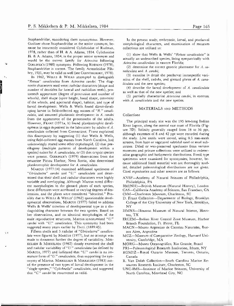


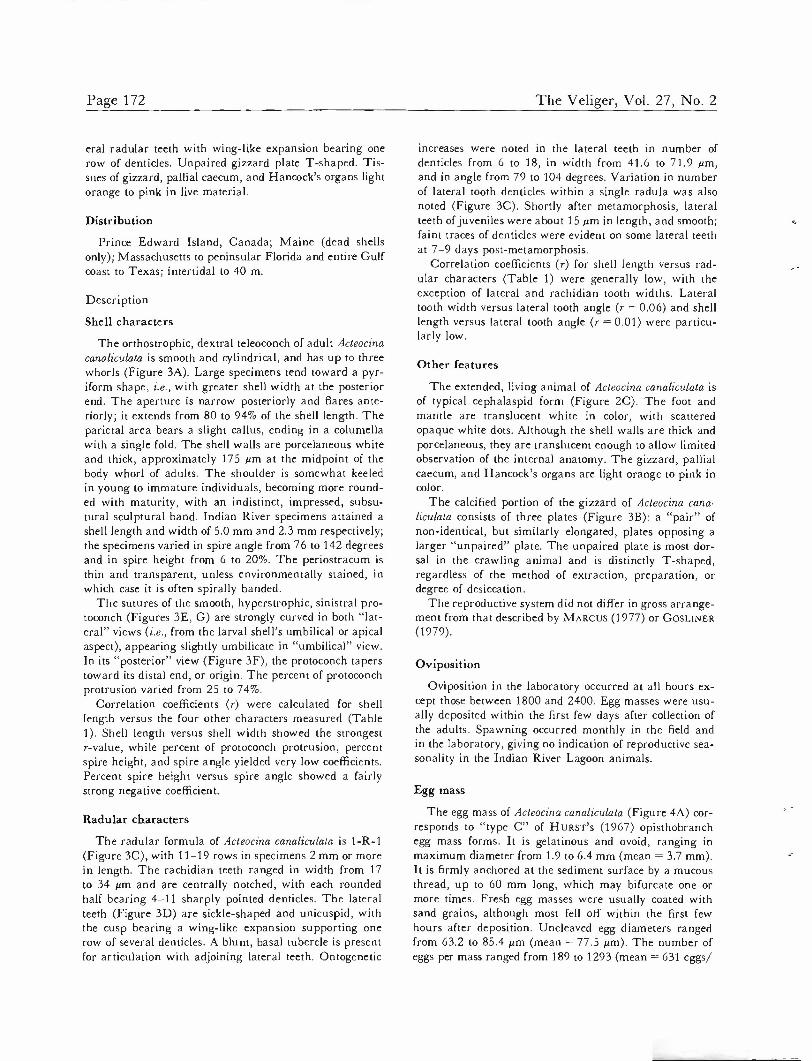
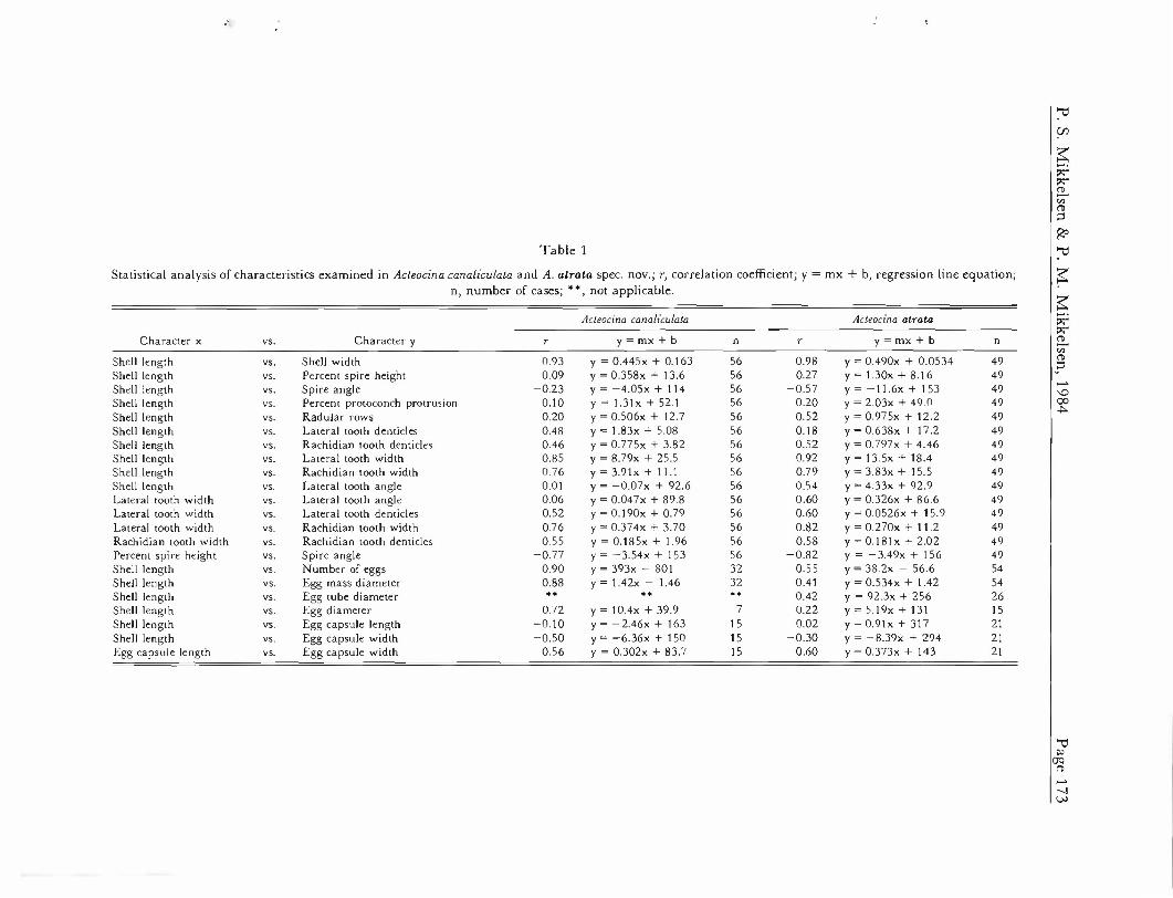


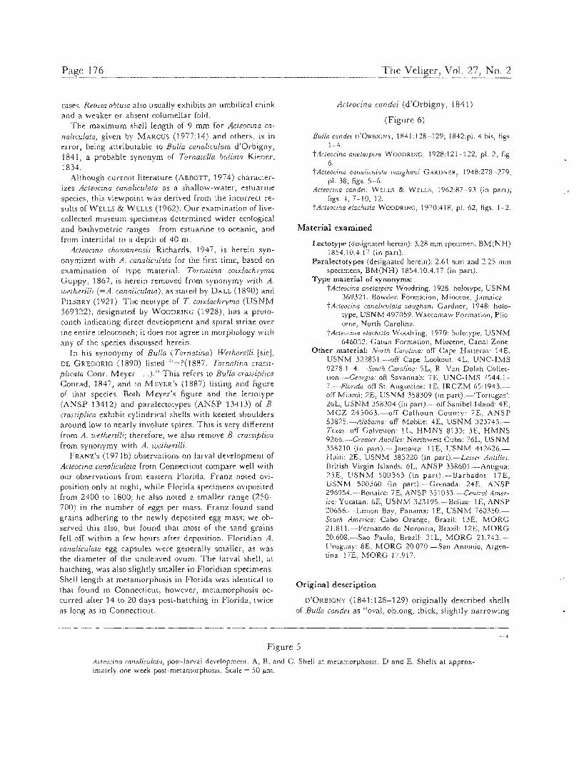
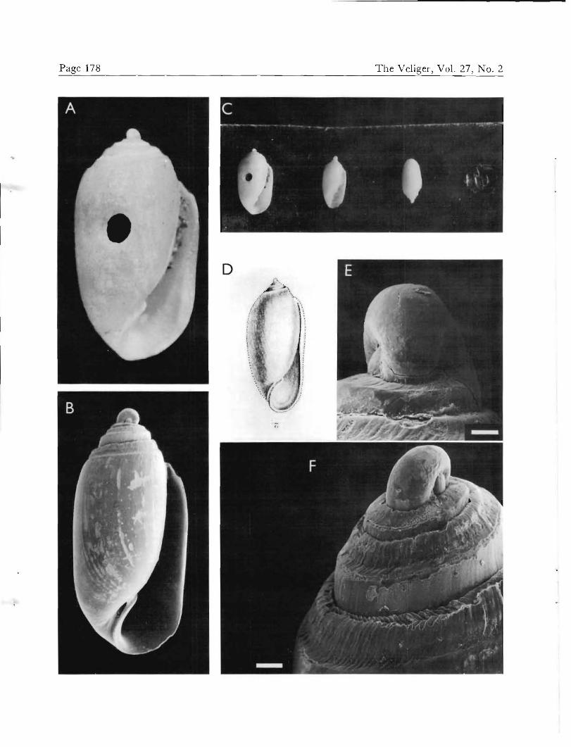
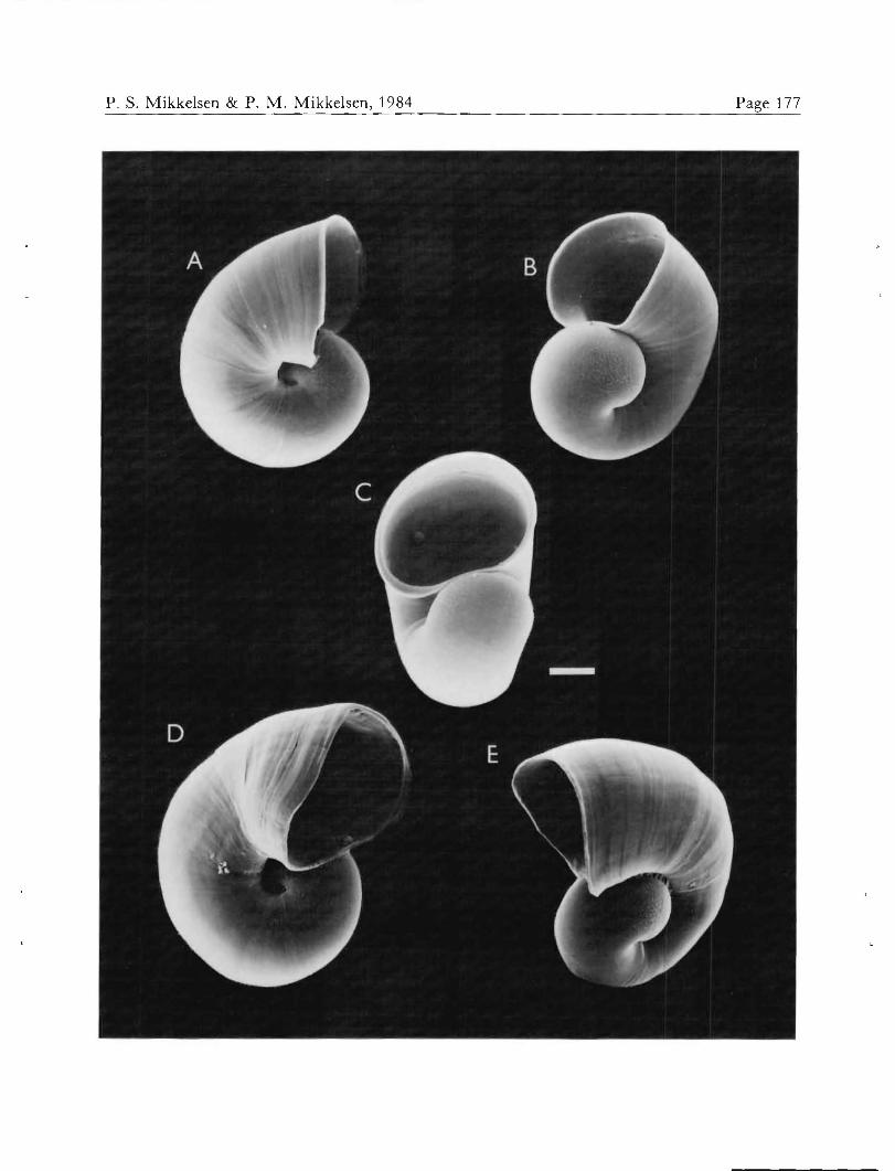


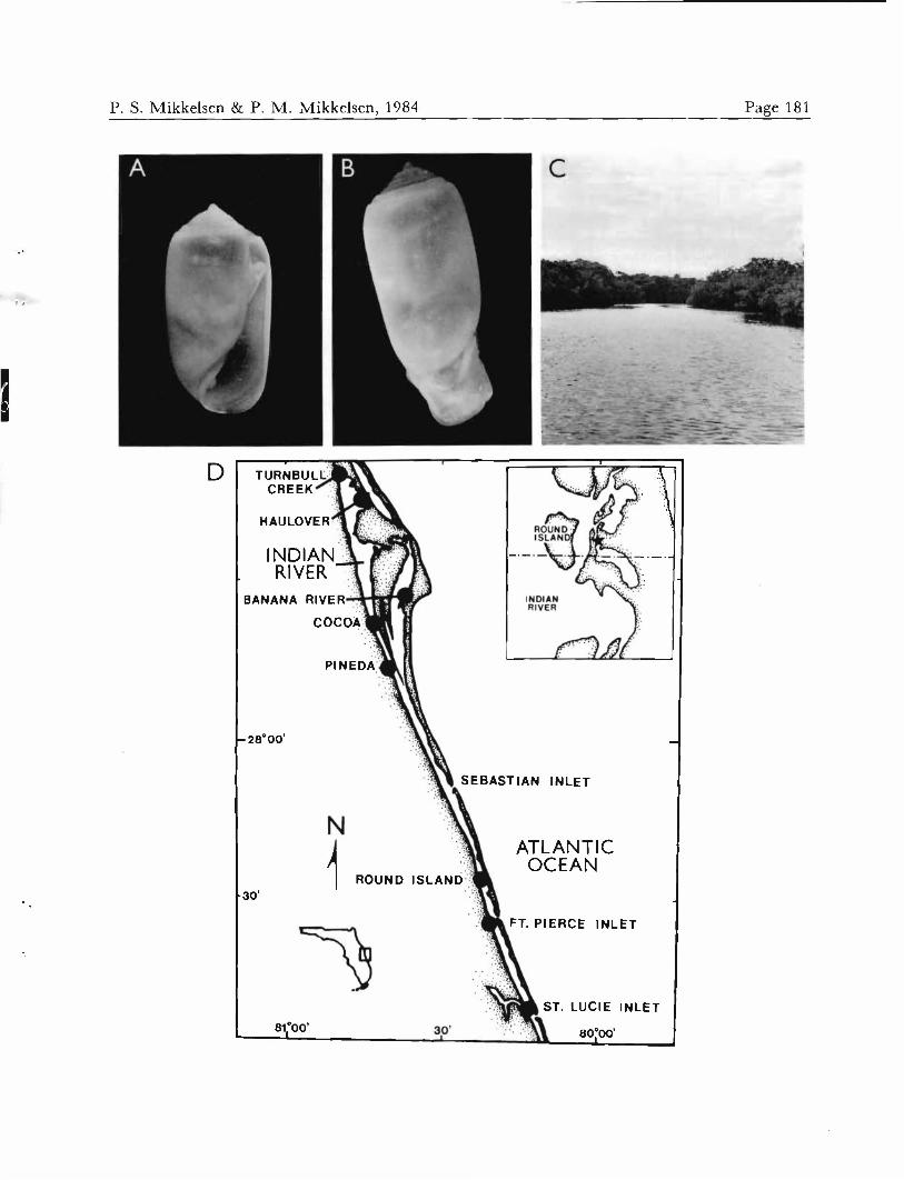


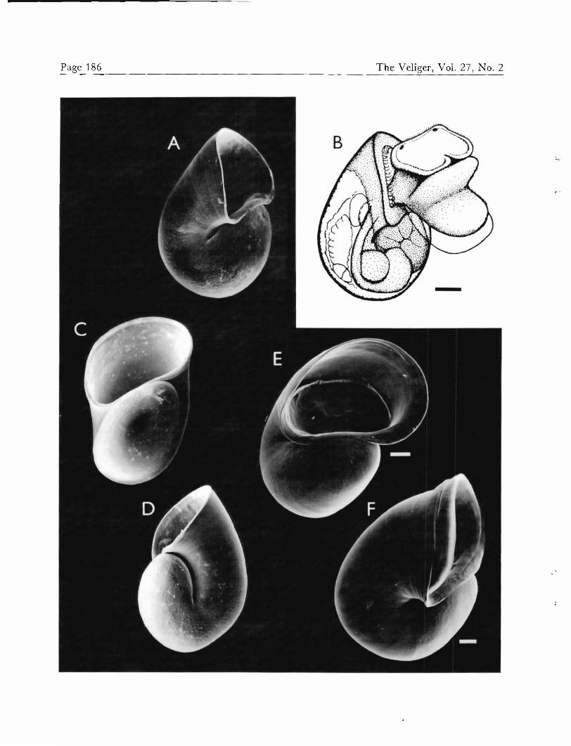

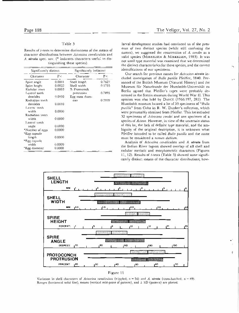
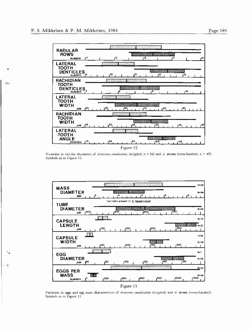


![1821 1841 2 Finished[1]](https://static.documents.pub/doc/80x56/558521ccd8b42ae4748b4cc9/1821-1841-2-finished1.jpg)


