+ All Categories
Home > Documents > Comparison of backbone dynamics of the type III antifreeze ...

Comparison of backbone dynamics of the type III antifreeze ...

Date post: 03-Dec-2021
Category:
Upload: others
View: 1 times
Download: 0 times
Share this document with a friend
14
ARTICLE Comparison of backbone dynamics of the type III antifreeze protein and antifreeze-like domain of human sialic acid synthase Yong-Geun Choi Chin-Ju Park Hee-Eun Kim Yeo-Jin Seo Ae-Ree Lee Seo-Ree Choi Shim Sung Lee Joon-Hwa Lee Received: 2 November 2014 / Accepted: 30 December 2014 / Published online: 10 January 2015 Ó Springer Science+Business Media Dordrecht 2015 Abstract Antifreeze proteins (AFPs) are found in a variety of cold-adapted (psychrophilic) organisms to pro- mote survival at subzero temperatures by binding to ice crystals and decreasing the freezing temperature of body fluids. The type III AFPs are small globular proteins that consist of one a-helix, three 3 10 -helices, and two b-strands. Sialic acids play important roles in a variety of biological functions, such as development, recognition, and cell adhesion and are synthesized by conserved enzymatic pathways that include sialic acid synthase (SAS). SAS consists of an N-terminal catalytic domain and a C-termi- nal antifreeze-like (AFL) domain, which is similar to the type III AFPs. Despite having very similar structures, AFL and the type III AFPs exhibit very different temperature- dependent stability and activity. In this study, we have performed backbone dynamics analyses of a type III AFP (HPLC12 isoform) and the AFL domain of human SAS (hAFL) at various temperatures. We also characterized the structural/dynamic properties of the ice-binding surfaces by analyzing the temperature gradient of the amide proton chemical shift and its correlation with chemical shift deviation from random coil. The dynamic properties of the two proteins were very different from each other. While HPLC12 was mostly rigid with a few residues exhibiting slow motions, hAFL showed fast internal motions at low temperature. Our results provide insight into the molecular basis of thermostability and structural flexibility in homologous psychrophilic HPLC12 and mesophilic hAFL proteins. Keywords NMR Backbone dynamics Antifreeze protein Antifreeze-like protein Ice-binding protein Abbreviations AFP Antifreeze protein QAE Quaternary-amino-ethyl SP Sulfopropyl TH Thermal hysteresis IBS Ice-binding surface AFL Domain antifreeze-like domain Sialic acid N-acetylneuraminic acid SAS Sialic acid synthase PEP Phosphoenolpyruvate ManNAc N-acetylmannosamine NeuNAc N-acetylneuraminic acid ManNAc-6P ManNAc 6-phosphate NeuNAc-9P NeuNAc 9-phosphate 3D Three-dimensional CSD Chemical shift deviation H-bond Hydrogen-bond NOE Nuclear Overhauser effect Yong-Geun Choi and Chin-Ju Park have contributed equally to this work. Electronic supplementary material The online version of this article (doi:10.1007/s10858-014-9895-2) contains supplementary material, which is available to authorized users. Y.-G. Choi H.-E. Kim Y.-J. Seo A.-R. Lee S.-R. Choi S. S. Lee J.-H. Lee (&) Department of Chemistry and Research Institute of Natural Science, Gyeongsang National University, Jinju, Gyeongnam 660-701, Republic of Korea e-mail: [email protected] C.-J. Park Division of Liberal Arts and Sciences and Department of Chemistry, Gwangju Institute of Science and Technology, Gwangju 500-712, Republic of Korea 123 J Biomol NMR (2015) 61:137–150 DOI 10.1007/s10858-014-9895-2
Transcript

ARTICLE

Comparison of backbone dynamics of the type III antifreezeprotein and antifreeze-like domain of human sialic acid synthase

Yong-Geun Choi • Chin-Ju Park • Hee-Eun Kim •

Yeo-Jin Seo • Ae-Ree Lee • Seo-Ree Choi •

Shim Sung Lee • Joon-Hwa Lee

Received: 2 November 2014 / Accepted: 30 December 2014 / Published online: 10 January 2015

� Springer Science+Business Media Dordrecht 2015

Abstract Antifreeze proteins (AFPs) are found in a

variety of cold-adapted (psychrophilic) organisms to pro-

mote survival at subzero temperatures by binding to ice

crystals and decreasing the freezing temperature of body

fluids. The type III AFPs are small globular proteins that

consist of one a-helix, three 310-helices, and two b-strands.

Sialic acids play important roles in a variety of biological

functions, such as development, recognition, and cell

adhesion and are synthesized by conserved enzymatic

pathways that include sialic acid synthase (SAS). SAS

consists of an N-terminal catalytic domain and a C-termi-

nal antifreeze-like (AFL) domain, which is similar to the

type III AFPs. Despite having very similar structures, AFL

and the type III AFPs exhibit very different temperature-

dependent stability and activity. In this study, we have

performed backbone dynamics analyses of a type III AFP

(HPLC12 isoform) and the AFL domain of human SAS

(hAFL) at various temperatures. We also characterized the

structural/dynamic properties of the ice-binding surfaces

by analyzing the temperature gradient of the amide proton

chemical shift and its correlation with chemical shift

deviation from random coil. The dynamic properties of the

two proteins were very different from each other. While

HPLC12 was mostly rigid with a few residues exhibiting

slow motions, hAFL showed fast internal motions at low

temperature. Our results provide insight into the molecular

basis of thermostability and structural flexibility in

homologous psychrophilic HPLC12 and mesophilic hAFL

proteins.

Keywords NMR � Backbone dynamics � Antifreeze

protein � Antifreeze-like protein � Ice-binding protein

Abbreviations

AFP Antifreeze protein

QAE Quaternary-amino-ethyl

SP Sulfopropyl

TH Thermal hysteresis

IBS Ice-binding surface

AFL Domain antifreeze-like domain

Sialic acid N-acetylneuraminic acid

SAS Sialic acid synthase

PEP Phosphoenolpyruvate

ManNAc N-acetylmannosamine

NeuNAc N-acetylneuraminic acid

ManNAc-6P ManNAc 6-phosphate

NeuNAc-9P NeuNAc 9-phosphate

3D Three-dimensional

CSD Chemical shift deviation

H-bond Hydrogen-bond

NOE Nuclear Overhauser effect

Yong-Geun Choi and Chin-Ju Park have contributed equally to this

work.

Electronic supplementary material The online version of thisarticle (doi:10.1007/s10858-014-9895-2) contains supplementarymaterial, which is available to authorized users.

Y.-G. Choi � H.-E. Kim � Y.-J. Seo � A.-R. Lee � S.-R. Choi �S. S. Lee � J.-H. Lee (&)

Department of Chemistry and Research Institute of Natural

Science, Gyeongsang National University, Jinju,

Gyeongnam 660-701, Republic of Korea

e-mail: [email protected]

C.-J. Park

Division of Liberal Arts and Sciences and Department of

Chemistry, Gwangju Institute of Science and Technology,

Gwangju 500-712, Republic of Korea

123

J Biomol NMR (2015) 61:137–150

DOI 10.1007/s10858-014-9895-2

Introduction

Antifreeze proteins (AFPs) are found in a variety of cold-

adapted organisms including fish, insects, plants, bacteria

and fungi and promote survival at subzero temperatures by

binding to ice crystals and decreasing the freezing tem-

perature of body fluids (Davies et al. 2002; Ewart et al.

1999; Fletcher et al. 2001; Jia and Davies 2002; Yeh and

Feeney 1996). Fish AFPs comprise several structurally

diverse classes of proteins and are grouped into four types

on the basis of their structures: type I AFPs are alanine-rich

a-helical proteins (Harding et al. 1999); type II AFPs are

cysteine-rich globular proteins containing five disulfide

bonds (Liu et al. 2007); type III AFPs are *7-kDa globular

proteins (Jia et al. 1996); and type IV AFPs are glutamate-

and glutamine-rich a-helical proteins (Gauthier et al.

2008).

One of most widely studied classes of AFPs is the type III

from arctic fish such as the ocean pout (Zoarces americanus)

and Japanese notched-fin eel pout (Zoarces elongates Kner).

The type III AFPs have been categorized into two subgroups,

quaternary-amino-ethyl (QAE) and sulfopropyl (SP)

Sephadex-binding, based on differences in their isoelectric

points (Fig. 1a) (Hew et al. 1988). The QAE proteins can be

further divided into two subgroups, QAE1 and QAE2

(Fig. 1a) (Nishimiya et al. 2005). Among them, only the

QAE1 isoforms exhibit full thermal hysteresis (TH) activi-

ties, whereas the others have extremely low or no such

activities (Nishimiya et al. 2005). The HPLC12 isoform from

the ocean pout, Z. americanus, which belongs to the QAE1

subgroup, has been extensively studied by NMR (Chao et al.

1994; Sonnichsen et al. 1996), X-ray crystallography (Jia

et al. 1996), mutagenesis (Chao et al. 1994; DeLuca et al.

1998; Graether et al. 1999; Jia et al. 1996), and molecular

dynamics (Antson et al. 2001; Chen and Jia 1999). The X-ray

structure of the HPLC12 isoform contains one a-helix (res-

idues 37–40), three 310-helices (residues 19–21, 33–36, and

57–59), and two b-strands (residues 4–7 and 22–25)

(Fig. 1a) (Jia et al. 1996). The ice-binding surface (IBS) is

comprising residues Q9, P12, N14, T15, A16, T18, V20,

M21 and Q44 (Jia et al. 1996) and the flatness of IBS is

thought to be important for antifreeze activity (Yang et al.

1998). The molecular dynamics study reported that five

additional residues, I13, L19, V41, S42, and K61 are also

potentially able to interact with ice (Chen and Jia 1999).

Among them, five hydrophilic residues (Q9, N14, T15, T18,

and Q44) have the potential to form hydrogen bonds

(H-bonds) with water molecule in the ice crystal

(Fig. 1b) (Sonnichsen et al. 1996; Jia et al. 1996). The resi-

dues, L10, I13, L19, V20, and V41 contributed to ice-binding

via hydrophobic interaction with ice and thus replacement

by alanine caused significant loss of antifreeze activity

(Baardsnes and Davies 2002). Recently, it was reported that

the IBS consists of two adjacent surfaces that are flat and

relatively hydrophobic (see Fig. 1b) (Garnham et al. 2012).

The QAE1 isoform binds both pyramidal and primary prism

planes of ice crystal and is able to halt the growth of ice,

whereas the SP and QAE2 isoforms exhibit low or no TH

activities by binding to only pyramidal ice plane (Garnham

et al. 2012). The inactive QAE2 isoform could be converted

into a fully-active QAE1-like isoform that is able to bind

primary prism ice plane by changing four residues (V9Q/

V19L/G20V/I41V) (Garnham et al. 2012).

Sialic acids (N-acetylneuraminic acids) play a vital role in

a variety of biological functions, such as transmembrane

signaling, cell growth, and cell adhesion (Schauer 2004).

Sialic acids are synthesized by a conserved enzymatic

pathway that includes sialic acid synthase (SAS). Bacterial

SAS converts phosphoenolpyruvate (PEP) and N-acetyl-

mannosamine (ManNAc) into N-acetylneuraminic acid

(NeuNAc, or sialic acid) (Masson and Holbein 1983).

Mammalian SAS catalyzes the condensation of PEP and

ManNAc 6-phosphate (ManNAc-6P) into NeuNAc 9-phos-

phate (NeuNAc-9P) which is dephosphorylated into sialic

acid (Lawrence et al. 2000). Both enzymes consist of two

distinct domains, an N-terminal catalytic NeuB domain

(*250 amino acid residues) and a C-terminal antifreeze-like

(AFL) domain (*75 amino acid residues). The crystal

structure of an SAS from Neisseria meningitides, in complex

with Mn2?, PEP, and ManNAc, revealed a unique domain-

swapped homodimer consisting of an N-terminal (a/b)8

barrel-type fold and a C-terminal AFL domain with high

structural similarity to the type III AFPs (Gunawan et al.

2005). The AFL domain of the N. meningitides SAS

(nmAFL) contributed to the substrate binding of the NeuB

domain of the opposite monomer via hydrogen bonding

(H-bonding) interaction between the side chain of R314 (see

Fig. 1a) and the acetyl oxygen of ManNAc (Gunawan et al.

2005). It was proposed that the deletion of the AFL domain in

human SAS (hAFL) would abolish the SAS activity, indi-

cating that the hAFL domain might play an important role in

substrate binding by the SAS (Hamada et al. 2006). An NMR

study of the hAFL domain found that the structure was

similar to those of the type III AFPs (Hamada et al. 2006).

Despite having very similar structures, the type III AFP

and AFL have very different temperature dependencies of

stability and activity. The type III AFP is considered as

psychrophilic protein because of the lower working tem-

perature (around 0 �C) while AFL is thought to be meso-

philic protein with the working temperature in the range

of 20–37 �C. Comparative molecular dynamics studies

have been used to elucidate temperature dependence of

the dynamics of psychrophilic and mesophilic proteins

(Brandsdal et al. 1999; Olufsen et al. 2007; Papaleo

et al. 2006, 2007, 2008). A recent molecular dynamics

138 J Biomol NMR (2015) 61:137–150

123

simulation study suggested that the type III AFP showed

increased overall flexibility at higher temperature com-

pared to the AFL protein (Kundu and Roy 2009). However,

the molecular basis of their distinctive behaviors is not well

understood.

Heteronuclear NMR spectroscopy is a suitable experi-

mental tool to compare the thermostability and structural

flexibility of homologous psychrophilic and mesophilic

proteins. In this study, we have investigated backbone

dynamics of the type III AFP from ocean pout, Z. amer-

icanus (HPLC12 isoform) and the AFL domain of human

SAS (hAFL) at various temperatures. We also character-

ized the structural/dynamic properties of the IBS of the

HPLC12 and hAFL proteins by analyzing the temperature

gradient of the amide proton chemical shift and its corre-

lation with chemical shift deviation (CSD) from random

coil. Our results show that the two proteins have entirely

different backbone dynamics, especially at low temperature

(5 �C). HPLC12 exhibits mostly rigid at low temperature

and this rigidity of IBS might be crucial for efficient

hydrophobic and H-bonding interaction with ice crystal.

Interestingly, most residues of hAFL are relatively rigid at

higher temperature (25 �C) while the substrate-binding

residues experience distinctive flexible backbone dynam-

ics. Based on our study, we suggest that the dynamic

properties of each protein give insight into the underlying

causes for the different biological working temperatures of

HPLC12 and hAFL. It is very likely that each protein

maintains a relatively rigid backbone structure overall, but

that the functionally important residues exhibit distinctive

dynamics for substrate binding (rigid for ice-binding or

flexible for sialic acid binding).

Fig. 1 a Multiple sequence alignment of the type III AFP family and

AFL domains. NfeAFP2, nfeAFP8, and nfeAFP11 are the isoforms 2,

8, and 11 of the Notched-fin eelpout type III AFP, respectively. hAFL,

mAFL, and nmAFL are the AFL domains of human, mouse, and

Neisseria meningitides SAS, respectively. Numbering and secondary

structure elements for HPLC12 are shown above the sequence. Red

and green bars indicate residues composing the pyramidal and

primary prism ice plane segments of the IBS, respectively. Five

residues (Q9, N14, T15, T18, and Q44) that can form H-bonds with

ice crystals are highlighted in yellow letters. The asterisk and dot

symbols represent invariant or nearly invariant residues in the type III

AFP family, respectively. Blue bars indicate the hydrophobic residues

that are invariant or nearly invariant residues in the type III AFP

family. Substrate-binding R314 in Neisseria meningitides SAS is

highlighted in orange. b Two IBS of HPLC12 and c the correspond-

ing surfaces of hAFL. Coordinates were derived from the HPLC12

X-ray crystal structure (PDB id = 1MSI) (Jia et al. 1996) and hAFL

NMR structure (PDB id = 1WVO) (Hamada et al. 2006). The

pyramidal and primary prism ice plane segments of the IBS in the

HPLC12 isoform and the corresponding segments in hAFL are shown

in red and green, respectively

J Biomol NMR (2015) 61:137–150 139

123

Materials and methods

Sample preparation

The DNA coding sequences for the HPLC12 (M1-A66)

and hAFL (L291–S359) were purchased from BIONEER

Inc. (Korea) and cloned into E. coli expression vectors

pET20b (Novagen, USA) with a C-terminal histidine-

affinity tag (His-tag) and pET28a (Novagen, USA) with an

N-terminal His-tag, respectively. To produce uniformly15N-labeled or 13C, 15N-labeled HPLC12, C41(DE3) cells

were grown in M9 minimal medium that contained 1 g/L15NH4Cl and/or 2 g/L 13C-glucose as the sole nitrogen and

carbon sources. Isotopically-labeled hAFL domain was

expressed in a similar manner but in BL21(DE3) cells. The

expressed proteins were purified by Ni–NTA affinity and

followed by Sephacryl S-100 gel filtration chromatography

(GE Healthcare, USA) on a GE AKTA Prime Plus. The

concentrations of both proteins were measured using a

Pierce BCA Protein Assay Kit (Thermo Scientific, USA).

For NMR experiments, the purified proteins were concen-

trated to 1 mM in a 90 % H2O/10 % D2O buffer containing

10 mM sodium phosphate (pH 8.0) and 100 mM NaCl.

NMR experiments

All of the 1H, 13C, and 15N NMR experiments were

performed on an Agilent DD2 700-MHz spectrometer

(GNU) equipped with a cold probe. All three-dimensional

(3D) NMR experiments were carried out with 1 mM 13C,15N-labeled HPLC12 and hAFL proteins at 25 �C. All 2D1H/15N-HSQC spectra for HPLC12 and hAFL were

obtained using 1 mM 15N-labeled protein samples at

various temperature. 1D NMR data were processed with

either VNMR J (Agilent, USA) or FELIX2004 (FELIX

NMR, USA) software, while the 2D and 3D data were

processed with NMRPIPE (Delaglio et al. 1995) and

analyzed with Sparky (Goddard and Kneller 2003). 2-2-

Dimethyl-2-silapentane-5-sulfonate was used as an exter-

nal reference.

The backbone resonance assignments for HPLC12 and

hAFL were obtained from the following 3D experiments:

CACB(CO)NH, HNCACB, HNCA, HNCO, and NOESY-1H/15N-HSQC. The 1H/15N-HSQC spectra of the HPLC12 and

hAFL were acquired at temperatures of 5–25 �C in 5 �C

increments. The Dd/DT values of the amide protons were

determined from linear variation of the NH chemical shifts

with temperature. The amide proton CSDs (dobs–drc) at 25 �C

were calculated from the observed chemical shifts (dobs) and

the corresponding random coil (drc) values taken from the

literature (Andersen et al. 1997).

Backbone dynamics parameters, longitudinal R1 relax-

ation rates, transverse R2 relaxation rates, and {1H}-15N

heteronuclear NOEs were measured using 15N-labeled

HPLC12 and hAFL protein samples at 5, 15, and 25 �C. 2D1H/15N-HSQC spectra for backbone dynamics measure-

ments were acquired with 2,048 (HN) 9 64 (N) data points

and 8 scans. Spectral widths of 7,716 Hz (11 ppm) and

2,414 Hz (34 ppm) were used in the HN and N dimensions,

respectively. R1 values were measured in a series of spectra

with relaxation delays of 50, 100, 150, 200, 250, 300, 350,

400, 500, and 600 ms. R2 measurements were taken with

relaxation delays of 10, 30, 50, 70, 90, 110, 130, 150, 190,

and 230 ms. For evaluation of {1H}-15N heteronuclear

NOE values, four different data sets with and without an

initial proton saturation (2.5 s period) were measured.

Reduced spectral density analysis was performed using the

following relationships as described by Bracken et al.

(1999):

rNH ¼ R1ðNOE � 1ÞcN=cH ð1Þ

Jð0:87xHÞ ¼ 4rNH=ð5d2Þ ð2Þ

JðxNÞ ¼ ð4R1 � 5rNHÞ=ð3d2 þ 4c2Þ ð3Þ

Jeð0Þ ¼ ð6R2 � 3R1 � 2:72rNHÞ=ð3d2 þ 4c2Þ ð4Þ

where d = (lohcNcH/8p2)(rNH-3 ) and c = xNDr/H3, lo is

the permeability of free space, h is Planck’s constant, cH

and cN are the gyromagnetic ratios of the 1H and 15N

nuclei, respectively, xH and xN are the Lamor frequencies,

rNH is the average 1H–15N bond length (1.02 A), and Dr is

the 15N chemical shift anisotropy (-160 ppm). The

effective J(0) [Je(0)] contains contributions from the

chemical exchange term, if present.

For well-ordered sites in a macromolecule, the correla-

tion time of overall tumbling motion (sm) is obtained from

the spectral density functions as (Bracken et al. 1999):

sm ¼ffiffiffiffiffiffiffiffiffiffiffiffiffiffiffiffiffiffiffiffiffiffiffiffiffiffiffiffiffiffiffiffiffiffiffiffiffiffiffiffiffiffiffiffiffiffi

fJð0Þ � JðxNÞg=JðxNÞp

xN

: ð5Þ

Results and discussion

NMR resonance assignments

The 1H/15N HSQC spectra of the HPLC12 isoform and

hAFL domain at 25 �C are shown in Supplementary

Fig. S1. Amide backbone resonance assignments for the

HPLC12 were made by heteronuclear three-dimensional

(3D) NMR experiments at pH 8.0. In hAFL, some amide

cross-peaks were missing at pH 8.0, so backbone resonance

assignments were conducted at pH 6.0 (see Supplementary

Fig. S2). Resonance assignments of hAFL at pH 8.0 were

made by comparing of 1H/15N-HSQC spectra acquired

at pH 6.0, 6.5, 7.0, and 8.0 and were confirmed by 3D

NOESY-1H/15N-HSQC and TOCSY-1H/15N-HSQC spec-

tra at pH 8.0.

140 J Biomol NMR (2015) 61:137–150

123

Temperature dependence of 1H/15N-HSQC spectra

of HPLC12 and hAFL

The NMR chemical shift is sensitive to the environment of

the observed nuclei as well as to temperature-related struc-

tural or dynamic changes (Hong et al. 2010; Ohnishi and

Urry 1969). As the temperature was lowered, significant

cross peak movements were observed in the 1H/15N HSQC

spectra of the HPLC12 and hAFL proteins (Supplementary

Fig. S3). Figure 2 shows the temperature gradients of amide

proton chemical shifts (Dd/DT) as a function of residue

number (in hAFL, the residue number is starting with residue

L291 of SAS as residue L1). In both proteins, most residues

have Dd/DT values [-4.6 ppb/K, indicating that these

amides are H-bonded (Cierpicki and Otlewski 2001; Cier-

picki et al. 2002). Residues with Dd/DT values\-4.6 ppb/K

(Fig. 2b, c) are in good agreement with the X-ray crystal

structure of HPLC12 (Jia et al. 1996) or the NMR structure of

hAFL (Hamada et al. 2006), where these amide protons were

not protected from exchange by H-bonding.

Residues N14, T15, I37, and V45 in HPLC12 have Dd/

DT values [0 ppb/K. Similarly, in hAFL, the correspond-

ing residues G14, I37, and V45 exhibit positive Dd/DT

values. Residues D20, G27, G42, and E50 in hAFL also

have Dd/DT values[0 ppb/K. The most surprising feature

is that some residues (V5, D20, N39, and N62) in hAFL

have significantly different Dd/DT values compared to the

corresponding residues in HPLC12 (Fig. 2a). For example,

the Dd/DT value of D20 in hAFL is 0.8 ppb/K, but the

corresponding residue V20 in HPLC12 has a Dd/DT value

of -4.7 ppb/K. Instead, the neighboring M19 amide proton

exhibits a large negative Dd/DT (-6.1 ppb/K).

Fig. 2 a Temperature dependence of the chemical shifts (Dd/DT) of

HPLC12 (closed blue circles) and hAFL (open red circles) as a

function of residue number. Secondary structure models for previ-

ously determined b X-ray crystal structure of HPLC12 (PDB

id = 1MSI) (Jia et al. 1996) and c NMR structure of hAFL (PDB

id = 1WVO) (Hamada et al. 2006). Residues with Dd/DT values

\-4.6 ppb/K are colored blue and residues with Dd/DT values

[0 ppb/K are in magenta. The amide proton Dd/DT–CSD correlation

plots of c HPLC12 and d hAFL. Solid lines indicate the statistical

cutoff, Dd/DT = –2.41 9 CSD-2.11. The residues composing the

pyramidal and primary prism ice plane segments of the IBS in the

HPLC12 isoform and the corresponding segments in hAFL are shown

in red and green, respectively. The hydrophobic residues that are

nearly invariant in the type III AFP family are highlighted in blue

J Biomol NMR (2015) 61:137–150 141

123

To characterize in detail the structural and dynamic

properties of the HPLC12 and hAFL proteins, a combined

analysis of Dd/DT values with CSD (Andersen et al. 1997)

was performed. Correlation plots of Dd/DT versus CSD are

shown in Figs. 2c, d. The hydrophobic residues, except I32

and V60, that nearly invariant in the type III AFP family

were all below the statistical correlation line between Dd/

DT and CSD (Dd/DT = -2.41 9 CSD-2.11) (blue in

Fig. 2c), suggesting that these residues are H-bonded and

in slow-exchange (Andersen et al. 1997). Similar results

were also observed in hAFL with the exception of V5, I55,

and V60 (Fig. 2d).

In HPLC12, residues I11 and V49 have Dd/DT values

significantly lower than the statistical cutoff (Fig. 2c),

reflecting strong H-bonding interactions between the s1 and

s5 strands [the strands s1–s6 were random coil in HPLC12

but were assigned as b strands in the type III AFP variant,

RD3, from Rhigophila dearborni (Miura et al. 2001)]

(Supplementary Fig. S4). Similarly, residues N14 and V45

contributed to strong H-bonding interactions between

the s2 and s4 strands via N14-NH$V45-CO and

V45-NH$T15-CO. In addition, four residues in the b1

strand (S4, V5, V6, and A7) and three residues in the

second 310 helix (A34, E35, and I37) had Dd/DT that

deviated significantly from random coil values (Fig. 2c,

Supplementary Fig. S4). In hAFL, significant deviations of

Dd/DT values from the statistical cutoff were observed for

I11, V49, G14, and V45 (Fig. 2d). However, residues S4

and V5 in the b1 strand and three residues in the a-helix

(residues 38, 39 and 41) have significantly higher Dd/DT

values compared to the statistical cutoff (Fig. 2d, Supple-

mentary Fig. S4), suggesting that these residues are not

strongly H-bonded but are in fast-exchange.

15N NMR relaxation dynamics of HPLC12 at various

temperatures

The longitudinal R1 relaxation rates, transverse R2 relaxa-

tion rates, and {1H}-15N heteronuclear NOEs for uniformly15N-labeled HPLC12 were measured at three different

temperatures (5, 15 and 25 �C) to gain insight into the

temperature-dependent dynamic motions of these two

proteins (see Figs. 3, 4, 5). R1 values of HPLC12 are fairly

uniform throughout the proteins and increase with tem-

perature (Fig. 3a; Table 1). The R2 values of HPLC12 are

also mostly uniform, with a few exceptions. For example,

R2 of T53 (25.39 Hz) and M59 (19.66 Hz) showed large

deviation from the average value at 5 �C (Fig. 3c). The

R2/R1 ratio was significantly larger than the average value,

which implies conformational exchange on the ls–ms

timescale (Hong et al. 2010; Tjandra et al. 1995).

Interestingly, residues I32, Q44, T53, and M59 exhibited

R2/R1 ratios above the upper cutoff line [upper/lower

cutoff = average ± 1.5 9 standard deviation (SD)] at

5 �C, whereas at 25 �C the R2/R1 ratios of these residues

were below the upper cutoff line (Fig. 4a).

With the exception of the termini, the {1H}-15N heter-

onuclear NOEs of HPLC12 were mostly [0.8 at all three

temperatures, except for V27 at 5 �C (Fig. 5a). This indi-

cates that there are no ps–ns timescale motion in the

temperature range studied here.

15N NMR relaxation dynamics of hAFL at various

temperatures

The R1 values of hAFL showed temperature dependence

similar to those of HPLC12 (Fig. 3b). Residues G27–G31

and E35 showed slightly elevated R1 values compared to

other residues except both termini at all three temperatures

(Fig. 3b). The R2 values increased and showed larger

deviation from the average values as the temperature

decreased (Fig. 3d; Table 1). Residues K30, G31, and I37

exhibited significantly larger R2/R1 ratios than the average

at 25 �C (Fig. 4b). Line broadening prevented the R2/R1

ratios for E28, K30, and G31 at 5 �C from being deter-

mined (Supplementary Fig. S3). At 5 �C, three more resi-

dues (K25, E58, and V60) show larger R2/R1 ratios than the

upper cutoff value (Fig. 4b). Residues K3, G27, and D51

have significantly smaller R2/R1 ratios than the average

value at 5 �C (Fig. 4b).

The {1H}-15N heteronuclear NOEs of hAFL exhibit very

different patterns from those of HPLC12 (Figs. 4, 5). Several

residues had NOE values B0.8 and this number increased

when the temperature dropped (Fig. 5d). At 25 �C, some

residues (K3, K10, V24, V26–G31, K44, E50, I55–E57 and

N62) had NOE values B0.8. The NOE values for residues

K3, K30, and G31 could not be determined due to line

broadening at 5 �C. Residues S4, E13, I16, D20, V26–G31,

L40–G42, K44, V45, T48–E50, D53, T54, and E57–N62 had

NOEs B0.8 or showed very weak resonances at 5 �C

(Fig. 5d). It is noteworthy that the structural flexibility of

hAFL on the ps–ns timescale increased as the temperature

decreased, in contrast to HPLC12. Interestingly, residues

K10 and K44 that showed NOEs \0.8 are located on the

corresponding surfaces of ice-binding site even at 25 �C

(Fig. 5d). Residues G7, V24, V26, G27, E28, K30 and G31

that showed NOEs\0.8 at 25 �C (Fig. 5d) are clustered on

the substrate-binding surface for bacterial SAS (Hamada

et al. 2006). Residue K44 of hAFL showed NOE values B0.8

at all three temperatures, while the corresponding Q44 of

HPLC12 had NOE values[0.8 through all temperatures. As

mentioned above, Q44 of HPLC12 may experience confor-

mational exchange on the ls–ms timescale at low tempera-

ture. Taken together, our data indicate that the IBS of

HPLC12 and the corresponding surface of hAFL have dif-

ferent dynamic properties at low temperature.

142 J Biomol NMR (2015) 61:137–150

123

Analysis of 15N NMR relaxation parameters in terms

of reduced spectral density

The 15N-relaxation data at three different temperatures

were used to calculate reduced spectral density function

J-values. Figure 6 shows Je(0), J(xN), and J(0.87xH) of

HPLC12 and hAFL determined at three temperatures.

The temperature dependence of Je(0) was similar to R2,

and that of J(xN) was similar to the R1 pattern. HPLC12

had fairly uniform Je(0) values with the average values

of 4.24 ns/rad at 5 �C, 3.22 ns/rad at 15 �C, and 2.63 ns/rad

at 25 �C (Table 1). Residues I32 (4.94 ns/rad), T53

(6.64 ns/rad), M59 (5.12 ns/rad), and Y63 (4.94 ns/rad)

had significantly larger Je(0) values than the average at

cβ1

310α310

310β2

Residue

a b

R 1 (

Hz)

HPLC12

β1310

α310310

β2 β1310

α310310

β2

hAFL

R 1 (

Hz)

15 oC

25 oC

5 oC

15 oC

5 oC

25 oC

D20 G31

G31D20E13

E28D61

G62E35

G62

G62

HPLC12

25 oCd

β1310

α310310

β2

hAFL

G31K30

G31

Residue

Residue

D61

V60E58V45

K25

R 2 (

Hz) 15 oC

5 oC

25 oC

T53

T53M59

A3

A3

A3

K3

K3

Residue

R 2 (

Hz) 15 oC

5 oC

E35

1.0

2.0

3.0

1.0

2.0

3.0

1.0

2.0

3.0

10 20 30 40 50 600.0

1.0

2.0

3.0

1.0

2.0

3.0

0 10 20 30 40 50 600.0

1.0

2.0

3.0

10

20

30

40

10

20

30

40

K30

10 20 30 40 50 600

10

20

30

40

10

20

30

40

10

20

30

40

10 20 30 40 50 600

10

20

30

40

Fig. 3 15N relaxation parameters of HPLC12 and hAFL as a function

of residue number. R1 relaxation rates of a HPLC12 and b hAFL are

determined at 5 �C (lower, blue), 15 �C (middle, green), and 25 �C

(upper, red). R2 relaxation rates of c HPLC12 and d hAFL are

determined at 5 �C (lower, blue), 15 �C (middle, green), and 25 �C

(upper, red). The error bars represent curve fitting errors during the

determination of R1 and R2 values from experiment data. The solid

lines indicate the average values. In hAFL, the R1 and R2 relaxation

rates of A7 at 5 and 15 �C could not be determined because of

overlapping with other resonance. The black square symbols indicate

residues whose relaxation data could not be determined because of

severe line-broadening at 5 �C

J Biomol NMR (2015) 61:137–150 143

123

5 �C (Fig. 6a). This indicates that these residues are

involved in ls–ms timescale motions (Lefevre et al.

1996). J(xN) and J(0.87xH) of HPLC12 were also

mostly uniform (Fig. 6a). The small J(0.87xH) values

imply that HPLC12 is well structured with limited

internal mobility at all three temperatures, which is

consistent with the uniformly high {1H}-15N heteronu-

clear NOE values.

Fig. 4 R2/R1 ratios of a HPLC12 and b hAFL as a function of residue

number are determined at 5 (lower, blue), 15 (middle, green),

and 25 �C (upper, red). The error bars represent the standard

deviations of R2/R1 ratios (rR2/R1) determined by using equation,

rR2=R1 ¼ R2=R1

ffiffiffiffiffiffiffiffiffiffiffiffiffiffiffiffiffiffiffiffiffiffiffiffiffiffiffiffiffiffiffiffiffiffiffiffiffiffiffiffiffiffiffiffiffiffi

ðrR2=R2Þ2 þ ðrR2=R2Þ2q

, where rR1 and rR2 are

curve fitting errors during the determination of R1 and R2 values,

respectively. The thick and two thin solid lines indicate the average

and upper/lower cutoff values (average ± 1.5 9 SD), respectively.

The black square symbols indicate residues whose relaxation data

could not be determined because of severe line-broadening at 5 �C.

Mapping of the R2/R1 ratios onto the secondary structure models of

c HPLC12 and d hAFL. Residues which have deviated R2/R1 ratios

than the upper/lower cutoff lines are shown in blue. Residues

composing the pyramidal and primary prism ice plane segments of

HPLC12 and corresponding residues of hAFL are shown in pink and

light green, respectively. Residue G27 of hAFL, which is corre-

sponding the substrate-binding R27 in Neisseria meningitides SAS, is

in orange

144 J Biomol NMR (2015) 61:137–150

123

In contrast, hAFL showed higher and more varied Je(0)

because of more deviation in R2. The average Je(0) values

were 5.48 ns/rad at 5 �C, 4.16 ns/rad at 15 �C, and

3.33 ns/rad at 25 �C (Table 1). The elevated average Je(0)

values of hAFL imply that there are more residues that

experience slow motions. Residues K25, E58, and V60

which showed larger R2/R1 ratios as well as significantly

higher Je(0) values than the average at 5 �C (Figs. 4b, 6b).

As mentioned previously, abnormally large values of Je(0)

indicate that these residues are experiencing ls–ms time-

scale motions. At all three temperatures, the J(0.87xH)

values of hAFL are much larger and show more deviation

than those HPLC12, implying that the hAFL domain

exhibits greater ps–ns internal motion compared to HPLC12.

The correlation times of overall tumbling motion (sm) of

HPLC12 and hAFL are obtained from the spectral density

functions as using Eq. 4 (Supplementary Fig. S5). The

residues that have R2/R1 ratio deviated from upper/lower

a bHPLC12

β1310

α310310

β2 β1310

α310310

β2

hAFL

0.6

0.8

1.0

NO

E

0.6

0.8

1.025 oC

15 oC

10 20 30 40 50 60

0.6

0.8

1.0

Residue

5 oCV27

G31E28V24 K44 E50

M56

K10K3N62

0.6

0.8

1.0

I55K30

0.6

0.8

1.0

G31

K30E28K3 V26

V26G27

G27

E50

N62

E57

M56

E57

10 20 30 40 50 60

0.6

0.8

1.0

Residue

NO

E

25 oC

15 oC

5 oC

I16G27

V26

E28

N62

D61E57D53V41 V45

c dHPLC12 hAFL

5 Co

V27

NOE < 0.8

M21

N14

T15

A16

T18

V20

L19

Q9

L10

P12 S42V41

bind to pyramidal

plane

bind to primary

prism plane

25 Co

T18

V9

K10

P12

G14

I16

T15K44

M21M19

G42V41

N62

E57

I13

D20A7

G31K30

P29

E28

V26G27

substratebinding

Q44

NOE < 0.75

I55

E13L46

M56

V24

K3

E50

Fig. 5 Heteronuclear {1H}-15N NOEs of a HPLC12 and b hAFL as a

function of residue number are determined at 5 �C (lower, blue),

15 �C (middle, green), and 25 �C (upper, red). The error bars of the

heteronuclear NOE data represent standard deviations of four

different data sets. In hAFL, the heteronuclear NOE of A7 at 5 and

15 �C could not be determined because of overlapping with other

resonance. The black square symbols indicate residues whose

relaxation data could not be determined because of severe line-

broadening at 5 �C. Mapping of the {1H}-15N NOEs onto the

secondary structure models of c HPLC12 and d hAFL. Residues

which have smaller heteronuclear NOE \0.8 or \0.75 are shown in

light blue and dark blue, respectively. Residues composing the

pyramidal and primary prism ice plane segments of HPLC12 and

corresponding residues of hAFL are shown in pink and light green,

respectively. Residue G27 of hAFL, which is corresponding the

substrate-binding R27 in Neisseria meningitides SAS, is in orange

J Biomol NMR (2015) 61:137–150 145

123

cutoff values or heteronuclear NOE \0.7 are excluded

from calculation of the average sm values. We determined

that the average sm values of HPLC12 were 9.74 ns at

5 �C, 7.30 ns at 15 �C, and 5.78 ns at 25 �C (Table 1).

Similarly, hAFL have the average sm values of 12.16 ns at

5 �C, 9.02 ns at 15 �C, and 7.15 ns at 25 �C (Table 1). If

HPLC12 and hAFL are monomeric, we would expect the

average sm values of approximately 8.1 ns for HPLC12

(8.1 kDa with C-terminal His-tag) and 9.9 ns for hAFL

(9.9 kDa with N-terminal His-tag) at 5 �C by using

b

eJ(0

) (ns

/rad)

J(ω N

) (ns

/rad)

J(0.

87ω H

) (ps

/rad)

hAFL

β1310

α310310

β2 β1310

α310310

β2 β1310

α310310

β2

J(0

) (ns

/rad)

e

Residue

T53

I32 M59

Y63

Residue

G62

J(ω N

) (ns

/rad)

Residue

D20G31

K25

E28D61

V60

E58

K30

G31

E28

A3 E35

Residue

Residue

G27

E28

K3

K30

I55

V27G62

a β1310

α310310

β2

HPLC12

β1310

α310310

β2 β1310

α310310

β2

10 20 30 40 50 600

2

4

6

8

1025 oC15 oC 5 oC

10 20 30 40 50 600.0

0.1

0.2

0.3

0.4

0.5

0.6

10 20 30 40 50 6002468

10121416

J(0.

87ω H

) (ps

/rad)

10 20 30 40 50 6002468

10121416

Residue

G31

10 20 30 40 50 600.0

0.1

0.2

0.3

0.4

0.5

0.6

10 20 30 40 50 600

2

4

6

8

10

Fig. 6 Reduced spectral density functions of HPLC12 and hAFL. Je(0) (left); J(xN) (middle); and J(0.87xH) (right) are calculated for a HPLC12

and b hAFL as a function of residue number determined at 5 �C (lower, blue), 15 �C (middle, green), and 25 �C (upper, red)

Table 1 The average 15N relaxation parameters of HPLC12 and hAFL determined at 5, 15, and 25 �C

HPLC12a hAFLb

5 �C 15 �C 25 �C 5 �C 15 �C 25 �C

R1 (Hz) 1.21 ± 0.10 1.60 ± 0.13 1.97 ± 0.15 1.07 ± 0.15 1.37 ± 0.12 1.70 ± 0.15

R2 (Hz) 16.41 ± 1.64 12.81 ± 0.90 10.80 ± 0.61 20.96 ± 3.17 15.85 ± 2.38 13.26 ± 1.57

R2/R1 13.66 ± 1.69 8.06 ± 0.79 5.50 ± 0.50 20.10 ± 4.13 11.71 ± 1.87 7.81 ± 0.89

Je(0) (ns/rad) 4.24 ± 0.44 3.22 ± 0.24 2.63 ± 0.16 5.48 ± 0.86 4.16 ± 0.97 3.33 ± 0.42

J(xN) (ns/rad) 0.21 ± 0.02 0.28 ± 0.02 0.35 ± 0.03 0.19 ± 0.02 0.24 ± 0.02 0.30 ± 0.03

J(0.87xH) (ps/rad) 2.56 ± 0.87 3.23 ± 0.74 4.17 ± 0.72 3.43 ± 1.63 4.05 ± 1.38 4.91 ± 1.30

sm (ns)c 9.74 ± 0.42 7.30 ± 0.32 5.78 ± 0.31 12.16 ± 0.95 9.02 ± 0.53 7.15 ± 0.36

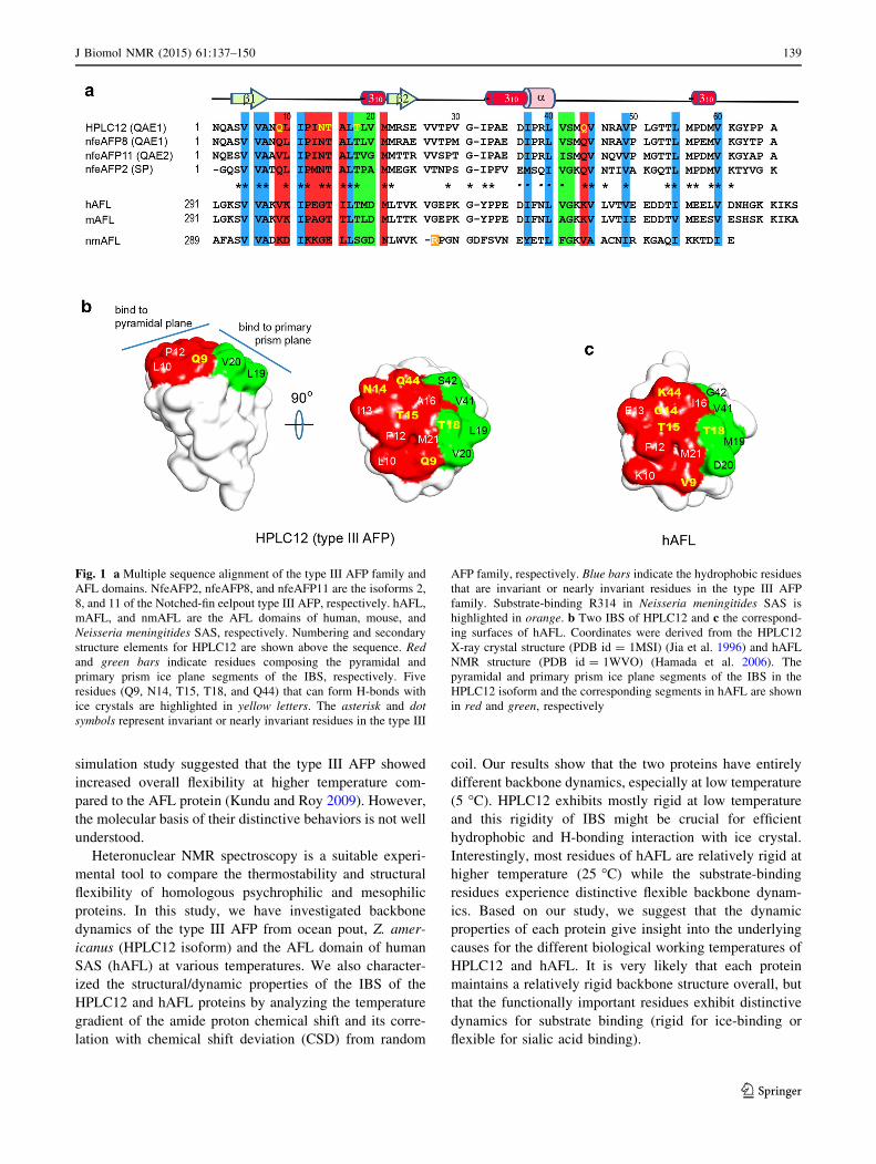
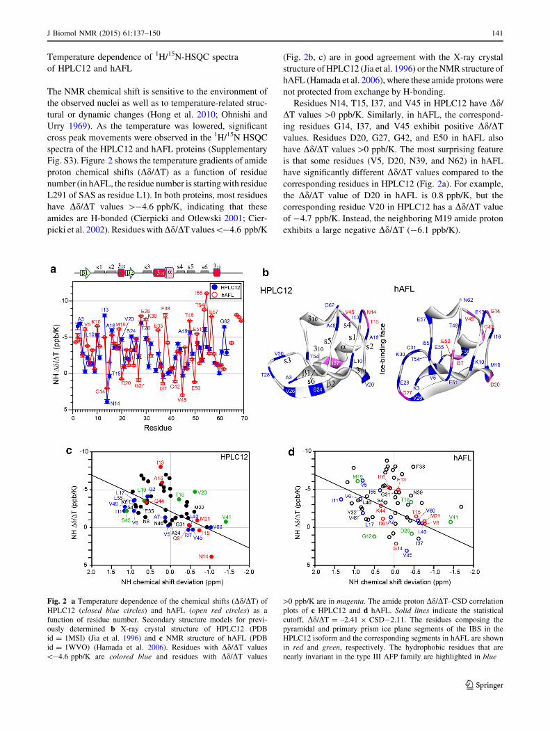
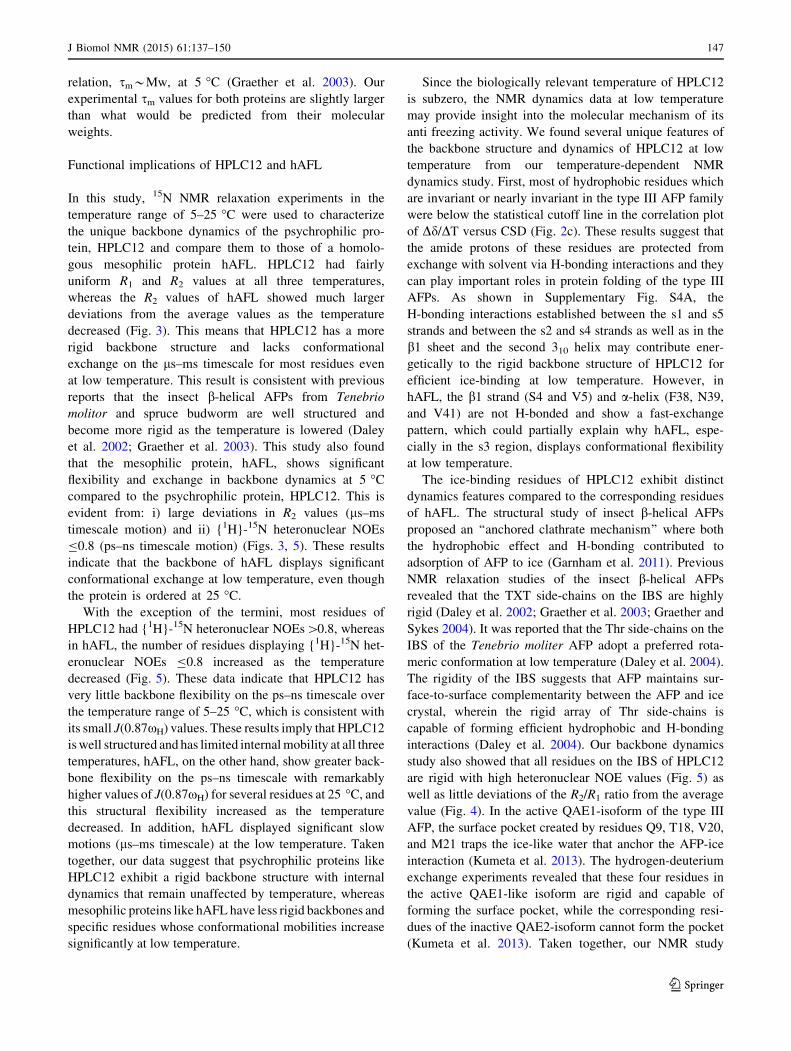
a The average values of residues 3–63b The average values of residues 3–62c Residues that exhibit an R2/R1 ratio deviated from upper/lower cutoff values or have an NOE\0.7 are excluded from calculation of the average

sm values

146 J Biomol NMR (2015) 61:137–150

123

relation, sm*Mw, at 5 �C (Graether et al. 2003). Our

experimental sm values for both proteins are slightly larger

than what would be predicted from their molecular

weights.

Functional implications of HPLC12 and hAFL

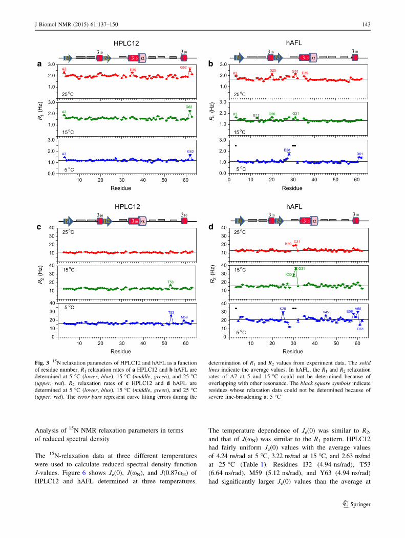
In this study, 15N NMR relaxation experiments in the

temperature range of 5–25 �C were used to characterize

the unique backbone dynamics of the psychrophilic pro-

tein, HPLC12 and compare them to those of a homolo-

gous mesophilic protein hAFL. HPLC12 had fairly

uniform R1 and R2 values at all three temperatures,

whereas the R2 values of hAFL showed much larger

deviations from the average values as the temperature

decreased (Fig. 3). This means that HPLC12 has a more

rigid backbone structure and lacks conformational

exchange on the ls–ms timescale for most residues even

at low temperature. This result is consistent with previous

reports that the insect b-helical AFPs from Tenebrio

molitor and spruce budworm are well structured and

become more rigid as the temperature is lowered (Daley

et al. 2002; Graether et al. 2003). This study also found

that the mesophilic protein, hAFL, shows significant

flexibility and exchange in backbone dynamics at 5 �C

compared to the psychrophilic protein, HPLC12. This is

evident from: i) large deviations in R2 values (ls–ms

timescale motion) and ii) {1H}-15N heteronuclear NOEs

B0.8 (ps–ns timescale motion) (Figs. 3, 5). These results

indicate that the backbone of hAFL displays significant

conformational exchange at low temperature, even though

the protein is ordered at 25 �C.

With the exception of the termini, most residues of

HPLC12 had {1H}-15N heteronuclear NOEs[0.8, whereas

in hAFL, the number of residues displaying {1H}-15N het-

eronuclear NOEs B0.8 increased as the temperature

decreased (Fig. 5). These data indicate that HPLC12 has

very little backbone flexibility on the ps–ns timescale over

the temperature range of 5–25 �C, which is consistent with

its small J(0.87xH) values. These results imply that HPLC12

is well structured and has limited internal mobility at all three

temperatures, hAFL, on the other hand, show greater back-

bone flexibility on the ps–ns timescale with remarkably

higher values of J(0.87xH) for several residues at 25 �C, and

this structural flexibility increased as the temperature

decreased. In addition, hAFL displayed significant slow

motions (ls–ms timescale) at the low temperature. Taken

together, our data suggest that psychrophilic proteins like

HPLC12 exhibit a rigid backbone structure with internal

dynamics that remain unaffected by temperature, whereas

mesophilic proteins like hAFL have less rigid backbones and

specific residues whose conformational mobilities increase

significantly at low temperature.

Since the biologically relevant temperature of HPLC12

is subzero, the NMR dynamics data at low temperature

may provide insight into the molecular mechanism of its

anti freezing activity. We found several unique features of

the backbone structure and dynamics of HPLC12 at low

temperature from our temperature-dependent NMR

dynamics study. First, most of hydrophobic residues which

are invariant or nearly invariant in the type III AFP family

were below the statistical cutoff line in the correlation plot

of Dd/DT versus CSD (Fig. 2c). These results suggest that

the amide protons of these residues are protected from

exchange with solvent via H-bonding interactions and they

can play important roles in protein folding of the type III

AFPs. As shown in Supplementary Fig. S4A, the

H-bonding interactions established between the s1 and s5

strands and between the s2 and s4 strands as well as in the

b1 sheet and the second 310 helix may contribute ener-

getically to the rigid backbone structure of HPLC12 for

efficient ice-binding at low temperature. However, in

hAFL, the b1 strand (S4 and V5) and a-helix (F38, N39,

and V41) are not H-bonded and show a fast-exchange

pattern, which could partially explain why hAFL, espe-

cially in the s3 region, displays conformational flexibility

at low temperature.

The ice-binding residues of HPLC12 exhibit distinct

dynamics features compared to the corresponding residues

of hAFL. The structural study of insect b-helical AFPs

proposed an ‘‘anchored clathrate mechanism’’ where both

the hydrophobic effect and H-bonding contributed to

adsorption of AFP to ice (Garnham et al. 2011). Previous

NMR relaxation studies of the insect b-helical AFPs

revealed that the TXT side-chains on the IBS are highly

rigid (Daley et al. 2002; Graether et al. 2003; Graether and

Sykes 2004). It was reported that the Thr side-chains on the

IBS of the Tenebrio moliter AFP adopt a preferred rota-

meric conformation at low temperature (Daley et al. 2004).

The rigidity of the IBS suggests that AFP maintains sur-

face-to-surface complementarity between the AFP and ice

crystal, wherein the rigid array of Thr side-chains is

capable of forming efficient hydrophobic and H-bonding

interactions (Daley et al. 2004). Our backbone dynamics

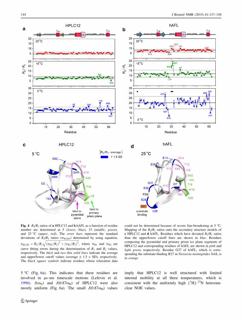
study also showed that all residues on the IBS of HPLC12

are rigid with high heteronuclear NOE values (Fig. 5) as

well as little deviations of the R2/R1 ratio from the average

value (Fig. 4). In the active QAE1-isoform of the type III

AFP, the surface pocket created by residues Q9, T18, V20,

and M21 traps the ice-like water that anchor the AFP-ice

interaction (Kumeta et al. 2013). The hydrogen-deuterium

exchange experiments revealed that these four residues in

the active QAE1-like isoform are rigid and capable of

forming the surface pocket, while the corresponding resi-

dues of the inactive QAE2-isoform cannot form the pocket

(Kumeta et al. 2013). Taken together, our NMR study

J Biomol NMR (2015) 61:137–150 147

123

support an ‘‘anchored clathrate mechanism’’ of the type III

AFP where the IBS exhibits rigid structure for efficient

hydrophobic and H-bonding interaction with ice crystal

and is capable of trapping ice-like water into surface pocket

that anchor the AFP-ice interaction.

Although HPLC12 has fairly uniform R1 and R2 values

at all three temperatures, some residues (I32, Q44, T53,

and M59) have significantly larger R2/R1 ratios than the

average value at 5 �C (Fig. 4a). As expected from R2/R1

ratios, these residues have significantly larger Je(0) values

than the average at 5 �C (Fig. 6a), which strongly support

the slow exchange of these residues on the ls–ms time-

scale. The internal motion of residue Q44 might be

required for efficient H-bonding of its side-chain with the

ice crystal. However, residues I32, T53, and M59 are far

from the IBS of HPLC12, indicating that the conforma-

tional exchange on the ls–ms timescale of these residues at

low temperature might not be crucial for interaction with

ice crystal.

At 25 �C, the R1 and R2 values of the mesophilic pro-

tein, hAFL, are uniform throughout and the {1H}-15N

heteronuclear NOEs[0.8 for most residues, indicating that

hAFL displays a rigid backbone structure at ambient tem-

perature. Interestingly, residues located at the loop region

(V26–Y32) between the b2 sheet and the second 310 helix

show significant flexibility and exchange in backbone

dynamics even at 25 �C evident from: i) large deviations in

R1 and R2 values (ls–ms timescale motion) and ii)

{1H}-15N heteronuclear NOEs B0.8 (ps–ns timescale

motion) (Figs. 3, 5). In contrast, the corresponding residues

of HPLC12 show no deviated R2/R1 ratios (Fig. 4a) as well

as heteronuclear NOE [0.8 (Fig. 5a) at 25 �C, indicating

this loop region of HPLC12 has rigid backbone structure.

In addition, residues I55–E58 also experience flexible

internal motion (see Figs. 4b, 5b). Among them, residues

V26, E28, and G31 are class-specific residues of the ver-

tebrate AFL domain (Hamada et al. 2006). The crystal

structure of an N. meningitides SAS found that the highly

conserved R314 (highlighted in orange as R27 in Fig. 1a)

protrudes to form direct of water-mediated H-bond with

ManNAc (Gunawan et al. 2005). The importance of R314

in the catalytic activity of SAS is supported by inhibition

study of Streptococcus agalactiae SAS through the addi-

tion of an arginine directed modifying reagent (Suryanti

et al. 2003). In the vertebrate AFL domains, this Arg res-

idue is missing from the ligand-binding site, but some

class-specific residues (K8 and K25 in Fig. 1a) with posi-

tively charged side-chains lie near the substrate (Hamada

et al. 2006). Interestingly, we found flexible regions clus-

tered around the corresponding residue in hAFL, G27

(Figs. 4d, 5d). Thus, it is reasonable to think that the

flexibility of these regions may contribute to the biological

function of the AFL domain in SAS. In contrast, the IBS of

the insect b-helical AFP exhibits rigid backbone structure

and the ice-binding Thr residues participate in intrastrand

H-bonds that stabilize the flat surface required for optimal

ice-binding (Daley et al. 2004). Instead, the Thr side-chains

on the IBS are flexible to adopt a preferred ice-binding

conformation without an entropic penalty (Daley and Sy-

kes 2003).

Conclusions

In this study, we have compared the dynamic properties of

the type III AFP (HPLC12 isoform) and the AFL domain of

human SAS (hAFL) by analyzing the temperature gradient

of the amide proton chemical shift, its correlation with

CSD from random coil, backbone dynamics, and reduced

spectral density. Our data show that the two homologous

proteins have distinctive backbone dynamics. Most resi-

dues of HPLC12 are rigid while a few residues on the IBS

experience conformational exchange on the ls–ms time-

scale at 5 �C. In contrast, hAFL is remarkably flexible at

5 �C, and its flexibility decreases at higher temperatures

(25 �C). At the same time, the residues that are involved in

substrate binding show fast internal motion. Based on our

study, we suggest that the dynamic properties of each

protein give insight into the basis for the different physi-

ological working temperatures of HPLC12 and hAFL. It is

very likely that each protein maintains a relatively rigid

backbone structure overall, but that the functionally

important residues exhibit distinctive dynamics for sub-

strate binding (rigid for ice-binding or flexible for sialic

acid binding).

Acknowledgments This work was supported by several National

Research Foundation of Korea (NRF) Grants funded by the Korean

Government (MSIP) [2010-0020480, 2013R1A2A2A05003837,

2012R1A4A1027750 (BRL)]. This work was also supported by a

Grant from the Next-Generation BioGreen 21 Program (SSAC,

No. PJ009041), Rural Development Administration, Korea. We thank

the GNU Central Instrument Facility for performing the NMR

experiments and Dr. Melissa Stauffer and Miss Laura Mizoue, of

Scientific Editing Solutions, for editing the manuscript.

References

Andersen NH, Neidigh JW, Harris SM et al (1997) Extracting

information from the temperature gradients of polypeptide NH

chemical shifts. 1. the importance of conformational averaging.

J Am Chem Soc 119:8547–8561

Antson AA, Smith DJ, Roper DI et al (2001) Understanding the

mechanism of ice binding by type III antifreeze proteins. J Mol

Biol 305:875–889

Baardsnes J, Davies PL (2002) Contribution of hydrophobic residues

to ice binding by fish type III antifreeze protein. Biochim

Biophys Acta 1601:49–54

148 J Biomol NMR (2015) 61:137–150

123

Bracken C, Carr PA, Cavanagh J, Palmer AG (1999) Temperature

dependence of intramolecular dynamics of the basic leucine

zipper of GCN4: implications for the entropy of association with

DNA. J Mol Biol 285:2133–2146

Brandsdal BO, Heimstad ES, Sylte I, Smalas AO (1999) Comparative

molecular dynamics of mesophilic and psychrophilic protein

homologues studied by 1.2 ns simulations. J Biomol Struct Dyn

17:493–506

Chao H, Sonnichsen FD, DeLuca CI et al (1994) Structure-function

relationship in the globular type III antifreeze protein: identifi-

cation of a cluster of surface residues required for binding to ice.

Protein Sci 3:1760–1769

Chen G, Jia Z (1999) Ice-binding surface of fish type III antifreeze.

Biophys J 77:1602–1608

Cierpicki T, Otlewski J (2001) Amide proton temperature coefficients

as hydrogen bond indicators in proteins. J Biomol NMR 21:

249–261

Cierpicki T, Zhukov I, Byrd RA, Otlewski J (2002) Hydrogen bonds

in human ubiquitin reflected in temperature coefficients of amide

protons. J Magn Reson 157:178–180

Daley ME, Sykes BD (2003) The role of side chain conformational

flexibility in surface recognition by Tenebrio molitor antifreeze

protein. Protein Sci 12:1323–1331

Daley ME, Spyracopoulos L, Jia Z et al (2002) Structure and

dynamics of a b-helical antifreeze protein. Biochemistry 41:

5515–5525

Daley ME, Graether SP, Sykes BD (2004) Hydrogen bonding on the

ice-binding face of a b-helical antifreeze protein indicated by

amide proton NMR chemical shifts. Biochemistry 43:13012–

13017

Davies PL, Baardsnes J, Kuiper MJ, Walker VK (2002) Structure and

function of antifreeze proteins. Philos Trans R Soc Lond B 357:

927–935

Delaglio F, Grzesiek S, Vuister GW et al (1995) NMRPipe: a

multidimensional spectral processing system based on UNIX

pipes. J Biomol NMR 6:277–293

DeLuca CI, Davies PL, Ye Q, Jia Z (1998) The effects of steric

mutations on the structure of type III antifreeze protein and its

interaction with ice. J Mol Biol 275:515–525

Ewart KV, Lin Q, Hew CL (1999) Structure, function and evolution

of antifreeze proteins. Cell Mol Life Sci 55:271–283

Fletcher GL, Hew CL, Davies PL (2001) Antifreeze proteins of

teleost fishes. Annu Rev Physiol 63:359–390

Garnham CP, Campbell RL, Davies PL (2011) Anchored clathrate

waters bind antifreeze proteins to ice. Proc Natl Acad Sci USA

108:7363–7367

Garnham CP, Nishimiya Y, Tsuda S et al (2012) Engineering a

naturally inactive isoform of type III antifreeze protein into one

that can stop the growth of ice. FEBS Lett 586:3876–3881

Gauthier SY, Scotter AJ, Lin FH et al (2008) A re-evaluation of the

role of type IV antifreeze protein. Cryobiology 57:292–296

Goddard TD, Kneller DG (2003) SPARKY 3. University of

California, San Francisco

Graether SP, Sykes BD (2004) Cold survival in freeze-tolerant

insects: structure and function of beta-helica proteins. Eur J

Biochem 271:3285–3296

Graether SP, DeLuca CI, Baardsnes J et al (1999) Quantitative and

qualitative analysis of type III antifreeze protein structure and

function. J Biol Chem 274:11842–11847

Graether SP, Gagne SM, Spyracopoulos L et al (2003) Spruce

budworm antifreeze protein: changes in structure and dynamics

at low temperature. J Mol Biol 327:1155–1168

Gunawan J, Simard D, Gilbert M et al (2005) Structural and

mechanistic analysis of sialic acid synthase NeuB from Neisseria

meningitidis in complex with Mn2?, phosphoenolpyruvate, and

N-acetylmannosaminitol. J Biol Chem 280:3555–3563

Hamada T, Ito Y, Abe T et al (2006) Solution structure of the

antifreeze-like domain of human sialic acid synthase. Protein Sci

15:1010–1016

Harding MM, Ward LG, Haymet AD (1999) Type I ‘antifreeze’

proteins. Structure-activity studies and mechanisms of ice

growth inhibition. Eur J Biochem 264:653–665

Hew CL, Wang NC, Joshi S et al (1988) Multiple genes provide the

basis for antifreeze protein diversity and dosage in the ocean

pout, Macrozoarces americanus. J Biol Chem 263:12049–12055

Hong J, Hu Y, Li C et al (2010) NMR characterizations of the ice

binding surface of an antifreeze protein. PLoS One 5:e15682

Jia Z, Davies PL (2002) Antifreeze proteins: an unusual receptor-

ligand interaction. Trends Biochem Sci 27:101–106

Jia Z, DeLuca CI, Chao H, Davies PL (1996) Structural basis for the

binding of a globular antifreeze protein to ice. Nature 384:

285–288

Kumeta H, Ogura K, Nishimiya Y et al (2013) NMR structure note: a

defective isoform and its activity-improved variant of a type III

antifreeze protein from Zoarces elongates Kner. J Biomol NMR

55:225–230

Kundu S, Roy D (2009) Comparative structural studies of psychro-

philic and mesophilic protein homologues by molecular dynam-

ics simulation. J Mol Graph Model 27:871–880

Lawrence SM, Huddleston KA, Pitts LR et al (2000) Cloning and

expression of the human N-acetylneuraminic acid phosphate

synthase gene with 2-keto-3-deoxy-D-glycero-D-galacto-nononic

acid biosynthetic ability. J Biol Chem 275:17869–17877

Lefevre JF, Dayie KT, Peng JW, Wagner G (1996) Internal mobility

in the partially folded DNA binding and dimerization domains of

GAL4: NMR analysis of the N-H spectral density functions.

Biochemistry 35:2674–2686

Liu Y, Li Z, Lin Q et al (2007) Structure and evolutionary origin of

Ca2?-dependent herring type II antifreeze protein. PLoS One 2:

e548

Masson L, Holbein BE (1983) Physiology of sialic acid capsular

polysaccharide synthesis in serogroup B Neisseria meningitidis.

J Bacteriol 154:728–736

Miura K, Ohgiya S, Hoshino T et al (2001) NMR analysis of type III

antifreeze protein intramolecular dimer. Structural basis for

enhanced activity. J Biol Chem 276:1304–1310

Nishimiya Y, Sato R, Takamichi M et al (2005) Co-operative effect of

the isoforms of type III antifreeze protein expressed in Notched-

fin eelpout, Zoarces elongatus Kner. FEBS J 272:482–492

Ohnishi M, Urry DW (1969) Temperature dependence of amide

proton chemical shifts: the secondary structures of gramicidin S

and valinomycin. Biochem Biophys Res Commun 36:194–202

Olufsen M, Brandsdal BO, Smalas AO (2007) Comparative unfolding

studies of psychrophilic and mesophilic uracil DNA glycosylase:

MD simulations show reduced thermal stability of the cold-

adapted enzyme. J Mol Graph Model 26:124–134

Papaleo E, Riccardi L, Villa C et al (2006) Flexibility and enzymatic

cold-adaptation: a comparative molecular dynamics investiga-

tion of the elastase family. Biochim Biophys Acta 1764:1397–

1406

Papaleo E, Olufsen M, De Gioia L, Brandsdal BO (2007) Optimi-

zation of electrostatics as a strategy for cold-adaptation: a case

study of cold- and warm-active elastases. J Mol Graph Model

26:93–103

Papaleo E, Pasi M, Riccardi L et al (2008) Protein flexibility in

psychrophilic and mesophilic trypsins. Evidence of evolutionary

conservation of protein dynamics in trypsin-like serine-prote-

ases. FEBS Lett 582:1008–1018

Schauer R (2004) Sialic acids: fascinating sugars in higher animals

and man. Zoology 107:49–64

Sonnichsen FD, DeLuca CI, Davies PL, Sykes BD (1996) Refined

solution structure of type III antifreeze protein: hydrophobic

J Biomol NMR (2015) 61:137–150 149

123

groups may be involved in the energetics of the protein–ice

interaction. Structure 4:1325–1337

Suryanti V, Nelson A, Berry A (2003) Cloning, over-expression,

purification, and characterisation of N-acetylneuraminate synthase

from Streptococcus agalactiae. Protein Expr Purif 27:346–356

Tjandra N, Feller SE, Pastor RW, Bax A (1995) Rotational diffusion

anisotropy of human ubiquitin from 15N NMR relaxation. J Am

Chem Soc 117:12562–12566

Yang DSC, Hon WC, Bubanko S et al (1998) Identification of the ice-

binding surface on a type III antifreeze protein with a ‘‘flatness

function’’ algorithm. Biophys J 74:2142–2151

Yeh Y, Feeney RE (1996) Antifreeze proteins: structures and

mechanisms of function. Chem Rev 96:601–618

150 J Biomol NMR (2015) 61:137–150

123


Recommended