Ann Lab Med 2017;37:267-271https://doi.org/10.3343/alm.2017.37.3.267
Brief CommunicationClinical Microbiology
Comparison of Luminex NxTAG Respiratory Pathogen Panel and xTAG Respiratory Viral Panel FAST Version 2 for the Detection of Respiratory VirusesChun Kiat Lee, M.S.1, Hong Kai Lee, Ph.D.1, Christopher Wei Siong Ng, B.S.1, Lily Chiu, M.S.1, Julian Wei-Tze Tang, M.D.2,3, Tze Ping Loh, M.D.1, and Evelyn Siew-Chuan Koay, Ph.D.1,4
Department of Laboratory Medicine1, National University Hospital, Singapore; Leicester Royal Infirmary2, University Hospitals of Leicester NHS Trust, Leicester, United Kingdom; Department of Infection, Immunity, Inflammation3, University of Leicester, Leicester, United Kingdom; Department of Pathology4, Yong Loo Lin School of Medicine, National University of Singapore, Singapore
Owing to advancements in molecular diagnostics, recent years have seen an increasing number of laboratories adopting respiratory viral panels to detect respiratory pathogens. In December 2015, the NxTAG respiratory pathogen panel (NxTAG RPP) was approved by the United States Food and Drug Administration. We compared the clinical performance of this new assay with that of the xTAG respiratory viral panel (xTAG RVP) FAST v2 using 142 clinical samples and 12 external quality assessment samples. Discordant results were resolved by using a laboratory-developed respiratory viral panel. The NxTAG RPP achieved 100% concordant negative results and 86.6% concordant positive results. It detected one coronavirus 229E and eight influenza A/H3N2 viruses that were missed by the xTAG RVP FAST v2. On the other hand, the NxTAG RPP missed one enterovirus/rhinovirus and one metapneumovirus that were detected by FAST v2. Both panels correctly identified all the pathogens in the 12 external quality assessment samples. Overall, the NxTAG RPP dem-onstrated good diagnostic performance. Of note, it was better able to subtype the influ-enza A/H3N2 viruses compared with the xTAG RVP FAST v2.
Received: August 10, 2016Revision received: October 27, 2016Accepted: January 23, 2017
Corresponding author: Evelyn Siew-Chuan KoayDepartment of Pathology, Yong Loo Lin School of Medicine, National University of Singapore, 21, Lower Kent Ridge Road, 119077, SingaporeTel: +65-6772-4564 Fax: +65-6772-4407 E-mail: [email protected]
This study was approved by the local institutional ethics board
(National Healthcare Group Domain-Specific Review Board A,
reference: 2016/00044) and was performed between May and
December 2015. Here, 142 de-identified clinical respiratory sam-
ples submitted to the Molecular Diagnosis Centre of the Singa-
pore National University Hospital were included (see Table 1 for
the list of viral pathogens included). Additionally, 12 external
quality assessment (EQA) samples from the College of Ameri-
can Pathologists (CAP) infectious disease respiratory panel, re-
ceived in year 2015, were tested (Table 2). Total nucleic acid
was extracted with the Qiagen EZ1 Virus Mini Kit v2.0 on the
BioRobot EZ1 extractor (Qiagen, Hilden, Germany).
All samples were initially tested with the xTAG RVP FAST v2
as part of our routine clinical service. In brief, the extracted nu-
cleic acid (10 µL) was used for target amplification by multiplex
Table 1. Summary of the performance of the NxTAG respiratory pathogen panel (NxTAG RPP) and the xTAG respiratory viral panel (xTAG RVP) FAST v2 for the detection of viral pathogens in 142 clinical samples
Viral targets
Number of samples with the following result Assay performance with the true-positive result*
*When NxTAG RPP and xTAG RVP FAST v2 results were discordant, a laboratory-developed respiratory viral panel was applied to the sample. A true-positive result was defined as one agreed by any two of the three assays.Abbreviations: CI, confidence interval; NA, not applicable; NxTAG RPP, NxTAG respiratory pathogen panel; xTAG RVP FAST v2, xTAG respiratory viral panel FAST v2; LDT, laboratory-developed test.
Table 2. Summary of the 12 College of American Pathologists 2015 external quality assessment samples used in the study
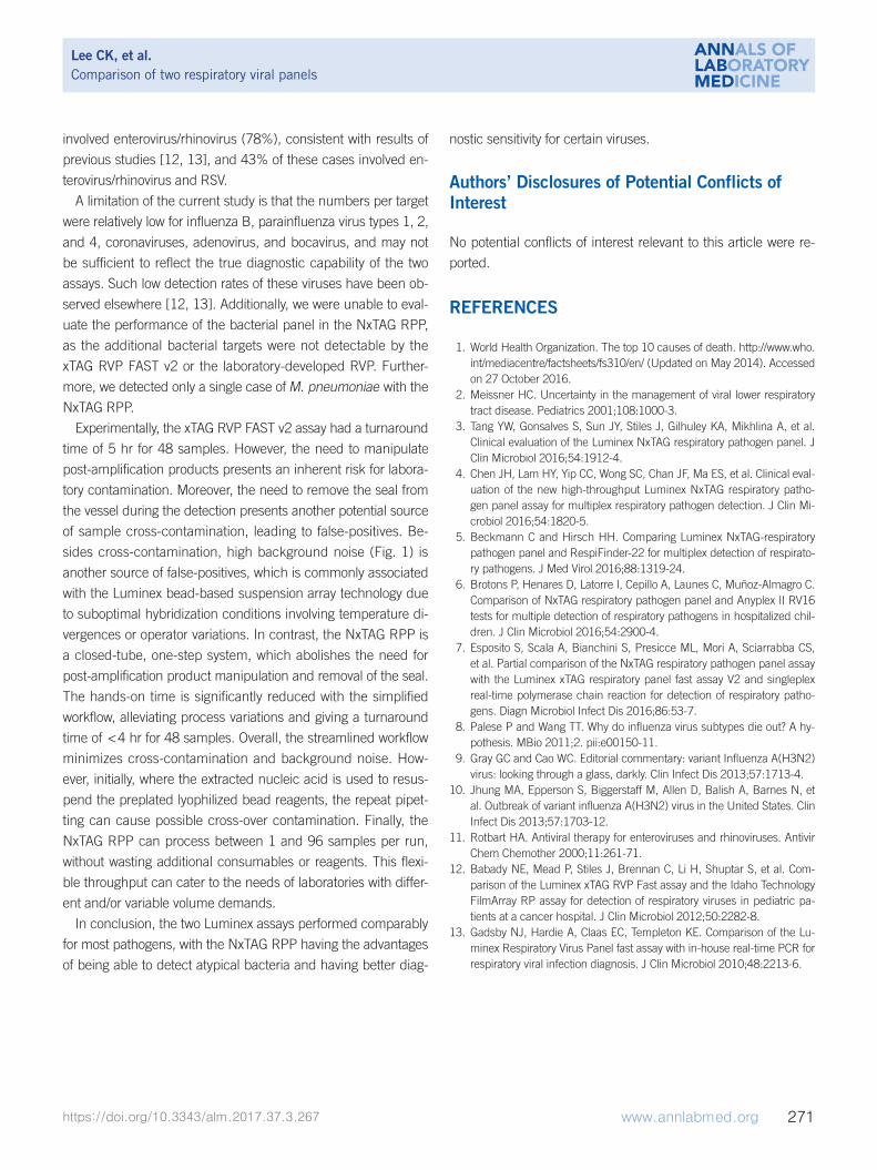
Fig. 1. High background noise observed with the Luminex bead hybridization technology in a run. (A) Sample A initially tested positive for coronavirus HKU1 with the xTAG respiratory viral panel (RVP) FAST v2 (top left). Of note, the internal control signal intensity was higher than that in previous runs. After repeating the bead hybridization step, sample A was negative for all viral targets (false-positive) and the internal control signal intensity was within the expected range (bottom left). (B) Sample B initially tested positive for seasonal influenza A/H1N1 vi-rus, influenza A/H1N1/2009 virus, and enterovirus/rhinovirus (top right). Again, the internal control signal intensity was higher than that in previous runs. After repeating the bead hybridization step, seasonal influenza A/H1N1 virus signal was found to be negative (false-positive), and the internal control signal intensity was within the expected range (bottom right). Subsequent investigation revealed that the high back-ground is likely due to operator variations.Abbreviations: Corona, coronavirus; RSV, respiratory syncytial virus; Para, parainfluenza virus; MFI, median fluorescence intensity.
A B
Lee CK, et al.Comparison of two respiratory viral panels
involved enterovirus/rhinovirus (78%), consistent with results of
previous studies [12, 13], and 43% of these cases involved en-
terovirus/rhinovirus and RSV.
A limitation of the current study is that the numbers per target
were relatively low for influenza B, parainfluenza virus types 1, 2,
and 4, coronaviruses, adenovirus, and bocavirus, and may not
be sufficient to reflect the true diagnostic capability of the two
assays. Such low detection rates of these viruses have been ob-
served elsewhere [12, 13]. Additionally, we were unable to eval-
uate the performance of the bacterial panel in the NxTAG RPP,
as the additional bacterial targets were not detectable by the
xTAG RVP FAST v2 or the laboratory-developed RVP. Further-
more, we detected only a single case of M. pneumoniae with the
NxTAG RPP.
Experimentally, the xTAG RVP FAST v2 assay had a turnaround
time of 5 hr for 48 samples. However, the need to manipulate
post-amplification products presents an inherent risk for labora-
tory contamination. Moreover, the need to remove the seal from
the vessel during the detection presents another potential source
of sample cross-contamination, leading to false-positives. Be-
sides cross-contamination, high background noise (Fig. 1) is
another source of false-positives, which is commonly associated
with the Luminex bead-based suspension array technology due
to suboptimal hybridization conditions involving temperature di-
vergences or operator variations. In contrast, the NxTAG RPP is
a closed-tube, one-step system, which abolishes the need for
post-amplification product manipulation and removal of the seal.
The hands-on time is significantly reduced with the simplified
workflow, alleviating process variations and giving a turnaround
time of <4 hr for 48 samples. Overall, the streamlined workflow
minimizes cross-contamination and background noise. How-
ever, initially, where the extracted nucleic acid is used to resus-
pend the preplated lyophilized bead reagents, the repeat pipet-
ting can cause possible cross-over contamination. Finally, the
NxTAG RPP can process between 1 and 96 samples per run,
without wasting additional consumables or reagents. This flexi-
ble throughput can cater to the needs of laboratories with differ-
ent and/or variable volume demands.
In conclusion, the two Luminex assays performed comparably
for most pathogens, with the NxTAG RPP having the advantages
of being able to detect atypical bacteria and having better diag-
nostic sensitivity for certain viruses.
Authors’ Disclosures of Potential Conflicts of Interest
No potential conflicts of interest relevant to this article were re-
ported.
REFERENCES
1. World Health Organization. The top 10 causes of death. http://www.who. int/mediacentre/factsheets/fs310/en/ (Updated on May 2014). Accessed on 27 October 2016.
2. Meissner HC. Uncertainty in the management of viral lower respiratory tract disease. Pediatrics 2001;108:1000-3.
3. Tang YW, Gonsalves S, Sun JY, Stiles J, Gilhuley KA, Mikhlina A, et al. Clinical evaluation of the Luminex NxTAG respiratory pathogen panel. J Clin Microbiol 2016;54:1912-4.
4. Chen JH, Lam HY, Yip CC, Wong SC, Chan JF, Ma ES, et al. Clinical eval-uation of the new high-throughput Luminex NxTAG respiratory patho-gen panel assay for multiplex respiratory pathogen detection. J Clin Mi-crobiol 2016;54:1820-5.
5. Beckmann C and Hirsch HH. Comparing Luminex NxTAG-respiratory pathogen panel and RespiFinder-22 for multiplex detection of respirato-ry pathogens. J Med Virol 2016;88:1319-24.
6. Brotons P, Henares D, Latorre I, Cepillo A, Launes C, Muñoz-Almagro C. Comparison of NxTAG respiratory pathogen panel and Anyplex II RV16 tests for multiple detection of respiratory pathogens in hospitalized chil-dren. J Clin Microbiol 2016;54:2900-4.
7. Esposito S, Scala A, Bianchini S, Presicce ML, Mori A, Sciarrabba CS, et al. Partial comparison of the NxTAG respiratory pathogen panel assay with the Luminex xTAG respiratory panel fast assay V2 and singleplex real-time polymerase chain reaction for detection of respiratory patho-gens. Diagn Microbiol Infect Dis 2016;86:53-7.
8. Palese P and Wang TT. Why do influenza virus subtypes die out? A hy-pothesis. MBio 2011;2. pii:e00150-11.
9. Gray GC and Cao WC. Editorial commentary: variant Influenza A(H3N2) virus: looking through a glass, darkly. Clin Infect Dis 2013;57:1713-4.
10. Jhung MA, Epperson S, Biggerstaff M, Allen D, Balish A, Barnes N, et al. Outbreak of variant influenza A(H3N2) virus in the United States. Clin Infect Dis 2013;57:1703-12.
11. Rotbart HA. Antiviral therapy for enteroviruses and rhinoviruses. Antivir Chem Chemother 2000;11:261-71.
12. Babady NE, Mead P, Stiles J, Brennan C, Li H, Shuptar S, et al. Com-parison of the Luminex xTAG RVP Fast assay and the Idaho Technology FilmArray RP assay for detection of respiratory viruses in pediatric pa-tients at a cancer hospital. J Clin Microbiol 2012;50:2282-8.
13. Gadsby NJ, Hardie A, Claas EC, Templeton KE. Comparison of the Lu-minex Respiratory Virus Panel fast assay with in-house real-time PCR for respiratory viral infection diagnosis. J Clin Microbiol 2010;48:2213-6.