Page 1
REVIEW ARTICLE
Complementary regulation of early B-lymphoid differentiationby genetic and epigenetic mechanisms
Takafumi Yokota • Takao Sudo • Tomohiko Ishibashi •
Yukiko Doi • Michiko Ichii • Kenji Orirani •
Yuzuru Kanakura
Received: 4 June 2013 / Revised: 21 August 2013 / Accepted: 23 August 2013 / Published online: 3 September 2013
� The Japanese Society of Hematology 2013
Abstract Although B lymphopoiesis is one of the best-
defined paradigms in cell differentiation, our knowledge
of the regulatory mechanisms underlying its earliest pro-
cesses, in which hematopoietic stem cells (HSCs) enter
the B lineage, is limited. However, recent methodological
advances in sorting progenitor cells and monitoring their
epigenetic features have increased our understanding of
HSC activities. It is now known that even the highly
enriched HSC fraction is heterogeneous in terms of lym-
phopoietic potential. While surface markers and reporter
proteins provide information on the sequential differenti-
ation of B-lineage progenitors, complex interactions
between transcription factors have also been shown to
play a major role in this process. Epigenetic regulation of
histones, nucleosomes, and chromatin appears to play a
crucial background role in this elaborate transcription
network. In this review, we summarize recent findings on
the physiological processes of early B-lineage differenti-
ation, which provides a new paradigm for understanding
the harmonious action of genetic and epigenetic
mechanisms.
Keywords Hematopoietic stem cells � Early B
lymphopoiesis � Developmental pathway �Transcription factors � Epigenetic regulation
Introduction
B lymphocytes are continuously replenished throughout
life, although their output ratios are influenced by infection
or age. Many studies have reported that their early differ-
entiation stages from hematopoietic stem cells (HSCs) are
largely influenced by the abovementioned specific cir-
cumstances [1, 2]. However, molecular mechanisms
underlying the age-related declines or the infection-related
changes in B lymphopoiesis remain elusive. If key mole-
cules responsible for the changes in early B-lymphoid
differentiation could be identified, manipulation of their
expression may facilitate in rejuvenating or activating the
immune system of elderly people and decrease the inci-
dence and/or impact of infections. The aim of this review is
to summarize recent findings in this field and to improve
our understanding of molecular mechanisms that regulate
the earliest steps involving B-lineage differentiation.
Developmental pathway of early B-lineage progenitors
Models on the relationships of B- and other lineage pro-
genitors have influenced the interpretations of experimental
results on changes in hematopoietic differentiation.
Extensive information has been gathered from studies on
surface molecules during early B-lineage differentiation. In
combination with genetically modified mice that show
changes in the expression of early B-lineage-related genes,
the developmental process of B-lineage cells has been
extensively studied.
An early study conducted by Hardy’s group fractionated
B-lineage cells from mouse bone marrow (BM) into
Fractions A to F based on the surface markers, CD45RA/
B220, CD43, heat-stable antigen, and BP1 [3]. Addition of
T. Yokota (&) � T. Sudo � T. Ishibashi � Y. Doi � M. Ichii �K. Orirani � Y. Kanakura
Department of Hematology and Oncology,
Osaka University Graduate School of Medicine,
2-2 Yamada-oka, Suita, Osaka 565-0871, Japan
e-mail: [email protected]
123
Int J Hematol (2013) 98:382–389
DOI 10.1007/s12185-013-1424-7
Page 2
AA4.1 and CD4 further sub-fractionated the early steps of
B-lineage differentiation in Fraction A [4], although later
studies showed that the earliest fraction, A, contained
progenitors for other lineages [5, 6]. Another study con-
ducted at the Basel Institute proposed a differentiation
scheme of the BM B-lineage cells based on immunoglob-
ulin gene rearrangements [7]. While those methods were
extremely useful in analyzing the molecular mechanisms
that precede B-lineage differentiation of committed pro-
genitors, investigations on earlier B-lineage differentiation
processes required an innovative method to discriminate
lineage-biased progenitors prior to cellular commitment.
Application of IL-7 receptor alpha (IL-7Ra) expression as
an early lymphoid-related marker brought about substantial
development. It was originally used by Kondo et al. [8] to
differentiate common lymphoid progenitors (CLPs) in mouse
BM. The definition of CLPs was based on the concept of a
binary fate decision in which B, T, and NK lymphoid cells
diverge from all other lineages, including myeloid, erythroid,
and megakaryocyte lineages. The IL-7Ra? cells in Lin-
c-KitLo Sca1? fraction met the criterion in terms of their
differentiation potential in culture and in the thymus [8].
While subsequent studies have shown that the divergence of
erythroid-megakaryocyte lineage occurred prior to that of
myeloid from lymphoid lineage [9] and that the separation
between myeloid and lymphoid potentials is rather gradual
[10], the expression of IL-7Ra has been consistently used to
study early events in B-lineage differentiation.
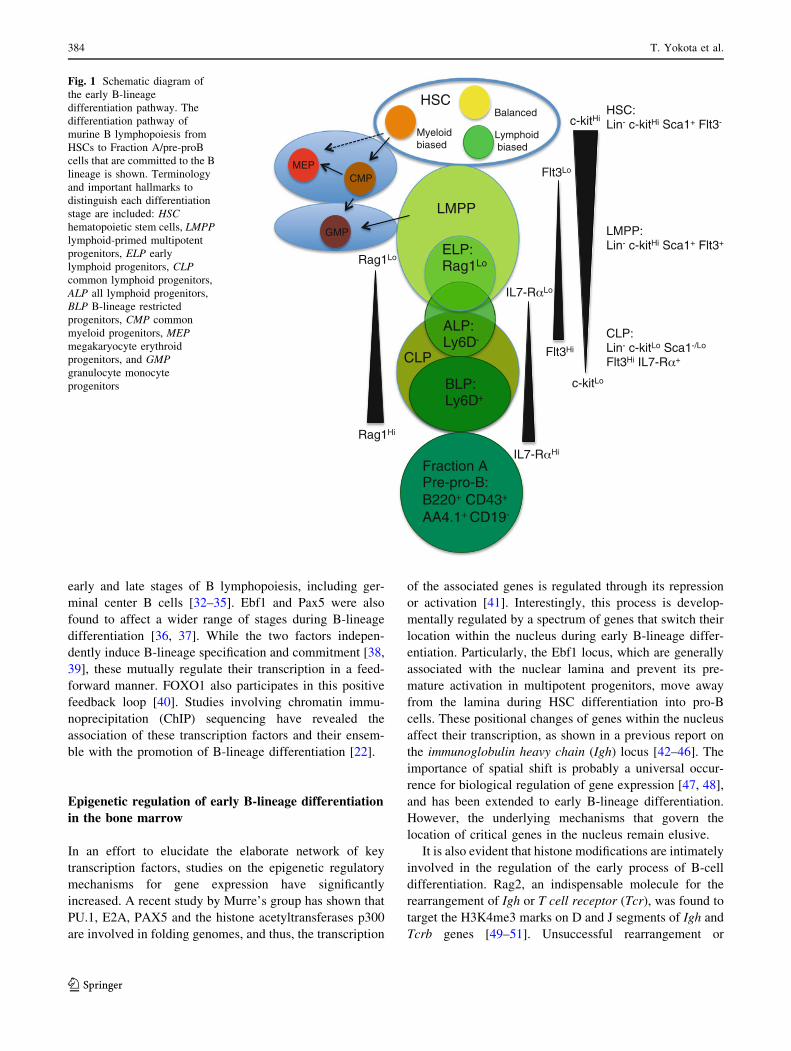
Recent improvements in immunodetection assays using
more sensitive antibodies and laser/fluorochrome/filter
combinations have enabled monitoring of IL-7Ra expres-
sion in c-KitHi cells [11]. In the same report, expression of
Ly6D was used to differentiate ‘‘all lymphoid progenitors
(ALPs)’’ and B-lineage restricted progenitors (named as
BLPs) among the IL-7Ra? progenitors, including the ori-
ginal CLPs. The observation supports our original claim
based on the data with Rag1-GFP knock-in mice, in which
the initiation of lymphoid-lineage differentiation occurs in
the Lin- c-KitHi Sca1? CD27? fraction, i.e., occurring in
more proximity to HSCs [12, 13]. This notion also corre-
sponds to a report demonstrating that an increased Flt3
expression is a characteristic of lymphoid-primed multi-
potent progenitors (LMPPs) present in the Lin- c-KitHi
Sca1? fraction [9]. Rag1-GFP? early lymphoid progenitors
(ELPs) are found in the Lin- c-KitHi Sca1? Flt3Hi fraction
[6, 14], suggesting the close and sequential relationship
between LMPPs and ELPs. Of note, those progenitors are
exclusively sensitive to steroid hormones among Lin-
c-KitHi Sca1? cells [14–16], whereas these generate B
lymphocytes in culture with slower kinetics than Lin-
c-KitLo Sca1Lo IL-7Ra? cells [17]. These evidences thus
suggest that an early differentiation pathway in B lineage is
likely to proceed sequentially from HSCs to BLPs (Fig. 1).
Network of transcription factors and cytokine signaling
Along with the recent progress in dissecting the differen-
tiation pathways of early B-lymphoid differentiation,
information on the role of transcription factors in regulat-
ing early stages has also increased. Five transcription fac-
tors namely, PU.1, Ikaros, E2A/E47, Ebf1, and Pax5 have
been traditionally recognized as essential regulators of
early B-lineage differentiation [18, 19]. Transcription fac-
tors PU.1, E2A, and Ikaros are expressed in primitive
progenitors and are involved in multiple lineage fate
determination, whereas Ebf1 and Pax5 regulate B-lineage-
specified progenitors.
Recent studies have identified the FOXO family as
crucial component in early B-lineage differentiation [20,
21]. The FOXO family enhances the expression of the IL7-
receptor, which transmits intracellular signals that result in
the inactivation of FOXO1. The pathway suggests the
involvement of an auto-regulatory loop for B-lineage
determination. Since FOXO1 is a direct target of E2A and
Ebf1, the family might act downstream of the two indis-
pensable factors of B-lineage differentiation [22, 23]. Early
B lymphopoiesis also depends on the function of c-Myb,
which acts upstream of Ebf1 and regulates the survival and
differentiation of pro-B cells [24, 25]. While c-Myb regu-
lates the expression of the IL7-receptor, the molecule also
affects pro-B cell survival independent of the IL7-receptor
signaling pathway.
Runx1 is a well-known critical factor in the develop-
ment of authentic HSCs. In addition to its role in hema-
topoiesis, a recent study by Taniuchi’s group has
demonstrated that loss of Runx1 in the mb1-cre conditional
knockout system causes a significant developmental block
in early B-lineage differentiation [26]. Expression of E2A,
Ebf1, and Pax5 was reduced in the Runx1-deficient pro-
genitors, indicating that the factor probably plays a role in
the early stages of B lymphopoiesis.
Based on the evidences on upstream or downstream
components of the signaling pathway, it is clear that the
sequential action of critical transcription factors plays an
important role in the early path of B-lineage differentiation.
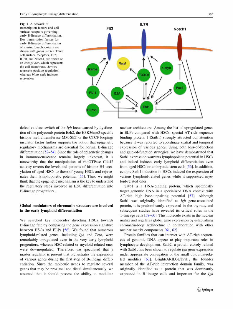
However, recent studies have also revealed that each
transcription factor affects a broader range of B-lineage
cells than previously expected. Furthermore, the regulatory
mechanisms of transcription factors are not influenced by a
simple hierarchy but by a cross-regulatory network (Fig. 2)
[27–29]. Ikaros plays an important role in establishing the
prime lymphoid-lineage fate of HSCs and this factor
associates with other B-lineage-related transcription factors
and upregulates various genes involved in the commitment
stage, in which CLPs differentiate into proB cells [30].
E2A induces T-lineage-related genes to act in concert with
Notch1 signaling [31], as well as plays critical roles in both
Early B-lymphocyte lineage differentiation 383
123
Page 3
early and late stages of B lymphopoiesis, including ger-
minal center B cells [32–35]. Ebf1 and Pax5 were also
found to affect a wider range of stages during B-lineage
differentiation [36, 37]. While the two factors indepen-
dently induce B-lineage specification and commitment [38,
39], these mutually regulate their transcription in a feed-
forward manner. FOXO1 also participates in this positive
feedback loop [40]. Studies involving chromatin immu-
noprecipitation (ChIP) sequencing have revealed the
association of these transcription factors and their ensem-
ble with the promotion of B-lineage differentiation [22].
Epigenetic regulation of early B-lineage differentiation
in the bone marrow
In an effort to elucidate the elaborate network of key
transcription factors, studies on the epigenetic regulatory
mechanisms for gene expression have significantly
increased. A recent study by Murre’s group has shown that
PU.1, E2A, PAX5 and the histone acetyltransferases p300
are involved in folding genomes, and thus, the transcription
of the associated genes is regulated through its repression
or activation [41]. Interestingly, this process is develop-
mentally regulated by a spectrum of genes that switch their
location within the nucleus during early B-lineage differ-
entiation. Particularly, the Ebf1 locus, which are generally
associated with the nuclear lamina and prevent its pre-
mature activation in multipotent progenitors, move away
from the lamina during HSC differentiation into pro-B
cells. These positional changes of genes within the nucleus
affect their transcription, as shown in a previous report on
the immunoglobulin heavy chain (Igh) locus [42–46]. The
importance of spatial shift is probably a universal occur-
rence for biological regulation of gene expression [47, 48],
and has been extended to early B-lineage differentiation.
However, the underlying mechanisms that govern the
location of critical genes in the nucleus remain elusive.
It is also evident that histone modifications are intimately
involved in the regulation of the early process of B-cell
differentiation. Rag2, an indispensable molecule for the
rearrangement of Igh or T cell receptor (Tcr), was found to
target the H3K4me3 marks on D and J segments of Igh and
Tcrb genes [49–51]. Unsuccessful rearrangement or
HSC:Lin- c-kitHi Sca1+ Flt3-
Balanced
Lymphoid biased
Myeloid biased
CMPMEP
GMP LMPP:Lin- c-kitHi Sca1+ Flt3+
Flt3Lo
Flt3Hi
c-kitLo
c-kitHi
IL7-R Lo
IL7-R Hi
CLP:Lin- c-kitLo Sca1-/Lo
Flt3Hi IL7-R +
ALP:Ly6D-
BLP:Ly6D+
ELP:Rag1LoRag1Lo
Rag1Hi
HSC
LMPP
CLP
Fraction A Pre-pro-B:B220+ CD43+ AA4.1+ CD19-
Fig. 1 Schematic diagram of
the early B-lineage
differentiation pathway. The
differentiation pathway of
murine B lymphopoiesis from
HSCs to Fraction A/pre-proB
cells that are committed to the B
lineage is shown. Terminology
and important hallmarks to
distinguish each differentiation
stage are included: HSC
hematopoietic stem cells, LMPP
lymphoid-primed multipotent
progenitors, ELP early
lymphoid progenitors, CLP
common lymphoid progenitors,
ALP all lymphoid progenitors,
BLP B-lineage restricted
progenitors, CMP common
myeloid progenitors, MEP
megakaryocyte erythroid
progenitors, and GMP
granulocyte monocyte
progenitors
384 T. Yokota et al.
123
Page 4
defective class switch of the Igh locus caused by dysfunc-
tion of the polycomb protein Ezh2, the H3K36me3-specific
histone methyltransferase MM-SET or the CTCF looping/
insulator factor further supports the notion that epigenetic
regulatory mechanisms are essential for normal B-lineage
differentiation [52–54]. Since the role of epigenetic changes
in immunosenescence remains largely unknown, it is
noteworthy that the manipulation of rhoGTPase Cdc42
activity reverts the levels and patterns of histone H4 acet-
ylation of aged HSCs to those of young HSCs and rejuve-
nates their lymphopoietic potential [55]. Thus, we might
think that the epigenetic mechanism is the key to understand
the regulatory steps involved in HSC differentiation into
B-lineage progenitors.
Global modulators of chromatin structure are involved
in the early lymphoid differentiation
We searched key molecules directing HSCs towards
B-lineage fate by comparing the gene expression signature
between HSCs and ELPs [56]. We found that numerous
lymphoid-related genes, including Igh and Tcrb, were
remarkably upregulated even in the very early lymphoid
progenitors, whereas HSC-related or myeloid-related ones
were downregulated. Therefore, we speculated that a
master regulator is present that orchestrates the expression
of various genes during the first step of B-lineage differ-
entiation. Since the molecule needs to regulate several
genes that may be proximal and distal simultaneously, we
assumed that it should possess the ability to modulate
nuclear architecture. Among the list of upregulated genes
in ELPs compared with HSCs, special AT-rich sequence
binding protein 1 (Satb1) strongly attracted our attention
because it was reported to coordinate spatial and temporal
expression of various genes. Using both loss-of-function
and gain-of-function strategies, we have demonstrated that
Satb1 expression warrants lymphopoietic potential in HSCs
and indeed induces early lymphoid differentiation even
from aged HSCs or embryonic stem cells [56]. In addition,
ectopic Satb1 induction in HSCs induced the expression of
various lymphoid-related genes while it suppressed mye-
loid-related ones.
Satb1 is a DNA-binding protein, which specifically
target genomic DNA in a specialized DNA context with
AT-rich high base-unpairing potential [57]. Although
Satb1 was originally identified as Igh gene-associated
protein, it is predominantly expressed in the thymus, and
subsequent studies have revealed its critical roles in the
T-lineage cells [58–60]. This molecule exists in the nuclear
matrix and regulates global gene expression by establishing
chromatin-loop architecture in collaboration with other
nuclear matrix components [61, 62].
Protein families that can interact with AT-rich sequen-
ces of genomic DNA appear to play important roles in
lymphocyte development. Satb2, a protein closely related
with Satb1, has been shown to regulate Igh gene expression
under appropriate conjugation of the small ubiquitin-rela-
ted modifier [63]. Bright/ARID3a/Dril1, the founder
member of the AT-rich interaction domain family, was
originally identified as a protein that was dominantly
expressed in B-lineage cells and important for the Igh
PU.1
Ikaros
E2A
Ebf1
Pax5
Runx1
FOXO1
Flt3 Notch1IL7R
Rag1c-Myb
Fig. 2 A network of
transcription factors and cell
surface receptors governing
early B-lineage differentiation.
Key transcription factors for
early B-lineage differentiation
of murine lymphopoiesis are
shown with green circles. Three
cell surface receptors, Flt3,
IL7R, and Notch1, are drawn on
an orange bar, which represents
the cell membrane. Arrows
represent positive regulation,
whereas blunt ends indicate
repression
Early B-lymphocyte lineage differentiation 385
123
Page 5
expression [64]. Subsequent studies have revealed that the
protein is expressed in very early B-lineage progenitors,
even HSCs, and is indispensable for normal cellular
development [65–67]. A recent paper has shown that
appropriate Satb1 expression in HSCs is critical for the
maintenance of cellular integrity [68]. Determining whe-
ther AT-rich sequence binding proteins interact in primi-
tive progenitors or independently play roles in lineage- and
stage-specific manners is, therefore, a very interesting
future research theme.
Complementary relationship between genetic
and epigenetic regulation
From the accumulated evidences mentioned, it is highly
likely that genetic and epigenetic regulations are comple-
mentarily involved in the early process of B-lineage dif-
ferentiation. As illustrated in Fig. 3, nuclear matrix
proteins such as Satb1 and histone modification seem to
construct a background frame for the expression of tran-
scription factors, which then work together on the tran-
scription of various lymphoid lineage-related genes.
Previous studies have employed the term ‘‘promiscu-
ous’’ in describing the expression of lineage-related genes
in hematopoietic progenitors and have collectively coined
the entire phenomenon as ‘‘lineage-priming’’ [69, 70].
Multipotent progenitors are generally capable of differen-
tiating into any of the possible lineage options and are
induced to follow a specific differentiation pathway based
on the organism’s physiological demand. Thus, the priming
process appears to exist inherently in those cells. However,
a recent investigation has otherwise observed that the
priming expression of lineage-related genes follows a
systemic approach, thus reflecting a reversible fluctuation
of transcriptome activities [71]. Novershtern et al. [72]
have subjected human HSCs to extensive gene arrays and
bioinformatics analyses. They have confirmed the antici-
patory loading of promoters of lineage-related genes even
with primitive HSCs, corresponding to the notion of
‘‘lineage-priming.’’ Furthermore, they have revealed that
such anticipatory loading of promoters becomes simple
when it simultaneously occurs with lineage specification.
Since both chromatin architecture and transcription factors
are important for multiple gene expression, epigenetic and
genetic regulations should be complementarily involved in
such lineage and stage-specific loading of promoters.
Epigenetic abnormality involved in early B-lineage
neoplasms
Recent revolutionary development in the DNA sequencing
technology has enabled us to reach comprehensive under-
standing of genome-wide abnormality in cancers [73, 74]. A
variety of novel gene mutations have been revealed in
hematopoietic malignancies. In particular, mutations of epi-
genetic regulators and RNA splicing factors have been
involved in the pathogenesis of myeloid-lineage neoplasms
including acute myeloid leukemia and myelodysplastic syn-
drome [75–77]. Compared with those advances in myeloid-
lineage neoplasms, the information of epigenetic abnormality
in lymphoid leukemia seems to be limited. However, there are
some important findings to be introduced.
Mullighan et al. [78] performed genome-wide analyses
for 242 pediatric acute lymphoid leukemia (ALL) patients.
They have identified high mutation frequencies of genes
encoding regulators for early B-cell lineage development in
acute lymphoblastic leukemia. Indeed, 40 % of pediatric
B-cell lineage ALL cases harbored mutations in the prin-
cipal regulators including PAX5, E2A/E47, EBF and I-
KAROS. Among them, mutations of IKZF1, a gene
Nucleus
Heterochromatin Euchromatin Nuclear matrix proteins
Chromatin loopTranscription factor
Transcription factor
Fig. 3 A proposed model of
Satb1 function on the chromatin
loop structure. Left a cartoon
showing the simplified nuclear
architecture consisting of
chromatin and the nuclear
matrix. Satb1 protein is present
in the nuclear matrix and binds
to the AT-rich DNA sequences
attached to nuclear matrix.
Right a schematic diagram of
the putative mechanism of
nuclear matrix proteins such as
Satb1 in regulating multiple
gene expressions through the
construction of chromatin loop
structures and transcriptional
complexes
386 T. Yokota et al.
123
Page 6
encoding IKAROS protein, have been drawing attention
because they are found to relate with a high relapse rate
and a poor prognosis [79]. Two recent studies have sug-
gested that the IKZF1 mutations are more involved in adult
rather than in pediatric B-ALL [80, 81]. Furthermore, the
IKZF1 gene abnormality is very frequent and severe in
BCR-ABL positive cases [82–84]. It is important to stress
here that, besides the essential role as a transcription factor
in B-lymphocyte differentiation, an IKAROS protein
serves as an integral component of chromatin-remodeling
networks and prevents leukemogenesis [85, 86]. Thus, the
loss of IKAROS function is assumed to cause aberrant
association between genetic and epigenetic networks,
thereby mediate high susceptibility to aggressive leukemia.
Future direction and concluding remarks
To date, it has become evident that the roles of transcrip-
tion factors are essential for physiological B lymphopoie-
sis. Substantial progress has been recently made in
understanding how epigenetic regulation has influenced
this process. More precise information of the comple-
mentary relation between the genetic and epigenetic
mechanisms may potentially boost lymphocyte production
after chemotherapy and stem cell transplantation in the
future. Environmental cues regulating the two mechanisms
during the early stage of B-lineage differentiation remain
elusive. Understanding the physiological process of early
differentiation is essential to appreciate when and how
aberrant incidences occur in cancer. Future research studies
can shed light on epigenetic problems associated with
malignant transformation.
Acknowledgments We would like to thank Dr. Noriko Saitoh
(Kumamoto University) for providing a figure of the cell nucleus and
Dr. Kiyoe Ura (Osaka University) for discussions and for critical
reading of this manuscript. We also thank Dr. Yusuke Satoh (Kobe
Shoin Women’s University) for the effort in Satb1-related
experiments.
Conflict of interest The authors declare that they have no conflict
of interest.
References
1. Allman D, Miller JP. The aging of early B-cell precursors.
Immunol Rev. 2005;205:18–29.
2. Zhang Q, Iida R, Shimazu T, Kincade PW. Replenishing B
lymphocytes in health and disease. Curr Opin Immunol.
2012;24:196–203.
3. Hardy RR, Carmack CE, Shinton SA, Kemp JD, Hayakawa K.
Resolution and characterization of Pro-B and Pre-Pro-B cell
stages in normal bone marrow. J Exp Med. 1991;173:1213–25.
4. Li Y-S, Wasserman R, Hayakawa K, Hardy RR. Identification of
the earliest B lineage stage in mouse bone marrow. Immunity.
1996;5:527–35.
5. Tudor KS, Payne KJ, Yamashita Y, Kincade PW. Functional
assessment of precursors from murine bone marrow suggests a
sequence of early B lineage differentiation events. Immunity.
2000;12:335–45.
6. Mansson R, Zandi S, Welinder E, et al. Single-cell analysis of the
common lymphoid progenitor compartment reveals functional
and molecular heterogeneity. Blood. 2010;115:2601–9.
7. Osmond DG, Rolink A, Melchers F. Murine B lymphopoiesis:
towards a unified model. Immunol Today. 1998;19:65–8.
8. Kondo M, Weissman IL, Akashi K. Identification of clonogenic
common lymphoid progenitors in mouse bone marrow. Cell.
1997;91:661–72.
9. Adolfsson J, Mansson R, Buza-Vidas N, et al. Identification of
Flt3? lympho-myeloid stem cells lacking erythro-megakaryo-
cytic potential. Cell. 2005;121:295–306.
10. Kawamoto H, Katsura Y. A new paradigm for hematopoietic cell
lineages: revision of the classical concept of the myeloid–lym-
phoid dichotomy. Trends Immunol. 2009;30:193–200.
11. Inlay MA, Bhattacharya D, Sahoo D, et al. Ly6d marks the earliest
stage of B-cell specification and identifies the branchpoint between
B-cell and T-cell development. Genes Dev. 2009;23:2376–81.
12. Igarashi H, Gregory SC, Yokota T, Sakaguchi N, Kincade PW.
Transcription from the RAG1 locus marks the earliest lympho-
cyte progenitors in bone marrow. Immunity. 2002;17:117–30.
13. Yokota T, Kouro T, Hirose J, et al. Unique properties of fetal
lymphoid progenitors identified according to RAG1 gene
expression. Immunity. 2003;19:365–75.
14. Yokota T, Oritani K, Garrett KP, et al. Soluble frizzled-related
protein 1 is estrogen inducible in bone marrow stromal cells and
suppresses the earliest events in lymphopoiesis. J Immunol.
2008;181:6061–72.
15. Medina KL, Garrett KP, Thompson LF, Rossi MI, Payne KJ, Kincade
PW. Identification of very early lymphoid precursors in bone marrow
and their regulation by estrogen. Nat Immunol. 2001;2:718–24.
16. Igarashi H, Medina KL, Yokota T, et al. Early lymphoid pro-
genitors in mouse and man are highly sensitive to glucocorti-
coids. Int Immunol. 2005;17:501–11.
17. Ichii M, Shimazu T, Welner RS, et al. Functional diversity of
stem and progenitor cells with B-lymphopoietic potential.
Immunol Rev. 2010;237:10–21.
18. Medina KL, Pongubala JM, Reddy KL, et al. Assembling a gene
regulatory network for specification of the B cell fate. Dev Cell.
2004;7:607–17.
19. Busslinger M. Transcriptional control of early B cell develop-
ment. Annu Rev Immunol. 2004;22:55–79.
20. Dengler HS, Baracho GV, Omori SA, et al. Distinct functions for
the transcription factor Foxo1 at various stages of B cell differ-
entiation. Nat Immunol. 2008;9:1388–98.
21. Amin RH, Schlissel MS. Foxo1 directly regulates the transcrip-
tion of recombination-activating genes during B cell develop-
ment. Nat Immunol. 2008;9:613–22.
22. Lin YC, Jhunjhunwala S, Benner C, et al. A global network of
transcription factors, involving E2A, EBF1 and Foxo1, that
orchestrates B cell fate. Nat Immunol. 2010;11:635–43.
23. Zandi S, Mansson R, Tsapogas P, Zetterblad J, Bryder D, Sig-
vardsson M. EBF1 is essential for B-lineage priming and estab-
lishment of a transcription factor network in common lymphoid
progenitors. J Immunol. 2008;181:3364–72.
24. Fahl SP, Crittenden RB, Allman D, Bender TP. c-Myb is required
for pro-B cell differentiation. J Immunol. 2009;183:5582–92.
25. Greig KT, de Graaf CA, Murphy JM, et al. Critical roles for
c-Myb in lymphoid priming and early B-cell development. Blood.
2010;115:2796–805.
Early B-lymphocyte lineage differentiation 387
123
Page 7
26. Seo W, Ikawa T, Kawamoto H, Taniuchi I. Runx1-Cbfbeta
facilitates early B lymphocyte development by regulating
expression of Ebf1. J Exp Med. 2012;209:1255–62.
27. Zhang Q, Iida R, Yokota T, Kincade PW. Early events in lym-
phopoiesis: an update. Curr Opin Hematol. 2013;20:265–72.
28. Mandel EM, Grosschedl R. Transcription control of early B cell
differentiation. Curr Opin Immunol. 2010;22:161–7.
29. Nutt SL, Kee BL. The transcriptional regulation of B cell lineage
commitment. Immunity. 2007;26:715–25.
30. Ferreiros-Vidal I, Carroll T, Taylor B, et al. Genome-wide
identification of Ikaros targets elucidates its contribution to
mouse B-cell lineage specification and pre-B-cell differentiation.
Blood. 2013;121:1769–82.
31. Ikawa T, Kawamoto H, Goldrath AW, Murre C. E proteins and
Notch signaling cooperate to promote T cell lineage specification
and commitment. J Exp Med. 2006;203:1329–42.
32. Semerad CL, Mercer EM, Inlay MA, Weissman IL, Murre C.
E2A proteins maintain the hematopoietic stem cell pool and
promote the maturation of myelolymphoid and myeloerythroid
progenitors. Proc Natl Acad Sci USA. 2009;106:1930–5.
33. Lazorchak AS, Wojciechowski J, Dai M, Zhuang Y. E2A pro-
motes the survival of precursor and mature B lymphocytes.
J Immunol. 2006;177:2495–504.
34. Kwon K, Hutter C, Sun Q, et al. Instructive role of the tran-
scription factor E2A in early B lymphopoiesis and germinal
center B cell development. Immunity. 2008;28:751–62.
35. Beck K, Peak MM, Ota T, Nemazee D, Murre C. Distinct roles
for E12 and E47 in B cell specification and the sequential rear-
rangement of immunoglobulin light chain loci. J Exp Med.
2009;206:2271–84.
36. Gyory I, Boller S, Nechanitzky R, et al. Transcription factor Ebf1
regulates differentiation stage-specific signaling, proliferation,
and survival of B cells. Genes Dev. 2012;26:668–82.
37. Schebesta A, McManus S, Salvagiotto G, Delogu A, Busslinger
GA, Busslinger M. Transcription factor Pax5 activates the chro-
matin of key genes involved in B cell signaling, adhesion,
migration, and immune function. Immunity. 2007;27:49–63.
38. Pongubala JMR, Northrup DL, Lancki DW, et al. Transcription
factor EBF restricts alternative lineage options and promotes B
cell fate commitment independently of Pax5. Nat Immunol.
2008;9:203–15.
39. Zandi S, Ahsberg J, Tsapogas P, Stjernberg J, Qian H, Sig-
vardsson M. Single-cell analysis of early B-lymphocyte devel-
opment suggests independent regulation of lineage specification
and commitment in vivo. Proc Natl Acad Sci USA. 2012;109:
15871–6.
40. Mansson R, Welinder E, Ahsberg J, et al. Positive intergenic
feedback circuitry, involving EBF1 and FOXO1, orchestrates
B-cell fate. Proc Natl Acad Sci USA. 2012;109:21028–33.
41. Lin YC, Benner C, Mansson R, et al. Global changes in the
nuclear positioning of genes and intra- and interdomain genomic
interactions that orchestrate B cell fate. Nat Immunol. 2012;13:
1196–204.
42. Roldan E, Fuxa M, Chong W, et al. Locus ‘decontraction’ and
centromeric recruitment contribute to allelic exclusion of the
immunoglobulin heavy-chain gene. Nat Immunol. 2005;6:31–41.
43. Goldmit M, Ji Y, Skok J, et al. Epigenetic ontogeny of the Igk
locus during B cell development. Nat Immunol. 2005;6:198–203.
44. Fuxa M, Skok J, Souabni A, Salvagiotto G, Roldan E, Busslinger
M. Pax5 induces V-to-DJ rearrangements and locus contraction
of the immunoglobulin heavy-chain gene. Genes Dev. 2004;18:
411–22.
45. Kosak ST, Skok JA, Medina KL, et al. Subnuclear compart-
mentalization of immunoglobulin loci during lymphocyte devel-
opment. Science. 2002;296:158–62.
46. Brown KE, Baxter J, Graf D, Merkenschlager M, Fisher AG.
Dynamic repositioning of genes in the nucleus of lymphocytes
preparing for cell division. Mol Cell. 1999;3:207–17.
47. Peric-Hupkes D, Meuleman W, Pagie L, et al. Molecular maps of
the reorganization of genome-nuclear lamina interactions during
differentiation. Mol Cell. 2010;38:603–13.
48. Schneider R, Grosschedl R. Dynamics and interplay of nuclear
architecture, genome organization, and gene expression. Genes
Dev. 2007;21:3027–43.
49. Ji Y, Resch W, Corbett E, Yamane A, Casellas R, Schatz DG.
The in vivo pattern of binding of RAG1 and RAG2 to antigen
receptor loci. Cell. 2010;141:419–31.
50. Matthews AG, Kuo AJ, Ramon-Maiques S, et al. RAG2 PHD
finger couples histone H3 lysine 4 trimethylation with V(D)J
recombination. Nature. 2007;450:1106–10.
51. Liu Y, Subrahmanyam R, Chakraborty T, Sen R, Desiderio S. A
plant homeodomain in RAG-2 that binds Hypermethylated lysine
4 of histone H3 is necessary for efficient antigen-receptor-gene
rearrangement. Immunity. 2007;27:561–71.
52. Su IH, Basavaraj A, Krutchinsky AN, et al. Ezh2 controls B cell
development through histone H3 methylation and Igh rear-
rangement. Nat Immunol. 2003;4:124–31.
53. Pei H, Wu X, Liu T, Yu K, Jelinek DF, Lou Z. The histone
methyltransferase MMSET regulates class switch recombination.
J Immunol. 2013;190:756–63.
54. Guo C, Yoon HS, Franklin A, et al. CTCF-binding elements mediate
control of V(D)J recombination. Nature. 2011;477:424–30.
55. Florian Maria C, Dorr K, Niebel A, et al. Cdc42 Activity regu-
lates hematopoietic stem cell aging and rejuvenation. Cell Stem
Cell. 2012;10:520–30.
56. Satoh Y, Yokota T, Sudo T, et al. The Satb1 protein directs
hematopoietic stem cell differentiation toward lymphoid lineages.
Immunity. 2013;38:1105–15.
57. Dickinson LA, Joh T, Kohwi Y, Kohwi-Shigematsu T. A tissue-
specific MAR/SAR DNA-binding protein with unusual binding
site recognition. Cell. 1992;70:631–45.
58. Alvarez JD, Yasui DH, Niida H, Joh T, Loh DY, Kohwi-Shig-
ematsu T. The MAR-binding protein SATB1 orchestrates tem-
poral and spatial expression of multiple genes during T-cell
development. Genes Dev. 2000;14:521–35.
59. Cai S, Lee CC, Kohwi-Shigematsu T. SATB1 packages densely
looped, transcriptionally active chromatin for coordinated
expression of cytokine genes. Nat Genet. 2006;38:1278–88.
60. Notani D, Gottimukkala KP, Jayani RS, et al. Global regulator
SATB1 recruits beta-catenin and regulates T(H)2 differentiation
in Wnt-dependent manner. PLoS Biol. 2010;8:e1000296.
61. Cai S, Han HJ, Kohwi-Shigematsu T. Tissue-specific nuclear
architecture and gene expression regulated by SATB1. Nat Genet.
2003;34:42–51.
62. Kumar PP, Bischof O, Purbey PK, et al. Functional interaction
between PML and SATB1 regulates chromatin-loop architecture and
transcription of the MHC class I locus. Nat Cell Biol. 2007;9:45–56.
63. Dobreva G, Dambacher J, Grosschedl R. SUMO modification of
a novel MAR-binding protein, SATB2, modulates immunoglob-
ulin mu gene expression. Genes Dev. 2003;17:3048–61.
64. Herrscher RF, Kaplan MH, Lelsz DL, Das C, Scheuermann R,Tucker PW. The immunoglobulin heavy-chain matrix-associating
regions are bound by Bright: a B cell-specific trans-activator that
describes a new DNA-binding protein family. Genes Dev.
1995;9:3067–82.
65. Webb CF, Smith EA, Medina KL, Buchanan KL, Smithson G,
Dou S. Expression of bright at two distinct stages of B lym-
phocyte development. J Immunol. 1998;160:4747–54.
66. Webb CF. The transcription factor, Bright, and immunoglobulin
heavy chain expression. Immunol Res. 2001;24:149–61.
388 T. Yokota et al.
123
Page 8
67. Webb CF, Bryant J, Popowski M, et al. The ARID family tran-
scription factor bright is required for both hematopoietic stem cell
and B lineage development. Mol Cell Biol. 2011;31:1041–53.
68. Will B, Vogler TO, Bartholdy B, et al. Satb1 regulates the self-
renewal of hematopoietic stem cells by promoting quiescence and
repressing differentiation commitment. Nat Immunol. 2013;14:
437–45.
69. Hu M, Krause D, Greaves M, et al. Multilineage gene expression
precedes commitment in the hemopoietic system. Genes Dev.
1997;11:774–85.
70. Miyamoto T, Iwasaki H, Reizis B, et al. Myeloid or lymphoid
promiscuity as a critical step in hematopoietic lineage commit-
ment. Dev Cell. 2002;3:137–47.
71. Chang HH, Hemberg M, Barahona M, Ingber DE, Huang S.
Transcriptome-wide noise controls lineage choice in mammalian
progenitor cells. Nature. 2008;453:544–7.
72. Novershtern N, Subramanian A, Lawton LN, et al. Densely
interconnected transcriptional circuits control cell states in human
hematopoiesis. Cell. 2011;144:296–309.
73. Meyerson M, Gabriel S, Getz G. Advances in understanding
cancer genomes through second-generation sequencing. Nat Rev
Genet. 2010;11:685–96.
74. Hudson TJ, Anderson W, Artez A, et al. International network of
cancer genome projects. Nature. 2010;464:993–8.
75. Yoshida K, Sanada M, Shiraishi Y, et al. Frequent pathway
mutations of splicing machinery in myelodysplasia. Nature.
2011;478:64–9.
76. Shih AH, Abdel-Wahab O, Patel JP, Levine RL. The role of
mutations in epigenetic regulators in myeloid malignancies. Nat
Rev Cancer. 2012;12:599–612.
77. Sashida G, Iwama A. Epigenetic regulation of hematopoiesis. Int
J Hematol. 2012;96:405–12.
78. Mullighan CG, Goorha S, Radtke I, et al. Genome-wide analysis
of genetic alterations in acute lymphoblastic leukaemia. Nature.
2007;446:758–64.
79. Mullighan CG, Su X, Zhang J, et al. Deletion of IKZF1 and
prognosis in acute lymphoblastic leukemia. N Engl J Med.
2009;360:470–80.
80. Mi JQ, Wang X, Yao Y, et al. Newly diagnosed acute lympho-
blastic leukemia in China (II): prognosis related to genetic abnor-
malities in a series of 1091 cases. Leukemia. 2012;26:1507–16.
81. Dupuis A, Gaub MP, Legrain M, et al. Biclonal and biallelic
deletions occur in 20% of B-ALL cases with IKZF1 mutations.
Leukemia. 2013;27:503–7.
82. Mullighan CG, Miller CB, Radtke I, et al. BCR-ABL1 lympho-
blastic leukaemia is characterized by the deletion of Ikaros.
Nature. 2008;453:110–4.
83. Iacobucci I, Lonetti A, Messa F, et al. Expression of spliced
oncogenic Ikaros isoforms in Philadelphia-positive acute lym-
phoblastic leukemia patients treated with tyrosine kinase inhibi-
tors: implications for a new mechanism of resistance. Blood.
2008;112:3847–55.
84. Iacobucci I, Storlazzi CT, Cilloni D, et al. Identification and
molecular characterization of recurrent genomic deletions on
7p12 in the IKZF1 gene in a large cohort of BCR-ABL1-positive
acute lymphoblastic leukemia patients: on behalf of Gruppo
Italiano Malattie Ematologiche dell’Adulto Acute Leukemia
Working Party (GIMEMA AL WP). Blood. 2009;114:2159–67.
85. Georgopoulos K. Haematopoietic cell-fate decisions, chromatin
regulation and ikaros. Nat Rev Immunol. 2002;2:162–74.
86. Zhang J, Jackson AF, Naito T, et al. Harnessing of the nucleo-
some-remodeling-deacetylase complex controls lymphocyte
development and prevents leukemogenesis. Nat Immunol.
2012;13:86–94.
Early B-lymphocyte lineage differentiation 389
123