Proc. Natl. Acad. Sci. USA Vol. 76, No. 4, pp. 1668-1672, April 1979 Biochemistry Complete amino acid sequence of a histidine-rich proteolytic fragment of human ceruloplasmin (protein structure/ferroxidase/copper binding/genetic polymorphism/evolution) I. BARRY KINGSTON*, BRIONY L. KINGSTONt, AND FRANK W. PUTNAMt Department of Biology, Indiana University, Bloomington, Indiana 47405 Contributed by Frank W. Putnam, January 22, 1979 ABSTRACT The complete amino acid sequence has been determined for a fragment of human ceruloplasmin [ferroxidase; iron(II)oxygen oxidoreductase, EC 1.16.3.1]. The fragment (designated Cp F5) contains 159 amino acid residues and has a molecular weight of 18,650; it lacks carbohydrate, is rich in histidine, and contains one free cysteine that may be part of a copper-binding site. This fragment is present in most commer- cial preparations of ceruloplasmin, probably owing to proteo- lytic degradation, but can also be obtained by limited cleavage of single-chain ceruloplasmin with plasmin. Cp F5 probably is an intact domain attached to the COOH-terminal end of sin- gle-chain ceruloplasmin via a labile interdomain peptide bond. A model of the secondary structure predicted by empirical methods suggests that almost one-third of the amino acid resi- dues are distributed in a helices, about a third in ,8-sheet structure, and the remainder in ft turns and unidentified struc- tures. Computer analysis of the amino acid sequence has not demonstrated a statistically significant relationship between this ceruloplasmin fragment and any other protein, but there is some evidence for an internal duplication. Ceruloplasmin [ferroxidase; iron(II):oxygen oxidoreductase, EC 1.16.3.1], is a blue, copper-containing a2-glycoprotein that is normally present in human plasma at a concentration of 20-40 mg/100 ml (1). Although the biological role of cerulo- plasmin (Cp) is not entirely clear, at least three functions have been ascribed: ferroxidase activity, copper transport and de- toxication, and maintenance of copper homeostasis in the tissues. These functions are not mutually exclusive, but the most important is thought to be the action of plasma ceruloplasmin as a ferroxidase in oxidizing ferrous iron to the ferric form which is then incorporated into transferrin (2). The only es- tablished biochemical abnormality involving ceruloplasmin is its deficiency in Wilson disease (hepatolenticular degeneration). The deficiency is due to a genetic defect in the rate of cerulo- plasmin synthesis that leads to abnormal copper metabolism and deposition of copper in the tissues (3). Despite several reports that it has a subunit structure (1, 4-6), ceruloplasmin has been shown to be a single polypeptide chain with a molecular weight of about 130,000 that is readily cleaved to large fragments by proteolytic enzymes (7, 8). We have iso- lated and characterized three fragments of ceruloplasmin that appear to be nonoverlapping and that have approximate mo- lecular weights of 20,000, 53,000, and 67,000 (8). We report here the complete amino acid sequence of the smallest frag- ment, which we call the histidine-rich fragment and designate Cp F5. This fragment contains 159 amino acid residues and lacks carbohydrate. It is present to a varying degree in all the commercial preparations we examined and can be prepared from single-chain ceruloplasmin by digestion with plasmin (8). Cp F5 is identical in NH2-terminal sequence to the so-called a chain of human ceruloplasmin studied by McCombs and Bowman (6) and probably corresponds to the a subunit pro- posed by Simons and Bearn (4) and the light chain of Freeman and Daniel (5). The relation of Cp F5 to the structure of the intact cerulo- plasmin chain has to be deduced from indirect observations because of the small amount of single-chain ceruloplasmin available to us and the strong interaction of the fragments. From the kinetics of the proteolytic cleavage of intact ceruloplasmin and the chemical properties of the fragments, we have sug- gested that Cp F5 is from the COOH terminus of the single chain (8). We have obtained a unique amino acid sequence and conclude that Cp F5 is probably an intact domain; it appears to be attached to the COOH-terminal end of the ceruloplasmin chain by a labile interdomain peptide bond. Prediction of the secondary structure by the empirical method of Chou and Fasman (9, 10) leads to a model in which approximately 30% of the residues occur in f3 sheets, 25% in a helices, and 45% in j turns and other structure. Because the ceruloplasmin preparation studied is derived from a pool of plasma from more than 10,000 donors from the ethnically mixed American population, the finding of a unique amino acid sequence indicates that the frequency of genetic polymorphism is too low to interfere with sequence determi- nation of this fragment. In a previous report (8) we identified a cysteine-containing sequence in human Cp F5 that appeared similar to a series of cysteine-containing sequences that are apparently homologous to each other and contain part of the copper-binding sites in bacterial azurins and plant plastocyanins. Computer analysis of an intersequence comparison of Cp F5 and azurin from Al- caligenes sp. indeed showed that the most similar segment of sequence was that which we had identified. However, no sta- tistically significant relationship could be demonstrated be- tween the ceruloplasmin fragment and any other protein, in- cluding azurin, plastocyanin, and superoxide dismutase, all of which are copper-containing proteins. Further computer analysis indicated a possible internal duplication in the ceru- loplasmin. MATERIALS AND METHODS Materials. The purified human ceruloplasmin from normal pooled sera used for sequence analysis was a preparation made by the method of Sgouris et al. (11), starting with ethanol fraction IV-1, and was generously provided by J. J. Hagan Abbreviation: Cp, ceruloplasmin. * Present address: National Institute of Agricultural Botany, Hun- tingdon Road, Cambridge, England. t Present address: Commonwealth Bureau of Plant Breeding and Genetics, Department of Applied Biology, Pembroke St., Cambridge, England. t To whom reprint requests should be addressed. 1668 The publication costs of this article were defrayed in part by page charge payment. This article must therefore be hereby marked "ad- vertisement" in accordance with 18 U. S. C. §1734 solely to indicate this fact.
Transcript
Proc. Natl. Acad. Sci. USAVol. 76, No. 4, pp. 1668-1672, April 1979Biochemistry
Complete amino acid sequence of a histidine-rich proteolyticfragment of human ceruloplasmin
I. BARRY KINGSTON*, BRIONY L. KINGSTONt, AND FRANK W. PUTNAMtDepartment of Biology, Indiana University, Bloomington, Indiana 47405
Contributed by Frank W. Putnam, January 22, 1979
ABSTRACT The complete amino acid sequence has beendetermined for a fragment of human ceruloplasmin [ferroxidase;iron(II)oxygen oxidoreductase, EC 1.16.3.1]. The fragment(designated Cp F5) contains 159 amino acid residues and hasa molecular weight of 18,650; it lacks carbohydrate, is rich inhistidine, and contains one free cysteine that may be part of acopper-binding site. This fragment is present in most commer-cial preparations of ceruloplasmin, probably owing to proteo-lytic degradation, but can also be obtained by limited cleavageof single-chain ceruloplasmin with plasmin. Cp F5 probably isan intact domain attached to the COOH-terminal end of sin-gle-chain ceruloplasmin via a labile interdomain peptide bond.A model of the secondary structure predicted by empiricalmethods suggests that almost one-third of the amino acid resi-dues are distributed in a helices, about a third in ,8-sheetstructure, and the remainder in ft turns and unidentified struc-tures. Computer analysis of the amino acid sequence has notdemonstrated a statistically significant relationship betweenthis ceruloplasmin fragment and any other protein, but thereis some evidence for an internal duplication.
Ceruloplasmin [ferroxidase; iron(II):oxygen oxidoreductase,EC 1.16.3.1], is a blue, copper-containing a2-glycoprotein thatis normally present in human plasma at a concentration of20-40 mg/100 ml (1). Although the biological role of cerulo-plasmin (Cp) is not entirely clear, at least three functions havebeen ascribed: ferroxidase activity, copper transport and de-toxication, and maintenance of copper homeostasis in thetissues. These functions are not mutually exclusive, but the mostimportant is thought to be the action of plasma ceruloplasminas a ferroxidase in oxidizing ferrous iron to the ferric formwhich is then incorporated into transferrin (2). The only es-tablished biochemical abnormality involving ceruloplasmin isits deficiency in Wilson disease (hepatolenticular degeneration).The deficiency is due to a genetic defect in the rate of cerulo-plasmin synthesis that leads to abnormal copper metabolismand deposition of copper in the tissues (3).
Despite several reports that it has a subunit structure (1, 4-6),ceruloplasmin has been shown to be a single polypeptide chainwith a molecular weight of about 130,000 that is readily cleavedto large fragments by proteolytic enzymes (7, 8). We have iso-lated and characterized three fragments of ceruloplasmin thatappear to be nonoverlapping and that have approximate mo-lecular weights of 20,000, 53,000, and 67,000 (8). We reporthere the complete amino acid sequence of the smallest frag-ment, which we call the histidine-rich fragment and designateCp F5. This fragment contains 159 amino acid residues andlacks carbohydrate. It is present to a varying degree in all thecommercial preparations we examined and can be preparedfrom single-chain ceruloplasmin by digestion with plasmin (8).Cp F5 is identical in NH2-terminal sequence to the so-called
a chain of human ceruloplasmin studied by McCombs andBowman (6) and probably corresponds to the a subunit pro-posed by Simons and Bearn (4) and the light chain of Freemanand Daniel (5).The relation of Cp F5 to the structure of the intact cerulo-
plasmin chain has to be deduced from indirect observationsbecause of the small amount of single-chain ceruloplasminavailable to us and the strong interaction of the fragments. Fromthe kinetics of the proteolytic cleavage of intact ceruloplasminand the chemical properties of the fragments, we have sug-gested that Cp F5 is from the COOH terminus of the singlechain (8). We have obtained a unique amino acid sequence andconclude that Cp F5 is probably an intact domain; it appearsto be attached to the COOH-terminal end of the ceruloplasminchain by a labile interdomain peptide bond. Prediction of thesecondary structure by the empirical method of Chou andFasman (9, 10) leads to a model in which approximately 30%of the residues occur in f3 sheets, 25% in a helices, and 45% inj turns and other structure.
Because the ceruloplasmin preparation studied is derivedfrom a pool of plasma from more than 10,000 donors from theethnically mixed American population, the finding of a uniqueamino acid sequence indicates that the frequency of geneticpolymorphism is too low to interfere with sequence determi-nation of this fragment.
In a previous report (8) we identified a cysteine-containingsequence in human Cp F5 that appeared similar to a series ofcysteine-containing sequences that are apparently homologousto each other and contain part of the copper-binding sites inbacterial azurins and plant plastocyanins. Computer analysisof an intersequence comparison of Cp F5 and azurin from Al-caligenes sp. indeed showed that the most similar segment ofsequence was that which we had identified. However, no sta-tistically significant relationship could be demonstrated be-tween the ceruloplasmin fragment and any other protein, in-cluding azurin, plastocyanin, and superoxide dismutase, all ofwhich are copper-containing proteins. Further computeranalysis indicated a possible internal duplication in the ceru-loplasmin.
MATERIALS AND METHODSMaterials. The purified human ceruloplasmin from normal
pooled sera used for sequence analysis was a preparation madeby the method of Sgouris et al. (11), starting with ethanolfraction IV-1, and was generously provided by J. J. Hagan
Abbreviation: Cp, ceruloplasmin.* Present address: National Institute of Agricultural Botany, Hun-tingdon Road, Cambridge, England.
t Present address: Commonwealth Bureau of Plant Breeding andGenetics, Department of Applied Biology, Pembroke St., Cambridge,England.
t To whom reprint requests should be addressed.1668
The publication costs of this article were defrayed in part by pagecharge payment. This article must therefore be hereby marked "ad-vertisement" in accordance with 18 U. S. C. §1734 solely to indicatethis fact.
Proc. Natl. Acad. Sci. USA 76 (1979) 1669
(Squibb). This was preparation Cp 1 of our previous paper (8);several other preparations described there were used for ref-erence purposes, including single-chain ceruloplasmin (Cp 3and Cp 5) supplied by Yu lee Hao (National FractionationLaboratory of the American National Red Cross).
Purification of Histidine-Rich Fragment Cp F5. FragmentCp F5 was prepared from Cp 1 after reduction and ami-noethylation. Cp 1 (420 mg) was dissolved in a buffer (20 ml)containing 6 M guanidine-HCI, 2 mM EDTA, 0.5 M Tris-HCI,at pH 8.0, and reduced under N2 at 50'C with dithiothreitol(360 mg). After cooling to 4°C, three aliquots (80 Al each) ofethylene imine (Pierce) were added at 10-min intervals. Thesolution was then dialyzed against deionized water (4 liters,three times) and lyophilized. The reduced aminoethylatedprotein was dissolved in 10 ml of 6 M urea/0.2 M formic acidand applied to a column (150 X 4 cm) of Sephadex G-150 orG-200 equilibrated with the same solution. The column effluentwas monitored at 280 nm, and good separation of Cp F5 wasobtained. After volume reduction by ultrafiltration, the frag-ment was desalted on Sephadex G-15 in 1 M acetic acid andIyophilized.
Methods. The methods for protein characterization, aminoacid analysis, and sequence determination were the same as inour previous report (8).
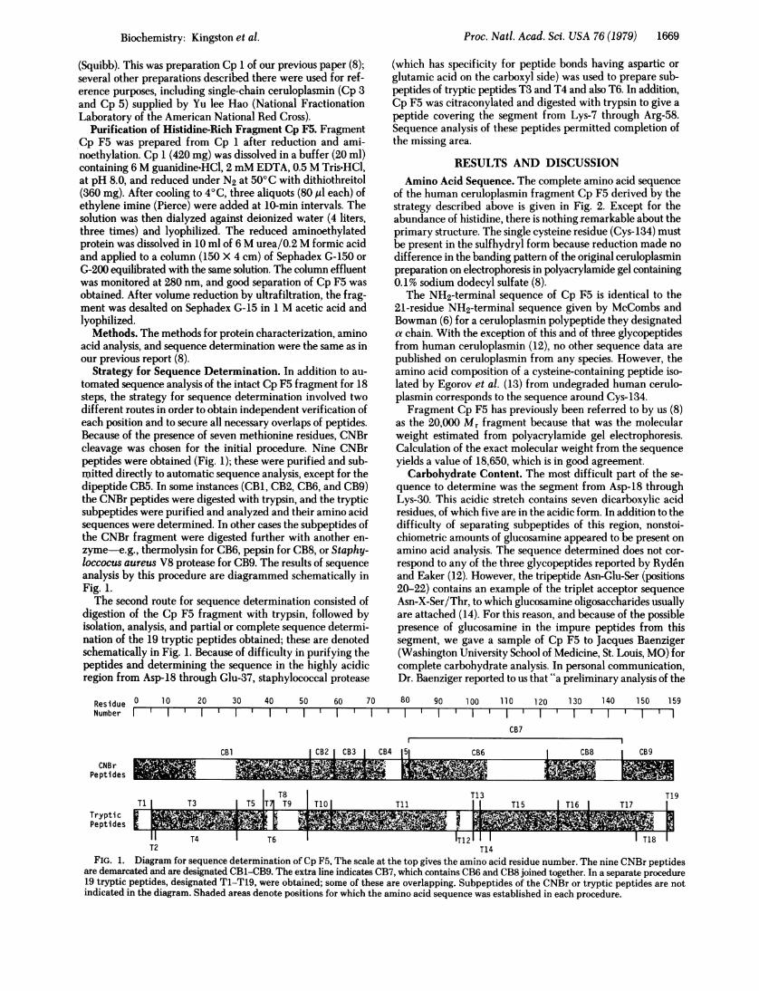
Strategy for Sequence Determination. In addition to au-tomated sequence analysis of the intact Cp F5 fragment for 18steps, the strategy for sequence determination involved twodifferent routes in order to obtain independent verification ofeach position and to secure all necessary overlaps of peptides.Because of the presence of seven methionine residues, CNBrcleavage was chosen for the initial procedure. Nine CNBrpeptides were obtained (Fig. 1); these were purified and sub-mitted directly to automatic sequence analysis, except for thedipeptide CB5. In some instances (CB1, CB2, CB6, and CB9)the CNBr peptides were digested with trypsin, and the trypticsubpeptides were purified and analyzed and their amino acidsequences were determined. In other cases the subpeptides ofthe CNBr fragment were digested further with another en-zyme-e.g., thermolysin for CB6, pepsin for CB8, or Staphy-loccocus aureus V8 protease for CB9. The results of sequenceanalysis by this procedure are diagrammed schematically inFig. 1.The second route for sequence determination consisted of
digestion of the Cp F5 fragment with trypsin, followed byisolation, analysis, and partial or complete sequence determi-nation of the 19 tryptic peptides obtained; these are denotedschematically in Fig. 1. Because of difficulty in purifying thepeptides and determining the sequence in the highly acidicregion from Asp-18 through Glu-37, staphylococcal protease
Residue 0 10 20 30 40 50 60 70Number I I II
(which has specificity for peptide bonds having aspartic orglutamic acid on the carboxyl side) was used to prepare sub-peptides of tryptic peptides T3 and T4 and also T6. In addition,Cp F5 was citraconylated and digested with trypsin to give apeptide covering the segment from Lys-7 through Arg-58.Sequence analysis of these peptides permitted completion ofthe missing area.
RESULTS AND DISCUSSIONAmino Acid Sequence. The complete amino acid sequence
of the human ceruloplasmin fragment Cp F5 derived by thestrategy described above is given in Fig. 2. Except for theabundance of histidine, there is nothing remarkable about theprimary structure. The single cysteine residue (Cys-134) mustbe present in the sulfhydryl form because reduction made nodifference in the banding pattern of the original ceruloplasminpreparation on electrophoresis in polyacrylamide gel containing0.1% sodium dodecyl sulfate (8).The NH2-terminal sequence of Cp F5 is identical to the
21-residue NH2-terminal sequence given by McCombs andBowman (6) for a ceruloplasmin polypeptide they designateda chain. With the exception of this and of three glycopeptidesfrom human ceruloplasmin (12), no other sequence data arepublished on ceruloplasmin from any species. However, theamino acid composition of a cysteine-containing peptide iso-lated by Egorov et al. (13) from undegraded human cerulo-plasmin corresponds to the sequence around Cys-134.Fragment Cp F5 has previously been referred to by us (8)
as the 20,000 Mr fragment because that was the molecularweight estimated from polyacrylamide gel electrophoresis.Calculation of the exact molecular weight from the sequenceyields a value of 18,650, which is in good agreement.
Carbohydrate Content. The most difficult part of the se-quence to determine was the segment from Asp-18 throughLys-30. This acidic stretch contains seven dicarboxylic acidresidues, of which five are in the acidic form. In addition to thedifficulty of separating subpeptides of this region, nonstoi-chiometric amounts of glucosamine appeared to be present onamino acid analysis. The sequence determined does not cor-respond to any of the three glycopeptides reported by Rydenand Eaker (12). However, the tripeptide Asn-Glu-Ser (positions20-22) contains an example of the triplet acceptor sequenceAsn-X-Ser/Thr, to which glucosamine oligosaccharides usuallyare attached (14). For this reason, and because of the possiblepresence of glucosamine in the impure peptides from thissegment, we gave a sample of Cp F5 to Jacques Baenziger(Washington University School of Medicine, St. Louis, MO) forcomplete carbohydrate analysis. In personal communication,Dr. Baenziger reported to us that "a preliminary analysis of the
80 90 100 110 120 130 140 150
T l ' l ' l ' l ' l ' l ' lCB7
159
CB1 ICB2 ICB3 I CB4 151 CB6 CB8 C9
TlT8 T13 lT1| T3 T5 T4 T9 T10ITll I T15 IT16 I T17
T1
_1 T4 I- T6 1-12 1 T T18 'B
CNB rPeptides
TrypticPeptides
T2 T14FIG. 1. Diagram for sequence determination of Cp F5, The scale at the top gives the amino acid residue number. The nine CNBr peptides
are demarcated and are designated CB1-CB9. The extra line indicates CB7, which contains CB6 and CB8 joined together. In a separate procedure19 tryptic peptides, designated T1-T19, were obtained; some of these are overlapping. Subpeptides of the CNBr or tryptic peptides are notindicated in the diagram. Shaded areas denote positions for which the amino acid sequence was established in each procedure.
FIG. 2. Amino acid sequence of the histidine-rich fragment of human Cp F5.
ceruloplasmin 20,000 Mr fragment would indicate that less than10% of the peptide could be glycosylated." Expressed as molesof carbohydrate per 20,000 daltons, the fragment contained:0.05 glucosamine, 0.12 galactose, 0.12 mannose, and 0.00 fu-cose. It is possible that Asn-20 is partially glycosylated and, al-though our calculation of the secondary structure of Cp F5 (seelater) places Asn-20 at the end of an a helix, residues 20-23 alsohave a high probability of being in a (3turn. Since it appearslikely that carbohydrate is frequently attached to glycoproteinsat (3turns (15, 16), Asn-20 may be in a favorable position forglycosylation. However, the bulky tryptophan residue just afterthe triplet (i.e., Asn-Glu-Ser-Trp) may hinder glycosylation.
Genetic Polymorphism. Genetic polymorphism of cerulo-plasmin is rare in whites, but in black Americans a variantdesignated Cp A occurs with an allele frequency of 0.052,compared to a frequency of 0.939 for the common form Cp Band 0.003 and 0.006 for the rare variants Cp C and Cp NH,respectively (1, 3). In white Americans the frequency of Cp Bis 0.994, whereas the frequency of Cp A is only 0.006, thoughCp A frequency rises to a high of 0.149 in Nigerians (1). Othervariants of ceruloplasmin have a very low incidence in allpopulations studied (1). Because blacks constitute about 11%of the American population, the incidence of Cp A and otherknown variants in pooled plasma collected by the AmericanRed Cross must be less than 0.6%. Such a value is too low to bedetected by the methodology of amino acid sequence analysis.We estimate that because of the cumulative errors in themethodology, genetic variants at an individual frequency ofless than 0.05 in pooled plasma would not be detectable byamino acid sequence analysis. It is thus not surprising that weobtained a unique amino acid sequence for the fragment CpF5. Furthermore, Cp F5 represents only a fragment of thewhole ceruloplasmin molecule and may not be the site of anypolymorphic substitution.
However, one observation suggests to us the possibility thatunidentified genetic variants not detectable by the usualmethod of electrophoretic screening may exist in the Americanpopulation. In early work with Cp 2 (8), a different preparationof ceruloplasmin than the one used for complete sequenceanalysis (Cp 1), we obtained a CNBr fragment§ correspondingto the sequence given in Fig. 2 as Phe-Gly-Asn-Leu-Gln-Gly-Leu-Thr-Met for positions 60-68; however, the compositionof the fragment showed 0.67 residue of phenylalanine and 0.33of tyrosine, and the dansyl method and sequenator analysisdemonstrated both amino acids at the NH2 terminus. Yet, inthe complete sequence study neither the CNBr fragment northe tryptic peptide covering this region gave evidence of ty-
§ The nonapeptide also had a bluish color and appeared to bindcopper.
rosine at position 60. We do not know whether the tyrosine-containing peptide was lost during purification or if it waspresent only in the first preparation of ceruloplasmin. It ispossible that a tyrosine-phenylalanine exchange at position 60represents a true genetic polymorphism, but it is unlikely thatthe tyrosyl variant is Cp A because the latter differs in chargefrom Cp B.Our evidence for proteolytic degradation coupled with the
observation of "gull-wing" patterns in immunoelectrophoresisof aged preparations suggests that some genetic variants pre-viously reported may be artifacts. Poulik and Weiss (1) havediscussed the questionable genetic significance of one reportedgenetic variant that has a band with identical electrophoreticmobility to one observed in aged serum that originally had onlythe Cp B type.
Role of Histidine in Histidine-Rich Fragment. Althoughexceeded in histidine content by the "histidine-rich a2 glyco-protein" of human plasma (17), which has a histidine contentof 9.9%, and by the a globin chains of some species that have10 or more histidines per a chain, the Cp F5 fragment ofhuman ceruloplasmin has a higher histidine content than mostother known proteins (8.82%, calculated from the sequence).Furthermore, 7 of the 12 histidines occur in an alternating se-quence of His-X-His-i.e., His-Phe-His-Gly-His, His-Cys-His,and His-Ile-His (beginning at positions 91, 133, and 139, re-spectively). This leads to two short segments that, when othernearby histidines are included, are exceedingly rich in histi-dine-i.e., there are four histidines in the eight-residue segmentfrom His-88 to His-95 and four more in the nine-residue seg-ment from His-l33 through His-141, which contains the freecysteine. In view of the role of histidine in the binding of copperby serum albumin (18), the proximity of so many histidines closeto Cys-134, which is assumed to be a copper-binding site (8),is probably significant. Because of the alternation, all of thehistidines could be exposed to the surface in the ( sheet postu-lated for residues 129-137 and the f turn following it.
Prediction of Secondary Structure. The problems of pre-dicting protein structure from amino acid sequence have re-cently been reviewed by Sternberg and Thornton (19). None-theless, they point out that "the method of Chou and Fasman(9, 10) has attracted most attention as it is simple to understand,can be applied without a computer, and has been relativelysuccessful." Hence, we have applied their rules for predictingsecondary structure, with the result depicted in Fig. 3.The model depicts a mixed a/,3 secondary structure. Almost
one-third of the residues appear to be distributed in five ahelices and about a third in ,B sheets. The remainder are in (turns, coils, or disordered structures, including one segment of15 residues (residues 100-115) that has no clear a or (3 initiationsites. The charged residues are concentrated at three sites: the
Proc. Natl. Acad. Sci. USA 76 (1979)
Proc. Natl. Acad. Sci. USA 76 (1979) 1671
6 7
84
108 116 120 123
His Cys139 134
144 146 152Met
159
FIG. 3. Diagram of the secondary structure ofCp F5 as predictedby the method of Chou and Fasman (9, 10). Residues are representedin helical (A.), ,3-sheet (A), and coil (-) conformational states. Chainreversals denote /-turn tetrapeptides (::.
NH2 terminus, the COOH terminus, and the two a helices andturn in the segment from Asp-34 through Lys-51. There are
several hydrophobic stretches, notably in the first helix fromPhe-10 through Phe-17. The alternation of the histidines re-
lieves the hydrophobic character of the sheet beginning atIle-129.
Noncovalent Interaction of Cp F5 with Other Fragments.The several fragments present in commercial preparationsinteract strongly, and harsh dissociating conditions are requiredto separate them. Thus, the blue color and the copper are lostduring the purification of Cp F5. The association is not due todisulfide bonds because ceruloplasmin preparations that exhibitmultiple bands in polyacrylamide gel electrophoresis in 0. 1%sodium dodecyl sulfate give similar patterns before and afterreduction although they sediment largely as single componentswith s2o = 6-7 S in analytical ultracentrifugation (8). The reason
for the strong interaction of Cp F5 with the 53,000 Mr and67,000 Mr fragments is not evident from the primary structure.However, one clue is offered by the model of the secondarystructure given in Fig. 3; this illustrates the concentration ofcharged groups in certain segments of the structure, notablythe NH2 terminus, the COOH terminus, and the two a helicesand the ,B turn proposed for residues 34-55. Half the residuesin the latter segment are either positively or negatively charged.If the largely hydrophobic structures from residues 55-144 are
mainly in the interior of the molecule and the ionized residuesare at the surface, as would be expected from the general rulesfor protein conformation (19), then the Cp F5 fragment wouldbe capable of strong electrostatic interaction with the other
fragments of ceruloplasmin. Probably this is its natural role, forit appears to be a separate domain structure of the intact mol-ecule.
Intersequence Comparison of Homology. We have pro-posed (8) that the cysteine-containing COOH-terminal se-quence of Cp F5 may be involved in binding one of the twotype 1 cupric ions in ceruloplasmin because the sequence fromGly-128 through Val-150 shows similarity to the cysteine-containing COOH-terminal sequence of blue copper-con-taining azurins and plastocyanins, which contain a single type1 copper ion. The cysteine, histidine, and methionine residues,which are invariant in the latter proteins and which have beenimplicated to be in the copper-binding site (8), could bematched by Cys-134, His-139, and Met-144 in Cp F5. Thisproposal has since been strengthened by the publication of theresults of x-ray crystallographic studies of plastocyanin (20) andazurin (21). In both cases, the copper atom appears to be boundto the cysteine and methionine residues which were matched(8) with Cys-134 and Met-144 in Cp F5 and by two histidineresidues one of which was matched with His-139. In our pre-dicted secondary structure shown in Fig. 3, the spacial ar-rangement of Cys-134, His-139, and Met-144 would give themthe potential to act as copper ligands. Furthermore, it is possibleto interpret Chou and Fasman's rules for predicting secondarystructure in a manner (L.-C. Lin, personal communication) thatpredicts that Cp F5 will be composed of two a-helical regionsat the NH2 terminus and six f-sheet strands separated into twogroups of three by an intervening a-helical or random region;these elements could then fold up to produce a structure rathersimilar to the cylindrical barrel shape formed largely by strandsof 3 sheet in poplar plastocyanin (20) and Pseudomonasaeruginosa azurin (21).To search for any additional similarity to other proteins, we
requested the National Biomedical Research Foundation toundertake a computer search comparing the sequence of CpF5 with all sequence data available, not only for the blue cop-per-containing proteins, but also for all other proteins. With theprogram RELATE (22), ceruloplasmin was compared to itselfand to plastocyanin, azurin, and superoxide dismutase, by usinga segment length of 15. In all cases the comparison of real se-quences gave a score less than 1 SD from the mean score with100 randomized sequences. Under the same conditions, azurinagainst plastocyanin gives a score of 3.9 SD. The ceruloplasminwas rerun against itself by using a segment length of 25. Thescore was 1.8 SD, which is interesting though not statisticallysignificant (Winona C. Barker, personal communication).However, as we had earlier identified by visual comparison ofthe segments from 1-53, 54-83, and 108-137, there is an indi-cation that the chain had duplicated. In the computer search,19 of the top 20 scores came from displacing the sequence by53 residues.Two 25-residue segments of ceruloplasmin (Met-79 through
Gly-i1S, which has six histidioes, and Pro-127 through Leu-151,which contains the cysteine) were searched against the entiredata collection of the Atlas of Protein Sequence and Structure(95,365 comparisons). The highest scoring segments retrievedwere from a variety of proteins. Nothing stood out significantlyfrom the rest. The cysteine-containing segment of the azurinsgave a good score, but the highest scores included a series offerredoxins and the a and.B globins of many species. The glo-bins and Cp F5 have a high histidine content in common. Theferredoxins are of interest because the highest scoring segmentis at the COOH terminus of both Cp F5 and ferredoxin, and thematching residues are Trp-130, Leu-132, and Cys-134, all closeto the site proposed for copper binding in Cp F5.
Biochemistry: Kingston et al.
1672 Biochemistry: Kingston et al.
We thank J. Dwulet, A. Galen, J. Madison, and S. Dorwin for tech-nical assistance, Dr. J. Baenziger for carbohydrate analysis, and Dr.W. C. Barker for computer analysis of the sequence homology. Thiswork was supported by National Institutes of Health Grant AM19221.
1. Poulik, M. D. & Weiss, M. L. (1975) in The Plasma Proteins, ed.Putnam, F. W. (Academic, New York), 2nd Ed., Vol. 2, pp.51-108.
2. Frieden, E. & Hsieh, H. S. (1976) Adv. Exp. Med. Biol. 74,505-529.
3. Gitlin, D. & Gitlin, J. D. (1975) in The Plasma Proteins, ed.Putnam, F. W. (Academic, New York), 2nd Ed., Vol. 2, pp.321-374.
4. Simons, K. & Bearn, A. G. (1969) Biochim. Blophys. Acta 175,260-270.
5. Freeman, S. & Daniel, E. (1973) Biochemistry 12,4806-4810.6. McCombs, M. L. & Bowman, B. H. (1976) Biochim. Biophys.
Acta 434,452-461.7. Ryden, L. (1972) Eur. J. Biochem. 26,380-386.8. Kingston, I. B., Kingston, B. L. & Putnam, F. W. (1977) Proc.
Natl. Acad. Sc$. USA 74,5377-5381.9. Chou, P. Y. & Fasman, G. D. (1974) Biochemistry 13, 211-
245.
10. Chou, P. Y. & Fasman, G. D. (1978) Annu. Rev. Biochem. 47,251-276.
11. Sgouris, J. T., Coryell, F. C., Gallick, M., Storey, R. W., McCall,K. B. & Anderson, H. D. (1962) Vox Sang. 7,394-405.
12. Ryden, L. & Eaker, D. (1974) Eur. J. Biochem. 44, 171-180.13. Egorov, T. A., Svenson, A., Ryden, L. & Carlsson, J. (1975) Proc.
NatI. Acad. Sci. USA 72,3029-3033.14. Clamp, J. R. (1975) in The Plasma Proteins, ed. Putnam, F. W.
(Academic, New York), 2nd Ed., Vol. 2, pp. 163-211.15. Aubert, J. P., Biserte, G. & Loucheux-Lefebvre, M. H. (1976)
Arch. Biochem. Biophys. 175,410-418.16. Huber, R., Deisenhofer, J., Colman, P. M., Matsushima, M. &
Palm, W. (1976) Nature (London) 264,415-420.17. Heimburger, N., Haupt, H., Kranz, T. & Baudner, S. (1972)
Hoppe-Seyler's Z. Physiol. Chem. 353, 1133-1140.18. Peters, T., Jr. (1975) in The Plasma Proteins, ed. Putnam, F. W.
(Academic, New York), 2nd Ed., Vol. 1, pp. 133-181.19. Sternberg, M. J. E. & Thornton, J. M. (1978) Nature (London)
271, 15-20.20. Colman, P. M., Freeman, H. C., Guss, J. M., Murata, M., Norris,
V. A., Ramshaw, J. A. M. & Venkatappa, M. P. (1978) Nature(London) 272, 319-324.
21. Adman, E. T., Stenkamp, R. E., Sieker, L. C. & Jensen, L. H.(1978) J. Mol. Biol. 123,35-47.
22. Barker, W. C., Ketcham, L. K. & Dayhoff, M. 0. (1978) J. Mol.Evol. 10, 265-281.


![Original DNA sequence GGC [TACGAGCTTCGAAATTTGCCGATC] CCA mRNA:AUG – CUC – GAA – GCU – UUA – AAC – GGC – UAG A.A.s MET – LEU – GLU – ALA - LEU – ASP – GLY.](https://static.documents.pub/doc/80x56/5a4d1ae17f8b9ab059977456/original-dna-sequence-ggc-tacgagcttcgaaatttgccgatc-cca-mrnaaug-cuc.jpg)


![Review: Amino Acid Side Chains Aliphatic- Ala, Val, Leu, Ile, Gly Polar- Ser, Thr, Cys, Met, [Tyr, Trp] Acidic (and conjugate amide)- Asp, Asn, Glu, Gln.](https://static.documents.pub/doc/80x56/56649cf65503460f949c5a07/review-amino-acid-side-chains-aliphatic-ala-val-leu-ile-gly-polar-ser.jpg)


