+ All Categories
Home > Documents > Complete Heart Block, Severe Ventricular Dysfunction and … · Keywords: Coronavirus, children,...

Complete Heart Block, Severe Ventricular Dysfunction and … · Keywords: Coronavirus, children,...

Date post: 03-Jul-2020
Category:
Upload: others
View: 3 times
Download: 0 times
Share this document with a friend
16
Journal Pre-proof Complete Heart Block, Severe Ventricular Dysfunction and Myocardial Inflammation in a Child with COVID-19 Infection Iqbal El Assaad, MD, M. Indriati Hood-Pishchany, MD PhD, John Kheir, MD, Kshitij Mistry, MD, Avika Dixit, MBBS MPH, Olha Halyabar, MD, Douglas Y. Mah, MD, Colin Meyer-Macaulay, MD, Henry Cheng, MD PII: S2666-0849(20)30486-1 DOI: https://doi.org/10.1016/j.jaccas.2020.05.023 Reference: JACCAS 529 To appear in: JACC Case Reports Please cite this article as: El Assaad I, Hood-Pishchany MI, Kheir J, Mistry K, Dixit A, Halyabar O, Mah DY, Meyer-Macaulay C, Cheng H, Complete Heart Block, Severe Ventricular Dysfunction and Myocardial Inflammation in a Child with COVID-19 Infection, JACC Case Reports (2020), doi: https:// doi.org/10.1016/j.jaccas.2020.05.023. This is a PDF file of an article that has undergone enhancements after acceptance, such as the addition of a cover page and metadata, and formatting for readability, but it is not yet the definitive version of record. This version will undergo additional copyediting, typesetting and review before it is published in its final form, but we are providing this version to give early visibility of the article. Please note that, during the production process, errors may be discovered which could affect the content, and all legal disclaimers that apply to the journal pertain. © 2020 Published by Elsevier on behalf of the American College of Cardiology Foundation.
Transcript

Journal Pre-proof

Complete Heart Block, Severe Ventricular Dysfunction and Myocardial Inflammationin a Child with COVID-19 Infection

Iqbal El Assaad, MD, M. Indriati Hood-Pishchany, MD PhD, John Kheir, MD, KshitijMistry, MD, Avika Dixit, MBBS MPH, Olha Halyabar, MD, Douglas Y. Mah, MD, ColinMeyer-Macaulay, MD, Henry Cheng, MD

PII: S2666-0849(20)30486-1

DOI: https://doi.org/10.1016/j.jaccas.2020.05.023

Reference: JACCAS 529

To appear in: JACC Case Reports

Please cite this article as: El Assaad I, Hood-Pishchany MI, Kheir J, Mistry K, Dixit A, Halyabar O,Mah DY, Meyer-Macaulay C, Cheng H, Complete Heart Block, Severe Ventricular Dysfunction andMyocardial Inflammation in a Child with COVID-19 Infection, JACC Case Reports (2020), doi: https://doi.org/10.1016/j.jaccas.2020.05.023.

This is a PDF file of an article that has undergone enhancements after acceptance, such as the additionof a cover page and metadata, and formatting for readability, but it is not yet the definitive version ofrecord. This version will undergo additional copyediting, typesetting and review before it is publishedin its final form, but we are providing this version to give early visibility of the article. Please note that,during the production process, errors may be discovered which could affect the content, and all legaldisclaimers that apply to the journal pertain.

© 2020 Published by Elsevier on behalf of the American College of Cardiology Foundation.

1

Complete Heart Block, Severe Ventricular Dysfunction and Myocardial Inflammation in a Child with COVID-19 Infection Iqbal El Assaad, MD1*; M. Indriati Hood-Pishchany, MD PhD2*; John Kheir, MD1; Kshitij Mistry, MD1; Avika Dixit, MBBS MPH2; Olha Halyabar, MD3; Douglas Y. Mah, MD1; Colin Meyer-Macaulay, MD1** ; Henry Cheng, MD1** Affiliations:

1. Department of Cardiology, Boston Children’s Hospital, Boston, Massachusetts. 2. Division of Infectious Disease, Department of Pediatrics, Boston Children’s Hospital,

Boston, Massachusetts. 3. Rheumatology Program, Division of Immunology, Boston Children’s Hospital, Boston,

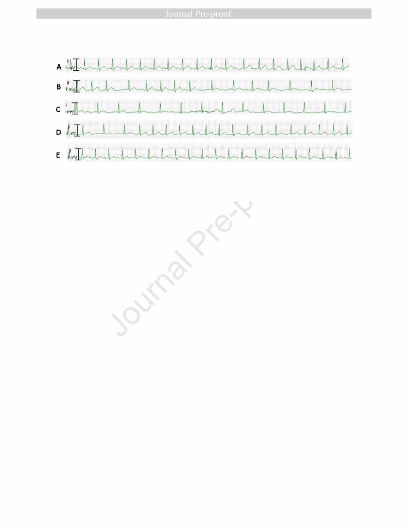
Massachusetts. * Dr. El-Assaad and Dr. Hood-Pishchany contributed equally to this article. ** Dr. Meyer-Macaulay and Dr. Cheng should be considered co-senior authors. Disclosure: None Funding: Dr. Hood Pishchany is supported through a Physician Scientist Fellowship from the Doris Duke Charitable Foundation (2019129) and a Child Health Research Career Development Award from the Eunice Kennedy Shriver National Institute of Child Health and Human Development (K12 HD052896 13). Dr. Dixit is supported through a Boston Children’s Hospital. OFD/BTREC/CTREC Faculty Career Development Fellowship and the Bushrod H. Campbell and Adah F. Hall Charity Fund/Charles A. King Trust Postdoctoral Fellowship. Dr. El-Assaad, Dr. Kheir, Dr. Mistry, Dr. Halyabar, Dr. Mah, Dr. Meyer-Macaulay, and Dr. Cheng have no funding support relevant to this article. Corresponding Author: Iqbal El-Assaad, MD Department of Cardiology, Boston Children’s Hospital, Boston, MA 02215. E-mail address: [email protected]. Abbreviations: COVID-19: Coronavirus Disease-2019, Complete Heart Block: CHB, Electrocardiogram: ECG. Keywords: Coronavirus, children, complete heart block. ventricular dysfunction, myocarditis, electrocardiogram. Abstract A young child presented with severe ventricular dysfunction and troponin leak in the setting of Coronavirus-19 disease (COVID-19). He developed intermittent, self-resolving, and hemodynamically insignificant episodes of complete heart block (CHB), which were diagnosed on telemetry and managed conservatively. This report is the first description of COVID-19 induced transient CHB in a child.

2

Case Presentation

A 10-year-old male presented with a 7-day history of fever and viral symptoms including

fatigue, cough, diarrhea, vomiting, myalgias and non-pruritic rash that spread to the trunk. His

vital signs were notable for sinus tachycardia (130 bpm), tachypnea (RR 24 bpm), hypotension

(84/40 mmHg) and normal oxygen saturation (98% on 2L nasal cannula). His physical

examination was notable for appearing drowsy but easily arousable, normal work of breathing, a

gallop on cardiac exam, cool extremities, and capillary refill was delayed at 4 seconds.

Past Medical History

Pityriasis lichenoides Chronica. No personal or family history of congenital heart disease,

immunodeficiency or autoimmune disease.

Differential Diagnosis

The differential diagnosis included viral induced myocarditis or underlying

cardiomyopathy unmasked by an acute viral illness.

Investigations

Laboratory evaluation was notable for elevated white blood cell count (17.1 K cells/µl)

with neutrophilic predominance and lymphopenia (0.91 K cells/uL, [ref 1.23-2.69 cells/uL]),

elevated high sensitivity troponin (84 ng/mL, [ref 0-14 ng/L]), brain natriuretic peptide (2000

pg/mL, [ref 0-100 pg/mL]) and inflammatory markers (C-reactive protein (CRP) 22 mg/dL [ref

<0.5 mg/dL], ferritin 1138 ng/mL[ref 10-320 ng/mL] and D-dimer 3.1 mcg/mL [ref <0.5

mcg/mL]). Chest x-ray was consistent with mild viral pneumonia (Figure 1). Electrocardiogram

(ECG) showed sinus tachycardia at 140 bpm and normal conduction interval (PR 140 ms).

Echocardiogram revealed normal left ventricular size (LVEDV 112 ml, Z score +0.8), and severe

3

left ventricular (LV) systolic dysfunction with an ejection fraction (EF) of 32% (Z score -6.8)

(Video 1). Coronary arteries were normal and there was no evidence of wall motion

abnormalities. Real-time Polymerase Chain Reaction was positive for SARS-CoV-2 from

nasopharyngeal swab. Evaluation for common infections associated with myocarditis identified

no other active infectious etiology (Table 1).

Management

He was started on Bilevel Positive Airway Pressure (BIPAP) for respiratory support and

afterload reduction. He was also commenced on epinephrine and norepinephrine infusions. He

received 2 doses of intravenous immune globulin (IVIG) at 1g/kg within the first 48 hours of

admission as an empiric treatment for potential myocarditis. In light of his hyper-inflammatory

state consistent with cytokine storm syndrome, multidisciplinary consultation led to the early

initiation of immunomodulatory therapy (anakinra 100 mg TID and methylprednisolone 2mg/kg

BID) and antiviral therapy (remdesivir 100mg daily after 200mg loading dose) on hospital day 1.

Due to evidence of hypercoagulability and concern for related thrombotic complications,

therapeutic anticoagulation was initiated with unfractionated heparin.

On day two of admission, his clinical status and hemodynamics began improving. He was

transitioned off of BIPAP to regular nasal cannula. He weaned off of norepinephrine on day two,

and off of epinephrine on day six of admission.

From a rhythm standpoint, he developed first degree AV block 24 hours into his

admission, which resolved spontaneously by the following morning. However, on day 3 of his

admission, he began to display bradyarrhythmias. Review of telemetry demonstrated transient,

self-resolving, episodes of CHB with a narrow junctional escape rhythm of approximately 90

bpm (Figure 2). The longest episode lasted for 4 minutes, during which the patient remained

4

hemodynamically stable and asymptomatic. Over the following days, he continued to have

repetitive, self-resolving, and hemodynamically insignificant episodes of conduction block

ranging from 1st degree AV block to 2nd and 3rd degree AV block. All episodes resolved within a

few minutes without intervention.

Discussion

Myocardial involvement with the novel coronavirus, ranging from mild troponin leak to

fulminant myocarditis, is well reported in adults and occurs in 8-28% of adults infected with

SARS-COV-2.1 Furthermore, acute cardiac injury portends poor outcomes.1-2 Children with

COVID-19 appear to have a much milder form of the disease compared to adults, and rarely

progress to severe disease and multi-organ dysfunction.3-5 The largest pediatric cohort from

China of 2135 patients (728 confirmed and 1407 suspected cases) showed that 94% of children

had moderate disease severity or less, and there was only a single death, while severe disease is

most common in neonates.5 Of 171 children with confirmed SARS-CoV-2 treated at the Wuhan

Children’s Hospital, only 3 patients with pre-existing conditions required intensive care support

and there was a single death.3 In a large systematic review of over 1000 children infected with

COVID-19, Castagnoli et al. reported a single case of severe COVID-19 infection occurring in a

13-month-old infant, who achieved full recovery.4 Due to its recent emergence, there is scarce

data on its effects on the myocardium in children, and more specifically, the conduction system.

A large retrospective single center case series of 138 hospitalized patients in China shows

that 7% of the total cohort developed acute cardiac injury and 16.7% had unspecified

arrhythmias.6 There is a single case report of an adult female admitted to the hospital with

COVID-19 and respiratory distress requiring intubation, who developed a single episode of

transient CHB with prolonged pause on day 14 of her illness. 7 She required cardiopulmonary

5

resuscitation prior to resumption of normal sinus rhythm. Compared to our patient, she had a

normal echocardiogram.

Our patient exhibited evidence of cytokine storm, with elevated ferritin, CRP and

procalcitonin, as well as markedly elevated soluble IL-2 receptor (14,800 pg/mL, ref ≤ 1033),

CXCL9 (1575 pg/mL, ref ≤ 121), IL-18 (1427 pg/mL, ref 89-540) and elevated IL-6 (44 pg/mL,

ref ≤ 5). The child’s only pre-existing condition was pityriasis lichenoides chronica. Although

there exists debate about the etiology of pityriasis lichenoides chronica, it can be characterized

by benign clonal T-cell lymphoproliferation, which may arise as an aberrant immune response to

an antigenic trigger, such as a viral infection.8 Whether this history suggests an underlying

immunologic phenotype that contributed to the patient’s hyperinflammatory state in response to

SARS-CoV-2 infection remains to be determined.

Our patient improved significantly after initiation of immunomodulatory and antiviral

therapy, though the relative contribution of either of these approaches cannot be discerned from

this single case. Positive response to systemic glucocorticoids and IVIG was reported in a single

case of an adult patient with coronavirus fulminant myocarditis.9 Remdesivir has shown promise

with in vitro activity against SARS-CoV-2, and early reports of positive clinical response;

however, trials are still underway to establish clinical efficacy.10 Additionally, anakinra has been

applied with a favorable safety profile in adults with myocarditis from other causes.11 Our patient

did not undergo a myocardial biopsy, so we cannot confirm that SARS-CoV-2 was directly

responsible for myocardial injury or myocarditis. However, testing for a number of common

infections often implicated in myocarditis was negative, except Parvovirus (low level viremia

with a positive IgG), and Epstein-Barr virus viremia (with positive antibody to NA and capsid

IgG), which both likely represent past infection with reactivation in the setting of acute illness.

6

The pathophysiology of myocardial injury secondary to SARS-CoV-2 is multifactorial,

including direct cardiotropic action of the virus mediated by its cellular receptor target:

Angiotensin Converting Enzyme-2 (ACE-2), which is heavily expressed in adult cardiac

pericytes.1,12 Furthermore, inhibition and downregulation of ACE2 results in an increased

circulating level of Angiotensin II, which has proinflammatory and vasoconstrictor properties,

leading to microangiopathy.1,12 In adults with coronary artery disease, an oxygen demand-supply

mismatch is suspected to play a role in the development of acute coronary syndromes, mediated

by hypoxic respiratory failure and increased metabolic demand.1,11 Finally, cytokine storm

secondary to downregulation of ACE-2 as well as direct endothelial injury may contribute to

myocardial dysfunction. 1,12

The mechanism for COVID-19 induced heart block is not well studied, but we suspect

that it is secondary to inflammation and edema of the conduction tissue, as part of the global

myocardial injury process. The two potential mechanisms resulting in inflammation are direct

viral invasion or immune mediated injury.1 In our case, the patient had significantly elevated

inflammatory markers, no other known risk factors for conduction disease, and a normal baseline

ECG. With therapy, his inflammatory markers trended down (figure 3), and his episodes of heart

block resolved, indicating that inflammation is likely the main etiology of conduction disease in

our patient.

Follow up:

Patient has had no recurrent episodes of CHB since day 4 of admission and a repeat

echocardiogram on day 12 of admission demonstrated low-normal biventricular systolic

function.

Conclusion:

7

Myocardial involvement in the form of dysfunction and conduction abnormalities appears

to be a rare manifestation of SARS-CoV-2 infection in children.3-5 Nonetheless, evaluation for

myocardial injury may be warranted in pediatric patients with symptomatic SARS-CoV-2

infection, particularly in patients whose clinical symptoms (e.g. dyspnea, hypoxia) seem out of

proportion to chest imaging findings. Furthermore, children infected with COVID-19 who have

myocardial involvement should be placed on continuous telemetry for close monitoring. Rhythm

management should be guided by the patient hemodynamics and clinical status. Additional

studies are needed in order to identify the spectrum of rhythm disorders and cardiac involvement

in pediatric patients with COVID-19 infections.

8

Learning Objectives:

• To be aware of the possibility of heart block in children admitted with COVID-19 and

myocardial dysfunction.

• To understand the benefit of a multidisciplinary approach in addressing potential

pathophysiologic components leading to the development of arrhythmia in COVID-19

patients with cardiac involvement.

9

References

1. Liu PP, Blet A, Smyth D, Li H. The science underlying COVID-19: implications for the

cardiovascular system. Circulation. 2020 Apr 15. doi:

10.1161/CIRCULATIONAHA.120.047549. [Epub ahead of print]

2. Li JW, Han TW, Woodward M, et al. The impact of 2019 novel coronavirus on heart

injury: a systemic review and meta-analysis. Prog Cardiovasc Dis 2020;20:30090-3

3. Lu X, Zhang L, Du H, et al. SARS-COV-2 infection in children. N. Engl. J. Med. (2020),

10.1056/nejmc2005073

4. Castagnoli R, Votto M, Licari A, et al. Severe acute respiratory syndrome coronavirus 2

(SARS-CoV-2) infection in children and adolescents. A systematic review. JAMA

Pediatr 2020, doi:10.1001/jamapediatrics.2020.1467.

5. Dong Y, Mo X, Hu Y, et al. Epidemiology of COVID-19 Among Children in China.

Pediatrics. 2020;e20200702. doi:10.1542/peds.2020-0702

6. Wang D, Hu B, Hu C, et al. Clinical Characteristics of 138 hospitalized patients with

2019 novel coronavirus-infected pneumonia in Wuhan, China. JAMA 2020; 323:1061-

1069

7. Azarkish M, Laleh Far V, Eslami M, Mollazadeh R. Transient complete heart block in a

patient with critical COVID-19. Eur Heart J 2020.

https://doi.org/10.1093/eurheartj/ehaa307

8. Magro C, Crowson AN, Kovatich A, Burns, F. Pityriasis lichenoides: A clonal T-cell

lymphoproliferative disorder. Human Pathology 2002;33:788–795

10

9. Hu, H, Ma, F, Wei, X, Fang, Y. Coronavirus fulminant myocarditis treated with

glucocorticoid and human immunoglobulin. Eur Heart J 2020.

doi:10.1093/eurheartj/ehaa190.

10. Grein J, Ohmagari N, Shin D, et al. Compassionate Use of Remdesivir for Patients with

Severe Covid-19. N Engl J Med. 2020 Apr 10. doi: 10.1056/NEJMoa2007016. [Epub

ahead of print] doi:10.1056/NEJMoa2007016

11. Cavalli G, Foppoli M, Cabrini L, Dinarello CA, Tresoldi M, Dagna L. Interleukin-1

Receptor Blockade Rescues Myocarditis-Associated End-Stage Heart Failure. Front

Immunol 2017;8:131.

12. Tersalvi G, Vicenzi M, Calabretta D, Biasco L. Elevated Troponin in Patients With

Coronavirus Disease 2019 : Possible Mechanisms. J Card Fail 2020.

doi: 10.1016/j.cardfail.2020.04.009

11

Table 1. Diagnostic evaluation for potential infectious etiologies or triggers of myocarditis. All serologies were collected prior to administration of IVIG.

Test Name Result Interpretation Reference range

CMV IgM <8.0 AU/mL

Not detected ≤ 29.9 AU/mL

CMV IgG 3.10 U/mL

Detected ≤ 0.59 units/mL

Cytomegalovirus PCR (blood)

Not detected

Not detected Not detected

EBV Capsid antigen, IgM <10.0 Not detected ≤ 35.9 U/mL EBV antibody to EA-D, IgG

<5.0 U/mL

Not detected ≤ 8.9 U/mL

EBV Capsid antigen IgG 717 U/mL Detected ≤ 17.9 U/mL EBV antibody to NA, IgG 331 Detected ≤ 17.9 U/mL Epstein-Barr virus PCR (blood)

21,257 copies/mL

Detected Not detected

HSV IgM 0.56 Not detected ≤ 0.89 U/mL HSV I/II Ab, IgG 0.28 Not detected ≤ 0.89 U/mL Parvovirus IgM 0.11 IV Negative ≤ 0.89 IV Parvovirus IgG 6.11 IV Positive ≤ 0.89 IV Parvovirus B19 PCR (blood)

<199 IU/mL

Detected, below limit of quantitation

Not detected

Coxsackie A9 virus IgG <1:8 Negative <1:32 Adenovirus PCR (blood) Not

detected Not detected Not detected

Enterovirus PCR (blood) Not detected

Not detected Not detected

Ehrlichia and Anaplasma PCR (blood)

Negative Not detected Not detected

Other (all negative or non-reactive): Blood culture; Respiratory pathogen PCR panel (includes Adenovirus, Coronavirus (HKU1, NL63, OC43, 229E), Human metapneumovirus, Human rhinovirus/enterovirus, Influenza A (2009H1N1, H1, H3), Influenza B, Parainfluenza virus types 1-4, Respiratory Syncytial Virus, Mycoplasma pneumoniae, Chlamydia pneumoniae, Bordetella pertussis, Bordetella parapertussis); Lyme IgG and IgM; Rapid Plasma Reagin; HIV combination antigen/antibody; Rapid Strep. PCR, Polymerase Chain Reaction

12

FIGURE TITLES/LEGENDS

Figure 1. Chest radiograph upon admission demonstrating coarsened interstitial lung

markings and hazy retrocardiac opacification.

Figure 2: Telemetry rhythm strips showing the progression from normal sinus rhythm to

CHB followed by resumption of normal conduction within 4 minutes. (A) normal PR

interval followed by 1st degree AV block with prolonged PR interval at 280 ms � (B) 2nd

degree Mobitz I � (C) CHB with a narrow junctional escape rate ~ 90 bpm � (D) 1st degree AV

block � (E) Sinus rhythm with normal PR interval at 160 ms.

Figure 3: NT-ProBNP and CRP plotted against day of admission. NT-ProBNP: N Terminal

Pro Brain Naturetic Peptide, CRP: C-Reactive Protein.

VIDEO TITLES/LEGENDS

Video 1. Echocardiogram upon admission to CICU. (A) Four chamber view and (B)

parasternal short axis view demonstrating severe left ventricular systolic dysfunction.


Recommended