Complications of Radiation Therapy to the Head and Neck Donita Dyalram DDS, MD Assistant Professor Associate Program Director Maxillofacial Oncology/Microvascular Surgery Department of Oral Maxillofacial Surgery University of Maryland
Transcript
Complications of Radiation Therapy to the Head and Neck
Donita Dyalram DDS, MD Assistant Professor
Associate Program Director Maxillofacial Oncology/Microvascular Surgery
Department of Oral Maxillofacial Surgery University of Maryland
Lecture Goals • Understand the use of Radiation in H&N • Early effects • Late effects • Dental Implications • Treatment protocol for:
– Candidiasis – Xerostomia – Dental Extractions in irradiated Jaw – Implants in the irradiated Jaw
Radiotherapy • Therapeutic radiation is
delivered by 2 main methods: – Electromagnetic (photons):
x-rays, gamma rays – Particulate radiation
• Electrons • Protons • Neutrons
• Depth of penetration required is the main criterion used in choosing which energy to employ
Radiotherapy
• Roentgenmeasure of ionization in air – Used for radiation safety
• Gray (Gy)
– Dose absorbed by the tissue (clinically relevant) – 1 Gy = the absorption of 1 joule/kg – 1 Gy = 100 cGy = 100 rad
• There is no absolute resistance to radiation
– Normal tissue tolerance limits the dose
Radiotherapy – Brachytherapy
• Radium • Cesium • iridium
Radiotherapy • Mechanism of action
– Interacts with atoms and molecules of the cells
– Produces free radicals – Damages DNA – Affects all phases of the
cell cycle but cells going mitosis are most affected
Radiation • The sensitivity of cells to radiation is most pronounced
shortly before and during mitosis; thus, the effect is greatest in rapidly dividing cells.
• Highly radiosensitive tissues are mucosa, skin, bone marrow, nerve, and muscle tissue.
• Of the bone cells, osteoblasts are more radiosensitive than are osteoclasts and osteocytes.
What orofacial tissues are affected by Radiation therapy?
• Oral mucosa • Skin • Subcutaneous tissue • Cartilage • Muscles of mastication • Temporomandibular joint • Teeth • Oral flora • Salivary glands • Nasolacrimal drainage system • Bone • Thyroid and parathyroid glands • Pituitary gland • Peripheral and cranial nerves • Lymphatics • Paranasal sinuses
Tissue Effect – Early • Acute skin reactions
• Hyperemia
• Reduced salivary gland
function
• Mucositis
• Loss of taste
8 weeks post radiotherapy
Tissue Effect – Long Term
• Seem not to occur when the tissues are
exposed to less than about 45 Gy
• Chronic damage to skin, muscle, nerves, and bone
• Seem not to occur when the tissues are
exposed to less than about 45 Gy
• Chronic damage to skin, muscle, nerves, and bone
Mucositis
• A term given for widespread oral erythema, ulceration and soreness
Mucositis World Health Organization Toxicity Grading
Grade Clinical features 0 - 1 Soreness/erythema 2 Erythema, Ulcers but able to eat solids 3 Ulcers, but requires liquid diet 4 Oral alimentation not possible
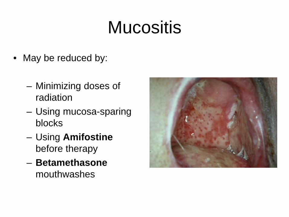
Mucositis • May be reduced by:
– Minimizing doses of
radiation – Using mucosa-sparing
blocks – Using Amifostine
before therapy – Betamethasone
mouthwashes
Mucositis • Opioids (MSO4 etc..) • Avoiding irritants (smoking, spirits, or spicy foods) • Good oral hygiene • Oral cooling using ice chips • Topical analgesics (especially before meals)
• Benadryl, Mylanta, and Carafate and viscous lidocaine in a 1:1:1:1
Mucositis
• The time to healing depends on the dose intensity and is usually complete within 3 weeks after the end of treatment.
Candidiasis
Candidiasis
• Infections by Candida albicans are commonly seen in irradiated patients
• Can be painful
Candidiasis
•Rinses
•Nystatin
•Amphotericin
• Clotrimazole (Mycelex) trouches
•Caution trouches contain sugars
•Oral/Systemic medications
•Diflucan
Xerostomia
Xerostomia
• Salivary Gland – Transient tenderness – Occasionally swelling – Occurs within a few hours after 1st dose – Decrease salivary flow noted within 24 hrs – May have ~50% decrease flow after 1st wk – Flow continues to decrease throughout treatment
Delanian S et al. Int J Radiat Oncol Biol Phys 2010
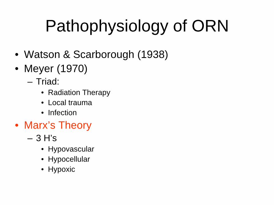
ORN treatment failure
Fibula Osteocutaneous Free Flap
ORN treatment failure
Dental Management Pre-Radiation
Dental Management Prior to Radiation
• Complete oral/dental examination and treatment plan
• Any necessary extraction and surgery • Maintenance of teeth and caries control • Restoration of restorable teeth • Prothetic examination to prevent postradiation
trauma from ill-fitting dentures
Dental Management Prior to Radiation
• Consider: – Condition of the
dentition – Level of oral hygiene
and patient attitude – Age of the patient – Radiation field and
dose – Urgency of radiation
treatment
Dental Management Prior to Radiation
• Caries control – Prophylactic care before and at the end of
therapy – Oral hygiene instructions – Daily administration of fluoride – Weekly follow-up during therapy and every 3-4
weeks afterward
Guideline for Extraction Prior to Radiation
• All carious teeth in the field of xrt (>60 Gy) should be extracted except in patients with excellent oral hygiene and dentition
• All questionable teeth should be extracted
• Full bony impacted teeth can be left in place
• Optimal time for extraction is 21 days before beginning xrt
Extractions
• Atraumatic extractions • perform an alveolectomy • smooth the bone • Perform a primary closure • Allow a minimum of 1 week to 10 days for
healing prior to beginning XRT • Preferable to allow 14 to 21 days
Dental Management Post-Radiation
Management of patient post radiation
• Obtain records of radiation fields and dose
• Recall for prophylaxis q 3 months
• Daily fluoride treatment for life
• Wait for mucositis to resolve prior to prosthesis placement
• Avoid invasive procedure involving irradiated bone