Email Phone Number Compressive Re-Sampling for Speckle Reduction in Medical Ultrasound Professor Richard Mammone Rutgers University Christine Podilchuk, Lev Barinov, Ajit Jairaj and William Hulbert ClearView Diagnostics Inc.
Transcript
Email Phone Number
Compressive Re-Sampling for Speckle
Reduction in Medical Ultrasound
Professor Richard Mammone
Rutgers University
Christine Podilchuk, Lev Barinov, Ajit Jairaj and William Hulbert
ClearView Diagnostics Inc.
Outline • Problem: Speckle Noise in Ultrasound Imaging
• Previous Methods for Speckle Noise Reduction
• Technical Approach: Compressive Re-Sampling
• Preliminary Results
– Simulated Data
– Clinical Data
– Recognition Performance
• Questions
The Problem: Breast Cancer Detection & Diagnosis
• Remains the “gold standard” for breast cancer screening
• Visualizes microcalcifications due to malignancies including early-stage
ductal carcinoma in situ (DCIS)
• Fewer false positives than Ultrasound
• Not effective at imaging women with dense breast tissue
• Ionizing
Mammography
Ultrasound
• FDA approved for diagnosis, not screening
• Speckle noise, inherent in all coherent imaging systems, results in lower
contrast and effectively lower resolution, poorer quality image
• More false positives than mammograms
• Effective in imaging dense breast tissue which is 4-6 times more likely to
develop cancer than non-dense tissue
• Non-ionizing
The Problem: Dense Breast Tissue
Imaging Sensitivity as a Function of BI-RADS Breast Density
Increasing BI-RADS density classification results in mammogram
sensitivity dropping from 100% to 47% while ultrasound remains in the
80-88% for all breast densities (Source: Berg, 2004)
0
20
40
60
80
100
Ultrasound
Mammography
Imaging Modality Diagnostic
Accuracy
Pos. Predictive Value of Biopsy
MAMMO .78 22.6
US 8.9
MAMMO + US .91 11.2
• Screening Breast Ultrasound Trial (ACRIN 6666) (JAMA 2008) showed
an increase in sensitivity from 78% with mammograms alone to 91% with
mammograms + ultrasound for women with dense breasts
• Screening US may depict small, node-negative breast cancers not seen
on mammography
• Ultrasound has not been able to replace mammography for screening
due to speckle noise which masks small, low-contrast lesions and
microcalcifications, a potential early indicator of breast cancer
The Problem: Speckle Noise in Ultrasound
Previous Methods for Speckle Reduction
• Fully formed speckle (FFS) is multiplicative noise modeled as a Raleigh
random variable with a constant SNR = m / s = 1.91 dB
• Speckle reduction techniques for ultrasound imaging include
compounding techniques and postprocessing (filtering) techniques.
• Frequency and spatial compounding:
• Additional hardware/acquisition time required.
• Frequency compounding averages images acquired in different frequency bands
• results in a loss in resolution due to the smaller bandwidth in each image.
• Spatial compounding averages images acquired in different scan directions
• results in a loss in resolution/accuracy due to spatial shifts between views.
• Reduces the noise by a factor of (L)1/2 where L is the number of estimates acquired.
• Filtering techniques such as Kaun and Lee, Diffusion, Median, Wavelet
(denoising by soft thresholding), Laplacian pyramid are based on
smoothing out the areas due to speckle noise while attempting to
preserve edges and other details. Smoothing can result in a loss of
small, high frequency details such as microcalcifications, a sign of ductal
carcinoma in situ (DCIS), an early stage of breast cancer.
Technical Approach
• Novel speckle reduction technique is inspired by fundamentals of
compressive sampling/sensing.
• Compressive sampling is motivated by reduced acquisition time or data
rate.
• Compressive Re-sampling takes advantage of the ability to recover an
estimate of the signal with fewer samples in order to provide multiple
estimates that can be used to reduce speckle noise and enhance tissue
and lesion detail.
• Compressive Re-sampling randomly samples the frequency components
over the entire bandwidth in order to provide an estimate that does not
reduce the overall resolution of the final image.
• The number of estimates L that can be acquired using this technique is
very large so that the reduction in noise by (L)1/2 is significant
Technical Approach
freq fMIN fMAX
…
X(f)+jY(f)
freq fMIN fMAX
…
X(f)+jY(f)
Iteration i
freq fMIN fMAX
…
X(f)+jY(f)
Iteration L-1 Iteration 0
Inverse DFT Inverse DFT Inverse DFT
Merging Module
Technical Approach
x(t): K-sparse ultrasound signal X(f): DFT{x(t)} B: Random binary mask of length N with M “1”s and (N-M) “0”s where M<<N L: Number of re-sampled estimates
B1 = [1, 0, 1… B(N)1]
B2 = [0, 0, 0… B(N)2]
BL = [1, 1, 0… B(N)L]
… …
X(f)1 = [X(1), 0, X(3) … X(N)*B(N)1]
X(f)2 = [0, 0, 0, … X(N)*B(N)2]
X(f)L = [X(1), X(2), 0, … X(N)*B(N)L]
x(t)i = x(t)i + j (x’(t)i)
xmag(t)i = ((x(t)i)2 + (x’(t)i)2)1/2
xmag(t) = 1/L Σi xmag(t)i
Initial Results: Simulated Data
Average SNR
improvement of 12 dB
on simulated data with
varying degrees of
speckle noise added
to the original
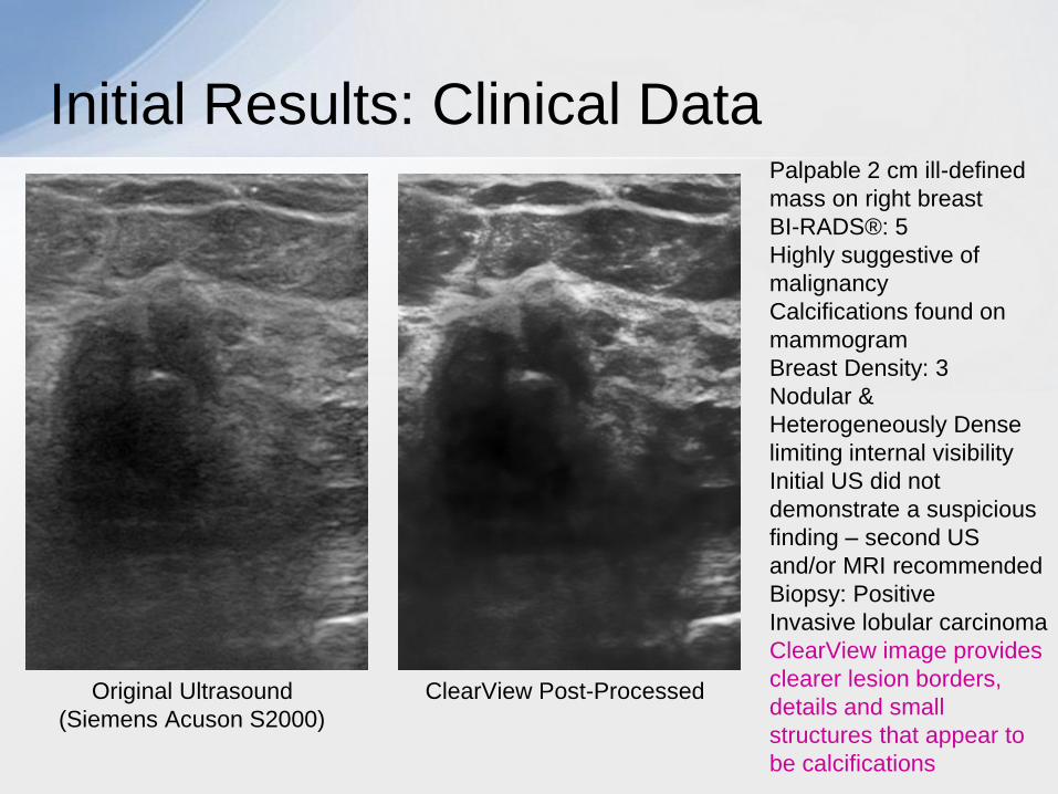
Initial Results: Clinical Data Palpable 2 cm ill-defined
mass on right breast
BI-RADS®: 5
Highly suggestive of
malignancy
Calcifications found on
mammogram
Breast Density: 3
Nodular &
Heterogeneously Dense
limiting internal visibility
Initial US did not
demonstrate a suspicious
finding – second US
and/or MRI recommended
Biopsy: Positive
Invasive lobular carcinoma
ClearView image provides
clearer lesion borders,
details and small
structures that appear to
be calcifications
Original Ultrasound ClearView Post-Processed
(Siemens Acuson S2000)
Initial Results: Clinical Data
Original Ultrasound ClearView Post-Processed
(Siemens Acuson S2000)
Palpable 2.1cm mass on
left breast and 1.7 cm
mass on right breast
BI-RADS®: 5
Highly suggestive of
malignancy
Calcifications found with
lesion on right breast
Breast Density: 3
Biopsy: Positive
Invasive ductal carcinoma
with calcifications
Image of lesion on right
breast pre-core ClearView image provides
clearer lesion borders,
details and small
structures that appear to
be calcifications
Initial Results: Clinical Data
Original Ultrasound ClearView Post-Processed Noise removed from original
Siemens Acuson S2000
Initial Results: Recognition Improvement
• Apply the Compressive Re-Sampling (CRS) Method as a preprocessor
to a CAD (Computer Aided Diagnostic) system to determine whether it
improves recognition performance
• Detection of microcalcifications using Neural Networks
• Roc curves were generated to determine the NN’s ability to detect
microcalcifications on the original (Siemens) images and the ClearView


![Non-Local Compressive Sampling Recoveryjyang29/papers/ICCP14_NLCS.pdf · Non-Local Compressive Sampling Recovery ... (3DCS) [20] significantly reduces the sampling rate of video](https://static.documents.pub/doc/80x56/5ad507a77f8b9a6d708c5a8a/non-local-compressive-sampling-jyang29papersiccp14nlcspdfnon-local-compressive.jpg)


