Computational and MR-guided Patient-Specific Laser Induced Thermal Therapy of Cancer D. Fuentes, J. T. Oden, K. R. Diller, A. Elliott, Y. Feng, J. D. Hazle, A. Shetty, and R. J. Stafford Abstract This chapter describes the development of a canonical dynamic data driven predictive control system for MR-guided laser induced thermal therapies (MRgLITT) of focal cancerous lesions within soft tissue. The predictive ability of computational models combined with advanced clinical imaging modalities is ex- ploited to plan, predict, control, and optimize the treatment outcome. The system is under continual development and embodies a cyberinfrastructure comprised of Magnetic Resonance Thermal Imaging (MRTI), computer visualization, laser op- tics, high-speed networks, nonlinear dynamic bioheat transfer models of heteroge- neous tissue, adaptive meshing, high-performance parallel computing, cell-damage models, inverse analysis, calibration, model validation, signal processing, optimal control algorithms, and error estimation and control. These diverse technologies and systems are connected across a high-speed computational grid connecting re- mote sites 150 miles apart and is an excellent example of a Dynamic Data Driven Application System (DDDAS). Webpage: http://wiki.ices.utexas.edu/dddas A. Elliott, D. Fuentes, J. D. Hazle, A. Shetty, and R. J. Stafford, The University of Texas M.D. Anderson Cancer Center, Department of Imaging Physics, Hous- ton TX 77030, USA, e-mail: [andrew.elliott,dtfuentes,jhazle,anil.shetty, jstafford]@mdanderson.org Y. Feng, Computational Bioengineering and Nanotechnology Lab, The University of Texas at San Antonio, San Antonio, TX 78749, USA, e-mail: [email protected]K. R. Diller Department of Biomedical Engineering, The University of Texas at Austin, Austin TX 78712, USA, e-mail: [email protected],[email protected]J. T. Oden Institute for Computational Engineering and Sciences, The University of Texas at Austin, Austin TX 78712, USA, e-mail: [email protected]1
Transcript
Computational and MR-guided Patient-SpecificLaser Induced Thermal Therapy of Cancer
D Fuentes J T Oden K R Diller A Elliott Y Feng J D Hazle A Shetty andR J Stafford
Abstract This chapter describes the development of a canonical dynamic datadriven predictive control system for MR-guided laser induced thermal therapies(MRgLITT) of focal cancerous lesions within soft tissue The predictive ability ofcomputational models combined with advanced clinical imaging modalities is ex-ploited to plan predict control and optimize the treatment outcome The systemis under continual development and embodies a cyberinfrastructure comprised ofMagnetic Resonance Thermal Imaging (MRTI) computer visualization laser op-tics high-speed networks nonlinear dynamic bioheat transfer models of heteroge-neous tissue adaptive meshing high-performance parallel computing cell-damagemodels inverse analysis calibration model validation signal processing optimalcontrol algorithms and error estimation and control These diverse technologiesand systems are connected across a high-speed computational grid connecting re-mote sites 150 miles apart and is an excellent example of a Dynamic Data DrivenApplication System (DDDAS)
Webpage httpwikiicesutexasedudddas
A Elliott D Fuentes J D Hazle A Shetty and R J StaffordThe University of Texas MD Anderson Cancer Center Department of Imaging Physics Hous-ton TX 77030 USA e-mail [andrewelliottdtfuentesjhazleanilshettyjstafford]mdandersonorg
Y FengComputational Bioengineering and Nanotechnology Lab The University of Texas at San AntonioSan Antonio TX 78749 USA e-mail yushengfengutsaedu
K R DillerDepartment of Biomedical Engineering The University of Texas at Austin Austin TX 78712USA e-mail odenicesutexasedukdillermailutexasedu
J T OdenInstitute for Computational Engineering and Sciences The University of Texas at Austin AustinTX 78712 USA e-mail odenicesutexasedu
1
2 Fuentes Oden Diller Elliott Feng Hazle Shetty Stafford
1 Introduction
Laser induced thermal therapy (LITT) is a minimally invasive procedure that re-places the scalpel of conventional surgery and the ionizing radiation of radiosurgerywith a lt2mm laser diode applicator minus the typical side effects and morbidity Aspecially designed laser fiber is delivered stereotactically or under real-time image-guidance to the site of a tumor and the fundamental idea is that when the laser heatsthe tumor cells to a certain point the cells are damaged and die The heating portionof the procedure takes only a few minutes MR-guided LITT (MRgLITT) is per-formed under thermal image monitoring using magnetic resonance thermal imaging(MRTI) The thermal images provide a quantitative treatment time estimate of thelethality of the thermal dose received by the tumor and surrounding healthy tissueMRgLITT has recently entered into patient use [4] multiple clinical trials withinthe United States are currently on-going for an FDA cleared MRgLITT system thatutilizes real-time temperature imaging feedback and dosimetry (Visualase Rcopy Visu-alase Inc Houston TX) and at least one trial for another system (AutoLITT Mon-teris Medical Cleveland Ohio) is being conducted under an investigational deviceexemption (IDE)
While real-time temperature monitoring provides invaluable treatment-timefeedback that makes the procedure safe and feasible once a laser applicatorhas been placed innovations in human assisted high performance computa-tional tools using this feedback are under development to plan control pre-dict and optimize the anticipated biological response to dramatically increasetreatment efficacy and reduce associated treatment morbidity and even reducerecurrence of the disease
DDDAS aims at developing a computational system that dynamically interactswith medical imaging technology for the predictive guidance and control of medicalprocedures It is a monumentally important development that could enhance manyfields of medical science The unique dynamic closed loop control system presentedby the predictive capabilities of computational simulation coupled with real-timemulti-planar image guidance as feedback has significant potential to facilitate areliable minimally invasive treatment modality that delivers an optimized thermaldose prescribed by the physician The control system developed in this work Fig-ure 1 employs a cyberinfrastructure [1] of magnetic resonance thermal imagingcomputer visualization laser optics high-speed networks nonlinear dynamic bio-heat transfer models of heterogeneous tissue adaptive meshing high-performanceparallel computing cell-damage and heat-shock protein models inverse analysiscalibration model validation signal processing optimal control algorithms and er-ror estimation and control A computational grid connects two remote arenas overa high bandwidth network an imaging and laser treatment arena that manages thethermal data and laser source and a computational arena that uses parallel com-puting algorithms to generate and solve computational models of bioheat transfer
Computational and MR-guided LITT 3
control
thermal image data
multi-planar
MRTI
treatment monitoring
data server compute server
computational prediction
temp profiles
treatment objective
FEM prediction
Temp oC
image acquisition
laser power
Fig 1 Computer driven MRgLITT communication architecture applied to in vivo canine prostateA data server is the central point of interaction with the physical surgical procedure The dataserver retrieves the multi-planar thermal imaging data and modulates the laser power The dataserver sends thermal images to the compute server and receives laser power updates over time Thethermal images are used by the compute server to update model predictions and optimize controlparameters A visualization of the multi-planar thermal images is provided at the top left Thefinite element prediction of the temperature field and the desired treatment objective with multipletemperature versus distance profiles is shown at the top right
and visualization software to interactively visualize the procedure The governingPennes bioheat transfer constrained optimization equations are reviewed in Sec-tion 2 The workflow and treatment protocols of the computer driven MRgLITTDDDAS research are described in the Section 3 Milestone results of current MR-gLITT DDDAS research is recapitulated in Section 4 This chapter concludes withthe authorsrsquo vision of the future direction towards realizing this technology within ageneral clinical setting
2 Governing Equations
The equations of bioheat transfer and light transport within laser-irradiated tissueare the fundamental equations used in this work Elements of continuum mechan-
4 Fuentes Oden Diller Elliott Feng Hazle Shetty Stafford
ics thermodynamics anatomy and physiology are coalesced within the field of bio-heat transfer Biological heat transfer may include conduction convection radiationmetabolism and evaporation However the defining characteristic is the biologicalheat transfer between blood and tissue blood flow through the complex vascula-ture networks embedded in tissue may act as a significant heat sink in MRgLITTThe seminal development of the equations of bioheat transfer are attributed to thework of Pennes [20] in 1948 The original Pennes model describes bioheat transferas the conservation of energy applied to a motionless non-deforming homogeneousmass of human tissue The model does not allow mass flux across the boundary andassumes a uniform heat source based on on the perfusion of blood throughout thetissue Pennes model has been shown to provide very accurate predictions of bio-logical heat transfer [5 8 10 18 22 27] We employ a nonlinear modification ofthe Pennes model and allow the thermal conductivity and perfusion model parame-ters to vary spatially The initial boundary value model is defined by the followingsystem
ρcppartupart tminusnabla middot (k(x)nablau)+ω(x)cblood(uminusua) = Qlaser(x t) in Ω
Qlaser(x t) = 3P(t)microamicrotrexp(minusmicroe f f xminusx0)
4πxminusx0microtr = microa +micros(1minusg)
microe f f =radic
3microamicrotr
minus k(ux)nablau middotn = h(uminusuinfin) on partΩC (1)
minusk(ux)nablau middotn = G on partΩN
u(x0) = u0 in Ω
The measured baseline body temperature is taken as the initial temperature field u0The density of the continuum ρ is homogeneous and the cblood denotes the specificheat On the Cauchy boundary partΩC uinfin is the ambient temperature and h is the co-efficient of cooling G denotes the prescribed heat flux on the Neumann boundarypartΩN The classical spherically symmetric isotropic solution to the transport equa-tion of light within a laser-irradiated tissue [26] is used to model optical-thermalresponse to the laser source Qlaser(x t) The anisotropic factor is denoted g and x0denotes the position of laser photon source P(t) is the laser power as a functionof time microa and micros are laser coefficients related to laser wavelength and give prob-ability of absorption and scattering of photons respectively The perfusion ω(x)and thermal conductivity k(x) are allowed to vary spatially within a local region ofinterest r asymp 1cm around the laser source
k0(x) =
k0 x isinBr(x)
k0(x) x isinBr(x)ω0(x)
ω0 x isinBr(x)
ω0(x) x isinBr(x)
The main problems of the control system are the optimal control of the lasersource and the calibration of the model parameters with respect to thermal imagingdata The mathematical structure of the calibration and optimal control problems
Computational and MR-guided LITT 5
both fall within the framework of PDE constrained optimization Find the set ofmodel parameters β lowast that minimizes a given objective function Q over a parametermanifold P
Find βlowast isin P st
Q(u(β lowast)β lowast) = infβisinP
Q(u(β )β )
Where β may represent any subset of the model parameters available for optimiza-tion perfusion thermal conductivity and laser parameters are highlighted in (1)and the objective function Q is of the form of the L2(0T L2(Ω)) norm of thedifference between the predicted temperature field u(x t) and an ideal temperaturefield uideal(x t)
Q(u(x t)) =12u(x t)minusuideal(x t)2
L2(∆T L2(Ω))
=12
intΩ
int∆T
(u(x t)minusuideal(x t)
)2dtdx
(2)
where dx = dx1dx2dx3 is a volume element and the time interval of interest is de-noted ∆T uideal may represent the thermal imaging data for the calibration problemor a desired thermal dose for the optimal control problem A quasi-Newton opti-mization solver [3] is used for the PDE constrained optimization problems Thegradient of the objective function (2) is computed using an adjoint method Thederivation of the gradient may be found in [11 19]
3 Simulation Guided MRgLITT Workflow
An overview of the continually evolving treatment workflow is provided in Table 1T1 and T2-weighted MRI is the definitive imaging modality for seeing the prostateanatomy and surrounding critical structures This in addition to the ability to pro-
Treatment Setup Registration Patient Specific CalibrationsModel Predictions Treatment Day Updates
Inter-operative Real-Time MonitoringModel Updates Model Control of Delivery
Post Treatment Prediction Validation
vide real-time feedback and post-treatment imaging verification of delivery makeit an ideal rdquoone-stop-shoprdquo for thermal therapy in the prostate Several days priorto treatment the anatomy the prostate in this case is scanned using a clinical MR
6 Fuentes Oden Diller Elliott Feng Hazle Shetty Stafford
scanner This pre-operative anatomical data is used to create a 3D finite elementrepresentation of the geometry of the anatomy Figure 2 A pipeline of software isused to segment the anatomy create a faceted surface representation and generatea high-quality finite element mesh An experienced user must identify and extractthe geometry from the pre-operative images The facetted surface represents a 3Dmanifold that is then used to create a volumetric finite element mesh
facetted surface
conforming hexahedral mesh
image segmentation
axial
hexahedra subset
and smoothingsurface projection
surfacetriangulation
intersectionbounding box
triangle
hexahedra
sagittalcoronal
3D prostate
Fig 2 Anatomical imaging to hexahedral finite element mesh pipeline The anatomy of interest islabeled for segmentation The prostate is labeled using a subset of the axial images The anatomylabels are displayed in the coronal and sagittal planes as well to ensure 3D conformity to theboundary of the anatomy The labeled voxels corresponding to the prostate are displayed in 3D anda facetted triangulation representing the boundary of the prostate is generated The intersection ofa structured grid and the volume enclosed by the interior of the facetted surface is the base of thehexahedral mesh The surface of the initial hexahedral mesh is projected toward the boundary ofthe prostate and the mesh is smoothed
Given the finite element mesh of the anatomy initial optimal laser parameters areidentified such as the location of the endpoint of the optical fiber and laser power asa function of time Prior to treatment mock simulations of the therapy are performedusing tabulated bioheat transfer data Figure 3 and allow the physician to tune thecomputed optimal delivery The laser is placed in the prostate using a stereotactic
Computational and MR-guided LITT 7
guide An actively cooled applicator with 980nm diffusing tip fiber is used to de-liver therapy Laser power is controlled by sending updated powers to the Visualasecontrol system in real-time The guide laser and Visualase control system are allmanufactured by BioTex Inc Houston TX The initial parameters are corrected
applicator
template
prostate
image guided targeting
canine
computer prediction
computer simulated treatment planning
MRI machine
Temp oC
Fig 3 Computer guided treatment planning A fiducial marked treatment template is registeredto planning images and used to guide the laser applicator Prior to the procedure trial simulationsof the thermal delivery may be simulated to evaluate the effect of the desired thermal dose tosurrounding critical structures Virtual repositioning of the laser reduces the morbidity associatedwith physically repositioning the applicator
during the calibration phase of the process using MRTI generated thermal imagingdata Developed over the past decade MRTI technology is a modification of ex-isting MRI technology to use temperature sensitive echo planar imaging sequencesto acquire larger imaging volumes in the same time with comparable temperaturesensitivity and to provide a time varying multi-planar temperature field in the livingtissue The treatment control system is guided by simulations performed at the com-putational modeling arena The simulation tools embed thermal imaging data withina Pennes bioheat transfer model constrained optimization framework Through ac-curate computer prediction the bioheat transfer response may be controlled througha collection of imaging based measurements about how the complex physiologicalsystem is responding to the surgery and make treatment plan updates based on an
8 Fuentes Oden Diller Elliott Feng Hazle Shetty Stafford
intelligent understanding of the physiological pathways to affect the surgical out-come
Temp oC
Temp oC
uncalibrated calibrated
temperature prediction
thermal image data
calibration
top cutline
parameter recoveryparameters
initialmiddle cutline
bottom cutline
applicatork0
Fig 4 Model calibration using homogeneous and heterogeneous model parameters A particulartime instance of the thermal imaging data used to drive the model calibrations is shown at the topThe uncalibrated model begins with homogeneous model parameters with different coefficientsin the neighborhood of the laser tip left center Isotherms of the initial uncalibrated temperatureprediction are spherical as expected from a homogeneous media with a isotropic source termlower left As the optimization process recovers spatially varying thermal parameters center rightisotherms of the model prediction are no longer spherical and are in significantly much betteragreement with the thermal imaging data The difference in the temperature profiles are providedin Figure 5
4 Results
The DDDAS infrastructure for MRgLITT has been successfully tested in vivo ca-nine prostate The laser induced thermal therapy was performed at MD AndersonCancer Center (MDACC) in Houston Texas A non-destructive calibration laserpulse was used to acquire intra-operative real time thermal imaging data of the heat-ing and cooling and calibrate the computational models of bioheat transfer The bio-heat transfer was controlled to within 5C of the predetermined treatment plan us-
Computational and MR-guided LITT 9
ing the calibrated models implemented on supercomputers over a distance of 150mifrom the treatment site Real-time remote visualization of the anatomical data ther-mal imaging data FEM prediction and model parameters of the on-going treatmentwas provided The computational requirements imposed an 18 minute treatmenttime 3 minutes for delivery of a low power training pulse 5 minutes of actual ther-apeutic exposure and the rest for synchronization and computational overhead Postoperative histology of the canine prostate reveals that the damage region was withinthe targeted 12cm diameter treatment objective See [11 13] for further technicaldetails
In vivo experiments thus far have utilized homogeneous parameter calibrationtechniques Heterogeneous model calibration involving thousands of model param-eters have been shown to deliver model predictions of unprecedented accuracy [6]Recent work has demonstrated the feasibility of converging to a solution of a het-erogeneous Pennes PDE constrained optimization problem with thousands of modelparameters on the scale of minutes [12] Figure 4 shows the effect of calibrating thetissue models as a heterogeneous linearly conductive media Allowing the biologi-cal thermal properties to vary spatially provides a means to achieve patient specificaccuracy in the model prediction Temperature profiles comparing the difference inthe thermal imaging data and model predictions is provided in Figure 5
top ǫuncalibrated
middle ǫuncalibrated
bottom ǫuncalibrated
top ǫcalibrated
middle ǫcalibrated
bottom ǫcalibrated
ǫ = uMRTI
minus u
distance [mm]
tem
per
ature
[C]
50-5-10-15-20-25-30-35-40
10
5
0
-5
-10
Fig 5 The difference in the uncalibrated and calibrated temperature profiles predicted by themodel The temperature difference and distance are given in units of degrees Celsius and millime-ters respectively The position of the top middle and bottom profiles are shown in Figure 4 Thepointwise difference between the thermal image data and finite element prediction pre- and post-calibration is compared Similar color graph lines represent corresponding profiles of the calibratedand uncalibrated predictions
10 Fuentes Oden Diller Elliott Feng Hazle Shetty Stafford
5 Discussion and Future Direction
Results have demonstrated the feasibility of designing simulation protocols andmethodologies that interact with thermal imaging modalities and provide real-timecontrol of thermal therapies for cancer treatment in a clinical setting Calibrationpulses prior to delivery of the thermal insult can be used to recover heterogeneousbiothermal parameters on a patient specific basis The predictive ability of com-putational models can be exploited to predict control and optimize the treatmentAll necessary technologies to realize computer driven MRgLITT within a clinicalsetting setting currently exist magnetic resonance thermal imaging computer vi-sualization laser optics high-speed networks nonlinear dynamic bioheat transfermodels of heterogeneous tissue adaptive meshing high-performance parallel com-puting cell-damage and heat-shock protein models inverse analysis calibrationmodel validation signal processing optimal control algorithms and error estima-tion and control A substantial effort is currently underway to package these tech-nologies into streamlined computational tools similar to those that exist for stereo-tactic radiosurgery [25]
A suite of hierarchical computational tools for MRgLITT is being developedFigure 6 Computational tools for prospective 3D treatment planning of MRgLITTforms the software foundation Tools for prospective treatment planning are a neces-sary precursor to existing online temperature monitoring technologies A significantsoftware development effort is needed to streamline protocols and computationalvisualization interfaces to interact with existing stereotactic technology for treat-ment time positioning of the thermal applicator For example for thermal therapy ofprostate fiducials on the applicator can be registered to planning images and threedimensional visualizations of the anatomy using either segmented surfaces or vol-ume visualization techniques The visualizations can provide depth perception forapplicator insertion superior to current methodologies that use a series of 2D slicesand have led to applicator insertion that can damage surrounding tissues Furthergiven the projected applicator position the thermal dose to the targeted lesion andother critical structures seminal vesicles rectum bladder may be simulated Visu-alization of the percentage of target tissue predicted to have a lethal thermal damageby an Arrhenius model or a two-state model [9] with respect to the desired plan andvisualization of surrounding structures Figure 7 may reveal the necessity of repo-sitioning the laser or early laser power cutoff The software interface is a crucialcomponent of the system A user-friendly and portable software infrastructure thatwill cleanly interact with a variety of commercial imaging modalities will providea reproducible means of therapy and allow the construction of multi-institutionaltrials to evaluate this therapy versus conventional modalities
A substantial amount of work is needed to retrospectively validate and verify thesoftware predictions in phantoms and in vivo the validated models can then be usedto answer important therapeutic questions and evaluate the efficacy of the tool fordeciding the placement and number of fibers needed to safely and effectively treata target volume and decrease the need for retreatment and repositioning Modelsthat recover the patient specific thermal parameters have demonstrated significant
Computational and MR-guided LITT 11
controlautomated
pre-treatment
planning
UQ
Fig 6 The translational research involved to realize computer driven MRgLITT within a clinicalsetting will build upon a hierarchy of methodologies and technologies each increasing in complex-ity The customized graphical user interface for visualizing and monitoring thermal image feedbackalong with computational predictions for pre-treatment planning provides the software foundationThe natural next layer of technology is provided by robust software for automated control of thethermal therapy delivery modality and updating the computational models on a patient specific ba-sis Software for uncertainty quantification-based decisions and control provides the final step thedegree of confidence in the treatment success including percentage of target lesion destroyed andan estimated damage to nearby critical structures will allow surgical oncologists to make informeddecisions
potential in accurately predicting the bioheat transfer and have even been shown tocompensate for modeling inaccuracies in the thermal source term Experiments canbe conducted that validate the spatially varying thermal parameters recovered byinverse problems against the actual local physical values These experiments willrequire accurate modeling of the laser fluence distribution beyond that provided theisotropic source term presented in this work either a Monte Carlo source [21] ordelta-P[7] model must be used Physiological factors that locally change the per-fusion levels can have a dramatic effect on the upper lesion size limits Given anexpected perfusion rate and the expected upper limit on lesion size the necessity ofusing multiple laser applicators may be evaluated Further the efficacy in terms ofconformal thermal dose and cost of multiple laser applicators can be compared tothat obtained using a high laser power from a single applicator Because of the uses
12 Fuentes Oden Diller Elliott Feng Hazle Shetty Stafford
of laser for therapy delivery the use of nanoshells may play an important role in thefuture an effective distribution of nanoshells that enhances the thermal propertiesof the media combined with a high laser power may prove to deliver an equivalentlethal conformal thermal dose as multiple applicators
multi-planar
canine
template
applicator
damage
Arrhenius
volumeprediction
T1 enhanced damage region
prostate
post contrast T1
Fig 7 Post Treatment validation Post treatment contrast that enhances the T1 properties of thetissue may be used to validate the damage predicted by an Arrhenius damage model Using 3Dvisualization techniques the damage region may be shown in perspective to the target tissue andsurrounding structures
As the planning software matures and is clinically validated the computationalperformance and methodologies will be optimized for computer guided real-timedelivery and control The target of real-time control is to deliver a lethal thermaldose that conforms to the target lesion Imaging feedback must be used with laserapplicator(s) to update the bioheat transfer models to the biological thermal proper-ties of the patient The models will modulate the power delivered by applicator(s)to deliver a conformal lethal dose Problems inherent to computer driven MRgLITThave been posed as PDE constrained optimization problems within the perspectiveof a clinical setting the overhead associated with the solution to these inverse prob-lems requires at least an order of magnitude speedup to allow treatment protocoldesign that is able to calibrate and recompute optimal parameters instantaneouslyAlternative frameworks are being explored such as state space control theory to
Computational and MR-guided LITT 13
achieve the required performance The likely solution will be a coalescence of mul-tiple frameworks
Finally the mathematical modeling and optimization of the treatment must beextended beyond traditional computational capabilities of single deterministic pre-diction to realistic stochastic systems Stochastic computational approaches em-body useful statistical information within expected values and standard deviationsof predicted treatment outcomes that are clinically familiar to physicians Computa-tional methodologies must be developed under an uncertainty quantification frame-work [24 14] that anticipates and accounts for potential complications concerning agiven treatment Uncertainty that may arise in the model prediction from any com-bination of possible inaccuracies in the probe placement unaligned registrationor inaccurate patient model parameters will be propagated in the model predictionto statistically characterize the treatment Under the stochastic framework the MR-gLITT computational tools will provide a degree of confidence in the computationalpredictions directly proportional to the quality of the patient specific model param-eters known at LITT time
DDDAS methodologies combined with minimally invasive approaches tosurgery have significant potential to dramatically improve cancer therapiesand enhance the quality of life of cancer patients A few of the many factorsand complexities that must be overcome to advance this particular DDDASimplementation within a clinical setting has been presented Current work fo-cuses on LITT and MRTI as the thermal and imaging modalities but the tech-nology developed is adaptable to other MR-guided thermal therapies such asfocused ultrasound Computationally changing the thermal source or imagingmodality amounts to utilizing a different source term in the governing PDE oradjoint problem respectively We are optimistic that these methodologies ofcomputer modeling and simulation interacting with medical technologies havethe potential to be extended to many target tissues and significantly enhancemany areas of thermal therapy including RF microwave ultrasound and evencryotherapy applicators
Acknowledgements The research in this chapter was supported in part through 5T32CA119930-03 and K25CA116291 NIH funding mechanisms and the National Science Foundation under grantCNS-0540033 The authors acknowledge the important support of DDDAS research by Dr Fred-erica Darema of NSF During the course of this work we benefited from advice and comments ofmany colleagues we mention in particular C Bajaj J C Browne I Babuska J Bass L BidautL Demkowicz S Goswami A Hawkins S Khoshnevis B Kwon and S Prudhomme The au-thors would like to thank Drs Ashok Gowda and Roger McNichols from BioTex Inc for providingthe Visualase RcopySystem and altering the software so that it may be remotely controlled The authorswould also like to thank the ITK [16] Paraview [15] PETSc [2] TAO [3] libMesh [17] and CU-BIT [23] communities for providing truly enabling software for real-time scientific computationand visualization
14 Fuentes Oden Diller Elliott Feng Hazle Shetty Stafford
References
1 D E Atkins K K Droegemeier S I Feldman H Garcia-Molina M L Klein D G Messer-schmitt P Messina J P Ostriker and M H Wright Revolutionizing science and engineeringthrough cyberinfrastructure Report of the national science foundation blue-ribbon advisorypanel on cyberinfrastructure Technical report National Science Foundation 2003
2 Satish Balay William D Gropp Lois C McInnes and Barry F Smith Petsc users manualTechnical Report ANL-9511 - Revision 215 Argonne National Laboratory 2003
3 Steven J Benson Lois Curfman McInnes Jorge More and Jason Sarich TAO user manual(revision 18) Technical Report ANLMCS-TM-242 Mathematics and Computer ScienceDivision Argonne National Laboratory 2005 httpwwwmcsanlgovtao
4 A Carpentier RJ McNichols RJ Stafford J Itzcovitz JP Guichard D Reizine S DelalogeE Vicaut D Payen A Gowda et al Real-time magnetic resonance-guided laser thermaltherapy for focal metastatic brain tumors Neurosurgery 63(1 Suppl 1)8 2008
5 CK Charny Mathematical models of bioheat transfer Adv Heat Trans 2219ndash155 19926 K R Diller J T Oden C Bajaj J C Browne J Hazle I Babuska J Bass L Bidaut
L Demkowicz A Elliott Y Feng D Fuentes S Goswami A Hawkins S KhoshnevisB Kwon S Prudhomme and R J Stafford Advances in Numerical Heat Transfer volume3 Numerical Implementation of Bioheat Models and Equations chapter 9 Computational In-frastructure for the Real-Time Patient-Specific Treatment of Cancer Taylor amp Francis Group2008
7 AM Elliott J Schwartz J Wang AM Shetty C Bourgoyne DP ONeal JD Hazle andRJ Stafford Quantitative comparison of delta P1 versus optical diffusion approximations formodeling near-infrared gold nanoshell heating Medical Physics 361351 2009
8 Y Feng D Fuentes A Hawkins J Bass M N Rylander A Elliott A Shetty R J Staffordand J T Oden Nanoshell-mediated laser surgery simulation for prostate cancer treatmentEngineering with Computers 25(1)3ndash13 2009
9 Y Feng J T Oden and MN Rylander A statistical thermodynamics based cell damagemodels and its validation in vitro J Biomech Eng 130(041016)1ndash10 2008
10 Y Feng M N Rylander J Bass J T Oden and K Diller Optimal design of laser surgeryfor cancer treatment through nanoparticle-mediated hyperthermia therapy In NSTI-Nanotech2005 volume 1 pages 39ndash42 2005
11 D Fuentes Computational Modeling and Real-Time Control of Patient-Specific Laser Treat-ment of Prostate Cancer PhD thesis The University of Texas at Austin 2008
12 D Fuentes Y Feng A Elliott A Shetty R J McNichols J T Oden and R J StaffordAdaptive Real-Time Bioheat Transfer Models for Computer Driven MR-guided Laser InducedThermal Therapy IEEE Trans BME 2009 submitted for publication
13 D Fuentes J T Oden K R Diller J Hazle A Elliott A Shetty and R J Stafford Compu-tational modeling and real-time control of patient-specific laser treatment cancer Ann BME37(4)763 2009 DOI 101007s10439-008-9631-8
14 MS Grewal and AP Andrews Kalman filtering theory and practice using MATLAB WileyNew York 2001
15 A Henderson and J Ahrens The ParaView Guide Kitware 200416 L Ibanez W Schroeder L Ng and J Cates The ITK Software Guide Kitware Inc ISBN
1-930934-15-7 httpwwwitkorgItkSoftwareGuidepdf second edition 200517 BS Kirk and JW Peterson libMesh-a C++ Finite Element Library CFDLab URL
httplibmesh sourceforge net 200318 J Liu L Zhu and L Xu Studies on the three-dimensional temperature transients in the
19 J T Oden K R Diller C Bajaj J C Browne J Hazle I Babuska J Bass L DemkowiczY Feng D Fuentes S Prudhomme M N Rylander R J Stafford and Y Zhang Dynamicdata-driven finite element models for laser treatment of prostate cancer Num Meth PDE23(4)904ndash922 2007
Computational and MR-guided LITT 15
20 H H Pennes Analysis of tissue and arterial blood temperatures in the resting forearm JAppl Physiol 193ndash122 1948
21 SA Prahl M Keijzer SL Jacques and AJ Welch A Monte Carlo model of light propagationin tissue Dosimetry of Laser Radiation in Medicine and Biology 155102ndash111
22 MN Rylander Design of Hyperthermia Protocols for Inducing Cardiac Protection and Tu-mor Destruction by Controlling Heat Shock Protein Expression PhD thesis The Universityof Texas at Austin 2005
23 T Blacker et al Cubit Users Manual 2008 httpcubitsandiagovdocumentation24 A Tarantola Inverse Problem Theory and Methods for Model Parameter Estimation Society
for Industrial and Applied Mathematics 200525 A Wambersie and T Landberg ICRU Report 62 Prescribing Recording and Reporting
Photon Beam Therapy (Supplement to ICRU Report 50) Bethesda MD International Com-mission on Radiation Units and Measurements 1999
26 A J Welch and M J C van Gemert Optical-Thermal Response of Laser-Irradiated TissueNew York Plenum Press 1995
27 E H Wissler Pennesrsquo 1948 paper revisited J Appl Physiol 8535ndash41 1998
Computational and MR-guided Patient-Specific Laser Induced Thermal Therapy of Cancer
D Fuentes J T Oden K R Diller A Elliott Y Feng J D Hazle A Shetty and R J Stafford
Introduction
Governing Equations
Simulation Guided MRgLITT Workflow
Results
Discussion and Future Direction
References
Computational and MR-guided LITT 15
20 H H Pennes Analysis of tissue and arterial blood temperatures in the resting forearm JAppl Physiol 193ndash122 1948
21 SA Prahl M Keijzer SL Jacques and AJ Welch A Monte Carlo model of light propagationin tissue Dosimetry of Laser Radiation in Medicine and Biology 155102ndash111
22 MN Rylander Design of Hyperthermia Protocols for Inducing Cardiac Protection and Tu-mor Destruction by Controlling Heat Shock Protein Expression PhD thesis The Universityof Texas at Austin 2005
23 T Blacker et al Cubit Users Manual 2008 httpcubitsandiagovdocumentation24 A Tarantola Inverse Problem Theory and Methods for Model Parameter Estimation Society
for Industrial and Applied Mathematics 200525 A Wambersie and T Landberg ICRU Report 62 Prescribing Recording and Reporting
Photon Beam Therapy (Supplement to ICRU Report 50) Bethesda MD International Com-mission on Radiation Units and Measurements 1999
26 A J Welch and M J C van Gemert Optical-Thermal Response of Laser-Irradiated TissueNew York Plenum Press 1995
27 E H Wissler Pennesrsquo 1948 paper revisited J Appl Physiol 8535ndash41 1998
Computational and MR-guided Patient-Specific Laser Induced Thermal Therapy of Cancer
D Fuentes J T Oden K R Diller A Elliott Y Feng J D Hazle A Shetty and R J Stafford

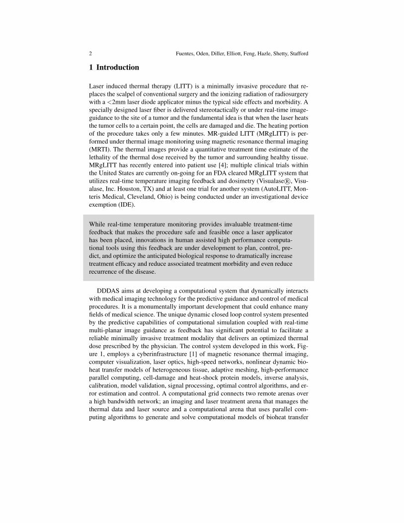


![Durum Wheat Stress Tolerance Induced by Endophyte Pantoea ... · locations confer to plants habitat-specific stress tolerance [7]. This ecological concept was recently validated](https://static.documents.pub/doc/80x56/5edde396ad6a402d66691df5/durum-wheat-stress-tolerance-induced-by-endophyte-pantoea-locations-confer-to.jpg)


