53
Computational Methods for Protein Structure Prediction Ying Xu Ying Xu
| Date post: | 19-Dec-2015 |
| Category: |
Documents |
| Upload: | lawrence-chase |
| View: | 226 times |
| Download: | 8 times |

Computational Methods for Protein Structure Prediction
Ying Xu Ying Xu

Outline introduction to protein structures
the problem of protein structure prediction
why we can predict protein structure
protein tertiary structure prediction – Ab initio folding– homology modeling
protein threading

Protein and Structure
>1MBN:_ MYOGLOBIN (154 AA) MVLSEGEWQLVLHVWAKVEADVAGHGQDILIRLFKSHPETLEKFDRFKHLKTEAEMKASEDLKKAGVTVLTALGAILKKKGHHEAELKPLAQSHATKHKIPIKYLEFISEAIIHVLHSRHPGNFGADAQGAMNKALELFRKDIAAKYKELGYQG
Protein sequenc
e
Protein structur
e
Oxygen storage
Protein function

Ball and stick
spacefill

Protein Structure protein sequence folds into a “unique” shape (“structure”) that
minimizes its free potential energy

Protein Structures Primary sequence
Secondary structure
MTYKLILNGKTKGETTTEAVDAATAEKVFQYANDNGVDGEWTYTE
-helix
-sheet
anti-parallel
parallel

Protein Structures Tertiary structure
Quaternary structure

Protein Structures Protein structure
– generally compact
Soluble protein structure– individual domains are generally globular
– they share various common characteristics, e.g. hydrophobic moment profile
Membrane protein structure
most of the amino acid sidechains of transmembrane segments are non-polar
polar groups of the polypeptide backbone of transmembrane segments generally participate in hydrogen bonds

Protein Tertiary Structures
Family: Clear evolutionary relationship, protein in the same family are homologous, sequence identity >=30%.
Superfamily: Low sequence identity, probable common evolutionary origin. Fold: May not have a common evolutionary origin. Major structural similarity.
Class: all-, all-, /, +, … http://scop.mrc-lmb.cam.ac.uk/scop/SCOP 1.65 release: 2327 families, 1294 superfamilies, 800 folds
SCOP: Structural Classification Of Proteins

Protein Tertiary Structures

Protein Structure Determination X-ray crystallography
– most accurate– in vitro– need crystals proteins – ~50K per structure
NMR – accurate– in vivo– no need for crystals– limited to small proteins
Cryo-EM– Imaging technology– Low-resolution

Protein Structure Prediction
Problem:Given the amino acid sequence of a protein,
what’s its 3-dimensional shape?
MVLSEGEWQLVLHVWAKVEADVAGHGQDILIRLFKSHPETLEKFDRFKHLKTEAEMKASEDLKKAGVTVLTALGAILKKKGHHEAELKPLAQSHATKHKIPIKYLEFISEAIIHVLHSRHPGNFGADAQGAMNKALELFRKDIAAKYKELGYQG
? ……..

Why Protein Structure Prediction? Importance of protein structure
– knowledge of the structure of a protein enable us to understand its function and functional mechanism
– design better mutagenesis experiments
– structure-based rational drug design Experimental methods for protein structure determination
Pros: high resolution Cons: time-consuming and very expensive

Why Protein Structure Prediction?
Big gap between the number of protein sequences and the number of protein structures– Uniprot/Swiss-prot, 283,454 protein sequences
– Uniprot/TrEMBL, 4,864,587 gene sequences
– PDB (Protein Data Bank), 48,385 protein structures
Fundamental, unsolved, challenging problem

Why We Can Predict Structure In theory, a protein structure can solved computationally
A protein folds into a 3D structure to minimizes its free potential energy
– Anfinsen’s classic experiment on Ribonuclease A folding in the 1960’s
– energy functions
This problem can be formulated as an optimization problem– protein folding problem, or ab initio folding

Why We Can Predict Structure While there could be billions of billions of proteins in nature, the
number of unique structural folds (shapes) might be small
90% of new structures submitted to PDB in the past three years have similar structural folds in PDB
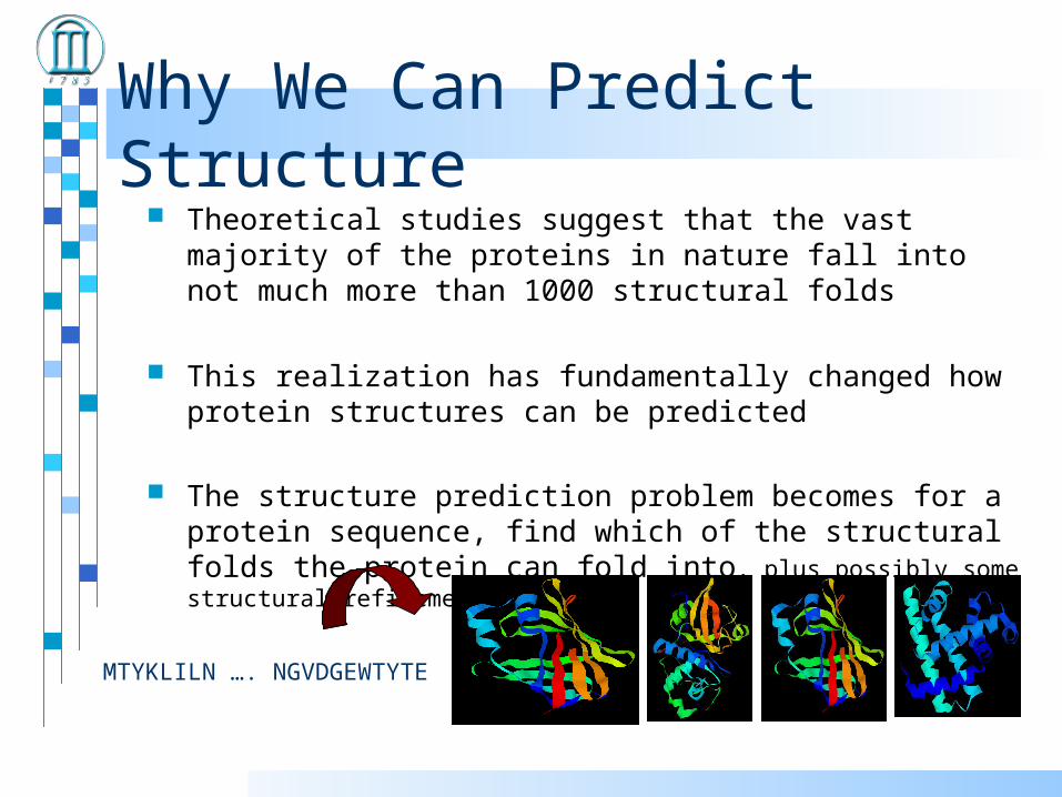
Why We Can Predict Structure Theoretical studies suggest that the vast majority of the proteins
in nature fall into not much more than 1000 structural folds
This realization has fundamentally changed how protein structures can be predicted
The structure prediction problem becomes for a protein sequence, find which of the structural folds the protein can fold into, plus possibly some structural refinement
MTYKLILN …. NGVDGEWTYTE

Computational Methods for Protein Structure Prediction
ab initio --use first principles to fold proteins --does not require templates --high computational complexity
•homology modeling --similar sequence similar structures --practically very useful, need homologues
• protein threading --many proteins share the same structural fold --a folding problem becomes a fold recognition problem
Need known protein structures

ab initio structure prediction
An energy function to describe the proteino bond energyo bond angle energyo dihedral angel energyo van der Waals energyo electrostatic energy
Efficient and reliable algorithms to search the conformational space to minimize the function and obtain the structure.

ab initio structure prediction
The problem is exceedingly difficult to solve– the search space is defined by psi/phi angles of backbone and side-
chain positions– the search space is enormous even for small proteins!– the number of local minima increases exponentially of the number
of residues
Theoretically solvable but practically infeasible!

ROSETTA (Dave Baker’s Lab)
Construct a library of small structure fragments, e.g. 9 AA
Cut a target sequence to sequence fragments. For each sequence fragment, choose structural candidate fragments from the fragment library
Assemble the fragment structures by Monte Carlo simulation
The generated structures are grouped into clusters
Clusters are ranked by their energy

Homology ModelingObservation: proteins with similar sequences tend to fold into similar structures.
1. Target sequence is aligned with the sequence of a known structure, they usually share sequence identity of 30% or higher
2. Superimpose target sequence onto the template, replacing equivalent side-chain atoms where necessary
3. Refine the model by minimizing an energy function.
Programs: Modeller http://salilab.org/modeller/
Swiss-Model http://swissmodel.expasy.org//SWISS-MODEL.html

Protein Threading Basic premise
Statistics from Protein Data Bank (~48,000 structures)
Chances for a protein to have a native-like structural fold in PDB are quite good (estimated to be 60-70%)
– Proteins with similar structural folds could be homologues or analogues
The number of unique structural (domain) folds in nature is fairly small (possibly a few thousand)
90% of new structures submitted to PDB in the past three years have similar structural folds in PDB

Protein Threading The goal: find the “correct” sequence-structure alignment
between a target sequence and its native-like fold in PDB
Energy function – knowledge (or statistics) based rather than physics based – Should be able to distinguish correct structural folds from
incorrect structural folds
– Should be able to distinguish correct sequence-fold alignment from incorrect sequence-fold alignments
MTYKLILN …. NGVDGEWTYTE

Protein Threading – four basic components
Structure database
Energy function
Sequence-structure alignment algorithm
Prediction reliability assessment

Protein Threading – structure database
Build a template database

Protein Threading – structure database
• Non-redundant representatives through structure-structure and/or sequence-sequence comparison
FSSP (http://www.bioinfo.biocenter.helsinki.fi:8080/dali/index.html)
(Families of Structurally Similar Proteins)
SCOP (http://scop.mrc-lmb.cam.ac.uk/scop/)
PDB-Select (http://www.sander.embl-heidelberg.de/pdbsel/)
Pisces (http://www.fccc.edu/research/labs/dunbrack/pisces/)

Protein Threading – energy function
MTYKLILNGKTKGETTTEAVDAATAEKVFQYANDNGVDGEWTYTE
how well a residue fits a structural environment: E_s
how preferable to put two particular residues nearby: E_p
alignment gap penalty: E_g
total energy: E_p + E_s + E_g
find a sequence-structure alignment to minimize the energy function

Protein Threading – energy function
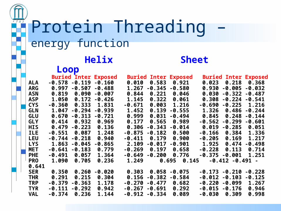
A singleton energy measures each residue’s preference in a specific structural environments– secondary structure
– solvent accessibility Compare actual occurrence against its “expected value” by
chance
Where

Protein Threading – energy function
A simple definition of structural environment– secondary structure: alpha-helix, beta-strand, loop– solvent accessibility: 0, 10, 20, …, 100% of accessibility– each combination of secondary structure and solvent
accessibility level defines a structural environment• E.g., (alpha-helix, 30%), (loop, 80%), …
E_s: a scoring matrix of 30 structural environments by 20 amino acids– E.g., E_s ((loop, 30%), A)
Singleton energy term
Protein Threading – energy function
Helix Sheet Loop Buried Inter Exposed Buried Inter Exposed Buried Inter ExposedALA -0.578 -0.119 -0.160 0.010 0.583 0.921 0.023 0.218 0.368ARG 0.997 -0.507 -0.488 1.267 -0.345 -0.580 0.930 -0.005 -0.032ASN 0.819 0.090 -0.007 0.844 0.221 0.046 0.030 -0.322 -0.487ASP 1.050 0.172 -0.426 1.145 0.322 0.061 0.308 -0.224 -0.541CYS -0.360 0.333 1.831 -0.671 0.003 1.216 -0.690 -0.225 1.216GLN 1.047 -0.294 -0.939 1.452 0.139 -0.555 1.326 0.486 -0.244GLU 0.670 -0.313 -0.721 0.999 0.031 -0.494 0.845 0.248 -0.144GLY 0.414 0.932 0.969 0.177 0.565 0.989 -0.562 -0.299 -0.601HIS 0.479 -0.223 0.136 0.306 -0.343 -0.014 0.019 -0.285 0.051ILE -0.551 0.087 1.248 -0.875 -0.182 0.500 -0.166 0.384 1.336LEU -0.744 -0.218 0.940 -0.411 0.179 0.900 -0.205 0.169 1.217LYS 1.863 -0.045 -0.865 2.109 -0.017 -0.901 1.925 0.474 -0.498MET -0.641 -0.183 0.779 -0.269 0.197 0.658 -0.228 0.113 0.714PHE -0.491 0.057 1.364 -0.649 -0.200 0.776 -0.375 -0.001 1.251PRO 1.090 0.705 0.236 1.249 0.695 0.145 -0.412 -0.491 -0.641SER 0.350 0.260 -0.020 0.303 0.058 -0.075 -0.173 -0.210 -0.228THR 0.291 0.215 0.304 0.156 -0.382 -0.584 -0.012 -0.103 -0.125TRP -0.379 -0.363 1.178 -0.270 -0.477 0.682 -0.220 -0.099 1.267TYR -0.111 -0.292 0.942 -0.267 -0.691 0.292 -0.015 -0.176 0.946VAL -0.374 0.236 1.144 -0.912 -0.334 0.089 -0.030 0.309 0.998

Protein Threading – energy function
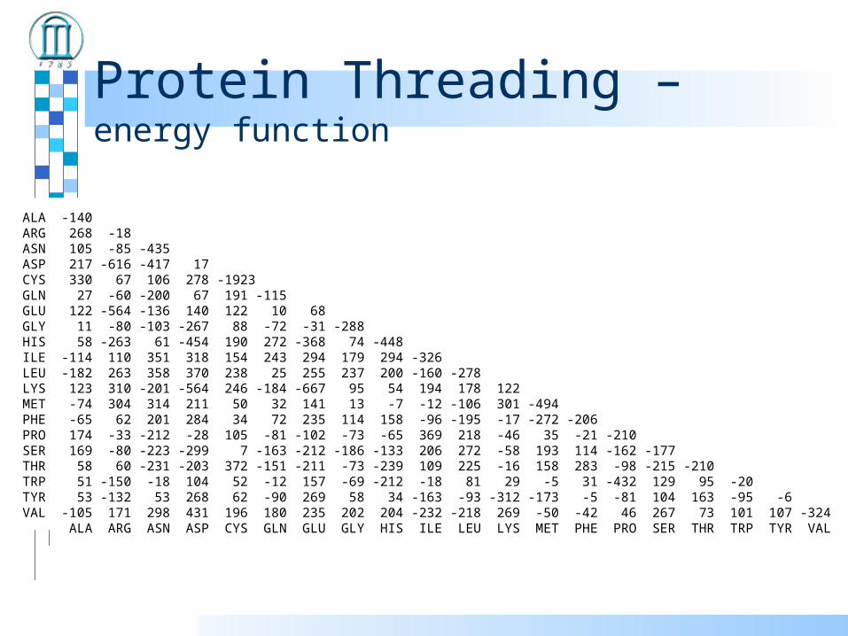
It measures the preference of a pair of amino acids to be close in 3D space.
How close is close?– distance dependent– single cutoff– C, C, or centroid of the sidechain
Observed occurrence of a pair compared with its “expected” occurrence
Pair-wise interaction energy term
Protein Threading – energy function
ALA -140ARG 268 -18ASN 105 -85 -435ASP 217 -616 -417 17CYS 330 67 106 278 -1923GLN 27 -60 -200 67 191 -115GLU 122 -564 -136 140 122 10 68GLY 11 -80 -103 -267 88 -72 -31 -288HIS 58 -263 61 -454 190 272 -368 74 -448ILE -114 110 351 318 154 243 294 179 294 -326LEU -182 263 358 370 238 25 255 237 200 -160 -278LYS 123 310 -201 -564 246 -184 -667 95 54 194 178 122MET -74 304 314 211 50 32 141 13 -7 -12 -106 301 -494PHE -65 62 201 284 34 72 235 114 158 -96 -195 -17 -272 -206PRO 174 -33 -212 -28 105 -81 -102 -73 -65 369 218 -46 35 -21 -210SER 169 -80 -223 -299 7 -163 -212 -186 -133 206 272 -58 193 114 -162 -177THR 58 60 -231 -203 372 -151 -211 -73 -239 109 225 -16 158 283 -98 -215 -210TRP 51 -150 -18 104 52 -12 157 -69 -212 -18 81 29 -5 31 -432 129 95 -20TYR 53 -132 53 268 62 -90 269 58 34 -163 -93 -312 -173 -5 -81 104 163 -95 -6VAL -105 171 298 431 196 180 235 202 204 -232 -218 269 -50 -42 46 267 73 101 107 -324 ALA ARG ASN ASP CYS GLN GLU GLY HIS ILE LEU LYS MET PHE PRO SER THR TRP TYR VAL
Protein Threading – energy function
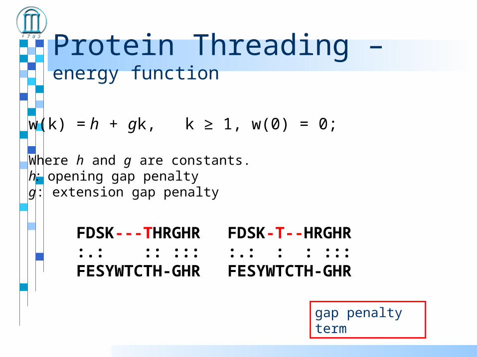
w(k) = h + gk, k ≥ 1, w(0) = 0;
Where h and g are constants.h: opening gap penaltyg: extension gap penalty
FDSK---THRGHR:.: :: :::FESYWTCTH-GHR
FDSK-T--HRGHR:.: : : :::FESYWTCTH-GHR
gap penalty term
Protein Threading – energy function
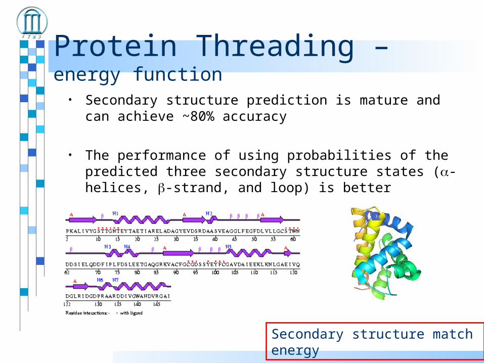
• Secondary structure prediction is mature and can achieve ~80% accuracy
• The performance of using probabilities of the predicted three secondary structure states (-helices, -strand, and loop) is better
Secondary structure match energy
Threading Parameter Optimization
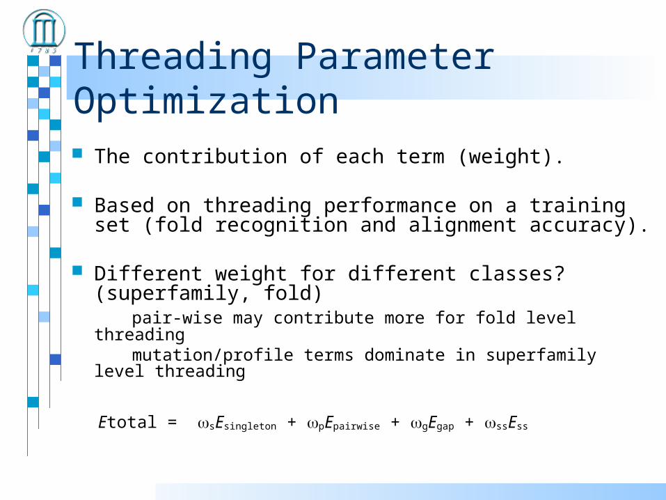
The contribution of each term (weight).
Based on threading performance on a training set (fold recognition and alignment accuracy).
Different weight for different classes? (superfamily, fold) pair-wise may contribute more for fold level threading mutation/profile terms dominate in superfamily level threading
Etotal = sEsingleton + pEpairwise + gEgap + ssEss

Protein Threading -- algorithm
Considering only singleton energy + gap penalty
Represent a structure a sequence of “structural environments”– (helix, 100%), (helix, 90%), ….. (strand, 0%)
Align a sequence MACKLPV …. with a structural sequence (helix, 100%), (helix, 90%), ….. (strand, 0%)

Protein Threading – dynamic programming
AAGG
AACG | | |
Two sequences: AACG and AAGG
A A C G
A
A
G
G
2
-1
-1
2
-1-12
2
3
4 3 2
5
2
3
5
Step #1: calculating alignment matrixRule:
1: initialization– fill the first row and column with matching scores
2: fill an empty cell based on scores of its left, upper and upper-left neighbors + the matching score of the current cell
3: chose the one giving the highest score

Protein Threading – dynamic programming
A A C G
A
A
G
G
2
-1
-1
2
-1-12
2
3
4 3 2
5
2
3
5
Step #2: Tracing back to recover the alignment
Rule:
1: start from the right-lower corner
2: trace back to left, upper or upper-left neighbor which gives the current cell’s score
3. Keep doing this until it cannot continue

Protein Threading – dynamic programming
Steps: 1. Initialization: construct an (n+1) x (m+1) matrix F for two sequences
of lengths n and m.
2. Matrix fill: for each cell in the matrix F, check all possible pathways back to the beginning of the sequence (allowing insertions and deletions) and give that cell the value of the maximum scoring.
3. Traceback: construct an alignment back from the last cell in the matrix (or the highest scoring) cell to give the highest scoring alignment.

Protein Threading -- dynamic programming
(helix, 100%)
(helix, 90%)
(helix, 80%)
(loop, 80%)
M
L
V
A
Protein Threading -- algorithm
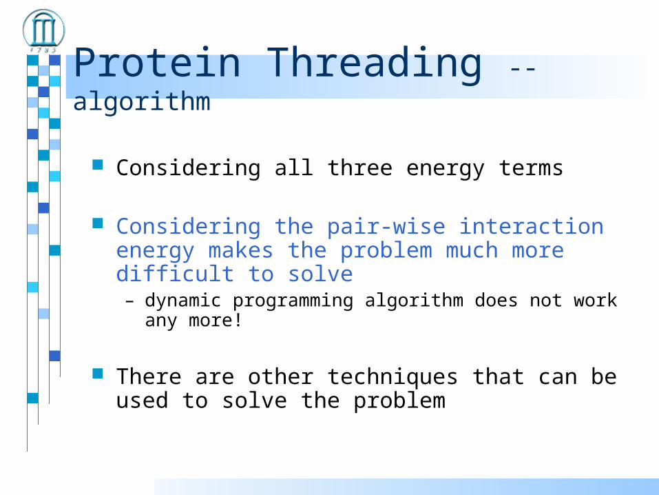
Considering all three energy terms
Considering the pair-wise interaction energy makes the problem much more difficult to solve – dynamic programming algorithm does not work any more!
There are other techniques that can be used to solve the problem

Protein Threading -- algorithm
Dynamic programming Heuristic algorithms for pair-wise interactions
– Frozen approximation algorithm (A. Godzik et al.)
– Double dynamic programming (D. Jones et al.)
– Monte carlo sampling (S.H. Bryant et al.)
Rigorous algorithms for pair-wise interactions– Branch-and-bound (R.H. Lathrop and T.F. Smith)
– Divide-and-conquer (Y. Xu et al.) --PROSPECT
– Linear programming (J. Xu et al.) –RAPTOR
– Tree decomposition (L. Cai et al.) Rigorous algorithm for treating backbone and side-chain
simultaneously (Li et al.)

Fold Recognition
MTYKLILNGKTKGETTTEAVDAATAEKVFQYANDNGVDGEWTYTE
Score = -1500 Score = -900Score = -1120Score = -720
Which one is the correct structural fold for the target sequence if any?
The one with the highest score ?
Fold Recognition
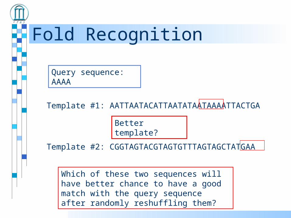
Template #1: AATTAATACATTAATATAATAAAATTACTGA
Query sequence: AAAA
Template #2: CGGTAGTACGTAGTGTTTAGTAGCTATGAA
Better template?
Which of these two sequences will have better chance to have a good match with the query sequence after randomly reshuffling them?
Fold Recognition
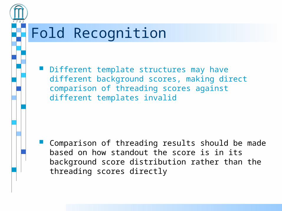
Different template structures may have different background scores, making direct comparison of threading scores against different templates invalid
Comparison of threading results should be made based on how standout the score is in its background score distribution rather than the threading scores directly

Fold Recognition
Threading 100,000 sequences against a template structure provides the baseline information about the background scores of the template
By locating where the threading score with a particular query sequence, one can decide how significant the score, and hence the threading result, is!
Not significant significant

Fold Recognition
Z-score = standard deviation
score - average
--randomly shuffle the query sequence and calculate the alignment score

Fold Recognition
Significance score versus prediction specificity/sensitivity
Z-score

Fold Recognition
Examine feature space of threading alignments: (singleton score, pair contact scores, secondary structure score, hydrophobic moment score, ......) versus true/false fold recognition
Separate true ones from false ones using support vector machine (SVM)
false
true
-2000, -500, -35, -90, ......, true
-1000, -201, -11, -500, ......, false
-5020, -900, -20, -75, ......, true
-1050, -185, -18, -320, ......, false
......

Fold Recognition
Each feature has somewhat different distributions in the true and false predictions
E.g., hydrophobic moments (Hydrophobic moments of protein structures: spatially profiling the
distribution, David Silverman, PNAS 2001 98: 4996-5001) is quite useful in distinguishing true from false threading predictions
Hydrophobic Moment profiles
-100
-80
-60
-40
-20
0
20
40
60
80
100
120
0 5 10 15 20 25 30 35
d (Angstromes)
H2
(d)

Application
Sequence preprocessing
Protein sequenceRemove signal peptide
Membrane/soluble??
Domain prediction
Database searching
Find homolog in PDB?
YESHomologymodeling
NO Fold recognition
3Dstructure
Sequence profile generationSecondary structure PredictionExperimental data constraints

Challenging Issues Much improved energy functions for threading
Considering side-chain information when doing protein threading
Effective integration of fragment-based methods and threading techniques


