Abstract A variety of natural or synthetic calcium phos-phate (CaP)-based scaffolds are currently produced for dentaland orthopaedic applications. These scaffolds have beenshown to stimulate bone formation due to their biocompat-ibility, osteoconductivity and osteoinductivity. The releaseof the Ca2+ ions from these scaffolds is of great interestin light of the aforementioned properties. It can depend ona number of biophysicochemical phenomena such as dis-solution, diffusion and degradation, which in turn dependon specific scaffold characteristics such as composition andmorphology. Achieving an optimal release profile can bechallenging when relying on traditional experimental workalone. Mathematical modelling can complement experimen-tation. In this study, the in vitro dissolution behaviour offour CaP-based scaffold types was investigated experimen-tally. Subsequently, a mechanistic finite element methodmodel based on biophysicochemical phenomena and specific
Electronic supplementary material The online version of thisarticle (doi:10.1007/s10237-016-0827-9) contains supplementarymaterial, which is available to authorized users.
1 Biomechanics Research Unit, GIGA In Silico Medicine, U.Liège, Chemin des Chevreuils 1, B52/3, 4000 Liège, Belgium
2 Biomechanics Section, Department of MechanicalEngineering, KU Leuven, Celestijnenlaan 300 C, Bus 2419,3001 Heverlee, Belgium
3 Prometheus, Division of Skeletal Tissue Engineering Leuven,KU Leuven, O&N 1, Herestraat 49, Bus 813, 3000 Leuven,Belgium
4 Tissue Engineering Unit, Skeletal Biology and EngineeringResearch Center, KU Leuven, O&N 1, Herestraat 49, Bus831, 3000 Leuven, Belgium
scaffold characteristics was developed to predict the experi-mentally observed behaviour. Before themodel could be usedfor local Ca2+ ions release predictions, certain parameterssuch as dissolution constant (kdc) and degradation constant(ksc) for each type of scaffold were determined by calibratingthemodel to the in vitro dissolution data. The resultingmodelshowed to yield release characteristics in satisfactory agree-ment with those observed experimentally. This suggests thatthe mathematical model can be used to investigate the localCa2+ ions release from CaP-based scaffolds.
Calcium phosphate (CaP) materials are widely used for den-tal and orthopaedic applications in the regeneration of bonedefects, because of their chemical similarity to native bonetissue and their ability to induce bone formation along withproviding structural support (Lanao et al. 2011). The releaseof calcium (Ca2+) and phosphate (Pi ) ions by dissolutionis believed to affect the bone cell chemotaxis, proliferationand differentiation. It has been reported in various in vitrostudies that calcium ion concentrations between 2 and 8 mMhave a profound impact on bone cell fate but optimum invivo calcium concentrations are still unknown (Bianchi et al.2014; Bléry et al. 2014; Carlier et al. 2011; Chai 2012a, b, c;Charles-Harris et al. 2008; Danoux 2015; Hong et al. 2014;Karadzic et al. 2015; Lobo et al. 2015; Mazón et al. 2015;Roberts et al. 2011; Shih et al. 2014). Once the CaP materi-als are implanted in vivo, they should induce bone formationbut should eventually completely degrade and be absorbed
by the body. Ideally, the degradation rate should be similarto the rate of bone formation (Wu and Ding 2005).
There are three major types of CaP materials usedfor bone tissue engineering applications: hydroxyapatite(HA,Ca10(PO4)6(OH)2), beta-tricalciumphosphate (β-TCP,Ca3(PO4)2) and biphasic calcium phosphate (BCP). HA cancause infections due to low degradation, whereas high degra-dation of β-TCP diminishes its structural function. Having amixture of HA and β-TCP results in BCP, which is moredegradable than HA whereas more stable than β-TCP. Asa result, the bioreactivity of BCPs can be controlled bymanipulating the HA/β-TCP ratio (Hong et al. 2014). Inaddition to the chemical composition (HA/β-TCP ratio),varying surface-structural properties, such as macro- andmicroporosity, specific surface area, roughness and overallgeometry, also affect bone formation (Bohner and Baum-gart 2004; Danoux 2015; Lobo et al. 2015; Mazón et al.2015). Moreover, it has been reported that the porosityor the pore size can influence the degradation rate. Scaf-folds with high porosity or small pore size degrade moreslowly than those with low porosity or large pore size (Wuand Ding 2005). Given their widespread medical use, thereremains a strong demand to design and develop CaP-basedbone substitutes that have optimal chemical compositionand surface-structural properties for the desired bone forma-tion (Bohner and Baumgart 2004). However, experimentallydetermining the ideal combinations of chemical compositionalong with surface-structural properties to maximize in vivobone formation for specific applications can be challenging.Nevertheless, testing these different combinations in silicocan save research time and costs.
Accurate mathematical models can be used to facilitatethe development of optimized degradable bone substitutesand other biomaterials by allowing rapid evaluation and val-idation of experimental parameters and also minimizing thenumber of required experimental studies (Frenning 2004;Kaunisto et al. 2013; Lao et al. 2011; Masaro and Zhu 1999;Siepmann et al. 1999; Siepmann and Siepmann 2013). Inaddition, they also allow quantitative understanding of thephysical, chemical and biological phenomena involved inthe controlled release of ions or molecules (Siepmann andGöpferich 2001; Snorradóttir et al. 2013). Due to significantadvances in information technology, mathematical mod-elling of biomaterial behaviour is increasing its academicand industrial importance, which makes it an integral partof future research and development in bone tissue engineer-ing. Concerning the development of optimized degradablescaffolds, the importance of such models lies in their rele-vance during the designing stage as well as the experimentalverification of degradation and release mechanism. How-ever, it is unlikely that there will be one mathematical modelthat will be able to describe any type of release of ions ormolecules from biomaterials. It is much more likely that
there will be different mathematical models, applicable tospecific types of systems that differ in geometry and com-position (Arifin et al. 2006; Brazel and Peppas 2000; Dashet al. 2010; Fredenberg et al. 2011; Frenning 2003; Frenninget al. 2005; Frenning and Strømme 2003; Frenning et al.2003; Polakovic et al. 1999; Siepmann and Peppas 2011;Siepmann and Siepmann 2008). Over the past few years, afew lattice-based three-dimensional (3D)mathematicalmod-els have been proposed to study the in vivo bone formationprocess in porous biodegradable CaP scaffolds (Adachi et al.2006; Byrne et al. 2007; Sun et al. 2013). Adachi et al.(2006) proposed a mathematical model of in vivo bone tis-sue regeneration that consisted of scaffold degradation dueto hydrolysis. Byrne et al. (2007) proposed a mathematicalmodel of in vivo tissue differentiation and bone regenerationin a degrading scaffold as a function of porosity, Young’smodulus, and dissolution rate. Sun et al. (2013) proposed amultiscale model of a biodegradable porous calcium phos-phate (CaP) scaffold to examine the effects of pore size andporosity on bone formation and angiogenesis. However, theabove-mentioned models being lattice-based did not capturethe actual geometry of the CaP scaffolds. Additionally, themodels had phenomenological description of the degrada-tion process as they were not interested in the degradationproducts themselves but rather in the changing stiffness andporosity of the scaffold.
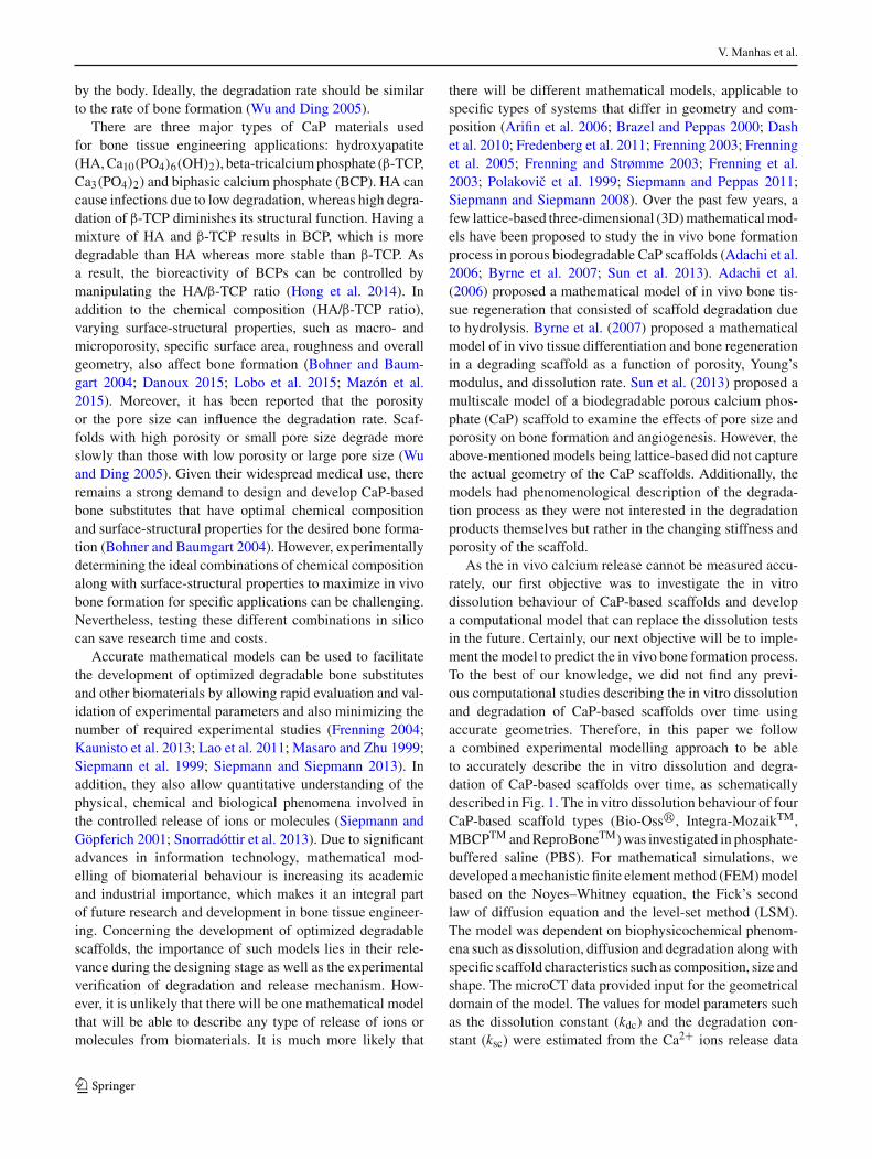
As the in vivo calcium release cannot be measured accu-rately, our first objective was to investigate the in vitrodissolution behaviour of CaP-based scaffolds and developa computational model that can replace the dissolution testsin the future. Certainly, our next objective will be to imple-ment themodel to predict the in vivo bone formation process.To the best of our knowledge, we did not find any previ-ous computational studies describing the in vitro dissolutionand degradation of CaP-based scaffolds over time usingaccurate geometries. Therefore, in this paper we followa combined experimental modelling approach to be ableto accurately describe the in vitro dissolution and degra-dation of CaP-based scaffolds over time, as schematicallydescribed in Fig. 1. The in vitro dissolution behaviour of fourCaP-based scaffold types (Bio-Oss�, Integra-MozaikTM,MBCPTM andReproBoneTM)was investigated in phosphate-buffered saline (PBS). For mathematical simulations, wedeveloped amechanistic finite elementmethod (FEM)modelbased on the Noyes–Whitney equation, the Fick’s secondlaw of diffusion equation and the level-set method (LSM).The model was dependent on biophysicochemical phenom-ena such as dissolution, diffusion and degradation along withspecific scaffold characteristics such as composition, size andshape. The microCT data provided input for the geometricaldomain of the model. The values for model parameters suchas the dissolution constant (kdc) and the degradation con-stant (ksc) were estimated from the Ca2+ ions release data
123
Computational modelling of local calcium ions release from calcium phosphate-based scaffolds
Fig. 1 Schematic diagram of combined experimental modellingapproach showing the initial estimation of the dissolution constant (kdc)anddegradation constant (ksc) from the in vitro dissolution tests and thenderiving the k∗
dc and the k∗sc by linking the kdc and the ksc, respectively, to
the structural properties measured in the microCT analysis using partialleast squares regression (PLSR). S denotes the CaP surface area in thescaffold
measured from dedicated dissolution experiments. Addition-ally, convergence and sensitivity analyses were carried outto investigate in more detail certain model parameters rele-vant to Ca2+ ions release. A partial least square regressions(PLSR) analysis was used to identify linear relationshipsbetween scaffold characteristics and the kdc and ksc valueswhich were used to estimate k∗
dc and k∗sc values. The model
outputs for both the sets of parameters were then compared.Finally, the linear relationships derived using PLSR wereused to investigate in silico the dissolution behaviour of CaPbiomaterials composed of different combinations of HA andβ-TCP.
2 Materials and methods
2.1 Calcium phosphate scaffolds
Four types of commercially available and clinically usedCaP or CaP/collagen composite bone void fillers wereused as scaffolds in this study (Table 1). Both Bio-Oss�(Geistlich, Wolhusen, Switzerland) and Integra-MozaikTM
(Integra LifeSciences, Plainsboro, USA) consist of CaP,which is distributed in an open collagen network, whereasMBCPTM (Biomatlante, Vigneux de Bretagne, France) andReproBoneTM (Ceramisys Ltd., Sheffield, UK) only consistof CaP. CaP of Bio-Oss� consists of natural bone mineral
made of bovine bone, whereas CaP of Integra-MozaikTM,MBCPTM and ReproBoneTM is of synthetic origin. Integra-MozaikTM is composed of 100% β-tricalcium phosphate(β-TCP), while MBCPTM and ReproBoneTM are composedof 60% hydroxyapatite (HA) and 40% β-TCP. All materi-als are described by the manufacturer as porous and in vivoresorbable. For this study, all the material blocks were cutwith a scalpel in cubes with a volume of 27mm3. All scaf-folds were stored at room temperature and used under sterileconditions for all experiments.
2.2 Structural characterization of the scaffolds usingmicrofocus computed tomography
The morphology and fractional composition (CaP/collagen/pores) of the scaffolds were characterized prior to (n = 5)and after (n = 1) the dissolution tests using microCT on aPhoenix NanoTom S (GE Measurements and Control Solu-tions, Germany) at an isotropic voxel size of 3μm3 (Table 2).The samples were scanned using a source voltage of 60 kVand current of 162μA, and an aluminium filter of 0.3 mmwas used to reduce beam hardening. The obtained 2400 radi-ographic images were reconstructed with Phoenix datos|xCT software, and the images were analysed using the in-house developed software tool Morphing CiTy (ULg, Liege,Belgium) and the commercially available tool CTAn (Brukermicro-CT Kontich, Belgium). Thresholding of the greyscalehistogram using a multilevel Otsu algorithm allowed thequantification of structural parameters for the complete scaf-fold and also for CaP and collagen separately (Sonnaert et al.2015). The dry weight normalized to the initial total scaffoldvolume (TSV) of each scaffold was recorded prior to (n = 5)and after (n = 1) the dissolution test (Table 2).
2.3 Calcium (Ca2+) dissolution kinetics of the scaffolds
The dissolution kinetics of the scaffolds (n = 3, see Table 2)in phosphate-buffered saline (PBS) solution (Biowhittaker�,without Ca2+ and Mg2+ ions) at 37 ◦C were investigated byplacing scaffolds inside a 15-ml Eppendorf tube placed onan orbital rotator rotating at 10 rpm. The PBS solution wasrefreshed at day 1, 3, 6 and 21. All the samples were storedat 4 ◦C. Before measurement, 240μl nitric acid was addedto the samples to ensure there was no precipitation in themedium (Roberts et al. 2011). The released Ca2+ over 21days was measured by a calcium micro ion electrode (LazarResearch Laboratories, Inc., USA).
2.4 Statistics
Data are expressed as mean ± standard deviation (SD). Sta-tistical significancewas determined using the 1-wayANOVAwith a post hoc Tukey test and the Student’s t test. Statistical
123
V. Manhas et al.
Table 1 Four commercially available scaffolds used in this study with their structural and material characteristics as provided by the manufacturers.No detailed data were provided for Integra-MozaikTM by the manufacturer
Scaffold CaP composition Polymeric matrix type Macropore size (μm) Micropore size (μm) Total porosity (%)
Bio-Oss� Bovine bone granules Col-I 200–600 0.1–1 83
Integra-MozaikTM 100% β-TCP Col-I – – –
MBCPTM 60% HA+40% β-TCP No polymeric matrix 300–600 <10 70
ReproBoneTM 60% HA+40% β-TCP No polymeric matrix 200–800 1–10 80
Table 2 Overview of theexperimental design used in thisstudy
Day (n)
Bio-Oss� Integra-MozaikTM MBCPTM Repro BoneTM
microCT analysis D0 5 5 5 5
D21 1 1 1 1
Dissolution experiments D0 3 3 3 3
D1 3 3 3 3
D3 3 3 3 3
D6 3 3 3 3
D21 3 3 3 3
Normalized weightmeasurements
D0 5 5 5 5
D21 1 1 1 1
The microCT data available for 1 scaffold at day 21 time point (after the dissolution test) was only used as aqualitative compliment to the study
significance is indicated on all graphs as follows: *: p< 0.05,**: p < 0.01, ***: p < 0.001. Partial least square regression(PLSR) analysis was performed in JMP� 11 (SAS InstituteInc., Cary, NC).
3 Mathematical model
3.1 Model formulation
In this paper, we developed a mathematical model to pre-dict the in vitro dissolution behaviour of CaP-based scaf-folds in PBS solution using the experimental dissolutionresults from Bio-Oss�, Integra-MozaikTM, MBCPTM andReproBoneTM.As the initial solid calcium concentrationwaslarger than the solubility of the Ca2+ ions in PBS solution,the solid and the dissolved Ca2+ coexisted at the same timeduring the dissolution process. The concentration changeof dissolved Ca2+ ions with time was described using adiffusion–dissolution equation (1). The Ca2+ ions diffusionprocess was implemented using Fick’s second law of diffu-sion,whereas the dissolution processwas implemented basedon theNoyes–Whitney equation (Frenning et al. 2005) wherethe rate of dissolution of a substance is described as a func-tion of, amongst others, the concentration of the substanceand its diffusion coefficient.
∂C
∂t= ∇ · (D∇C) + kdc
SD
VPBS(Csol − C) (1)
where C is the dissolved Ca2+ ions concentration (mM),D is the diffusion coefficient (m2/s), kdc is the dissolutionconstant (m−1), S is the CaP surface area (m2) calculatedfrom the input microCT image (Table 3), VPBS is the PBSvolume (1.5×10⁻⁵ m3) andCsol is the solubility of the Ca2+ions in PBS (mM). As the thickness of the boundary layercould not be measured experimentally, it was considered asa mathematical parameter that was incorporated in the kdcin Eq. (1). The diffusion coefficient of Ca2+ ions in PBSsolution at 37 ◦C was considered to be constant and it wascalculated using Stokes–Einstein equation (2) (Young et al.1980) given below:
D = kBT
6πηr= 2.83 × 10⁻⁹ m2/s (2)
where kB is the Boltzmann’s constant (1.38 × 10⁻²³m2kgs−2K−1), T is the absolute temperature (37 ◦C = 310.5K ),η is the viscosity of PBS at 37 ◦C (0.0007 Pa s) (Horbett et al.1988) and r is the radius of Ca2+ ion (114 pm) (Shannon1976). The diffusion coefficient (D) for calcium chloride(CaCl2) in aqueous solutions at 310.15Khas been reported tobe (1.601±0.02)×10−9m2/s (Ribeiro et al. 2008)which is ofthe same order ofmagnitude that was calculated theoreticallyin this study.
To implement degradation of the scaffold, we used thelevel-set method (LSM) to track implicitly the movement ofthe scaffold interfaceΓ with velocity v during the dissolution
123
Computational modelling of local calcium ions release from calcium phosphate-based scaffolds
Fig. 2 Computational domain. Left Eppendorf tube filled with PBS and the scaffold. a Iso-levels of ϕ function (thick line represents the CaPinterface Γ where ϕ = 0). b CaP indicator function (IΩ1) computed over the domain
process. To implement the LSM, a microCT cross-sectionalimage of a scaffold was projected on a 2D finite elementdomain Ω ⊂ R
2. The domain was then decomposed intotwo sub-domains Ω1 and Ω2, and the interface/boundarybetween two partitions was denoted by Γ . The level-setfunction (ϕ), a continuous scalar distance function, was thencomputed over the domain; ϕ was positive in Ω1 (scaffold),negative in Ω2 (PBS solution) and zero on Γ (boundary ofthe scaffold) (Fig. 2). The motion of the interface (Γ ) in thenormal direction was described by the level-set equation (3)with a velocity field v (Guyot et al. 2014):
∂ϕ
∂t= v |∇ϕ| (3)
The degradation velocity v was derived by dividing the dis-solution term in Eq. (1) by S · Csol/VPBS, where S is theCaP surface area (m2), Csol is the solubility of Ca2+ ions inPBS (mM) and VPBS is the PBS volume (m3), thus resultingin degradation velocity as shown below in Eq. (4), with kscbeing a dimensionless degradation constant.
v = ksckdcD (Csol − C) /Csol (4)
In this model, the solubility (Csol) of Ca2+ ions was esti-mated to be 0.17 mM. The modelling of precipitation wasnot considered in this model as we did not detect precipitateformation during the experiments. The model also did notconsider explicitly the influence of the CaP crystallinity orcrystal size on the dissolution behaviour. The influence ofcollagen distribution around CaP in Bio-Oss� and Integra-MozaikTM on the Ca2+ ion dissolution was not modelled.
3.2 Model implementation
Themathematical domain for dissolution test simulation wasrepresented by the front view of a 15-ml Eppendorf tube(Fig. 2). Themodelwas implemented using the finite elementmethod (FEM) with an open-source partial differential equa-tion (PDE) solver FreeFem++ (http://freefem.org) (Hecht2012). This solver is well adapted for the type of modeldeveloped in this study due to its ability to deal with com-plex geometries and meshes and its simplified way to treatany type of PDE by implementing its variational form.
To implement the Dirichlet-type boundary condition forCa2+ ions on a penalized FEM domain, we used an indicatorfunction (IΩ1) which is described by Eq. (5), where ϕ is thelevel-set function.
IΩ1 :={1 if x ∈ Ω1 (ϕ > 0)0 if x /∈ Ω1 (ϕ ≤ 0)
(5)
The initial condition for dissolved Ca2+ ions was set as:
C(0,Ω) = 0 for dissolved Ca2+ ions (6)
To impose no-flux boundary condition, we implemented thehomogenous Neumann boundary condition on ∂Ω as shownbelow:
∂C
∂n= 0 (7)
Essentially, a convergence study (Online resource 1) wasexecuted to determine a time step as well as a mesh-size-
independent numerical solution. This resulted in a time stepof 3600 s and a number of elements being 115,460.
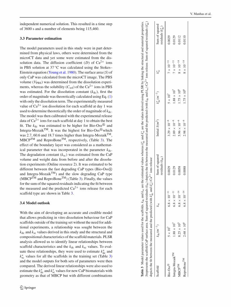
3.3 Parameter estimation
The model parameters used in this study were in part deter-mined from physical laws, others were determined from themicroCT data and yet some were estimated from the dis-solution data. The diffusion coefficient (D) of Ca2+ ionsin PBS solution at 37 ◦C was calculated using the Stokes–Einstein equation (Young et al. 1980). The surface area (S) ofonly CaP was calculated from the microCT image. The PBSvolume (VPBS) was determined from the dissolution experi-ments, whereas the solubility (Csol) of the Ca2+ ions in PBSwas estimated. For the dissolution constant (kdc), first theorder of magnitude was theoretically calculated using Eq. (1)with only the dissolution term. The experimentally measuredvalue of Ca2+ ion dissolution for each scaffold at day 1 wasused to determine theoretically the order of magnitude of kdc.The model was then calibrated with the experimental releasedata of Ca2+ ions for each scaffold at day 1 to obtain the bestfit. The kdc was estimated to be higher for Bio-Oss� andIntegra-MozaikTM. It was the highest for Bio-Oss�whichwas 2.7, 60.6 and 18.7 times higher than Integra-MozaikTM,MBCPTM and ReproBoneTM, respectively, (Table 3). Theeffect of the boundary layer was considered as a mathemat-ical parameter that was incorporated in the parameter kdc.The degradation constant (ksc) was estimated from the CaPvolume and weight data from before and after the dissolu-tion experiments (Online resource 2). It was estimated to bedifferent between the fast degrading CaP types (Bio-Oss�and Integra-MozaikTM) and the slow degrading CaP type(MBCPTM and ReproBoneTM) (Table 3). Finally, the valuesfor the sum of the squared residuals indicating the fit betweenthe measured and the predicted Ca2+ ions release for eachscaffold type are shown in Table 3.
3.4 Model outlook
With the aim of developing an accurate and credible modelthat allows predicting in vitro dissolution behaviour for CaPscaffolds outside of the training set without the need for addi-tional experiments, a relationship was sought between thekdc and ksc values derived in this study and the structural andcompositional characteristics of the scaffoldmaterials. PLSRanalysis allowed us to identify linear relationships betweenscaffold characteristics and the kdc and ksc values. To eval-uate these relationships, they were used to estimate k∗
dc andk∗sc values for all the scaffolds in the training set (Table 3)and the model outputs for both sets of parameters were thencompared. The derived linear relationships were also used toestimate the k∗
dc and k∗sc values for newCaP biomaterials with
geometry as that of MBCP but with different combinations Table3
Modelparameter
values
used
forthe
scaffolds.k d
candk scaretheestim
ated
values
whereas
k∗ dcandk∗ sc
arethevalues
derivedusingPL
SRby
linking
thestructuralandmaterialp
roperties.
SrepresentsthescaffoldCaP
surfacearea.S
umof
squaredresidu
als(k
dc)depictsthefit
betw
eenthemeasuredandthepredicted(w
ithk d
candk sc)Ca2
+ions
release.Su
mof
squaredresidu
als(k
∗ dc)
depictsthefit
betw
eenthemeasuredandthepredicted(w
ithk∗ dc
andk∗ sc
)Ca2
+ions
release
Scaffold
k dc(m
−1)
k sc
Sum
ofsquared
residu
als(k
dc)
Initial
S(m
2)
k∗ dc(m
−1)
k∗ scSu
mof
squared
residu
als(k
∗ dc)
Bio-O
ss�
5×
107
6.8
×10
−11
0.0029
3.29
×10
−65
×10
77
×10
−11
0.0029
Integra-MozaikT
M1.88
×10
76.8
×10
−11
0.0129
2.89
×10
−61.88
×10
77
×10
−11
0.0129
MBCPT
M8.25
×10
56.8
×10
−14
0.0058
5.96
×10
−61.75
×10
68
×10
−14
0.0112
ReproBoneT
M2.68
×10
66.8
×10
−14
0.0088
2.98
×10
−61.75
×10
68
×10
−14
0.0110
123
Computational modelling of local calcium ions release from calcium phosphate-based scaffolds
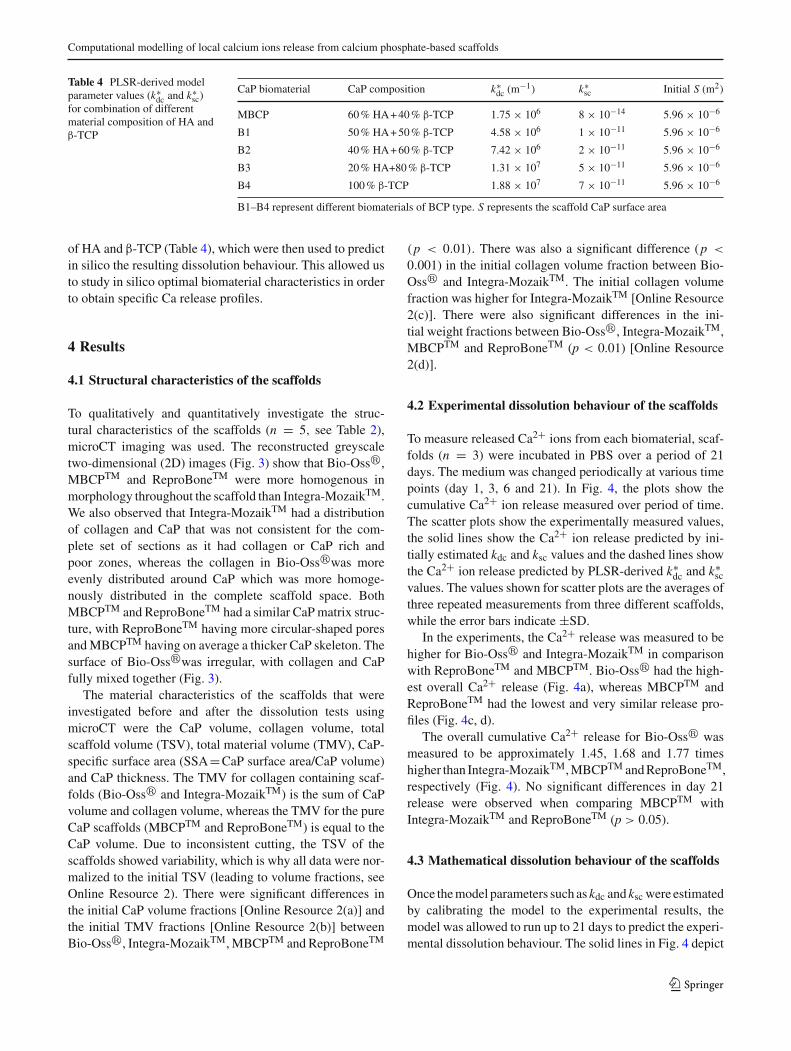
Table 4 PLSR-derived modelparameter values (k∗
dc and k∗sc)
for combination of differentmaterial composition of HA andβ-TCP
B1–B4 represent different biomaterials of BCP type. S represents the scaffold CaP surface area
of HA and β-TCP (Table 4), which were then used to predictin silico the resulting dissolution behaviour. This allowed usto study in silico optimal biomaterial characteristics in orderto obtain specific Ca release profiles.
4 Results
4.1 Structural characteristics of the scaffolds
To qualitatively and quantitatively investigate the struc-tural characteristics of the scaffolds (n = 5, see Table 2),microCT imaging was used. The reconstructed greyscaletwo-dimensional (2D) images (Fig. 3) show that Bio-Oss�,MBCPTM and ReproBoneTM were more homogenous inmorphology throughout the scaffold than Integra-MozaikTM.We also observed that Integra-MozaikTM had a distributionof collagen and CaP that was not consistent for the com-plete set of sections as it had collagen or CaP rich andpoor zones, whereas the collagen in Bio-Oss�was moreevenly distributed around CaP which was more homoge-nously distributed in the complete scaffold space. BothMBCPTM and ReproBoneTM had a similar CaPmatrix struc-ture, with ReproBoneTM having more circular-shaped poresandMBCPTM having on average a thicker CaP skeleton. Thesurface of Bio-Oss�was irregular, with collagen and CaPfully mixed together (Fig. 3).
The material characteristics of the scaffolds that wereinvestigated before and after the dissolution tests usingmicroCT were the CaP volume, collagen volume, totalscaffold volume (TSV), total material volume (TMV), CaP-specific surface area (SSA=CaP surface area/CaP volume)and CaP thickness. The TMV for collagen containing scaf-folds (Bio-Oss� and Integra-MozaikTM) is the sum of CaPvolume and collagen volume, whereas the TMV for the pureCaP scaffolds (MBCPTM and ReproBoneTM) is equal to theCaP volume. Due to inconsistent cutting, the TSV of thescaffolds showed variability, which is why all data were nor-malized to the initial TSV (leading to volume fractions, seeOnline Resource 2). There were significant differences inthe initial CaP volume fractions [Online Resource 2(a)] andthe initial TMV fractions [Online Resource 2(b)] betweenBio-Oss�, Integra-MozaikTM,MBCPTM and ReproBoneTM
(p < 0.01). There was also a significant difference (p <
0.001) in the initial collagen volume fraction between Bio-Oss� and Integra-MozaikTM. The initial collagen volumefraction was higher for Integra-MozaikTM [Online Resource2(c)]. There were also significant differences in the ini-tial weight fractions between Bio-Oss�, Integra-MozaikTM,MBCPTM and ReproBoneTM (p < 0.01) [Online Resource2(d)].
4.2 Experimental dissolution behaviour of the scaffolds
To measure released Ca2+ ions from each biomaterial, scaf-folds (n = 3) were incubated in PBS over a period of 21days. The medium was changed periodically at various timepoints (day 1, 3, 6 and 21). In Fig. 4, the plots show thecumulative Ca2+ ion release measured over period of time.The scatter plots show the experimentally measured values,the solid lines show the Ca2+ ion release predicted by ini-tially estimated kdc and ksc values and the dashed lines showthe Ca2+ ion release predicted by PLSR-derived k∗
dc and k∗sc
values. The values shown for scatter plots are the averages ofthree repeated measurements from three different scaffolds,while the error bars indicate ±SD.
In the experiments, the Ca2+ release was measured to behigher for Bio-Oss� and Integra-MozaikTM in comparisonwith ReproBoneTM and MBCPTM. Bio-Oss� had the high-est overall Ca2+ release (Fig. 4a), whereas MBCPTM andReproBoneTM had the lowest and very similar release pro-files (Fig. 4c, d).
The overall cumulative Ca2+ release for Bio-Oss� wasmeasured to be approximately 1.45, 1.68 and 1.77 timeshigher than Integra-MozaikTM,MBCPTM andReproBoneTM,respectively (Fig. 4). No significant differences in day 21release were observed when comparing MBCPTM withIntegra-MozaikTM and ReproBoneTM (p > 0.05).
4.3 Mathematical dissolution behaviour of the scaffolds
Once themodel parameters such as kdc and ksc were estimatedby calibrating the model to the experimental results, themodel was allowed to run up to 21 days to predict the experi-mental dissolution behaviour. The solid lines in Fig. 4 depict
123
V. Manhas et al.
Fig. 3 Typical cross-sectional microCT images of the scaffolds. The CaP phase is clearly visible in light grey tones. Arrows indicate the collagen(dark grey) present in Bio-Oss� and Integra-MozaikTM. Scale bar 1mm
the predicted amount of Ca2+ ions released over the exper-imental measurement time. The values for the sum of thesquared residuals indicating the fit between themeasured andthe predicted Ca2+ ions release (using kdc and ksc) for eachscaffold type are shown in Table 3. It is evident that there is anagreement between the predicted (solid lines) and the experi-mentallymeasured (scatter plots) cumulative release (Fig. 4).Themodelwas also able to predict the spatio-temporal degra-dation and the dissolution of scaffolds over time. Figure 5a–cdepicts the degradation of Bio-Oss�predicted on day 0, day10 and day 21, whereas Fig. 5d–f depicts the local concen-tration of Ca2+ ions predicted during dissolution at the sametime points.
Afterwards, the estimated kdc and ksc were linked to thestructural andmaterial properties obtained from themicroCTanalysis, using PLSR (Table 3). From Eqs. (1), (8) and(9), it can be inferred that the dissolution and degrada-tion of CaP in scaffolds are primarily governed by scaffold
composition (% bovine bone granules, % β-TCP, % HA).Structural parameters such as the CaP surface area andporosity are taken into account through the input microCTimage.
kdc∗ = 105(−95.83 + 5.95 ∗ % Bovine bone
+ 2.83 ∗ % β-TCP) (8)
ksc∗ = 10−11(6.8 − 0.1132 ∗ %HA) (9)
With the initially estimated kdc and ksc values, the modelpredicted CaP degradation between 9 and 18% for Bio-Oss� and Integra-MozaikTM whereas the predicted CaPdegradation was between 0.5 and 5% for MBCPTM andReproBoneTM (Fig. 6a). The microCT CaP volume changedata normalized to the initial total scaffold volume (TSV)qualitatively confirmed the degradation prediction by themodel (Fig. 6b).
123
Computational modelling of local calcium ions release from calcium phosphate-based scaffolds
Fig. 4 Dissolution behaviour of different scaffolds observed exper-imentally (scatter plot), predicted mathematically [solid lines (kdc)]and model corroboration [dashed lines (k∗
dc)]. a Bio-Oss�. b Integra-
MozaikTM. c MBCPTM. d ReproBoneTM. In a and b the dotted linecoincides with the solid lines. Error bars indicate ±SD (n = 3)
4.4 Model sensitivity analysis and outlook
We carried out the single-parameter sensitivity analysis toinvestigate the effect of a number of estimated parameterson the simulation outcome, which is shown in Fig. 7. Weinvestigated the effect of altering the value for solubility(Csol) and observed that it altered the final outcome. A 10 %change in this value resulted in a corresponding 10% changein the predicted calcium release (Fig. 7a). Additionally, weinvestigated the influence of using other representative 2Dimages of Bio-Oss� and Integra-MozaikTM in the modeldomain. We observed no significant differences in simula-tion results when using other images from the same stackfor the Bio-Oss� scaffold (Fig. 7b); however, we did findlarger differences for Integra-MozaikTM which has a moreheterogeneous structure (Fig. 7c).
Additionally, the obtained relationships between the kdcand ksc and the compositional characteristics were tested byusing the k∗
dc and the k∗sc values calculated from Eqs. (8) and
(9) for each scaffold type to predict the experimental dis-
solution behaviour. Figure 4 shows a satisfactory agreementbetween the predicted (solid lines), PLSR derived (dashedlines) and experimentally measured (scatter plots) releaseof Ca2+ ions, which demonstrates acceptable accuracy ofthe model’s representation of the real in vitro dissolutionexperimental set-up. The values for the sum of the squaredresiduals indicating the fit between themeasured and the pre-dicted Ca2+ ions release (using k∗
dc and k∗sc) for each scaffold
type are shown in Table 3. In addition, for scaffold degra-dation, the PLSR-derived k∗
dc and k∗sc values were similar
to the initially estimated values, as was the model outcomeobtained with those PLSR-derived values (Fig. 6). Finally,using the PLSR-derived linear relationships, the model wasused to investigate the dissolution behaviour of a biomaterialwith geometry as that of MBCP but with different combi-nations of HA and β-TCP (Fig. 8). It is clearly evident thatthe increase of β-TCP in biomaterial composition resultedin higher dissolution (Fig. 8a) along with higher degradation(Fig. 8b)whereas the effect ofHA in biomaterial compositionwas completely opposite to β-TCP.
123
V. Manhas et al.
Fig. 5 Dissolution behaviour of Bio-Oss� predicted mathematically. a Scaffold shape at day 0, b at day 10, and c at day 21. d–f Local Ca2+ ionconcentration during dissolution at day 0, 10 and 21. The circles in images a–c highlight degradation of the scaffold
Fig. 6 a Change in the scaffold surface area (2D) as predicted by themodel using kdc and k∗
dc. D0, day 0; D21, day 21. b Quantitative resultsof the scaffold CaP volume fraction (%). In b, the data have been nor-
malized to the initial total scaffold volume (TSV) leading to fractions.Statistical significance: **p < 0.01; ***p < 0.001 (n = 5 for day 0 andn = 1 for day 21). D0, day 0; D21, day 21
5 Discussion
In this study, a combined experimental-modelling approachwas used to describe the in vitro dissolution of CaP-basedscaffolds over time. First, the in vitro dissolution kinetics ofchemically and structurally different CaP biomaterials wereinvestigated in PBS. Proceeding from the in vitro experi-ments, a 2D FEM model based on the diffusion-dissolutionequation along with the level-set equation was developedto predict the in vitro dissolution behaviour of CaP-based
scaffolds in PBS solution. The model parameters were eitherdetermined from the experimental results or derived from theresults described in the literature or estimated. The modelwas able to capture the in vitro dissolution of Ca2+ ionsin accordance with the experimental reports. Additionally,a single-parameter sensitivity analysis was carried out todetermine the influence of specific model parameters on thepredicted outcome. This obtained model is able to predictthe dissolution behaviour of CaP-based scaffolds as well asthe local calcium concentration inside the scaffold based on
123
Computational modelling of local calcium ions release from calcium phosphate-based scaffolds
Fig. 7 Parameter sensitivity analyses—a influence of the solubility(Csol) on the Ca2+ ions release for Bio-Oss�. In a, scatter plot repre-sents the experimental results. Influence of using different images withvarying initial CaP surface area (S) on the modelling outcome b Bio-
Oss� and c Integra-MozaikTM. Boxed values indicate the values usedin the analyses. In b and c the lines that are not visible coincide withthe solid line
Fig. 8 Investigating the dissolution and degradation behaviour of a biomaterial with multiple combinations of β-TCP and HA. a Dissolution andb degradation behaviour for scaffolds with a geometry as that of MBCP but a composition that is a mix of β-TCP and HA
the parameters that can be obtained from microCT imaging.In order to limit the amount of experimental work needed tomake predictions for scaffold materials within the trainingset, relations were derived between the scaffold composi-tional characteristics and the kdc and ksc values and thentested on the training set itself. Finally, using the PLSR-derived linear relationships, themodelwas used to investigatethe dissolution behaviour of a biomaterialwith different com-binations of HA and β-TCP.
In this study, the in vitro dissolution kinetics of bio-materials broadly classified as fast degrading (Bio-Oss�and Integra-MozaikTM) and slow degrading (MBCPTM andReproBoneTM) scaffolds were investigated in PBS. The fourbiomaterials contained different chemical formation and vol-ume percentages of CaP, along with different structuralcharacteristics such as collagen volume, total scaffold vol-ume (TSV) and total material volume (TMV). The observedchemical and structural variations between scaffold typesallowed for differences in release kinetics of Ca2+ ions in
PBS medium, where the fast degrading scaffolds were foundto have higher in vitro dissolution than the slow degradingscaffolds (Fig. 4). It is worth mentioning that the dissolutionof bovine bone granules in Bio-Oss� is higher when com-pared to synthetic HA (Mezahi et al. 2009).We did not detectprecipitate formation.
The reported mathematical analysis of in vitro dissolutionbehaviour of CaP-based biomaterials is themost mechanisticmodel published to date. Previous models describing CaP-based scaffold dissolution behaviour were either theoreticalin nature (Bohner and Baumgart 2004) or lattice-based thatdid not capture the actual geometry of the CaP scaffolds.Additionally, the lattice based models used a phenomeno-logical description of the degradation process (Adachi et al.2006; Byrne et al. 2007; Sun et al. 2013). However, by explic-itly incorporating dedicated experimental results along withtaking into account the accurate geometrical shape of thescaffolds, this study takes an important step towards the use ofin silico modelling in biomaterial design. This study focuses
123
V. Manhas et al.
on measurements and modelling of Ca2+ ions dissolution, soit did not consider the influenceof themicroporosity, collagendistribution, CaP crystallinity or crystal size, precipitation,phosphate (PO3−
4 ) ions dissolution or changing collagen vol-ume on the diffusivity behaviour of Ca2+ ions. Ultimately,these influences need to be included comprehensively in themodel (Gao and Fagerness 1995). Additional simplificationsmade in the course of this study pertain mainly to the mod-elling aspects. The first simplification to be dealt within thefuture is the fact that the model only looks at dissolutionkinetics in 2D, thereby neglecting any 3D spatial aspects,which might influence the overall numerical solution. It hasbeen reported that the parameter values estimated by the 2Dmodel could be overestimated with respect to the ones pre-dicted by the 3D model, which could influence the overalloutput of the model (Nava et al. 2013). The second simpli-fication included in the model was exclusion of solving thefluid flow due to rotation during the dissolution experimentas well as the advection of Ca2+ ions due to fluid flow andthe movement of scaffold due to rotation. This was done tokeep the model computationally inexpensive and is in linewith other models studying drug release from carriers underdynamic conditions (Arifin et al. 2006; Frenning et al. 2003;Snorradóttir et al. 2013). It isworth stating that thismodel canbe adapted to potentially recapitulate the local in vivo (wherethe fluid flow is not so important) Ca2+ ion concentrationsduring the dissolution process by making adjustments to theexisting parameters and including additional parameters spe-cific to the in vivo environment.
The values for unknown parameters such as dissolutionconstant (kdc) and degradation constant (ksc) for each scaf-fold type were determined by calibrating the model to theexperimental release data of Ca2+ ions. It is evident fromFig. 4 that there was overall an acceptable though not perfectagreement between the predicted (solid lines) and the exper-imentally measured (scatter plots) release profiles. It is alsoevident from Fig. 4 that the model is not able to accuratelycapture the initial release. This could be due to the fact thatthe model does not consider advection of Ca2+ ions due tofluid flow. Moreover, we did observe that using a higher kdcvalue captured the initial release but greatly overestimated therelease at day 21. Therefore, the value of kdc was estimated bykeeping the best possible balance between underestimationof initial and overestimation of final calcium release profile.Due to a substantial lack of relevant data on this matter, werestricted ourselves to the use of a fixed value for kdc inthis study. However, using the value of kdc as an exponen-tially decreasing function rather than a constant value wouldgive a better fit with experimental data. The use of kdc as anexponentially decreasing function could take into account theeffect of adhesion of precipitated ions to the surface as wellas the effect of changing pH during the dissolution process.
Additionally, the model was able to predict the spatial-temporal degradation and dissolution of the scaffolds(Fig. 5a–f). This spatial information represents one of theclear added values of computational modelling in the con-text of designing new biomaterials. Nomeasurement devicesexist to date that allow this spatial representation of the localcalcium concentration—even though these local concentra-tions are what is felt by the cells seeded onto the scaffold.Computational models therefore allow to link the insightfrom in vitro experiments by studying the effect of Ca2+ ionson cellular behaviour (Arifin et al. 2006) to the design of opti-mized biomaterials. Furthermore, themodel predicted higherdegradation of CaP for Bio-Oss� and Integra-MozaikTM,whichwas expected due to their higher dissolution behaviour(Fig. 6). Moreover, the single-parameter sensitivity analysessuch as the one carried out in this study was able to identifythe influence of a few estimatedmodel parameters on the finaloutcome. We found that the solubility (Csol) had a quantita-tive influence on the simulation results (Fig. 7a). In addition,we observed that themodel results were not influenced by thechoice in representative 2D image for the investigated scaf-folds, except for Integra-MozaikTM (Fig. 7b, c). This wasmainly due to heterogeneous structure of Integra-MozaikTM
which resulted in variability of surface area (S) of CaP acrossdifferent image slices. Subsequently, the estimated values forkdc and ksc were connected to the material composition prop-erties (such as% bovine bone granules, % β-TCP and%HA)of the scaffolds through PLSR to derive k∗
dc and k∗sc values.
This type of relationships allows estimating dissolution prop-erties of scaffolds based on material characteristics withoutperforming in vitro tests. The current relationships, being lin-ear in nature and derived from a small sample size, are onlyindicative and have to be re-evaluated when additional databecomes available (additional CaP-based scaffold types andn > 3 for heterogeneous scaffolds). Yet, the approach takenis simple and easily allows for continuous improvement. Inaddition to allowing to estimating the in vitro behaviour ofCaP containing scaffolds, the current model can also be usedin a more proactive way, namely in the design of new CaPcontaining biomaterials. If a specific calcium releasewindowis desired (in order to obtain a specific biological function),the model can be used to find the specific geometrical andcompositional properties of the scaffold(s) that would leadto the desired degradation behaviour. Yet, a limitation of thecurrently used simple equation for k∗
sc is it’s incapacity topredict a relevant k∗
sc value for materials containing morethan 60% HA. Finally, Fig. 8 shows that a higher amount ofβ-TCP in the biomaterial resulted in higher dissolution anddegradation whereas the effect of HA on biomaterial disso-lution and degradation was completely the opposite, which isin line with common knowledge on CaP-based biomaterials(Hong et al. 2014).
123
Computational modelling of local calcium ions release from calcium phosphate-based scaffolds
6 Conclusion
This study illustrates the integrative experimental-modellingapproach to elucidate mathematically the local in vitro disso-lution kinetics of CaP-based scaffolds. The model is capableof predicting experimentally unmeasurable local in vitroCa2+ ion concentrations that affect the bone cell chemotaxisand proliferation. Even though we designed and conducteddedicated experiments to determine specificmodel parametervalues, themathematical model still contains some estimatedparameter values due to the absence of accurate, quantitativedata in literature and the complexity of making quantitativein vitro measurements. But despite the simplifications andestimations, the model is able to recapitulate experimentalfindings qualitatively. The mathematical modelling of disso-lution of CaP-based biomaterials has significant potential tofacilitate understanding and development of complex bonetissue engineering constructs (i.e. scaffolds with cells and/orgrowth factors) in the future. It can be used to study numer-ous design parameters and avoid excessive experimentation,thus saving time and resources.
Acknowledgments Céline Smekens is gratefully acknowledged forher work as a master student on measuring the Ca2+ ions release usingmicro ion electrode. Bachelor students Antoine Delacroix and SimonBruneau (ESEO, Angers, France) are gratefully acknowledged for theirwork on development of Morphing CiTy (ULg, Liege, Belgium). VarunManhas and Yann Guyot are funded by Belgian National Fund for Sci-entific Research (FNRS) Grant FRFC 2.4564.12. Greet Kerckhofs andYoke Chin Chai are financed by the postdoctoral Grant of the ResearchFoundation—Flanders (FWO/12R4315Nand1.5.172.13N-Interdisc.—http://www.fwo.be/en/). The research leading to these results hasreceived funding from the European Research Council under the Euro-pean Union’s Seventh Framework Programme (FP/2007-2013)/ERCGrant Agreement No. 279100. The microCT images have been gener-ated on the X-ray computed tomography facilities of the Departmentof Materials Engineering of the KU Leuven, financed by the HerculesFoundation (Project AKUL 09/001: Micro- and nanoCT for the hier-archical analysis of materials). This work is part of Prometheus, theKULeuven R&DDivision of Skeletal Tissue Engineering (http://www.kuleuven.be/prometheus).
Compliance with ethical standards
Conflict of interest The authors declare that they have no conflict ofinterest.
References
Adachi T, Osako Y, TanakaM, Hojo M, Hollister SJ (2006) Frameworkfor optimal design of porous scaffold microstructure by compu-tational simulation of bone regeneration. Biomaterials 27:3964–3972
Arifin DY, Lee LY, Wang C-H (2006) Mathematical modeling and sim-ulation of drug release from microspheres: implications to drugdelivery systems. Adv Drug Deliv Rev 58:1274–1325
Bianchi M et al (2014) Substrate geometry directs the in vitro mineral-ization of calciumphosphate ceramics. Acta Biomater 10:661–669
Bléry P et al (2014) Evaluation of new bone formation in irradiatedareas using association of mesenchymal stem cells and total freshbone marrow mixed with calcium phosphate scaffold. J Mater SciMater Med 25:2711–2720
Bohner M, Baumgart F (2004) Theoretical model to determine theeffects of geometrical factors on the resorption of calcium phos-phate bone substitutes. Biomaterials 25:3569–3582
Brazel CS, Peppas NA (2000) Modeling of drug release from swellablepolymers. Eur J Pharm Biopharm 49:47–58
ByrneDP, LacroixD, Planell JA,KellyDJ, Prendergast PJ (2007) Simu-lation of tissue differentiation in a scaffold as a function of porosity.Young’s modulus and dissolution rate: application of mechanobio-logical models in tissue engineering. Biomaterials 28:5544–5554
Carlier A, Chai YC, MoesenM, Theys T, Schrooten J, Van OosterwyckH, Geris L (2011) Designing optimal calcium phosphate scaffold-cell combinations using an integrativemodel-based approach.ActaBiomater 7:3573–3585
Chai YC et al (2012a) Current views on calcium phosphate osteogenic-ity and the translation into effective bone regeneration strategies.Acta Biomater 8:3876–3887
Chai YC et al (2012b) Ectopic bone formation by 3D porous calciumphosphate-Ti6Al4V hybrids produced by perfusion electrodeposi-tion. Biomaterials 33:4044–4058
Chai YC et al (2012c)Mechanisms of ectopic bone formation by humanosteoprogenitor cells on CaP biomaterial carriers. Biomaterials33:3127–3142
Charles-HarrisM,KochMA,NavarroM,LacroixD,Engel E, Planell JA(2008) A PLA/calcium phosphate degradable composite materialfor bone tissue engineering: an in vitro study. J Mater Sci MaterMed 19:1503–1513
Danoux CB et al (2015) Elucidating the individual effects of calciumand phosphate ions on hMSCs by using composite materials. ActaBiomater 17:1–15
Dash S, Murthy PN, Nath L, Chowdhury P (2010) Kinetic modelingon drug release from controlled drug delivery systems. Acta PolPharm 67:217–223
Fredenberg S, Wahlgren M, ReslowM, Axelsson A (2011) The mecha-nisms of drug release in poly (lactic-co-glycolic acid)-based drugdelivery systems—a review. Int J Pharm 415:34–52
Frenning G (2003) Theoretical investigation of drug release from pla-nar matrix systems: effects of a finite dissolution rate. J ControlRelease 92:331–339
Frenning G (2004) Theoretical analysis of the release of slowly dis-solving drugs from spherical matrix systems. J Control Release95:109–117
Frenning G, Strømme M (2003) Drug release modeled by dissolution,diffusion, and immobilization. Int J Pharm 250:137–145
Frenning G, Tunón Å, Alderborn G (2003) Modelling of drug releasefrom coated granular pellets. J Control Release 92:113–123
Frenning G, Brohede U, Strømme M (2005) Finite element analysisof the release of slowly dissolving drugs from cylindrical matrixsystems. J Control Release 107:320–329
Gao P, Fagerness PE (1995) Diffusion in HPMC gels. I. Determinationof drug and water diffusivity by pulsed-field-gradient spin-echoNMR. Pharm Res 12:955–964
Guyot Y, Papantoniou I, Chai YC, Van Bael S, Schrooten J, Geris L(2014) A computational model for cell/ECM growth on 3D sur-faces using the level set method: a bone tissue engineering casestudy. Biomech Model Mechanobiol 13:1361–1371
Hecht F (2012) New development in freefem++. J NumerMath 20:251–266
Hong M-H, Kim S-M, Om J-Y, Kwon N, Lee Y-K (2014) Seedingcells on calcium phosphate scaffolds using hydrogel enhancedosteoblast proliferation and differentiation. Ann Biomed Eng42:1424–1435
Horbett T, Waldburger J, Ratner B, Hoffman A (1988) Cell adhesionto a series of hydrophili-hydrophobic copolymers studies with aspinning disc apparatus. J Biomed Mater Res 22:383–404
Karadzic I, Vucic V, Jokanovic V, Debeljak-Martacic J, Markovic D,Petrovic S, Glibetic M (2015) Effects of novel hydroxyapatite-based 3D biomaterials on proliferation and osteoblastic differen-tiation of mesenchymal stem cells. J Biomed Mater Res Part A103:350–357
Kaunisto E, Tajarobi F, Abrahmsen-Alami S, Larsson A, Nilsson B,Axelsson A (2013) Mechanistic modelling of drug release froma polymer matrix using magnetic resonance microimaging. Eur JPharm Sci 48:698–708
Lanao RPF, Leeuwenburgh SC, Wolke JG, Jansen JA (2011) Boneresponse to fast-degrading, injectable calcium phosphate cementscontaining PLGA microparticles. Biomaterials 32:8839–8847
Lao LL, Peppas NA, Boey FYC, Venkatraman SS (2011) Modeling ofdrug release from bulk-degrading polymers. Int J Pharm 418:28–41
Lobo SE, Glickman R, da Silva WN, Arinzeh TL, Kerkis I (2015)Response of stem cells from different origins to biphasic calciumphosphate bioceramics. Cell Tissue Res 361: 477–495
Masaro L, Zhu X (1999) Physical models of diffusion for polymersolutions, gels and solids. Prog Polym Sci 24:731–775
Mazón P, García-Bernal D, Meseguer-Olmo L, Cragnolini F, PiedadN (2015) Human mesenchymal stem cell viability, proliferationand differentiation potential in response to ceramic chemistry andsurface roughness. Ceram Int 41:6631–6644
Mezahi F, Oudadesse H, Harabi A, Lucas-Girot A, Le Gal Y, Chaair H,Cathelineau G (2009) Dissolution kinetic and structural behaviourof natural hydroxyapatite vs. thermal treatment. J Therm AnalCalorim 95:21–29
Nava MM, Raimondi MT, Pietrabissa R (2013) A multiphysics 3Dmodel of tissue growth under interstitial perfusion in a tissue-engineering bioreactor. Biomech Model Mechanobiol 12:1169–1179
Polakovic M, Görner T, Gref R, Dellacherie E (1999) Lidocaine loadedbiodegradable nanospheres: II. Modelling of drug release. J Con-trol Release 60:169–177
Ribeiro AC, Barros MC, Teles AS, Valente AJ, Lobo VM, Sobral AJ,Esteso M (2008) Diffusion coefficients and electrical conductivi-ties for calcium chloride aqueous solutions at 298.15 K and 310.15K. Electrochimica Acta 54:192–196
Roberts SJ, Geris L, Kerckhofs G, Desmet E, Schrooten J, Luyten FP(2011) The combined bone forming capacity of human periostealderived cells and calcium phosphates. Biomaterials 32:4393–4405
Shannon RT (1976) Revised effective ionic radii and systematic studiesof interatomic distances in halides and chalcogenides. Acta Crys-tallogr Sect A Cryst Phys Diffr Theor Gen Crystallogr 32:751–767
Shih Y-RV et al (2014) Calcium phosphate-bearing matrices induceosteogenic differentiation of stem cells through adenosine signal-ing. Proc Natl Acad Sci 111:990–995
Siepmann J, Göpferich A (2001) Mathematical modeling of bio-erodible, polymeric drug delivery systems. Adv Drug Deliv Rev48:229–247
Siepmann J, Siepmann F (2008) Mathematical modeling of drug deliv-ery. Int J Pharm 364:328–343
Siepmann J, Peppas NA (2011) Higuchi equation: derivation, applica-tions, use and misuse. Int J Pharm 418:6–12
Siepmann J, Siepmann F (2013) Mathematical modeling of drug disso-lution. Int J Pharm 453:12–24
Siepmann J, Kranz H, Bodmeier R, Peppas N (1999) HPMC-matricesfor controlled drug delivery: a new model combining diffusion,swelling, and dissolution mechanisms and predicting the releasekinetics. Pharm Res 16:1748–1756
Sonnaert M, Luyten FP, Schrooten J, Papantoniou I (2015) Bioreactor-based online recovery of human progenitor cells with uncompro-mised regenerative potential: a bone tissue engineering perspec-tive. PloS ONE 10:e0136875
Sun X, Kang Y, Bao J, Zhang Y, Yang Y, Zhou X (2013) Modelingvascularized bone regenerationwithin a porous biodegradable CaPscaffold loaded with growth factors. Biomaterials 34:4971–4981
Wu L, Ding J (2005) Effects of porosity and pore size on in vitrodegradation of three-dimensional porous poly (D, L-lactide-co-glycolide) scaffolds for tissue engineering. J Biomed Mater ResPart A 75:767–777
Young M, Carroad P, Bell R (1980) Estimation of diffusion coefficientsof proteins. Biotechnol Bioeng 22:047–955


![Scientific Studies · calcium and phosphate ions, and precipitation of biological apatite). This phenomenon was largely known and published for biphasic calcium phosphate [12,13].](https://static.documents.pub/doc/80x56/60772edce0335e343572d1a7/scientific-studies-calcium-and-phosphate-ions-and-precipitation-of-biological-apatite.jpg)
![Research Article Properties of Calcium Acetate …downloads.hindawi.com/journals/amse/2016/2920370.pdfcalcium ions or increasing the hydration of amphoteric aluminum ions [ ]. Generally,](https://static.documents.pub/doc/80x56/5b0061877f8b9a65618bf844/research-article-properties-of-calcium-acetate-ions-or-increasing-the-hydration.jpg)


