Computational Prediction of One-Electron Reduction Potentials andAcid Dissociation Constants for Guanine Oxidation Intermediatesand ProductsBrian T. Psciuk and H. Bernhard Schlegel*
Department of Chemistry, Wayne State University, Detroit, Michigan 48202, United States
*S Supporting Information
ABSTRACT: Reduction potentials and pKa values were calculated for intermediates and products along three major pathwaysfor guanine oxidation using the B3LYP and CBS-QB3 levels of theory with the SMD implicit solvation model. N-methylatednucleobases were used as models for nucleoside species. Ensemble averaged reduction potentials at pH 7 (E7) were obtained bycombining calculated standard reduction potentials with calculated pKa values in addition to accounting for tautomerizationenergies. Calculated pKa values are reasonable based on experimental estimates and chemical intuition. Pathway A leads toguanidinohydantoin (Gh) and spiroiminodihydantoin (Sp). The first step is the oxidation of 8-oxoguanine which proceeds by theloss of an electron followed by the loss of two protons and loss of another electron, yielding 8-oxopurine. The calculated E7values for the remaining intermediates and products are at least 0.3 V higher than that of guanine, indicating that furtheroxidation of these species is unlikely. Pathway B leads to two formamidopyrimidine isomers (FAPyG and 2,5FAPyG). Speciesalong this pathway have calculated reduction potentials that are much lower than the oxidation potential for guanine and wouldlikely be very short-lived in an oxidatively stressed environment. Pathway C leads to reduced spiroiminodihydantoin and 5-carboxamido-5-formamido-2-iminohydantoin (2Ih). Similar to pathway A, the calculated reduction potentials for species alongthis pathway are at least 0.4 V higher than that of guanine.
■ INTRODUCTION
Ionizing radiation and reactive oxygen species are nearly constantsources of oxidative stress to the DNA of living organisms.Oxidative damage to DNA has been implicated in the process ofaging, neurological diseases, carcinogenesis, and cellular death.1−9
Among the canonical nucleobases, guanine is well-known tobe the most susceptible to oxidative damage.10 Extensive researchhas been carried out over the last few decades to map thereaction pathways following guanine oxidation and to identify themajor intermediates and products both experimentally4,7,11−17
and theoretically.18−22 The key intermediate and product speciesexamined in this study are based on these experimental andtheoretical findings. Figure 1 shows three significant reactionpathways that are available after the initial oxidation of guanine.Determining the reduction potentials for nucleobase species
has been a specific area of research interest to investigatorsstudying oxidative damage to DNA. While there have beennumerous experimental studies attempting to accurately measure
a set of absolute reduction potentials for the canonical nucleicacid residues,10,23−29 the studies by Seidel et al.25 and Steenkenet al.26,27 are the most widely cited. Standard potentials (E°) forthe canonical nucleosides were measured in acetonitrile solutionby Seidel et al. using cyclic voltammetry. When the potentialsin acetonitrile were measured, the possibilities for protonation/deprotonation events were eliminated and the redox pair wasstrictly between the reduced neutral and the oxidized radicalcation. Measurements by Steenken et al. were made in aqueoussolution at a specific buffered pH to obtain reduction potentialsat a constant pH of 7 (E7). Redox potentials were obtainedby chemical oxidation and kinetic rate measurements of ref-erence compounds reacting with the adenosine, guanosine, and8-oxoguanosine nucleosides. Environmental factors can further
Received: June 24, 2013Revised: July 14, 2013Published: July 22, 2013
complicate the interpretation of redox potentials in connectionwith oxidative damage to DNA. The redox potentials of guanine,guanosine, and guanosine monophosphate differ by 0.15 V.30
Phosphate groups can stabilize neighboring radicals by as muchas 10 kcal/mol.31 Base stacking and pairing can change redoxpotentials by up to 0.3 V.32,33
In a previous publication, we developed a computationalscheme34 that reliably reproduced the relative trends in the E°values measured by Seidel et al. and the E7 values measured bySteenken et al. E° values were obtained from calculated solu-tion phase Gibbs free energies for the redox reactions. E7 valueswere computed using the Nernst half-cell equation, whichtakes into account the physiologically relevant acid dissociation
constants (Ka) for each species in addition to the E° values.Because measured pKa values were unavailable for many of theone-electron oxidized nucleic acid species, we calculated allthe pKa values needed for the reduced and oxidized species.Using the SMD implicit solvation model with solvent cavityscaling, we achieved good agreement between calculated andmeasured pKa values and used the same solvent scalingparameters to reproduce the experimental trends for the E° andE7 values of nucleosides. For the present study, we employ thesame methodology to predict unknown pKa and E7 values forkey intermediate and product species in the reaction pathwaysfollowing guanine oxidation. These intermediate and productspecies may be transient and difficult to isolate experimentally.The thermodynamic properties of many of these species arecurrently unknown. Accurate prediction of these unknownvalues could help provide a better understanding of the reactionpathways and mechanisms of oxidative damage.
■ METHODSA reduction potential under standard conditions is directly propor-tional to Gibbs free energy of the reaction under the same standardconditions. For a one-electron reduction of the radical cation
+ ⎯ →⎯⎯⎯⎯⎯⎯⎯+• − Δ *B e B
G(sol) (sol)
red(sol)
(1)
the standard reduction potential is
=−Δ *
◦EG
Fred(sol)red(sol)
(2)
where F is Faraday’s constant (23.06 kcal/mol). The Gibbs energyof reducing the radical cation species in solution
Δ * = * − * −+• ◦ −G G B G B G e( ) ( ) ( )red(sol) (sol) (sol) (g) (3)
is calculated with the aid of a thermodynamic cycle shown inScheme 1. The standard state Gibbs energy in solution
* = + Δ + *◦ →G G G G( )(sol) (g)1atm 1M
solv (4)
is the sum of the standard state Gibbs energy in the gas phase G(g)°and the standard state Gibbs energy of solvation Gsolv* with anadditional term, ΔG1 atm→1M = 1.89 kcal/mol, for converting fromthe standard state concentration of 1 atm in the gas phase to thestandard state concentration of 1 mol/L in the solution phase.Using the notation introduced by Ben-Naim and Marcus,35 thestandard state at 1 atm is denoted by a degree symbol (°) and thestandard state at 1 mol/L is denoted by an asterisk (*). As outlinedin Scheme 1, the Gibbs energy for the reduction reaction insolution is
Δ * = + Δ + Δ *
− + Δ + Δ * −
◦ →
◦ +• → +• ◦ −
G G B G G B
G B G G B G e
( ( ) ( ))
( ( ) ( )) ( )
red(sol) (g)1atm 1M
solv
(g)1atm 1M
solv (g) (5)
Figure 1. Key intermediates and products along three significantreaction pathways following guanine oxidation. Pathway A starts withthe 9-methyl-8-oxoguanine (8oxoG) intermediate structure and leadsto the spiroiminodihydantoin (Sp) and guanidinohydantoin (Gh)products. Pathway B goes through the hemiaminal intermediate andproduces the formamidopyrimidine (FAPyG) products. Pathway Cstarts at 5-hydroxy-9-methylguanine (5OHG) and goes through thedeoxospiroiminodihydantoin (Spred) intermediate and ends at the5-carboxamido-5-formamido-2-iminohydantoin (2Ih) product.
Scheme 1. Thermodynamic Cycle Used in the Calculation ofReduction Potentials
The Journal of Physical Chemistry B Article
dx.doi.org/10.1021/jp4062412 | J. Phys. Chem. B 2013, 117, 9518−95319519
Experimental potentials are measured or referenced against astandard electrode and reported as relative half-cell potentials.To convert calculated absolute reduction potentials to standardreduction potentials, the estimated absolute potential of thestandard hydrogen electrode (SHE = 4.281 V)36−39 is subtractedfrom the computed potential calculated using eq 2. Our previousinvestigation found that there were still systematic differences in thecalculated potentials relative to the SHE compared to the measuredpotentials. In this study, all calculated potentials will be reportedrelative to the calculated guanine potential.The calculated gas phase Gibbs energy is
= + + Δ◦→
◦G E GZPE(g) el 0 298K (6)
where Eel is the computed electronic energy including nuclearrepulsion, ZPE the zero point vibrational energy, and ΔG0→298K°the calculated increase in the Gibbs energy from 0 to 298 Kbased on ideal gas approximations. Gas phase structures areoptimized using the B3LYP hybrid density functional40−44
with the 6-31+G(d,p) basis set.45−50 Vibrational frequencycalculations at this geometry are used to compute the ZPE andΔG0→298K° energies. Eel is obtained from a single-point calcula-tion with the larger aug-cc-pVTZ basis set51 using the gas phaseoptimized geometry. The computational procedure for obtainingthe gas phase Gibbs energy is
= +
+ Δ
◦ ‐ ‐ ‐ + ‐ +
→◦ ‐ +
G E
G
ZPE(g) elB3LYP/aug cc pVTZ//B3LYP/6 31 G(d,p) B3LYP/6 31 G(d,p)
0 298KB3LYP/6 31 G(d,p) (7)
A more accurate approach for computing gas phase Gibbsenergies is also employed using the CBS-QB3 compound modelchemistry,52,53 which has been shown to produce nearlychemically accurate gas phase thermodynamic energies (meanabsolute error of 1.1 kcal/mol).The Gibbs energy of solvation for a given molecule is the
difference between the solution phase Gibbs energy of a solutionphase optimized molecule (R′) and the gas phase Gibbs energyof a gas phase optimized molecule (R).
Δ * = * ′ − *G G R G R( ) ( )solv (sol) (g) (8)
Solution phase Gibbs energies are computed at the B3LYP/6-31+G(d,p) level of theory using the SMD implicit solvationmodel.54 The SMD implicit solvation model54 includes electro-static, cavitation, and dispersion energies. SMD uses the integralequation formalism of the polarizable continuum model(IEF-PCM)55−58 with a parametrized set of atomic radii to cal-culate the bulk electrostatic energy contribution. Solute−solventshort-range interactions are calculated using a modified solvent-accessible area with parameters for atomic and molecular surfacetensions and hydrogen bond acidity and basicity. An averagetesserae area of 0.2 Å2 is used for the tessellated solute−solventboundary.Combining the gas phase Gibbs energies with the SMD
solvation Gibbs energies calculated yields the B3LYP solutionphase Gibbs energies
* =
+ + Δ + Δ
+ Δ *
‐ ‐ ‐ +
‐ +→
‐ + →
‐ +
G E
G G
G
ZPE K
(sol) elB3LYP/aug cc pVTZ//B3LYP/6 31 G(d,p)
B3LYP/6 31 G(d,p)0 298B3LYP/6 31 G(d,p) 1atm 1M
solvSMD/B3LYP/6 31 G(d,p)
(9)
and the CBS-QB3 solution phase Gibbs energies
* = + Δ + Δ *‐ → ‐ +G G G G(sol) (g)CBS QB3 1atm 1M
solvSMD/B3LYP/6 31 G(d,p)
(10)
All calculations in this study were performed with the develop-ment version of the Gaussian series of programs.59
Our previous study34 found that reliable relative reduc-tion potentials and pKa values could be computed usingN-methylated nucleobases (N9 for pyrimidines and N1 forpurines shown in Scheme 2) as models for nucleosides and
nucleotides. While the effect of the sugar moiety on the absolutereduction potential for the nucleic acid species is debatable,the influence on the relative reduction potentials should besmall because the valence molecular orbitals involved in theredox process are localized on the base. Because the hydroxylgroups of the sugar are deprotonated only under very basicconditions (pH > 12), N-methylated nucleobases should alsobe good models for the protic equilibria of nucleosides nearphysiological pH.For biological environments, the reduction potentials under
standard conditions at pH 0, E°, are not as relevant as thereduction potentials at pH 7, E7. The Nernst half-cell equation
= −◦ ⎛⎝⎜
⎞⎠⎟E E
RTF
ln[Red][Ox]1/2
(11)
can be used convert standard potentials to other conditions.For the reduction potential at pH 7 in aqueous solution, theequilibrium concentrations of the physiologically relevantprotonation states must be obtained using the acid dissociationconstants (Ka). Assuming dilute concentrations of the soluteand low ionic strength, an example equation of a pH-dependentpotential is60,61
= +
++ +
+
◦ • +
− −
−
⎛⎝⎜
⎞⎠⎟
⎛⎝⎜
⎞⎠⎟
E ERTF
KK
RTF
K K KK
(X , H /XH) ln
ln10 10
10
pHa1o
a1r
a1r a2r a1rpH 2pH
a1opH
(12)
The standard redox pair in eq 12 involves a one-electron redoxevent and a change in protonation state where XH is thereduced neutral form of the nucleobase species and X• is theneutral oxidized radical that has one less proton than thereduced neutral species. In a physiological environment wherethe pH of the aqueous solution is near 7, the neutral forms ofthe reduced and oxidized nucleobases tend to be dominant;therefore, the redox reaction for E° is immediately followedby a proton transfer. The acid dissociation constants for theoxidized and reduced species are identified by subscripts “o”and “r”, respectively. The Ka subscript number indicates theordering of the deprotonation events from acidic to basic; forexample, the first physiologically relevant deprotonation event
Scheme 2. Atomic Numbering for Purine and PyrimidineNucleobases
The Journal of Physical Chemistry B Article
dx.doi.org/10.1021/jp4062412 | J. Phys. Chem. B 2013, 117, 9518−95319520
for the reduced species of a given nucleobase is signified by Ka1r.Further details about the derivation of eq 12 and additionaldiscussions regarding pH-dependent potentials can be found inthe literature.60,61
For most of the intermediate and product species along theguanine oxidation pathways, experimental measurements ofpKa values are not available. This is especially true for oxidizedspecies. Because these pKa values are required for evaluatingthe E7 values, we calculated the relevant pKa values for eachof the intermediate and product species. Similar to reductionpotentials, the pKa values are directly proportional to the Gibbsenergy for deprotonation
=Δ *
KG
RTp
2.303adeprot(aq)
(13)
Scheme 3 describes the thermodynamic cycle used to calculateΔGdeprot (aq)* . In the calculation of the Gibbs energy for aqueousdeprotonation of a given species, HA,
Δ * = + Δ + Δ *
+ + Δ + Δ *
− + Δ + Δ *
◦ − → −
◦ + → +
◦ →
G G G G
G G G
G G G
( (A ) (A ))
( (H ) (H ))
( (HA) (HA))
deprot(aq) (g)1atm 1M
solv
(g)1atm 1M
solv
(g)1atm 1M
solv (14)
the aqueous solvation Gibbs energy of a proton is given by theliterature value ΔGsolv* (H+) = 265.9 kcal/mol.62
Nucleobases contain multiple sites for protonation or deprotona-tion. In an aqueous solvent environment, multiple tautomers ofa given nucleobase will be present in equilibrium concentra-tions for both the reduced and oxidized species (Scheme 4).
To model experimental measurements in aqueous solution, all ofthe significantly populated tautomers must be taken into account.Using the Boltzmann populations for each tautomer
∑=∑
=
− *
− *
⎜ ⎟
⎜ ⎟
⎛⎝
⎞⎠
⎛⎝
⎞⎠
f fexp
expwhere 1
G
RT
nG
RT
n1n
1(sol)
(sol)
(15)
the ensemble averaged reduction potential is
= + − ′◦ ◦E ERTF
fRTF
fln( ) ln( )iji jred(sol) red(sol) (16)
where E°red(sol)ij is the tautomer specific reduction potential, f i the
population of the i-th tautomer of the oxidized species, and f j′the j-th tautomer of the reduced species. Similarly, the ensemblepKa value is
= − + ′K K f fp p log( ) log( )xyx ya a (17)
where pKaxy is the tautomer specific pKa and f x is the population
of the species that is protonated relative to the deprotonatedspecies that has a population f y′.For the tautomeric sampling, as many as four low-energy
tautomers were examined per protonation state for each reducedand oxidized species; though in most cases, only one or twotautomers needed to be considered. Tautomers that included
Figure 2. Calculated pKa values and reduction potentials for9-methylguanine (G). Where applicable, relative Gibbs free energydifferences in kilocalories per mole (black) with the tautomer popula-tions (black italics) are shown near individual tautomers. Tautomerspecific pKa values (red) are shown between individual isomers.Ensemble averaged pKa values (red) are shown at the top and bottomof the figure. Ensemble oxidation potentials E° and E7 (blue) areshown between the reduced and oxidized species on the right. Figures3−15 report oxidation potentials relative to guanine (E°rel and E7 rel).Experimentally measured values are shown in parentheses.
Scheme 3. Thermodynamic Cycle Used in the Calculation ofpKa Values
Scheme 4. Multiple Tautomers Contribute to the EnsembleReduction Potential
The Journal of Physical Chemistry B Article
dx.doi.org/10.1021/jp4062412 | J. Phys. Chem. B 2013, 117, 9518−95319521
protonated amino groups (e.g., −NH3+) and enol forms of keto
structures were typically found to be much higher in energyand were not included in the tautomeric sampling. Multiplycharged species were not considered because they are unlikely tobe relevant.Our previous study34 found that standard implicit solvent
methods were not sufficiently accurate for redox and pKa calcula-tions because of specific solvent interactions with chargedsolutes. To overcome these inaccuracies in solvation modeling,we scaled the solute cavity for charged species to obtain a bestfit between our calculated pKa values and well-establishedexperimentally measured pKa values for the nucleobases. Cavityscaling parameters for B3LYP were 1.00 for cations and 0.90for anions; the scaling parameters for CBS-QB3 were 0.975 forcations and 0.925 for anions; cavities for neutral molecules were
not scaled. Because the guanine oxidation pathway intermediateand product species are sufficiently similar to the canonicalnucleobases from the previous study, we used the same cavityscaling parameters for all the species in this study.
■ RESULTS AND DISCUSSION
Details of the present calculations of pKa values and reductionpotentials of the intermediates and products along the guanineoxidation pathways are presented in Figures 2−15, and aresummarized in Table 1. The calculated pKa values for guanine(pKa1 3.20, pKa2 9.34 at B3LYP and pKa1 3.46, pKa2 9.17 atCBS-QB3)34,34,34,32,31,30 are in excellent agreement with othercalculated values (pKa1 3.4,63 3.15;64 pKa2 9.6,63 9.6064) andexperimental measurements (pKa1 3.1, pKa2 9.5).65 Similarly,our calculated pKa values for oxidized guanine (pKa1 ox 3.34,
Table 1. Calculated Reduction Potentials and pKa values for Reactant, Intermediate, and Product Species Using B3LYP andCBS-QB3 Methodologies
aNumbers in parentheses are for reduction potentials referenced to SHE.
The Journal of Physical Chemistry B Article
dx.doi.org/10.1021/jp4062412 | J. Phys. Chem. B 2013, 117, 9518−95319522
pKa2 ox 10.78) are also in excellent agreement with calculated(pKa1 ox 4.01
66) and experimental values (pKa1 ox 3.9,67 pKa2 ox
10.967). The contributing tautomers were also consistent withthose found experimentally, where the pKa1 is a deprotonationat the N7 position, the pKa2 and pKa1 ox are deprotonationsfrom the N1 position, and the pKa2 ox is a deprotonation fromthe N2 amino group.Guanine is the most easily oxidized of the canonical nucleo-
bases. The most widely cited value for guanine is 1.29 V,26
obtained by Steenken from chemical oxidation and kinetic ratemeasurements on the nucleoside in aqueous solution. Bycontrast, Faraggi et al.24 measured 1.06 V for 1-methylguanineby cyclic voltammetry. Our calculations on 9-methyl guanineat the CBS-QB3 and B3LYP levels of theory yield 0.95 and0.96 V. As discussed in our previous study,34 better agreementbetween computed and experimental values can be obtained forreduction potential differences between the nucleobases thanfor reduction potentials relative to SHE. The computations havedifficulty in obtaining accurate estimates of solvation energiesfor ionic species; the experiments are hampered by problemswith solubility and irreversibility; environmental effects routinelyshift the redox potentials. When differences are taken, many ofthese systematic biases cancel. For example, the calculated dif-ference between the reduction potentials of methyl-substitutedguanine and 8-oxoguanine (−0.53 V at CBS-QB3 and −0.56 Vat B3LYP) is in very good agreement with experiment forguanosine and 8-oxoguanosine (−0.55 V).26,27 Therefore,discussion of reduction potentials in this study will be basedon values relative to the reduction potential of guanine at pH 7,and are denoted as E7 rel. Because all reduction potentials andpKa values were computed using two theoretical methods, thenumbers are reported as a range of values in the discussion.Following the initial oxidation of guanine, three significant
reaction pathways are considered in the present study, identifiedas A, B, and C in Figure 1. Key intermediates and productswere chosen based on pathways deduced from experimentalinvestigations4,7,11,13−17 and computational investigations of theenergetics of the guanine oxidation reaction pathways (forleading computational references, see work by Wetmore et al.19
and by Munk et al.20−22).Pathway A. This pathway starts with 9-methyl-8-oxoguanine
(8oxoG), which can be formed by the attack of a reactiveoxygen species at the C8 position of guanine (G). As shown inTable 1 and Figure 3, the calculated pKa values of 8oxoG(pKa1 −0.12, pKa2 8.13, pKa1 ox −0.28, and pKa2 ox 5.50)
34 are ingood agreement with experimental measurements (pKa1 0.1,
68
pKa2 8.6,68 and pKa2 ox 6.627) and other calculated results(pKa1 −0.4,63 pKa2 8.0,63 pKa1 0.22,69 and pKa2 8.6969). Ringnitrogen atoms in cationic 8oxoG are all protonated, andthe first deprotonation occurs at the N3 position. The reducedneutral and radical cation species are identical isomers, butdeprotonation for pKa2 occurs at N1 while deprotonation forpKa1 ox occurs at N7. The pKa2 ox involves the deprotonationof the last protonated ring nitrogen at N1. Our calculated E7value for 8oxoG is 0.53−0.56 V lower than that for G,34 in verygood agreement with the experimental difference in reductionpotentials (0.55 V).26,27 Although 8oxoG is a stable intermediatealong the path, its low reduction potential relative to G indicatesthat 8oxoG can easily undergo further oxidation. At pH7, theoxidation of the canonical nucleobases is accompanied by theloss of a proton. The calculated pKa values for oxidized 8oxoGshow that it will lose not just one proton but also a secondproton to form the radical anion at physiological pH. Loss of an
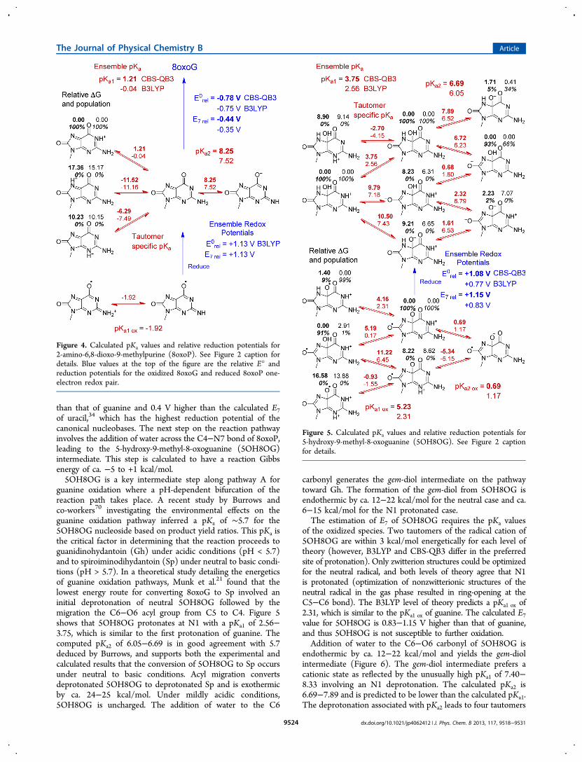
additional electron from oxidized 8oxoG yields 2-amino-6,8-dioxo-9-methylpurine (8oxoP). The calculated reductionpotential of oxidized 8oxoG is 0.35−0.44 V lower than that of G.This indicates the oxidation of 8oxoG to 8oxoP can occurreadily by the removal of an electron, followed by the loss of twoprotons and then removal of one more electron.The pKa values for 8oxoP (Figure 4) are very similar to those
of 8oxoG: pKa1 is 0.0−1.2, pKa2 7.5−8.2, and pKa1 ox ca. −2.However, the protonation sites of 8oxoP differ from 8oxoG:for pKa1, deprotonation takes place at N1, while for pKa2 andpKa1 ox, it occurs at the exocyclic N2 amine position. The CBS-QB3 calculations of pKa1 ox and the reduction potentials areunreliable because of severe spin contamination. Because 8oxoPhas already been oxidized twice from 8oxoG, the reductionpotential is expected to be comparatively high, and furtheroxidation is unlikely. The calculated E7 of 8oxoP is 1.1 V higher
Figure 3. Calculated pKa values and relative reduction potentials for9-methyl-8-oxoguanine (8oxoG). See Figure 2 caption for details. Bluevalues at the bottom of the figure are the relative E° and E7 values forthe oxidized 8oxoG and reduced 8oxoP one-electron redox pair.
The Journal of Physical Chemistry B Article
dx.doi.org/10.1021/jp4062412 | J. Phys. Chem. B 2013, 117, 9518−95319523
than that of guanine and 0.4 V higher than the calculated E7of uracil,34 which has the highest reduction potential of thecanonical nucleobases. The next step on the reaction pathwayinvolves the addition of water across the C4−N7 bond of 8oxoP,leading to the 5-hydroxy-9-methyl-8-oxoguanine (5OH8OG)intermediate. This step is calculated to have a reaction Gibbsenergy of ca. −5 to +1 kcal/mol.5OH8OG is a key intermediate step along pathway A for
guanine oxidation where a pH-dependent bifurcation of thereaction path takes place. A recent study by Burrows andco-workers70 investigating the environmental effects on theguanine oxidation pathway inferred a pKa of ∼5.7 for the5OH8OG nucleoside based on product yield ratios. This pKa isthe critical factor in determining that the reaction proceeds toguanidinohydantoin (Gh) under acidic conditions (pH < 5.7)and to spiroiminodihydantoin (Sp) under neutral to basic condi-tions (pH > 5.7). In a theoretical study detailing the energeticsof guanine oxidation pathways, Munk et al.21 found that thelowest energy route for converting 8oxoG to Sp involved aninitial deprotonation of neutral 5OH8OG followed by themigration the C6−O6 acyl group from C5 to C4. Figure 5shows that 5OH8OG protonates at N1 with a pKa1 of 2.56−3.75, which is similar to the first protonation of guanine. Thecomputed pKa2 of 6.05−6.69 is in good agreement with 5.7deduced by Burrows, and supports both the experimental andcalculated results that the conversion of 5OH8OG to Sp occursunder neutral to basic conditions. Acyl migration convertsdeprotonated 5OH8OG to deprotonated Sp and is exothermicby ca. 24−25 kcal/mol. Under mildly acidic conditions,5OH8OG is uncharged. The addition of water to the C6
carbonyl generates the gem-diol intermediate on the pathwaytoward Gh. The formation of the gem-diol from 5OH8OG isendothermic by ca. 12−22 kcal/mol for the neutral case and ca.6−15 kcal/mol for the N1 protonated case.The estimation of E7 of 5OH8OG requires the pKa values
of the oxidized species. Two tautomers of the radical cation of5OH8OG are within 3 kcal/mol energetically for each level oftheory (however, B3LYP and CBS-QB3 differ in the preferredsite of protonation). Only zwitterion structures could be optimizedfor the neutral radical, and both levels of theory agree that N1is protonated (optimization of nonzwitterionic structures of theneutral radical in the gas phase resulted in ring-opening at theC5−C6 bond). The B3LYP level of theory predicts a pKa1 ox of2.31, which is similar to the pKa1 ox of guanine. The calculated E7value for 5OH8OG is 0.83−1.15 V higher than that of guanine,and thus 5OH8OG is not susceptible to further oxidation.Addition of water to the C6−O6 carbonyl of 5OH8OG is
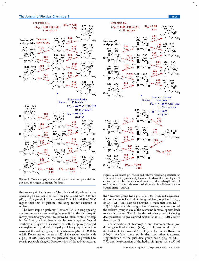
endothermic by ca. 12−22 kcal/mol and yields the gem-diolintermediate (Figure 6). The gem-diol intermediate prefers acationic state as reflected by the unusually high pKa1 of 7.40−8.33 involving an N1 deprotonation. The calculated pKa2 is6.69−7.89 and is predicted to be lower than the calculated pKa1.The deprotonation associated with pKa2 leads to four tautomers
Figure 5. Calculated pKa values and relative reduction potentials for5-hydroxy-9-methyl-8-oxoguanine (5OH8OG). See Figure 2 captionfor details.
Figure 4. Calculated pKa values and relative reduction potentials for2-amino-6,8-dioxo-9-methylpurine (8oxoP). See Figure 2 caption fordetails. Blue values at the top of the figure are the relative E° andreduction potentials for the oxidized 8oxoG and reduced 8oxoP one-electron redox pair.
The Journal of Physical Chemistry B Article
dx.doi.org/10.1021/jp4062412 | J. Phys. Chem. B 2013, 117, 9518−95319524
that are very similar in energy. The calculated pKa values for theoxidized gem-diol are 1.48−5.53 for pKa1 ox and 5.07−5.85 forpKa2 ox. The gem-diol has a calculated E7 which is 0.48−0.78 Vhigher than that of guanine, indicating further oxidation isunlikely.The next step on pathway A toward Gh is a ring-opening
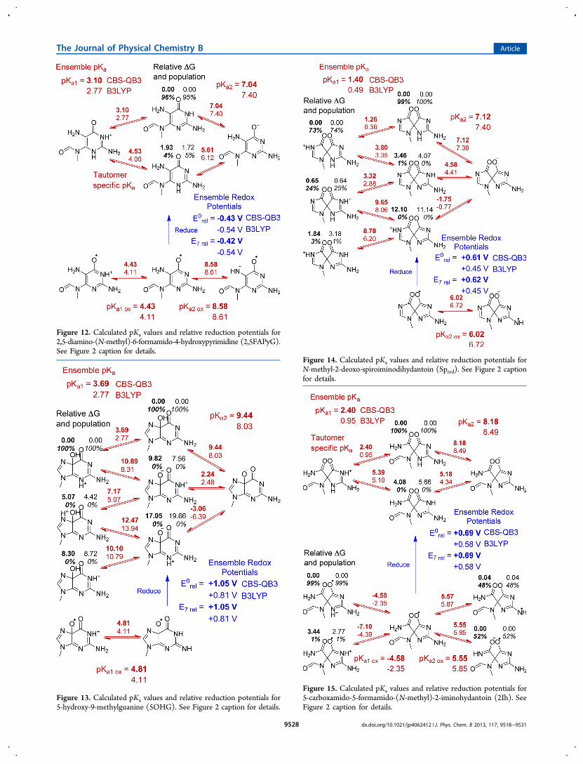
and proton transfer, converting the gem-diol to the 4-carboxy-9-methylguanidinohydantoin (4carboxyGh) intermediate. This stepis 15−23 kcal/mol exothermic for the neutral species. Neutral4carboxyGh (Figure 7) is a zwitterion with a negatively chargedcarboxylate and a positively charged guanidine group. Protonationoccurs at the carboxyl group with a calculated pKa1 of −0.44 to−2.59. Deprotonation occurs at N7 of the neutral species witha pKa2 of 6.07−6.66, and the guanidine group is predicted toremain positively charged. Deprotonation of the radical cation at
the 4-hydroxyl group has a pKa1 ox of 3.88−7.61, and deprotona-tion of the neutral radical at the guanidine group has a pKa2oxof 7.81−9.11. This leads to a nominal E7 value that is ca. 1.11−1.23 V higher than that of guanine. However, deprotonation ofthe carboxyl group in any of the 4carboxyGh radical species leadsto decarboxylation. The E7 for the oxidative process includingdecarboxylation to give oxidized neutral Gh is 0.01−0.10 V lowerthan E7 for G.Decarboxylation of 4carboxyGh and tautomerization pro-
duces guanidinohydantoin (Gh), and is exothermic by ca.30 kcal/mol. For neutral Gh (Figure 8), the zwitterion is3.6−5.1 kcal/mol more stable than the other tautomers.Deprotonation of the guanidine group has a pKa1 of 6.11−7.77, and deprotonation of the hydantoin group has a pKa2 of
Figure 7. Calculated pKa values and relative reduction potentials for4-carboxy-1-methylguanidinohydantoin (4carboxyGh). See Figure 2caption for details. Calculations show that if the carboxylic acid ofoxidized 4carboxyGh is deprotonated, the molecule will dissociate intocarbon dioxide and Gh.
Figure 6. Calculated pKa values and relative reduction potentials forgem-diol. See Figure 2 caption for details.
The Journal of Physical Chemistry B Article
dx.doi.org/10.1021/jp4062412 | J. Phys. Chem. B 2013, 117, 9518−95319525
9.14−9.58. There is no mention of any experimental measure-ments for the pKa values of Gh in the literature. The pKa valuesof guanidines can vary significantly with substituents (e.g.,guanidine pKa 13.7,
71 acetylguanidine pKa 8.3 (experimental72)versus 8.5 (present calculations)) but the pKa values of substitutedhydantoins fall in a much narrower range (pKa 8−10).73−75 Thecalculated pKa1 ox of 0.70−2.29 can be compared to pKa1 ox valuescomputed for guanine (2.53−3.34), 4carboxyGh (3.88−7.61), and5OH8OG (2.31−5.23). Oxidized Gh is predicted to be neutral at pH7, and E7 is calculated to be 0.38−0.49 V higher than that of guanine.As summarized in Figure 9, Sp is readily deprotonated, with
pKa1 −0.06 to 1.32 and pKa2 2.64−3.30. The values are inagreement with those calculated by Verdolino et al. (pKa1 0.5and pKa2 4.8).
63 However, they pointed out that a pKa2 of 4.8 is“surprisingly low for a substituted hydantoin”.63 They attributethe low pKa2 value to through-space interactions of the C6carbonyl group (confirmed by replacing C6−O6 with CCH2and CH2) and greater solvent stabilization of the N7 anion(tautomers with an N7 anion have a dipole moment of 15−20debye compared to 5−15 debye for other tautomers).63 Thepresent calculations may underestimate pKa2, and a better treat-ment of solvent effects may be needed. Verdolino et al. haveestimated the pKa2 of Sp to be around 7. The calculated pKavalues of oxidized Sp are probably also too low because ofsimilar effects described above for the pKa2. Oxidized Sp is mostlikely an anion at pH 7, and E7 is calculated to be 0.78−0.93 Vhigher than that of guanine.
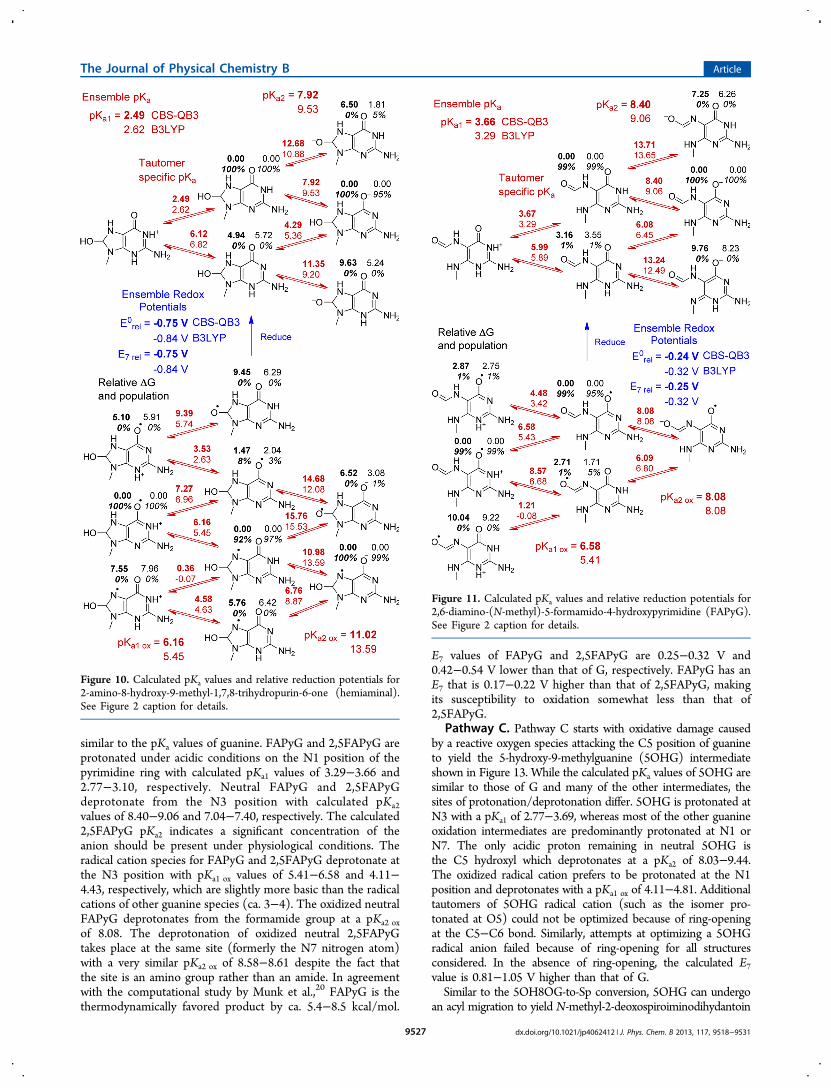
Pathway B. A key intermediate on the pathway to FAPyG is2-amino-8-hydroxy-9-methyl-1,7,8-trihydropurin-6-one (hemi-aminal), shown in Figure 10. The pKa values of the hemiaminal(pKa1 2.49−2.62, pKa2 7.92−9.53) are similar to the pKa valuesof guanine (pKa1 3.20−3.50, pKa2 9.10−9.34). All the ringnitrogens are protonated in the cation species, and the pKa1corresponds to a deprotonation at N3. The next deprotonationfor the pKa2 occurs at N1. The pKa1 ox of the radical cationinvolves a deprotonation at the N7 position. Under very basicconditions, the neutral radical is predicted to deprotonate atthe N1 position. The hemiaminal intermediate is very readilyoxidized, and its E7 value is 0.75−0.84 V lower than that ofguanine. The hemiaminal intermediate is even more susceptibleto oxidation than is 8oxoG. Tautomers with the C8-hydroxylspecies deprotonated were difficult to obtain as many of theoptimizations progressed toward ring-opening. Ring-opening atthe C8−N9 bond of the hemiaminal and tautomerism leads to2,6-diamino-(N-methyl)-5-formamido-4-hydroxypyrimidine(FAPyG) and is exothermic by ca. 9−11 kcal/mol. Breaking theN7−C8 bond followed by tautomerism yields 2,5-diamino-(N-methyl)-6-formamido-4-hydroxypyrimidine (2,5FAPyG) and isexothermic by ca. 4−6 kcal/mol.The pKa values for FAPyG (Figure 11) and 2,5FAPyG
(Figure 12) are similar to one another in addition to being
Figure 9. Calculated pKa values and relative reduction potentials forN-methylspiroiminodihydantoin (Sp). See Figure 2 caption for details.
Figure 8. Calculated pKa values and relative reduction potentials for1-methylguanidinohydantoin (Gh). See Figure 2 caption for details.
The Journal of Physical Chemistry B Article
dx.doi.org/10.1021/jp4062412 | J. Phys. Chem. B 2013, 117, 9518−95319526
similar to the pKa values of guanine. FAPyG and 2,5FAPyG areprotonated under acidic conditions on the N1 position of thepyrimidine ring with calculated pKa1 values of 3.29−3.66 and2.77−3.10, respectively. Neutral FAPyG and 2,5FAPyGdeprotonate from the N3 position with calculated pKa2values of 8.40−9.06 and 7.04−7.40, respectively. The calculated2,5FAPyG pKa2 indicates a significant concentration of theanion should be present under physiological conditions. Theradical cation species for FAPyG and 2,5FAPyG deprotonate atthe N3 position with pKa1 ox values of 5.41−6.58 and 4.11−4.43, respectively, which are slightly more basic than the radicalcations of other guanine species (ca. 3−4). The oxidized neutralFAPyG deprotonates from the formamide group at a pKa2 oxof 8.08. The deprotonation of oxidized neutral 2,5FAPyGtakes place at the same site (formerly the N7 nitrogen atom)with a very similar pKa2 ox of 8.58−8.61 despite the fact thatthe site is an amino group rather than an amide. In agreementwith the computational study by Munk et al.,20 FAPyG is thethermodynamically favored product by ca. 5.4−8.5 kcal/mol.
E7 values of FAPyG and 2,5FAPyG are 0.25−0.32 V and0.42−0.54 V lower than that of G, respectively. FAPyG has anE7 that is 0.17−0.22 V higher than that of 2,5FAPyG, makingits susceptibility to oxidation somewhat less than that of2,5FAPyG.
Pathway C. Pathway C starts with oxidative damage causedby a reactive oxygen species attacking the C5 position of guanineto yield the 5-hydroxy-9-methylguanine (5OHG) intermediateshown in Figure 13. While the calculated pKa values of 5OHG aresimilar to those of G and many of the other intermediates, thesites of protonation/deprotonation differ. 5OHG is protonated atN3 with a pKa1 of 2.77−3.69, whereas most of the other guanineoxidation intermediates are predominantly protonated at N1 orN7. The only acidic proton remaining in neutral 5OHG isthe C5 hydroxyl which deprotonates at a pKa2 of 8.03−9.44.The oxidized radical cation prefers to be protonated at the N1position and deprotonates with a pKa1 ox of 4.11−4.81. Additionaltautomers of 5OHG radical cation (such as the isomer pro-tonated at O5) could not be optimized because of ring-openingat the C5−C6 bond. Similarly, attempts at optimizing a 5OHGradical anion failed because of ring-opening for all structuresconsidered. In the absence of ring-opening, the calculated E7value is 0.81−1.05 V higher than that of G.Similar to the 5OH8OG-to-Sp conversion, 5OHG can undergo
an acyl migration to yield N-methyl-2-deoxospiroiminodihydantoin
Figure 11. Calculated pKa values and relative reduction potentials for2,6-diamino-(N-methyl)-5-formamido-4-hydroxypyrimidine (FAPyG).See Figure 2 caption for details.
Figure 10. Calculated pKa values and relative reduction potentials for2-amino-8-hydroxy-9-methyl-1,7,8-trihydropurin-6-one (hemiaminal).See Figure 2 caption for details.
The Journal of Physical Chemistry B Article
dx.doi.org/10.1021/jp4062412 | J. Phys. Chem. B 2013, 117, 9518−95319527
Figure 14. Calculated pKa values and relative reduction potentials forN-methyl-2-deoxo-spiroiminodihydantoin (Spred). See Figure 2 captionfor details.
Figure 15. Calculated pKa values and relative reduction potentials for5-carboxamido-5-formamido-(N-methyl)-2-iminohydantoin (2Ih). SeeFigure 2 caption for details.
Figure 12. Calculated pKa values and relative reduction potentials for2,5-diamino-(N-methyl)-6-formamido-4-hydroxypyrimidine (2,5FAPyG).See Figure 2 caption for details.
Figure 13. Calculated pKa values and relative reduction potentials for5-hydroxy-9-methylguanine (5OHG). See Figure 2 caption for details.
The Journal of Physical Chemistry B Article
dx.doi.org/10.1021/jp4062412 | J. Phys. Chem. B 2013, 117, 9518−95319528
(i.e., reduced spiroiminodihydantoin or Spred). This reaction isexothermic by 20−21 kcal/mol. Spred has three ring nitrogens thatare reasonable protonation sites under physiological conditions.Two of three combinations of two protons on the three ringnitrogen sites are calculated to be similar in energy (Figure 14).Spred protonates at the guanidine group with a pKa1 of 0.49−1.40.Neutral Spred deprotonates at the former N3 with a pKa2 of 7.12−7.40. An oxidized radical cation isomer of Spred could not beoptimized because of the instabilities in the rings leading to ring-opening. Oxidation coupled with loss of a proton leads to theneutral radical of Spred, which can be optimized. The calculatedpKa2 ox for the neutral radical is 6.02−6.72, indicating that it wouldlose another proton at physiological conditions, yielding the radical
anion species. The calculated E7 for Spred is 0.45−0.62 V higherthan the calculated potential for G.The final product in pathway C involves a water addition
across the C2−N3 bond of the deaminated hydantoin ringfollowed by proton transfer and ring-opening to the 5-carboxamido-5-formamido-(N-methyl)-2-iminohydantoin (2Ih)product. This reaction is calculated to be endothermic by3−6 kcal/mol. Similar to Sp and Spred, the predicted pKa1 of2Ih is acidic with a calculated value of 0.95−2.40, and 2Ih ispredicted to deprotonate from the former N1 position (Figure 15).2Ih deprotonates from the former N3 position with a calculatedpKa2 of 8.18−8.49. The oxidized radical cation also has a predictedacidic pKa1 ox of −4.58 to −2.35 for deprotonation at the formerN3 position. The neutral 2Ih radical species can deprotonate fromeither of the exocyclic amino groups with almost equal energy.The calculated pKa2 ox is 5.55−5.85 and is similar to the calculatedpKa2 ox of Spred. The calculated E7 value of 2Ih is also very similar tothat of the Spred species and is 0.58−0.69 V higher than that ofguanine, indicating that further oxidation of 2Ih is unlikely.
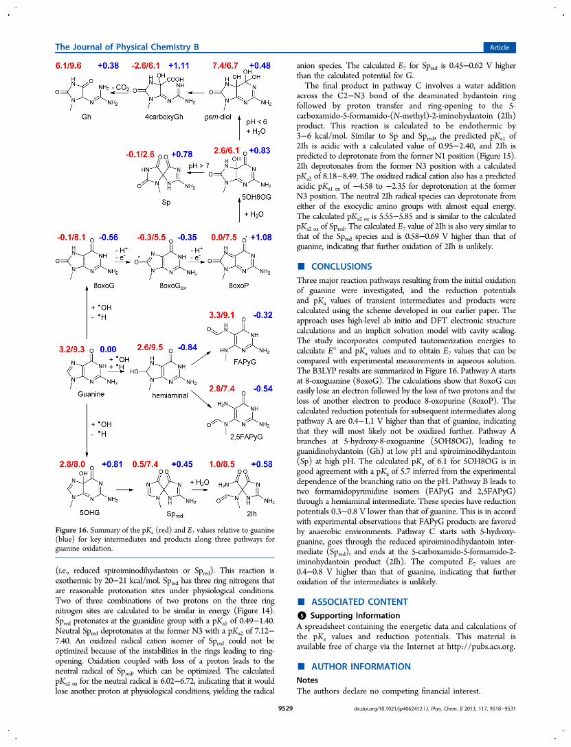
■ CONCLUSIONSThree major reaction pathways resulting from the initial oxidationof guanine were investigated, and the reduction potentialsand pKa values of transient intermediates and products werecalculated using the scheme developed in our earlier paper. Theapproach uses high-level ab initio and DFT electronic structurecalculations and an implicit solvation model with cavity scaling.The study incorporates computed tautomerization energies tocalculate E° and pKa values and to obtain E7 values that can becompared with experimental measurements in aqueous solution.The B3LYP results are summarized in Figure 16. Pathway A startsat 8-oxoguanine (8oxoG). The calculations show that 8oxoG caneasily lose an electron followed by the loss of two protons and theloss of another electron to produce 8-oxopurine (8oxoP). Thecalculated reduction potentials for subsequent intermediates alongpathway A are 0.4−1.1 V higher than that of guanine, indicatingthat they will most likely not be oxidized further. Pathway Abranches at 5-hydroxy-8-oxoguanine (5OH8OG), leading toguanidinohydantoin (Gh) at low pH and spiroiminodihydantoin(Sp) at high pH. The calculated pKa of 6.1 for 5OH8OG is ingood agreement with a pKa of 5.7 inferred from the experimentaldependence of the branching ratio on the pH. Pathway B leads totwo formamidopyrimidine isomers (FAPyG and 2,5FAPyG)through a hemiaminal intermediate. These species have reductionpotentials 0.3−0.8 V lower than that of guanine. This is in accordwith experimental observations that FAPyG products are favoredby anaerobic environments. Pathway C starts with 5-hydroxy-guanine, goes through the reduced spiroiminodihydantoin inter-mediate (Spred), and ends at the 5-carboxamido-5-formamido-2-iminohydantoin product (2Ih). The computed E7 values are0.4−0.8 V higher than that of guanine, indicating that furtheroxidation of the intermediates is unlikely.
■ ASSOCIATED CONTENT*S Supporting InformationA spreadsheet containing the energetic data and calculations ofthe pKa values and reduction potentials. This material isavailable free of charge via the Internet at http://pubs.acs.org.
■ AUTHOR INFORMATIONNotesThe authors declare no competing financial interest.
Figure 16. Summary of the pKa (red) and E7 values relative to guanine(blue) for key intermediates and products along three pathways forguanine oxidation.
The Journal of Physical Chemistry B Article
dx.doi.org/10.1021/jp4062412 | J. Phys. Chem. B 2013, 117, 9518−95319529
The authors thank Prof. Cynthia Burrows for stimulatingdiscussions and encouragement. We also thank Profs. BarbaraMunk and Richard Lord for their comments and suggestions.This work was supported by a grant from the National ScienceFoundation (CHE1212281). We thank Wayne State Univer-sity’s computing grid for computer time.
■ REFERENCES(1) Ames, B. N.; Shigenaga, M. K.; Hagen, T. M. Oxidants,Antioxidants, and the Degenerative Diseases of Aging. Proc. Natl. Acad.Sci., U.S.A. 1993, 90, 7915−7922.(2) Breen, A. P.; Murphy, J. A. Reactions of Oxyl Radicals with DNA.Free Radical Biol. Med. 1995, 18, 1033−1077.(3) Cadet, J.; Berger, M.; Douki, T.; Morin, B.; Raoul, S.; Ravanat, J.L.; Spinelli, S. Effects of UV and Visible Radiation on DNA: Final BaseDamage. Biol. Chem. 1997, 378, 1275−1286.(4) Burrows, C. J.; Muller, J. G. Oxidative Nucleobase ModificationsLeading to Strand Scission. Chem. Rev. 1998, 98, 1109−1151.(5) Cooke, M. S.; Olinski, R.; Evans, M. D. Does Measurement ofOxidative Damage to DNA Have Clinical Significance? Clin. Chim.Acta 2006, 365, 30−49.(6) Gimisis, T.; Cismas, C. Isolation, Characterization, andIndependent Synthesis of Guanine Oxidation Products. Eur. J. Org.Chem. 2006, 1351−1378.(7) Pratviel, G.; Meunier, B. Guanine Oxidation: One- and Two-Electron Reactions. Chem.Eur. J. 2006, 12, 6018−6030.(8) Son, J.; Pang, B.; McFaline, J. L.; Taghizadeh, K.; Dedon, P. C.Surveying the Damage: The Challenges of Developing Nucleic AcidBiomarkers of Inflammation. Mol. Biosyst. 2008, 4, 902−908.(9) Lonkar, P.; Dedon, P. C. Reactive Species and DNA Damage inChronic Inflammation: Reconciling Chemical Mechanisms andBiological Fates. Int. J. Cancer 2011, 128, 1999−2009.(10) Steenken, S. Purine Bases, Nucleosides, and Nucleotides:Aqueous Solution Redox Chemistry and Transformation Reactions ofTheir Radical Cations and e− and OH Adducts. Chem. Rev. 1989, 89,503−520.(11) Candeias, L. P.; Steenken, S. Reaction of HO● with GuanineDerivatives in Aqueous Solution: Formation of Two Different Redox-Active OH-Adduct Radicals and Their Unimolecular TransformationReactions. Properties of G(-H)●. Chem.Eur. J. 2000, 6, 475−484.(12) Luo, W. C.; Muller, J. G.; Rachlin, E. M.; Burrows, C. J.Characterization of Spiroiminodihydantoin as a Product of One-Electron Oxidation of 8-Oxo-7,8-dihydroguanosine. Org. Lett. 2000, 2,613−616.(13) Llano, J.; Eriksson, L. A. Oxidation Pathways of Adenine andGuanine in Aqueous Solution from First Principles Electrochemistry.Phys. Chem. Chem. Phys. 2004, 6, 4707−4713.(14) Crean, C.; Geacintov, N. E.; Shafirovich, V. Oxidation ofGuanine and 8-Oxo-7,8-dihydroguanine by Carbonate Radical Anions:Insight from Oxygen-18 Labeling Experiments. Angew. Chem., Int. Ed.2005, 44, 5057−5060.(15) Fleming, A. M.; Muller, J. G.; Ji, I. S.; Burrows, C. J.Characterization of 2′-Deoxyguanosine Oxidation Products Observedin the Fenton-like System Cu(II)/H2O2/Reductant in Nucleoside andOligodeoxynucleotide Contexts. Org. Biomol. Chem. 2011, 9, 3338−3348.(16) Delaney, S.; Jarem, D. A.; Volle, C. B.; Yennie, C. J. Chemicaland Biological Consequences of Oxidatively Damaged Guanine inDNA. Free Radical Res. 2012, 46, 420−441.(17) Jena, N. R.; Mishra, P. C. Formation of Ring-Opened andRearranged Products of Guanine: Mechanisms and BiologicalSignificance. Free Radical Biol. Med. 2012, 53, 81−94.(18) Wetmore, S. D.; Boyd, R. J.; Eriksson, L. A. Comparison ofExperimental and Calculated Hyperfine Coupling Constants. WhichRadicals Are Formed in Irradiated Guanine? J. Phys. Chem. B 1998,102, 9332−9343.
(19) Wetmore, S. D.; Boyd, R. J.; Llano, J.; Lundqvist, M. J.;Eriksson, L. A. Hydroxyl Radical Reactions in Biological Media. InRecent Advances in Density Functional Methods; Barone, V., Bencini, A.,Fantucci, P., Eds.; World Scientific: Singapore, 2000; Vol. 3, pp 387−415.(20) Munk, B. H.; Burrows, C. J.; Schlegel, H. B. Exploration ofMechanisms for the Transformation of 8-Hydroxy Guanine Radical toFAPyG by Density Functional Theory. Chem. Res. Toxicol. 2007, 20,432−444.(21) Munk, B. H.; Burrows, C. J.; Schlegel, H. B. An Exploration ofMechanisms for the Transformation of 8-Oxoguanine to Guanidino-hydantoin and Spiroiminodihydantoin by Density Functional Theory.J. Am. Chem. Soc. 2008, 130, 5245−5256.(22) Ye, Y.; Munk, B. H.; Muller, J. G.; Cogbill, A.; Burrows, C. J.;Schlegel, H. B. Mechanistic Aspects of the Formation ofGuanidinohydantoin from Spiroiminodihydantoin under AcidicConditions. Chem. Res. Toxicol. 2009, 22, 526−535.(23) Jovanovic, S. V.; Simic, M. G. One-Electron Redox Potentials ofPurines and Pyrimidines. J. Phys. Chem. 1986, 90, 974−978.(24) Faraggi, M.; Broitman, F.; Trent, J. B.; Klapper, M. H. One-Electron Oxidation Reactions of Some Purine and Pyrimidine Bases inAqueous Solutions. Electrochemical and Pulse Radiolysis Studies. J.Phys. Chem. 1996, 100, 14751−14761.(25) Seidel, C. A. M.; Schulz, A.; Sauer, M. H. M. Nucleobase-Specific Quenching of Fluorescent Dyes. 1. Nucleobase One-ElectronRedox Potentials and Their Correlation with Static and DynamicQuenching Efficiencies. J. Phys. Chem. 1996, 100, 5541−5553.(26) Steenken, S.; Jovanovic, S. V. How Easily Oxidizable Is DNA?One-Electron Reduction Potentials of Adenosine and GuanosineRadicals in Aqueous Solution. J. Am. Chem. Soc. 1997, 119, 617−618.(27) Steenken, S.; Jovanovic, S. V.; Bietti, M.; Bernhard, K. The TrapDepth (in DNA) of 8-Oxo-7,8-dihydro-2′-deoxyguanosine as Derivedfrom Electron-Transfer Equilibria in Aqueous Solution. J. Am. Chem.Soc. 2000, 122, 2373−2374.(28) Oliveira-Brett, A. M.; Piedade, J. A. P.; Silva, L. A.; Diculescu, V.C. Voltammetric Determination of All DNA Nucleotides. Anal.Biochem. 2004, 332, 321−329.(29) Fukuzumi, S.; Miyao, H.; Ohkubo, K.; Suenobu, T. Electron-Transfer Oxidation Properties of DNA Bases and DNA Oligomers. J.Phys. Chem. A 2005, 109, 3285−3294.(30) Langmaier, J.; Samec, Z.; Samcova, E.; Hobza, P.; Reha, D.Origin of Difference between One-Electron Redox Potentials ofGuanosine and Guanine: Electrochemical and Quantum ChemicalStudy. J. Phys. Chem. B 2004, 108, 15896−15899.(31) Gryn’ova, G.; Marshall, D. L.; Blanksby, S. J.; Coote, M. L.Switching Radical Stability by pH-Induced Orbital Conversion. Nat.Chem. 2013, 5, 474−481.(32) Caruso, T.; Carotenuto, M.; Vasca, E.; Peluso, A. DirectExperimental Observation of the Effect of the Base Pairing on theOxidation Potential of Guanine. J. Am. Chem. Soc. 2005, 127, 15040−15041.(33) Crespo-Hernandez, C. E.; Close, D. M.; Gorb, L.; Leszczynski, J.Determination of Redox Potentials for the Watson−Crick Base Pairs,DNA Nucleosides, and Relevant Nucleoside Analogues. J. Phys. Chem.B 2007, 111, 5386−5395.(34) Psciuk, B. T.; Lord, R. L.; Munk, B. H.; Schlegel, H. B.Theoretical Determination of One-Electron Oxidation Potentials forNucleic Acid Bases. J. Chem. Theory Comput. 2012, 8, 5107−5123.(35) Bennaim, A.; Marcus, Y. Solvation Thermodynamics ofNonionic Solutes. J. Chem. Phys. 1984, 81, 2016−2027.(36) Lewis, A.; Bumpus, J. A.; Truhlar, D. G.; Cramer, C. J.Molecular Modeling of Environmentally Important Processes:Reduction Potentials. J. Chem. Educ. 2004, 81, 596−604.(37) Lewis, A.; Bumpus, J. A.; Truhlar, D. G.; Cramer, C. J. J. Chem.Educ. 2004, 81, 596−604. J. Chem. Educ. 2007, 84, 934−934.(38) Kelly, C. P.; Cramer, C. J.; Truhlar, D. G. Aqueous SolvationFree Energies of Ions and Ion−Water Clusters Based on an AccurateValue for the Absolute Aqueous Solvation Free Energy of the Proton.J. Phys. Chem. B 2006, 110, 16066−16081.
The Journal of Physical Chemistry B Article
dx.doi.org/10.1021/jp4062412 | J. Phys. Chem. B 2013, 117, 9518−95319530
(39) Isse, A. A.; Gennaro, A. Absolute Potential of the StandardHydrogen Electrode and the Problem of Interconversion of Potentialsin Different Solvents. J. Phys. Chem. B 2010, 114, 7894−7899.(40) Vosko, S. H.; Wilk, L.; Nusair, M. Accurate Spin-DependentElectron Liquid Correlation Energies for Local Spin-DensityCalculations: A Critical Analysis. Can. J. Phys. 1980, 58, 1200−1211.(41) Becke, A. D. Density-Functional Exchange-Energy Approx-imation with Correct Asymptotic Behavior. Phys. Rev. A 1988, 38,3098−3100.(42) Lee, C. T.; Yang, W. T.; Parr, R. G. Development of the Colle−Salvetti Correlation-Energy Formula into a Functional of the Electron-Density. Phys. Rev. B 1988, 37, 785−789.(43) Becke, A. D. Density-Functional Thermochemistry. 3. The Roleof Exact Exchange. J. Chem. Phys. 1993, 98, 5648−5652.(44) Stephens, P. J.; Devlin, F. J.; Chabalowski, C. F.; Frisch, M. J. AbInitio Calculations of Vibrational Absorption and Circular-DichroismSpectra Using Density Functional Force Fields. J. Phys. Chem. 1994,98, 11623−11627.(45) Ditchfield, R.; Hehre, W. J.; Pople, J. A. Self-ConsistentMolecular-Orbital Methods. IX. An Extended Gaussian-Type Basis forMolecular-Orbital Studies of Organic Molecules. J. Chem. Phys. 1971,54, 724−728.(46) Hehre, W. J.; Ditchfield, R.; Pople, J. A. Self-ConsistentMolecular-Orbital Methods. XII. Further Extensions of Gaussian-TypeBasis Sets for Use in Molecular Orbital Studies of Organic Molecules.J. Chem. Phys. 1972, 56, 2257.(47) Hariharan, P. C.; Pople, J. A. Influence of Polarization Functionson Molecular-Orbital Hydrogenation Energies. Theor. Chim. Acta1973, 28, 213−222.(48) Hariharan, P. C.; Pople, J. A. Accuracy of AH EquilibriumGeometries by Single Determinant Molecular-Orbital Theory. Mol.Phys. 1974, 27, 209−214.(49) Gordon, M. S. The Isomers of Silacyclopropane. Chem. Phys.Lett. 1980, 76, 163−168.(50) Francl, M. M.; Pietro, W. J.; Hehre, W. J.; Binkley, J. S.; Gordon,M. S.; Defrees, D. J.; Pople, J. A. Self-Consistent Molecular-OrbitalMethods. XXIII. A Polarization-Type Basis Set for Second-RowElements. J. Chem. Phys. 1982, 77, 3654−3665.(51) Kendall, R. A.; Dunning, T. H.; Harrison, R. J. Electron-Affinities of the First-Row Atoms Revisited. Systematic Basis-Sets andWave-Functions. J. Chem. Phys. 1992, 96, 6796−6806.(52) Montgomery, J. A.; Frisch, M. J.; Ochterski, J. W.; Petersson, G.A. A Complete Basis Set Model Chemistry. VI. Use of DensityFunctional Geometries and Frequencies. J. Chem. Phys. 1999, 110,2822−2827.(53) Montgomery, J. A.; Frisch, M. J.; Ochterski, J. W.; Petersson, G.A. A Complete Basis Set Model Chemistry. VII. Use of the MinimumPopulation Localization Method. J. Chem. Phys. 2000, 112, 6532−6542.(54) Marenich, A. V.; Cramer, C. J.; Truhlar, D. G. UniversalSolvation Model Based on Solute Electron Density and on aContinuum Model of the Solvent Defined by the Bulk DielectricConstant and Atomic Surface Tensions. J. Phys. Chem. B 2009, 113,6378−6396.(55) Cances, E.; Mennucci, B.; Tomasi, J. A New Integral EquationFormalism for the Polarizable Continuum Model: TheoreticalBackground and Applications to Isotropic and Anisotropic Dielectrics.J. Chem. Phys. 1997, 107, 3032−3041.(56) Mennucci, B.; Tomasi, J. Continuum Solvation Models: A NewApproach to the Problem of Solute’s Charge Distribution and CavityBoundaries. J. Chem. Phys. 1997, 106, 5151−5158.(57) Cossi, M.; Barone, V.; Mennucci, B.; Tomasi, J. Ab Initio Studyof Ionic Solutions by a Polarizable Continuum Dielectric Model.Chem. Phys. Lett. 1998, 286, 253−260.(58) Scalmani, G.; Frisch, M. J. Continuous Surface ChargePolarizable Continuum Models of Solvation. I. General Formalism. J.Chem. Phys. 2010, 132, 114110.
(59) Frisch, M. J.; Trucks, G. W.; Schlegel, H. B.; Scuseria, G. E.;Robb, M. A.; et al. Gaussian Development Version, revision H.20+;Gaussian, Inc.: Wallingford, CT, 2010.(60) Clark, W. M. Oxidation-Reduction Potentials of Organic Systems;Williams & Wilkins: Baltimore, MD, 1960.(61) Wardman, P. Reduction Potentials of One-Electron CouplesInvolving Free-Radicals in Aqueous-Solution. J. Phys. Chem. Ref. Data1989, 18, 1637−1755.(62) Camaioni, D. M.; Schwerdtfeger, C. A. Comment on “AccurateExperimental Values for the Free Energies of Hydration of H+, OH−,and H3O
+”. J. Phys. Chem. A 2005, 109, 10795−10797.(63) Verdolino, V.; Cammi, R.; Munk, B. H.; Schlegel, H. B.Calculation of pKa Values of Nucleobases and the Guanine OxidationProducts Guanidinohydantoin and Spiroiminodihydantoin UsingDensity Functional Theory and a Polarizable Continuum Model. J.Phys. Chem. B 2008, 112, 16860−16873.(64) Jang, Y. H.; Goddard, W. A.; Noyes, K. T.; Sowers, L. C.;Hwang, S.; Chung, D. S. pKa Values of Guanine in Water: DensityFunctional Theory Calculations Combined with Poisson−BoltzmannContinuum−Solvation Model. J. Phys. Chem. B 2003, 107, 344−357.(65) Song, B.; Zhao, J.; Griesser, R.; Meiser, C.; Sigel, H.; Lippert, B.Effects of (N7)-Coordinated Nickel(II), Copper(II), or Platinum(II)on the Acid-Base Properties of Guanine Derivatives and Other RelatedPurines. Chem.Eur. J. 1999, 5, 2374−2387.(66) Baik, M. H.; Silverman, J. S.; Yang, I. V.; Ropp, P. A.; Szalai, V.A.; Yang, W. T.; Thorp, H. H. Using Density Functional Theory toDesign DNA Base Analogues with Low Oxidation Potentials. J. Phys.Chem. B 2001, 105, 6437−6444.(67) Christensen, J. J.; Rytting, J. H.; Izatt, R. M. ThermodynamicpK, ΔH°, ΔS°, and ΔCp° Values for Proton Dissociation from SeveralPurines and Their Nucleosides in Aqueous Solution. Biochemistry1970, 9, 4907−4913.(68) Cho, B. P. Structure of Oxidatively Damaged Nucleic-AcidAdducts: pH-Dependence of the C-13 NMR-Spectra of 8-Oxoguanosine and 8-Oxoadenosine. Magn. Reson. Chem. 1993, 31,1048−1053.(69) Jang, Y. H.; Goddard, W. A.; Noyes, K. T.; Sowers, L. C.;Hwang, S.; Chung, D. S. First Principles Calculations of the Tautomersand pKa Values of 8-Oxoguanine: Implications for Mutagenicity andRepair. Chem. Res. Toxicol. 2002, 15, 1023−1035.(70) Fleming, A. M.; Muller, J. G.; Dlouhy, A. C.; Burrows, C. J.Structural Context Effects in the Oxidation of 8-Oxo-7,8-dihydro-2′-deoxyguanosine to Hydantoin Products: Electrostatics, Base Stacking,and Base Pairing. J. Am. Chem. Soc. 2012, 134, 15091−15102.(71) Shugar, D.; Fox, J. J. Spectrophotometric Studies of NucleicAcid Derivatives and Related Compounds as a Function of pH. I.Pyrimidines. Biochim. Biophys. Acta 1952, 9, 199−218.(72) Albert, A.; Goldacre, R.; Phillips, J. The Strength of HeterocyclicBases. J. Chem. Soc. 1948, 2240−2249.(73) Edward, J. T.; Nielsen, S.; Thiohydantoins, I. Ionization andultraviolet absorption. J. Chem. Soc. 1957, 5075−5079.(74) Budavari, S. The Merck Index, 12th ed.; Merck: WhitehouseStation, NJ, 1996.(75) Nosheen, E.; Shah, A.; Badshah, A.; Zia ur, R.; Hussain, H.;Qureshi, R.; Ali, S.; Siddiq, M.; Khan, A. M. Electrochemical Oxidationof Hydantoins at Glassy Carbon Electrode. Electrochim. Acta 2012, 80,108−117.
The Journal of Physical Chemistry B Article
dx.doi.org/10.1021/jp4062412 | J. Phys. Chem. B 2013, 117, 9518−95319531