Computer-aided diagnosis system for the Acute Respiratory Distress Syndrome from chest radiographs Nesrine Zaglam a,b,n , Philippe Jouvet a , Olivier Flechelles a , Guillaume Emeriaud a , Farida Cheriet a,b a Sainte-Justine Hospital Research Center, Montreal, QC, Canada H3T 1C5 b The Department of Computer Engineering, Ecole Polytechnique de Montréal, Montreal, Canada H3T 1J4 article info Article history: Received 3 March 2014 Accepted 12 June 2014 Keywords: Chest radiographs Acute Respiratory Distress Syndrome (ARDS) Texture analysis Automatic classification Computer-aided diagnosis system abstract This paper presents a computer-aided diagnosis (CAD) system for the assessment of Acute Respiratory Distress Syndrome (ARDS) from chest radiographs. Our method consists in automatically extracting intercostal patches from chest radiographs belonging to the test database using a semiautomatic segmentation method of the ribs. Statistical and spectral features are computed from each patch then a method of feature transformation is applied using the Linear Discriminant Analysis (LDA). A training database of 321 patches was classified by an expert in two classes, a class of normal patches and a class of abnormal patches. Patches belonging to the test database are then classified using the SVM classifier. Finally, the rate of abnormal patches is calculated for each quadrant to decide if the chest radiograph presents an ARDS. The method has been evaluated on 90 radiographs where 53 images present ARDS. The results show a sensitivity of 90.6% at a specificity of 86.5%. & 2014 Elsevier Ltd. All rights reserved. 1. Introduction The Acute Respiratory Distress Syndrome (ARDS) is an acute inflammatory disease of the lungs associated with severe hypox- emia mostly due to pneumonia, sepsis, aspiration of gastric contents and major trauma [1,2]. Despite a mortality rate between 18% and 35% [3,4], ARDS is still underdiagnosed in intensive care [5]. Early diagnosis of ARDS counts among the measures that should improve the management of this disease including an early initiation of a lung protective ventilator support. The diagnosis of ARDS is based on four criteria [6]: Acute situation. PaO2/FiO2 o200 mmHg. No clinical evidence of left heart failure. Fluid overload and bilateral infiltrates (alveolar damage) on chest radiograph (white spots as shown in Fig. 1) [7]. The alveolar damage appears in chest radiograph in the form of opacities. This is due to the fact that the alveoli, which are responsible for air transfer between the outside and the blood, are filled with inflammatory fluid and/or are collapsed. These criteria are difficult to highlight simultaneously, which usually leads to a false diagnosis, and among them, Angoulvant et al. [8] demonstrated that the inter-observer variability on the chest radiograph, which corresponds to the fourth criteria, was very high (kappa ¼ 0.3) and was a limiting factor in the early diagnosis of ARDS. In fact, the difficulty of diagnosing this disease is probably due to both the quality of the chest radiograph and the intensi- vist's interpretation which usually depends on his own experience. In order to assist clinicians in the early diagnosis of ARDS, the development of an automatic system has proven to be essential. 1.1. Computer-aided diagnosis system for chest radiograph The chest radiograph is an essential tool for diagnosing ARDS. Its low cost, portability, speed, and its use of a moderate dose of radiation make it the most adequate imaging tool. A computer- aided diagnosis (CAD) system to detect ARDS (diffuse abnormal- ities) in chest radiographs is a new area that has not been previously studied, even though CAD is one of the major research areas in medical imaging and diagnostic radiology [9] where several CAD systems have already been developed in order to help radiologists in chest radiographs diagnosis [10]. The compu- terized analysis of chest radiograph in these CAD systems is based on size measurements, detection of nodules (low-contrast white circular objects) or texture analysis [10]. Texture analysis is the best approach that assesses diffuse patterns in chest radiographs and can be used for ARDS diagnosis. Contents lists available at ScienceDirect journal homepage: www.elsevier.com/locate/cbm Computers in Biology and Medicine http://dx.doi.org/10.1016/j.compbiomed.2014.06.006 0010-4825/& 2014 Elsevier Ltd. All rights reserved. n Corresponding author at: The Department of Computer Engineering, Ecole Polytechnique de Montréal, Montreal, QC, Canada H3T 1J4. E-mail address: [email protected](N. Zaglam). Computers in Biology and Medicine 52 (2014) 41–48
Transcript
Computer-aided diagnosis system for the Acute Respiratory DistressSyndrome from chest radiographs
Nesrine Zaglam a,b,n, Philippe Jouvet a, Olivier Flechelles a,Guillaume Emeriaud a, Farida Cheriet a,b
a Sainte-Justine Hospital Research Center, Montreal, QC, Canada H3T 1C5b The Department of Computer Engineering, Ecole Polytechnique de Montréal, Montreal, Canada H3T 1J4
a r t i c l e i n f o
Article history:Received 3 March 2014Accepted 12 June 2014
Keywords:Chest radiographsAcute Respiratory DistressSyndrome (ARDS)Texture analysisAutomatic classificationComputer-aided diagnosis system
a b s t r a c t
This paper presents a computer-aided diagnosis (CAD) system for the assessment of Acute RespiratoryDistress Syndrome (ARDS) from chest radiographs. Our method consists in automatically extractingintercostal patches from chest radiographs belonging to the test database using a semiautomaticsegmentation method of the ribs. Statistical and spectral features are computed from each patch then amethod of feature transformation is applied using the Linear Discriminant Analysis (LDA). A trainingdatabase of 321 patches was classified by an expert in two classes, a class of normal patches and a classof abnormal patches. Patches belonging to the test database are then classified using the SVM classifier.Finally, the rate of abnormal patches is calculated for each quadrant to decide if the chest radiographpresents an ARDS. The method has been evaluated on 90 radiographs where 53 images present ARDS.The results show a sensitivity of 90.6% at a specificity of 86.5%.
& 2014 Elsevier Ltd. All rights reserved.
1. Introduction
The Acute Respiratory Distress Syndrome (ARDS) is an acuteinflammatory disease of the lungs associated with severe hypox-emia mostly due to pneumonia, sepsis, aspiration of gastriccontents and major trauma [1,2]. Despite a mortality rate between18% and 35% [3,4], ARDS is still underdiagnosed in intensive care[5]. Early diagnosis of ARDS counts among the measures thatshould improve the management of this disease including an earlyinitiation of a lung protective ventilator support.
The diagnosis of ARDS is based on four criteria [6]:
� Acute situation.� PaO2/FiO2 o200 mmHg.� No clinical evidence of left heart failure.� Fluid overload and bilateral infiltrates (alveolar damage) on
chest radiograph (white spots as shown in Fig. 1) [7].
The alveolar damage appears in chest radiograph in the form ofopacities. This is due to the fact that the alveoli, which areresponsible for air transfer between the outside and the blood,are filled with inflammatory fluid and/or are collapsed. These
criteria are difficult to highlight simultaneously, which usuallyleads to a false diagnosis, and among them, Angoulvant et al. [8]demonstrated that the inter-observer variability on the chestradiograph, which corresponds to the fourth criteria, was veryhigh (kappa¼0.3) and was a limiting factor in the early diagnosisof ARDS. In fact, the difficulty of diagnosing this disease is probablydue to both the quality of the chest radiograph and the intensi-vist's interpretation which usually depends on his own experience.In order to assist clinicians in the early diagnosis of ARDS, thedevelopment of an automatic system has proven to be essential.
1.1. Computer-aided diagnosis system for chest radiograph
The chest radiograph is an essential tool for diagnosing ARDS.Its low cost, portability, speed, and its use of a moderate dose ofradiation make it the most adequate imaging tool. A computer-aided diagnosis (CAD) system to detect ARDS (diffuse abnormal-ities) in chest radiographs is a new area that has not beenpreviously studied, even though CAD is one of the major researchareas in medical imaging and diagnostic radiology [9] whereseveral CAD systems have already been developed in order tohelp radiologists in chest radiographs diagnosis [10]. The compu-terized analysis of chest radiograph in these CAD systems is basedon size measurements, detection of nodules (low-contrast whitecircular objects) or texture analysis [10]. Texture analysis is thebest approach that assesses diffuse patterns in chest radiographsand can be used for ARDS diagnosis.
Contents lists available at ScienceDirect
journal homepage: www.elsevier.com/locate/cbm
Computers in Biology and Medicine
http://dx.doi.org/10.1016/j.compbiomed.2014.06.0060010-4825/& 2014 Elsevier Ltd. All rights reserved.
n Corresponding author at: The Department of Computer Engineering, EcolePolytechnique de Montréal, Montreal, QC, Canada H3T 1J4.
The texture analysis plays a very important role in CAD systems[11]. There are several approaches to represent the texture. Themain ones are divided into three categories namely statistical,structural, and spectral [12]. In the 1970s, the texture analysis hasbeen applied in chest radiographs in order to detect the pneumo-coniosis using the Fourier spectrum [13]. More recent work hasrealized a fully automatic CAD scheme for pneumoconiosis usingthe histogram and co-occurrence matrices features [14]. Ginnekenet al. [15] used as features the moments of responses to a multi-scale filter bank in order to detect tuberculosis (TB) and interstitialdisease. Chest radiographs also include normal structures that maydistort the analysis of the lungs. To address this, several studieshave performed their analysis exclusively on the Regions OfInterest (ROI) that may contain the lesions. Kruger et al. [16] usedthe characteristics of the co-occurrence matrix and the Fourierspectrum extracted from the manually selected intercostal regions(ROI). Katsuragawa et al. [17] used the variation of Root MeanSquare (RMS) and the first moment of the Fourier spectrum asfeatures extracted from small automatically selected intercostalregions. They developed an algorithm to eliminate the posteriorribs by first selecting a large number of squares (ROI) in the chestradiograph and then eliminate those that contain sharp rib edges,using edge gradient analysis. However, in [16,17], the areascontaining anterior ribs were not eliminated, when selectingintercostal regions for analysis. This can distort the ensuinganalysis because these areas are visually similar to the lesions.
1.3. Aim of our study
In the present work, we developed a computer-aided diagnosissystem for the detection of ARDS based on statistical and spectraltexture features and using an SVM classifier. We applied a methodof semiautomatic segmentation of the ribs in order to excludethem because of their similarity with opacities. This allowed anautomatic selection of intercostal regions for analysis.
2. Materials
Chest radiographs have been selected from a previous clinicaldatabase (TARD study: Transfusion Associated Respiratory Distress),
which included children in Pediatric Intensive Care [18]. Patients whoparticipated in the TARD study were aged between 7 days (new-borns) and 18 years. Given the inter-observer variability in thediagnosis of ARDS shown in the literature, a study of the variabilityin reading chest radiographs for the diagnosis of ARDS in childrenwas necessary in order to optimize the reading method and to createin the end, a test set of chest radiographs diagnosed with consensusto validate precisely our automatic decision system.
2.1. Optimization of reading
To optimize the diagnosis of our database, in the first evalua-tion, two intensivists have initially agreed on the criteria for chestradiographs reading based on the 1994 definition of the ConsensusConference on ARDS [6] and from a panel of X-rays from theliterature [19]. Then, they classified a set of 120 chest radiographsas ARDS positive or negative. In the same process, they determinedthe positions of the affected quadrants for each chest radiograph[20]. To establish the basis of a diagnosis consensus, chest radio-graphs showing disagreement between the two intensivists werereviewed by a third one. In case of disagreement among the threeintensivists on the location of the damage, the three observersperformed an evaluation together to determine the final diagnosis.
3. Methods
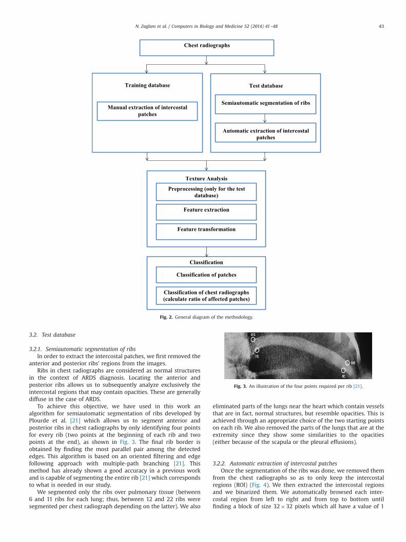
The methods for the development of a CAD to detect ARDS aredescribed in this section and shown in Fig. 2.
3.1. Training database
3.1.1. Manual extraction of intercostal patchesOpacities in ARDS are bilateral and diffuse. To analyze the
normal and abnormal regions, we manually selected normal andabnormal patches of size 32�32 pixels in the intercostal regionswith the help of an intensivist (PJ) in order to create the trainingset. 321 patches were thus selected and diagnosed (avoiding theanterior and posterior ribs) from 9 radiographs. 4 radiographspresented ARDS and 5 others did not (4 normal cases and 1 casewith left lung affected). A patch is considered abnormal if about90% of its surface contains opacity.
Fig. 1. Left chest radiograph: normal case; right chest radiograph: ARDS case (Ref: Sainte Justine Hospital).
N. Zaglam et al. / Computers in Biology and Medicine 52 (2014) 41–4842
3.2. Test database
3.2.1. Semiautomatic segmentation of ribsIn order to extract the intercostal patches, we first removed the
anterior and posterior ribs' regions from the images.Ribs in chest radiographs are considered as normal structures
in the context of ARDS diagnosis. Locating the anterior andposterior ribs allows us to subsequently analyze exclusively theintercostal regions that may contain opacities. These are generallydiffuse in the case of ARDS.
To achieve this objective, we have used in this work analgorithm for semiautomatic segmentation of ribs developed byPlourde et al. [21] which allows us to segment anterior andposterior ribs in chest radiographs by only identifying four pointsfor every rib (two points at the beginning of each rib and twopoints at the end), as shown in Fig. 3. The final rib border isobtained by finding the most parallel pair among the detectededges. This algorithm is based on an oriented filtering and edgefollowing approach with multiple-path branching [21]. Thismethod has already shown a good accuracy in a previous workand is capable of segmenting the entire rib [21] which correspondsto what is needed in our study.
We segmented only the ribs over pulmonary tissue (between6 and 11 ribs for each lung; thus, between 12 and 22 ribs weresegmented per chest radiograph depending on the latter). We also
eliminated parts of the lungs near the heart which contain vesselsthat are in fact, normal structures, but resemble opacities. This isachieved through an appropriate choice of the two starting pointson each rib. We also removed the parts of the lungs that are at theextremity since they show some similarities to the opacities(either because of the scapula or the pleural effusions).
3.2.2. Automatic extraction of intercostal patchesOnce the segmentation of the ribs was done, we removed them
from the chest radiographs so as to only keep the intercostalregions (ROI) (Fig. 4). We then extracted the intercostal regionsand we binarized them. We automatically browsed each inter-costal region from left to right and from top to bottom untilfinding a block of size 32�32 pixels which all have a value of 1
Chest radiographs
Training database
Classification
Test database
Texture Analysis
Automatic extraction of intercostal patches
Manual extraction of intercostalpatches
Preprocessing (only for the test database)
Feature extraction
Feature transformation
Classification of patches
Classification of chest radiographs (calculate ratio of affected patches)
Semiautomatic segmentation of ribs
Fig. 2. General diagram of the methodology.
Fig. 3. An illustration of the four points required per rib [21].
N. Zaglam et al. / Computers in Biology and Medicine 52 (2014) 41–48 43
(white). This means that the patch contains only the pixelsbelonging to the lung tissue between both ribs. Once we find theposition of the white patch in the binary picture, we extract it fromthe original intercostal area for analysis as shown in Fig. 5.
The 32�32 pixel patch's size was chosen because it is generallythe optimal size that can be extracted from the intercostal area(smaller than or equal to the width of the intercostal regions).However, for smaller intercostal areas, 16�16 pixel patches wereextracted as shown in Fig. 6.
3.3. Texture analysis
3.3.1. PreprocessingDuring the diagnosis, doctors take into account the variation of
contrast between the skin tissue and the background. If it is large,then the patient is considered to be underexposed (white chestradiograph) and poorly penetrated. In this case, the chest radio-graph is too bright and does not have enough contrast.
To solve this problem, we first calculated automatically thecontrast between the skin tissue and the background for eachchest radiograph by averaging neighboring pixels of the mostlateral point of the fourth left rib, which is close enough to the skintissue of the left lung. The background in a chest radiograph isnormally black. If the difference in intensity is high, we apply theCLAHE (Contrast-Limited Adaptive Histogram Equalization) pre-processing technique to improve the local contrast.
3.3.2. Feature extractionIn this work, we used both statistical features (the histogram
and the co-occurrence matrix) and spectral features (the Fouriertransform).
Using the central moments can be a good method to representquantitatively a histogram. This can be represented by the follow-ing formulas:
μn ¼ ∑L�1
i ¼ 0ðzi�mÞnpðziÞ ð1Þ
where n is the order of the moment, zi is a random variablerepresenting the intensity, p(z) is the histogram of the intensitylevels in the image, L is the number of possible intensity levels andm is the mean intensity with
m¼ ∑L�1
i ¼ 0zipðziÞ ð2Þ
Six texture descriptors based on the intensity histogramwere usedas shown in Table 1.
However, the histogram-based features do not capture thespatial relationships between the pixels' intensities. These rela-tionships can be represented numerically using grayscale spatialdependence matrices, also called co-occurrence matrices. These
Fig. 4. The image on the left shows a chest radiograph, the middle image shows posterior and anterior segmented ribs and the image on the right shows the chestradiograph after removal of ribs.
Fig. 5. Intercostal region extracted from the X-ray (left), image after binarization and extraction of the white patches (middle), patches on the original intercostal region(right).
Fig. 6. Patches automatically selected from a chest radiograph (size 32�32 or16�16).
N. Zaglam et al. / Computers in Biology and Medicine 52 (2014) 41–4844
determine the frequency of occurrence of a “pattern” formed oftwo pixels separated by a certain distance dis in a particulardirection θ relative to the horizontal axis [22]. Co-occurrencematrices can show the degree of similarity or dissimilarity ofadjacent pixels. A region of interest with opacity will containadjacent pixels that have similar high intensities. Four features areextracted from the co-occurrence matrix: energy, contrast, homo-geneity and correlation.
Spectral texture measurements for our study are based on theFourier spectrum. In the frequency spectrum, the high frequenciesshow the strong variations between dark areas and opacities,which make them useful for our work. To facilitate interpretationof spectrum features, the spectrum is expressed in polar coordi-nates by the function Sðr;θÞ, where S is the function of spectrumand r and θ are the independent variables in the polar system [23].The global description of the spectral energy is obtained bysumming the following two functions:
SðrÞ ¼ ∑π
θ ¼ 0SθðrÞ ð3Þ
SðθÞ ¼ ∑R0
r ¼ 1SrðθÞ ð4Þ
where SθðrÞ is a 1D function representing the spectrum Sðr;θÞ foreach direction θ and SrðθÞ is another 1D function representing thespectrum Sðr;θÞ for each radius r. S(r) and SðθÞ represent adescription of the texture spectral energy for either the wholeimage or a part of it.
The descriptors of these functions can be computed to char-acterize their quantitative behavior. Typical descriptors used forthis purpose are the mean and variance of the function as well asthe distance between the mean and the maximum value of thefunction [23].
3.4. Feature transformation
Linear Discriminant Analysis (LDA) is a method with theobjective of finding an optimal linear transformation of featuresthat separates 2 or more classes. The best transformation is theone that shows a maximum distance between the different classesand a minimum distance between elements of each class [24]. Ourtraining database X is partitioned into two classes as X ¼ fX1;X2gwhere X1 containing n1 patches diagnosed as abnormal and a classof normal patches X2 containing n2 patches diagnosed as normal.
The objective of LDA is to find a projection direction wn thatmaximizes the ratio of between-class scatter to within-class
scatter [25], where
wn ¼ S�1w ðm1�m2Þ ð5Þ
The between- and within-class scatter matrices Sb and Sw aredefined as
Sb ¼ ðm1�m2Þðm1�m2ÞT ð6Þ
Sw ¼ ∑iA1;2
∑xAXi
ðx�miÞðx�miÞT ð7Þ
where mi ¼ ð1=niÞ∑xAXix is the mean of the ith class.
3.5. Classification
3.5.1. Classification of patchesWe used as a classifier, the SVM (Support Vector Machine) with
a Gaussian kernel, to classify patches as normal or abnormal. TheSVM is a technique developed by Vapnik [26] and has shown itseffectiveness in the field of medical imaging and other fields. AnSVM classifier looks for an optimal hyperplane as a decisionfunction in a high-dimensional space, i.e., the one with themaximum distance from the nearest training patterns [27].
To effectively implement the SVM, it is necessary to optimizethe values of the hyper-parameters C and γ of the Gaussian kernel.C is the constant of soft margin. This parameter is used to fix thetradeoff between minimizing the training error and maximizingthe margin, while γ is the parameter of the Gaussian kernel. Tochoose the best hyper-parameters, we applied the SVM classifieron the training database using the leave-one-out validation strat-egy. The classification results were evaluated using measures ofsensitivity and specificity. Then we plot the Received OperatingCharacteristic (ROC) curve, which shows the true positive fraction(sensitivity) as a function of the false positive fraction (1-specifi-city). The ROC curve is used to select the optimal hyper-parameters that give the best classification performance.
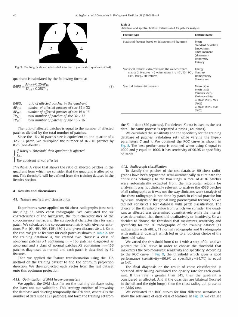
3.5.2. Classification of chest radiographsARDS is defined as the presence of bilateral opacities. Before
making a diagnosis of ARDS, doctors generally divide each lunginto two parts called quadrants (Fig. 7). If at least two quadrants(one on the left lung and one on the right lung) are affected, thenthey consider the radiograph as an ARDS case. After havingclassified the patches, the ratio of affected patches in each
Table 1Descriptors of texture based on histograms.
Descriptors Expression Measure of texture
Mean m¼ ∑L�1
i ¼ 0zipðziÞ A measure of average intensity
Standard deviation μ2 A measure of average contrast
Smoothness R¼ 1� 11þσ2
Measures the relative smoothness of theintensity
Third moment(skewness)
μ3 Measures the skewness of a histogram
Uniformity U ¼ ∑L�1
i ¼ 0p2ðziÞ Measures the uniformity
Entropy e¼ � ∑L�1
i ¼ 0pðziÞ log 2 pðziÞ A measure of randomness
N. Zaglam et al. / Computers in Biology and Medicine 52 (2014) 41–48 45
quadrant is calculated by the following formula:
RAPQ ¼ AP32þ0:25AP16
TP32þ0:25TP16ð8Þ
RAPQ: ratio of affected patches in the quadrantAP32: number of affected patches of size 32�32AP16: number of affected patches of size 16�16TP32: total number of patches of size 32�32TP16: total number of patches of size 16�16
The ratio of affected patches is equal to the number of affectedpatches divided by the total number of patches.
Since the 16�16 patch's size is equivalent to one-quarter of a32�32 patch, we multiplied the number of 16�16 patches by0.25 (one-fourth):
If RAPQ4Threshold then quadrant is affected
Else
The quadrant is not affected
8><>:
Threshold: A value that shows the ratio of affected patches in thequadrant from which we consider that the quadrant is affected ornot. This threshold will be defined from the training dataset in theResults section.
4. Results and discussions
4.1. Texture analysis and classification
Experiments were applied on 90 chest radiographs (test set),including 53 ARDS chest radiographs. We calculated the sixcharacteristics of the histogram, the four characteristics of theco-occurrence matrix and the six spectral characteristics for eachpatch. We calculated the co-occurrence matrix with given orienta-tions θ A {01, 451, 901, 1351, 1801} and given distance dis¼1. So atthe end, we got 32 features for each patch as shown in Table 2. Forthe training database X, we created two classes: a class ofabnormal patches X1 containing n1 ¼ 165 patches diagnosed asabnormal and a class of normal patches X2 containing n2¼156patches diagnosed as normal and each patch is described by 32features.
Then we applied the feature transformation using the LDAmethod on the training dataset to find the optimum projectiondirection. We then projected each vector from the test datasetonto this optimum projection.
4.1.1. Optimization of SVM hyper-parametersWe applied the SVM classifier on the training database using
the leave-one-out validation. This strategy consists of browsingthe database and deleting temporarily the Kth data, where K is thenumber of data used (321 patches), and form the training set from
the K�1 data (320 patches). The deleted K data is used as the testdata. The same process is repeated K times (321 times).
We calculated the sensitivity and the specificity for the trainingdatabase of patches (validation set) while varying the hyper-parameters C and γ. We obtained the ROC curve as shown inFig. 8. The best performance is obtained when using C equal to1000 and γ equal to 1000. It has sensitivity of 90.9% at specificityof 94.9%.
4.1.2. Radiograph classificationTo classify the patches of the test database, 90 chest radio-
graphs have been segmented semi-automatically to eliminate theentire ribs belonging to the two lungs. A total of 4536 patcheswere automatically extracted from the intercostal regions foranalysis. It was not clinically relevant to analyze the 4536 patchesof all radiographs as it was not the way clinicians work (analysis ofthe chest radiograph is not done by patch in clinical practice butby visual analysis of the global lung parenchymal texture). So wedid not construct a test database with patch classification. Thechoice of the threshold value from which we consider the quad-rant as affected was determined quantitatively while the intensi-vists determined that threshold qualitatively or intuitively. So wedecided to choose the threshold that maximizes sensitivity andspecificity for the 30 radiographs of the training dataset (11radiographs with ARDS, 11 normal radiographs and 8 radiographswith unilateral opacity), which led us to a judicious choice of thethreshold value.
We varied the threshold from 0 to 1 with a step of 0.1 and weplotted the ROC curve in order to choose the threshold thatmaximizes the two measures: sensitivity and specificity. Accordingto the ROC curve in Fig. 9, the threshold which gives a goodperformance (sensitivity¼90.9% at specificity¼94.7%) is equalto 34%.
The final diagnosis or the result of chest classification isobtained after having calculated the opacity rate for each quad-rant. If this rate is greater than 34%, then the quadrant isconsidered as affected. And if the opacities are bilateral (locatedin the left and the right lungs), then the chest radiograph presentsan ARDS case.
We evaluated the ROC curves for four different scenarios toshow the relevance of each class of features. In Fig. 10, we can see
Fig. 7. The lung fields are subdivided into four regions called quadrants (1–4).
Table 2Statistical and spectral texture features used for patch's analysis.
Feature type Feature name
Statistical features based on histograms (6 features) MeanStandard deviationSmoothnessThird moment(skewness)UniformityEntropy
Statistical features extracted from the co-occurrencematrix (4 features �5 orientations θ A {01, 451, 901,1351, 1801}¼20 features)
EnergyContrastHomogeneityCorrelation
Spectral features (6 features) Mean ðSðrÞÞMean ðSðθÞÞVariance ðSðrÞÞVariance ðSðθÞÞΔ(Mean ðSðrÞÞ, MaxðSðrÞÞ)Δ(Mean ðSðθÞÞ, MaxðSðθÞÞ)
N. Zaglam et al. / Computers in Biology and Medicine 52 (2014) 41–4846
that texture analysis based on the combination of the three typesof texture features outperforms the use of one kind of feature only(histogram or co-occurrence matrix or spectral features). For theautomatic analysis of the 90 chest radiographs based on statisticaland spectral texture features, the results of classification gave asensitivity of 90.6% at a specificity of 86.5% (Table 3).
These results show that our system has a good performance.We also note that our automatic system is more sensitive thanspecific and therefore there are less false negative cases. Theintensivists prefer to be more sensitive, since early diagnosis ofARDS has the advantage of using a reduced tidal volume andthereby avoiding the complications that can be caused by thetreatment (mechanical ventilation).
According to our knowledge, this computer-aided diagnosissystem for the detection of ARDS is considered as the firstsuch system that evaluates this disease. ARDS introduces high
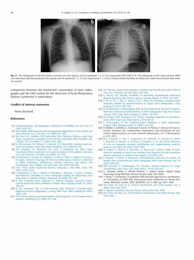
inter-observer variability in chest radiograph interpretation whichmakes the development of an automatic system challenging.However our study of the variability of diagnosis between inten-sivists and the reconstruction of chest radiographs databasediagnosed with consensus allowed to realize such a system. Thissystem aims to interpret the chest radiograph, which is the mostlimiting factor among the four diagnostic criteria of ARDS definedby the AECC. The other three criteria (Acute situation, PaO2/FiO2o200 mmHg and no clinical evidence of left heart failure) shouldbe taken into consideration to establish a global diagnosis which isnot studied in this work. The semiautomatic segmentation wasused to analyze the intercostal regions (ROI) and eliminate otherunnecessary areas for the diagnosis (ribs, heart, skin tissue,diaphragm, etc.). In addition the analysis of intercostal patchesallows the intensivists to locate abnormalities on the chest radio-graph by framing affected patches in black and the other patchesin white (Fig. 11). Locating parts of the lungs that need to beventilated to help the patient breathing can be very useful duringtreatment.
5. Conclusion
We have presented a computer-aided diagnosis system for thedetection of Acute Respiratory Distress Syndrome (ARDS) in chestradiographs. Our method is based on texture analysis applied onintercostal patches which are selected automatically after havingsegmented the entire ribs. Statistical and spectral features areextracted from each patch and classified with an SVM. Finally, theclassification of the chest radiographs is realized after calculatingthe opacity rate for each quadrant. Experiments were performedon a test dataset containing 90 chest radiographs, and thereforethis tool can be used by intensivists to get a second opinion ofdiagnosis and elaborate an early diagnosis of the disease forimproving its treatment. To show the utility of such a system, a
0
0.1
0.2
0.3
0.4
0.5
0.6
0.7
0.8
0.9
1
0 0.2 0.4 0.6 0.8 1
Tru
e po
sitiv
e fr
actio
n (S
ensi
tivity
)
False postive fraction (1-Specificity)
Fig. 8. ROC curve for the training set using the three kinds of features to selecthyper-parameters C and γ.
0
0.1
0.2
0.3
0.4
0.5
0.6
0.7
0.8
0.9
1
0 0.1 0.2 0.3 0.4 0.5 0.6 0.7
Tru
e po
sitiv
e fr
actio
n (S
ensi
tivity
)
False postive fraction (1-Specificity)
Fig. 9. ROC curve for the 30 radiographs of the training dataset to select threshold.
0
0.1
0.2
0.3
0.4
0.5
0.6
0.7
0.8
0.9
1
0 0.1 0.2 0.3 0.4 0.5 0.6 0.7 0.8 0.9 1
Tru
e po
sitiv
e fr
actio
n (S
ensi
tivity
)
False postive fraction (1-Specificity)
Combined
Coocurrence
Histogram
Spectral
Fig. 10. ROC curves for the test set using the three kinds of features.
Table 3Results of classification for the different kinds of texture features.
N. Zaglam et al. / Computers in Biology and Medicine 52 (2014) 41–48 47
comparison between the intensivist's assessment of chest radio-graphs and the CAD system for the detection of Acute RespiratoryDistress Syndrome is undertaken.
Conflict of interest statement
None Declared.
References
[1] K. Raghavendran, L.M. Napolitano, Definition of ALI/ARDS, Crit. Care Clin. 27(2011) 429–437.
[2] M.M. Taylor, Ards diagnosis and management: implications for the critical carenurse, Dimens. Crit. Care Nurs. 24 (2005) 197–207.
[3] H.R. Flori, D.V. Glidden, G.W. Rutherford, M.A. Matthay, Pediatric acute lunginjury: prospective evaluation of risk factors associated with mortality, Am.J. Respir. Crit. Care Med. 171 (2005) 995–1001.
[4] J.J. Zimmerman, S.R. Akhtar, E. Caldwell, G.D. Rubenfeld, Incidence and out-comes of pediatric acute lung injury, Pediatrics 124 (2009) 87–95.
[5] M.C. Kneyber, A.G. Brouwers, J.A. Caris, S. Chedamni, F.B. Plötz, Acuterespiratory distress syndrome: is it underrecognized in the pediatric intensivecare unit? Intensiv. Care Med. 34 (2008) 751–754.
[6] G.R. Bernard, A. Artigas, K.L. Brigham, J. Carlet, K. Falke, L. Hudson, M. Lamy, J.R. Legall, A. Morris, R. Spragg, The American-0European consensus conferenceon ARDS. Definitions, mechanisms, relevant outcomes, and clinical trialcoordination, Am. J. Respir. Crit. Care Med. 149 (1994) 818–824.
[8] F. Angoulvant, J. Llor, C. Alberti, A. Kheniche, I. Zaccaria, C. Garel, S. Dauger,Inter-observer variability in chest radiograph reading for diagnosing acutelung injury in children, Pediatr. Pulmonol. 43 (2008) 987–991.
[9] K. Doi, Computer-aided diagnosis in medical imaging: historical review,current status and future potential, Comput. Med. Imaging Graph. 31 (2007)198–211.
[10] B. Van Ginneken, B.M. ter Haar Romeny, M.A. Viergever, Computer-aideddiagnosis in chest radiography: a survey, IEEE Trans. Med. Imaging 20 (2001)1228–1241.
[11] G.D. Tourassi, Journey toward computer-aided diagnosis: role of image textureanalysis1, Radiology 213 (1999) 317–320.
[12] A.P. Dhawan, Image Representation, Analysis, and Classification, John Wiley &Sons, Inc., Hoboken, NJ, USA (2011) 265–309.
[13] G. Revesz, H.L. Kundel, Feasibility of classifying disseminated pulmonarydiseases based on their Fourier spectra, Investig. Radiol. 8 (1973) 345–349.
[14] P. Yu, H. Xu, Y. Zhu, C. Yang, X. Sun, J. Zhao, An automatic computer-aideddetection scheme for pneumoconiosis on digital chest radiographs, J. Dig.Imaging 24 (2011) 382–393.
[15] B. van Ginneken, S. Katsuragawa, B.M. ter Haar Romeny, K. Doi, M.A. Viergever,Automatic detection of abnormalities in chest radiographs using local textureanalysis, IEEE Trans. Med. Imaging 21 (2002) 139–149.
[16] R.P. Kruger, W.B. Thompson, A.F. Turner, Computer diagnosis of pneumoco-niosis, IEEE Trans. Syst. Man Cybern. (1974) 40–49.
[17] S. Katsuragawa, K. Doi, Computer-aided diagnosis in chest radiography,Comput. Med. Imaging Graph. 31 (2007) 212–223.
[18] N. Kleiber, E. Lefebvre, G. Emeriaud, P. Jouvet, N. Poitras, T. Ducruet, M. Tucci, J.Lacroix, Incidence des complications respiratoires post-transfusion de con-centrés érythrocytaires aux soins intensifs pédiatriques, vol. 2, Réanimation,p. S131–S131.
[19] N.D. Ferguson, E. Fan, L. Camporota, M. Antonelli, A. Anzueto, R. Beale,L. Brochard, R. Brower, A. Esteban, L. Gattinoni, et al., The berlin definitionof ards: an expanded rationale, justification, and supplementary material,Intensiv. Care Med. 38 (2012) 1573–1582.
[20] N. Zaglam, F. Cheriet, O. Flechelles, G. Emeriaud, P. Jouvet, Study of inter-observer variability in chest X-ray reading in the diagnosis of acute respiratorydistress syndrome in children, Can. Respir. J. 20 (2012) e34.
[21] F. Plourde, F. Cheriet, J. Dansereau, Semiautomatic detection of scoliotic ribborders from posteroanterior chest radiographs, IEEE Trans. Biomed. Eng. 59(2012) 909–919.
[22] R.M. Haralick, K. Shanmugam, I.H. Dinstein, Textural features for imageclassification, IEEE Trans. Syst. Man Cybern. (1973) 610–621.
[23] C. Gonzalez Rafael, E. Woods Richard, L. Eddins Steven, Digital ImageProcessing Using MATLAB, Pearson Prentice Hall, USA, 2004.
[24] T. Xiong, V. Cherkassky, A combined SVM and LDA approach for classification,in: Proceedings of 2005 IEEE International Joint Conference on Neural Net-works, Montreal, Canada, 2005 (IJCNN'05), vol. 3. IEEE, pp. 1455–1459.
[25] R.O. Duda, P.E. Hart, et al., Pattern Classification and Scene Analysis, vol. 3,Wiley, New York, 1973.
[26] V. Vapnik, Statistical Learning Theory, Wiley, New York, 1998.[27] R.O. Duda, P.E. Hart, D.G. Stork, Pattern Classification, John Wiley & Sons, New
York, 2001.
Fig. 11. The radiograph on the left shows a normal case (the opacity rate for quadrants 1, 2, 3, 4 is respectively 0.09, 0.08, 0, 0). The radiograph on the right presents ARDScase with three affected quadrants (the opacity rate for quadrants 1, 2, 3, 4 are respectively 1, 1, 1, 0.15). Patches framed by black are abnormal, while those framed with whiteare normal.
N. Zaglam et al. / Computers in Biology and Medicine 52 (2014) 41–4848