63
Concepts of cancer immunotherapy

Concepts of cancer immunotherapy
History • Paul Ehrlich first conceived the idea that tumor cells can be recognized as
“foreign” and eliminated by the immune system. • Subsequently, Lewis Thomas and Macfarlane Burnet formalized this
concept by coining the term immune surveillance, which implies that a normal function of the immune system is to constantly “scan” the body for emerging malignant cells and destroy them.
• This idea has been supported by many observations – the presence of lymphocytic infiltrates around tumors and reactive changes in
lymph nodes draining sites of cancer – experimental results, mostly with transplanted tumors; – the increased incidence of some cancers in immunodeficient people and mice; – the direct demonstration of tumor-specific T cells and antibodies in patients; – most recently and most directly, the response of advanced cancers to
therapeutic agents that act by stimulating latent host T-cell responses
Cancer immunoediting • The fact that cancers occur in immunocompetent
individuals indicates that immune surveillance is imperfect • it follows that the tumors that do grow out must be
composed of cells that are either invisible to the host immune system or that release factors that actively suppress host immunity.
• The term cancer immunoediting has been used to describe the ability of the immune system to shape and mold the immunogenic properties of tumor cells in a fashion that ultimately leads to the darwinian selection of subclones that are best able to avoid immune elimination.
Tumor Antigens
• Product of mutated genes • Consequence of enhanced or aberrant
expression • Product of oncogenic viruses • Oncofetal antigens • Altered cell surface glycolipids and
glycoproteins • Differentiation antigens
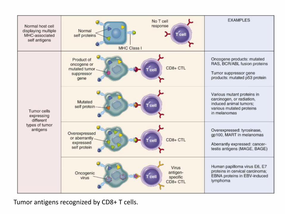
Tumor antigens recognized by CD8+ T cells.
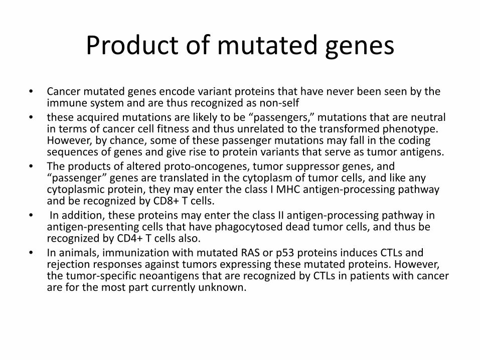
Product of mutated genes • Cancer mutated genes encode variant proteins that have never been seen by the
immune system and are thus recognized as non-self • these acquired mutations are likely to be “passengers,” mutations that are neutral
in terms of cancer cell fitness and thus unrelated to the transformed phenotype. However, by chance, some of these passenger mutations may fall in the coding sequences of genes and give rise to protein variants that serve as tumor antigens.
• The products of altered proto-oncogenes, tumor suppressor genes, and “passenger” genes are translated in the cytoplasm of tumor cells, and like any cytoplasmic protein, they may enter the class I MHC antigen-processing pathway and be recognized by CD8+ T cells.
• In addition, these proteins may enter the class II antigen-processing pathway in antigen-presenting cells that have phagocytosed dead tumor cells, and thus be recognized by CD4+ T cells also.
• In animals, immunization with mutated RAS or p53 proteins induces CTLs and rejection responses against tumors expressing these mutated proteins. However, the tumor-specific neoantigens that are recognized by CTLs in patients with cancer are for the most part currently unknown.

Overexpressed and aberrantly expressed proteins
• Tumor antigens may also be normal cellular proteins that are abnormally expressed in tumor cells.
• Examples: tyrosinase, expressed only in normal melanocytes and melanomas – tyrosinase is normally produced in such small amounts and in
so few normal cells that it is not recognized by the immune system and fails to induce tolerance.
• Cancer-testis antigens, are encoded by genes that are silent in all adult tissues except germ cells in the testis. – sperm do not express MHC class I antigens, so these proteins
are not immunogenic normally. – Melanoma antigen gene (MAGE) family. Although originally
described in melanomas, MAGE antigens are expressed by a variety of tumor types.
Products of oncoviruses
• Oncoviruses produce proteins that are recognized as foreign by the immune system.
• Examples in humans include human papilloma virus (HPV) and Epstein-Barr virus (EBV). – Abundant evidence that CTLs recognize antigens of
these viruses and that a competent immune system plays a role in surveillance against virus-induced tumors
– the concept of immune surveillance against tumors is best established for DNA virus-induced tumors.
Oncofetal proteins • Oncofetal antigens are proteins that are expressed at high levels on
cancer cells and in normal developing (fetal) tissues. • Amounts of these proteins are increased in tissues and in the
circulation in various inflammatory conditions, and they are even found in small quantities in normal tissues.
• There is no evidence that oncofetal antigens are important inducers or targets of antitumor immunity.
• Oncofetal proteins are sufficiently specific that they can serve as markers that aid in tumor diagnosis and clinical management.
• The two most thoroughly characterized oncofetal antigens are carcinoembryonic antigen (CEA) and α-fetoprotein (AFP). These are used extensively as tumor markers in clinics.

Cell surface glycolipids and glycoproteins
• Most tumors express higher than normal levels and/or abnormal forms of surface glycoproteins and glycolipids
• These altered molecules include: – gangliosides – blood group antigens – mucins.
• Mucins are high-molecular-weight glycoproteins containing numerous carbohydrate side chains on a core polypeptide. Tumors often have dysregulated expression of the enzymes that synthesize these carbohydrate side chains, which leads to the appearance of tumor-specific epitopes on the carbohydrate side chains or on the abnormally exposed polypeptide core.
• Several mucins have been the focus of diagnostic and therapeutic studies, including CA-125 and CA-19-9, expressed on ovarian carcinomas, and MUC-1, expressed on both ovarian and breast carcinomas.
• MUC-1 is an integral membrane protein that is normally expressed only on the apical surface of breast ductal epithelium. In ductal carcinomas of the breast, however, the molecule is expressed in an unpolarized fashion and contains new, tumor-specific carbohydrate and peptide epitopes that induce both antibody and T-cell responses in cancer patients and are therefore considered candidates for tumor vaccines in patients with breast cancer and possibly ovarian cancer as well.

Cell-type specific differentiation antigens
• Tumors express molecules that are normally present on the cells of origin, called differentiation antigens because they are specific for particular lineages or differentiation stages of various cell types.
• Differentiation antigens are typically normal self-antigens, and therefore they do not induce immune responses in tumor-bearing hosts. – Their importance is as potential targets for
immunotherapy and for identifying the tissue of origin of tumors.

An example: CD20 • CD20 is a transmembrane protein that is expressed on
the surface of all normal mature B cells • Antibodies against CD20 have broad cytocidal activity
against mature B-cell lymphomas and leukemias and are widely used in the treatment of these tumors.
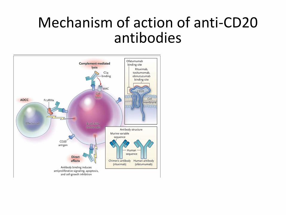
• These antibodies are believed to induce cell killing through several mechanisms, including opsonization and phagocytosis of tumor cells, antibody-dependent cell-mediated cytotoxicity and complement fixation.
• Anti-CD20 antibodies also kill normal B cells, but because hematopoietic stem cells are spared, normal B cells reemerge following treatment.
Mechanism of action of anti-CD20 antibodies

Mechanism of action of anti-CD20 antibodies

Other approaches • Monoclonal antibodies may also be covalently coupled to drugs,
toxins, or radiochemicals – the antibody serves as guided missile that delivers a therapeutic
warhead to cancers expressing particular surface antigens • Anti-CD30 antibodies:
– CD30 is a member of the TNF receptor family of transmembrane proteins that is expressed by particular T cell lymphomas and most Hodgkin lymphomas.
– Antibodies against CD30 linked to a cytotoxic drug have recently produced remarkable responses in patients with CD30-positive lymphomas that have failed conventional therapies.
• Bispecific antibodies engineered to have two different antigen recognition surfaces, one that binds tumor antigens and a second that binds to the CD3 signaling molecule on T cells, have produced some promising results in clinical trials.
Antitumor Effector Mechanisms
• Humoral immunity: negligible • Cellular immunity: main mechanism
Cytotoxic T-lymphocytes
• The antitumor effect of cytotoxic T cells reacting against tumor antigens is well established in experimentally induced tumors.
• In humans, CD8+ CTLs have a clear protective role against virus-associated neoplasms (e.g., EBV- and HPV-induced tumors)
• Several studies have shown that the number of tumor-infiltrating CD8+ T cells and the presence of a “gene signature” associated with CD8+ CTLs correlates with a better prognosis in a variety of cancers, not only those caused by oncogenic viruses.

Natural Killer cells
• NK cells are lymphocytes that are capable of destroying tumor cells without prior sensitization and thus may provide the first line of defense against tumor cells.
• After activation with IL-2 and IL-15, NK cells can lyse a wide range of human tumors, including many that seem to be nonimmunogenic for T cells.
• While the importance of NK cells in host response against spontenous tumors is still not well established, cytokines that activate NK cells are being used for immunotherapy.
Macrophages
• Activated macrophages exhibit cytotoxicity against tumor cells in vitro.
• T cells, NK cells, and macrophages may collaborate in antitumor reactivity, because interferon-γ, a cytokine secreted by T cells and NK cells, is a potent activator of macrophages.
• Activated macrophages may kill tumors by mechanisms similar to those used to kill microbes (e.g., production of reactive oxygen species)
Immune surveillance against cancer • Increased frequency of cancers in the setting of
immunodeficiency. – Persons with congenital immunodeficiencies develop cancers at about
200 times the rate in immunocompetent individuals. – Immunosuppressed transplant recipients and persons with AIDS also
have an increased incidence of malignancies. – Particularly illustrative is the rare X-linked recessive immunodeficiency
disorder termed XLP (X-linked lymphoproliferative syndrome), caused by mutations in the gene encoding an adapter protein, SAP, which participates in NK and T-cell signaling pathways. In affected boys, EBV infection does not take the usual self-limited form of infectious mononucleosis but instead evolves into a chronic or sometimes fatal form of infectious mononucleosis or, even worse, a lymphoma comprised of EBV-infected B cells.
• Most cancers occur in persons who do not suffer from any overt immunodeficiency. It is evident, then, that tumor cells must develop mechanisms to escape or evade the immune system in immunocompetent hosts.

The 3 “E”s: Elimination

The 3 “E”s: Equilibrium

The 3 “E”s: Escape

Evasion of the immune response

Mechanisms of evasion of the immune response
• Selective outgrowth of antigen-negative variants. – During tumor progression, strongly immunogenic
subclones may be eliminated, an example of immunoediting that has already been discussed.
• Loss or reduced expression of MHC molecules.
– Tumor cells may fail to express normal levels of HLA class I molecules, thereby escaping attack by cytotoxic T cells. Such cells, however, may trigger NK cells if the tumor cells express ligands for NK cell activating receptors.

Mechanisms of evasion of the immune response
• Secretion of immunosuppressive factors by cancer cells. – Tumors may secrete products that inhibit the host immune response.
• TGF-β is secreted in large quantities by many tumors and is a potent immunosuppressant.
• Other tumors secrete galectins, sugar-rich lectin-like factors that skew T-cell responses so as to favor immunosuppression.
• Many other soluble factors produced by tumors are also suspected of inhibiting the host immune response, including interleukin-10, prostaglandin E2, certain metabolites derived from tryptophan, and VEGF, which can inhibit the diapedesis of T cells from the vasculature into the tumor bed.
• Induction of regulatory T cells (Tregs).
– Some studies suggest that tumors produce factors that favor the development of immunosuppressive regulatory T cells, which could also contribute to “immunoevasion.”
Mechanisms of evasion of the immune response
• Activation of immunoregulatory pathways. – tumor cells actively inhibit tumor immunity by engaging normal
pathways of immune regulation that serve as “checkpoints” in immune responses.
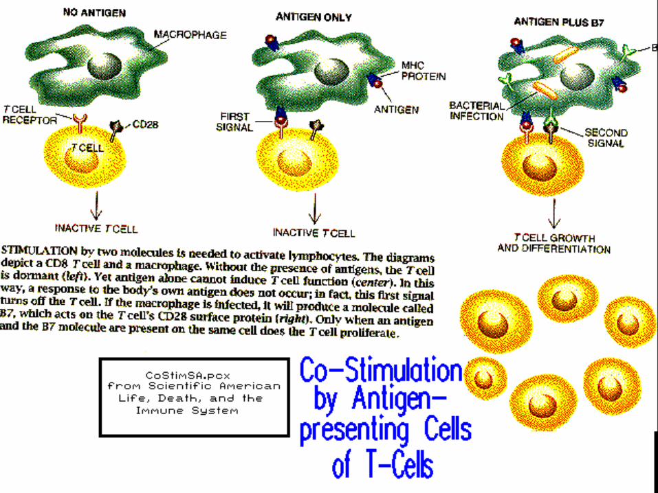
• Tumor cells may downregulate the expression of costimulatory factors on antigen-presenting cells, such as dendritic cells
• as a result, the antigen presenting cells fail to engage the stimulatory receptor CD28 and instead activate the inhibitory receptor CTLA-4 on effector T cells.
• This not only prevents sensitization but also may induce long-lived unresponsiveness in tumor-specific T cells.
• Tumor cells also may upregulate the expression of PD-L1 and PD-L2, cell surface proteins that activate the programmed death-1 (PD-1) receptor on effector T cells.
• PD-1, like CTLA-4, may inhibit T cell activation.
Forms of Cancer Immunotherapy
• Non-Specific: Generalized, Non-Antigen-Specific Immune Activation
• Specific: Antigen-specific Response Induced in the Mouse or Patient or Passively Transferred in from Donor Source
Forms of Cancer Immunotherapy Active: Induced Directly in the Tumor-Bearing
Animal or in the Patient • Can be Specific or Non Specific
Passive or Adoptive: Immunologically Active Material Transferred into Mouse or Patient as a Passive Recipient
• Can be Specific (Antibodies, T-Cells, Antigen-presenting cells – Dendritic Cell Vaccines)
• Or Non-Specific (Non-specifically-activated T-Cells; Cytokines
Active Non-Specific Immunotherapy
Bacterial Extracts: Non-Specific Immune Adjuvants • BCG: Bacillus Calmette-Guerin (Attenuated Bovine
Tuberculosis Bacterium) • Membrane Extracts of BCG • C Parvum: Corynebacterium parvum (related to diphtheria
bacillus) Bacterial Endotoxins: Muramyl Dipeptide Chemical Adjuvants: • Levamisole • Poly IC (Poly-inosinic-Poly-cytidyllic acid) Cytokines: (Can be actively induced or passively transferred) • Interferons • Interleukin 2 (IL2) • Tumor Necrosis Factor (TNF)
Induced in the Patient or Mouse: Non-Antigen-specific

Tumor Necrosis Factor (TNHa) in Immunotherapy of Cancer (Passive or Active)

Adoptive Immunotherapy of Cancers (Passive: Donor to Recipient)
Non-Specific: • Lymphokine-activated Killer Cells (LAK Cells)\ • Cytokines (TNF alpha; IL2; Interferon) Specific: Molecular Transfer • Monoclonal Antibodies (antibodies are specific) Specific: Cellular Transfer (antigen-specific) • Tumor-Infiltrating Lymphocytes (TIL Cells) • Engineered Antigen-Presenting Cells (Dendritic
Cells)


Structure of interleukin 2
Schematic overview of the high–affinity interleukin–2 receptor complex, including the receptor chains, downstream signaling components and target genes
Fig. 1
Fig. 2
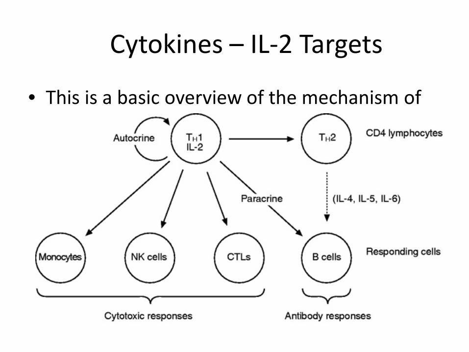
Cytokines – IL-2 Targets
• This is a basic overview of the mechanism of IL-2 activation
Adoptive Immunotherapy
• Immunotherapy – IL–2, alone, can be used as a cancer treatment by
activation of cells which are cytotoxic for the tumor
• Some success has been obtained with renal cell carcinoma and metastatic melanoma. – Rosenberg study

Adoptive Immunotherapy using TILs
• Technique involves isolating tumor-infiltrating lymphocytes (TIL’s) – Primarily activated cytotoxic T-lymphocytes – Lymphocytes with antitumor reactivity found within the tumor
• Expanding their number artificially in cell culture by means of human recombinant interleukin-2.
• The TILs are then put back into the bloodstream, along with IL-2, where they can bind to and destroy the tumor cells.

A.I. This figure shows adoptive immunotherapy isolation techniques
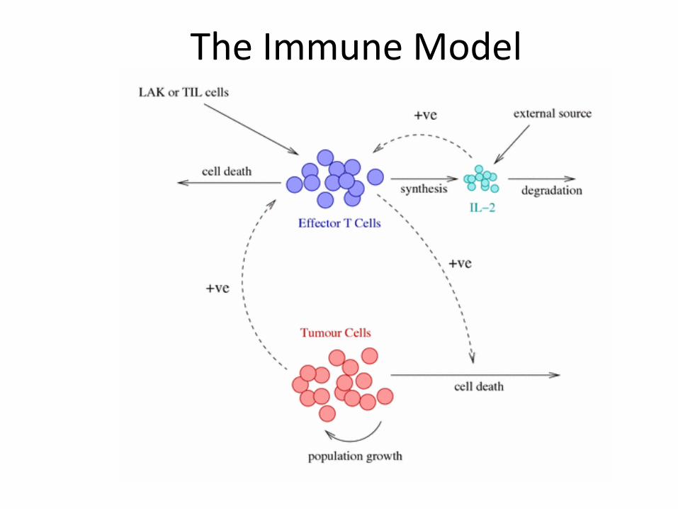
The Immune Model
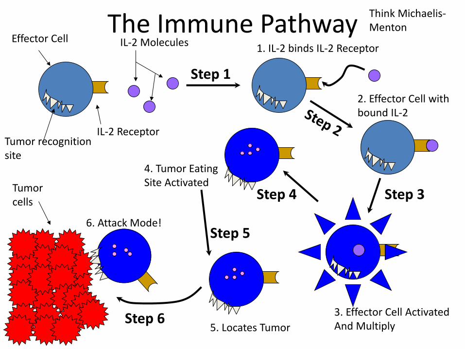
The Immune Pathway Effector Cell IL-2 Molecules
IL-2 Receptor Tumor recognition site
Tumor cells
Step 1
1. IL-2 binds IL-2 Receptor
2. Effector Cell with bound IL-2
3. Effector Cell Activated And Multiply
4. Tumor Eating Site Activated
6. Attack Mode!
Think Michaelis- Menton
5. Locates Tumor
Step 3 Step 4
Step 5
Step 6
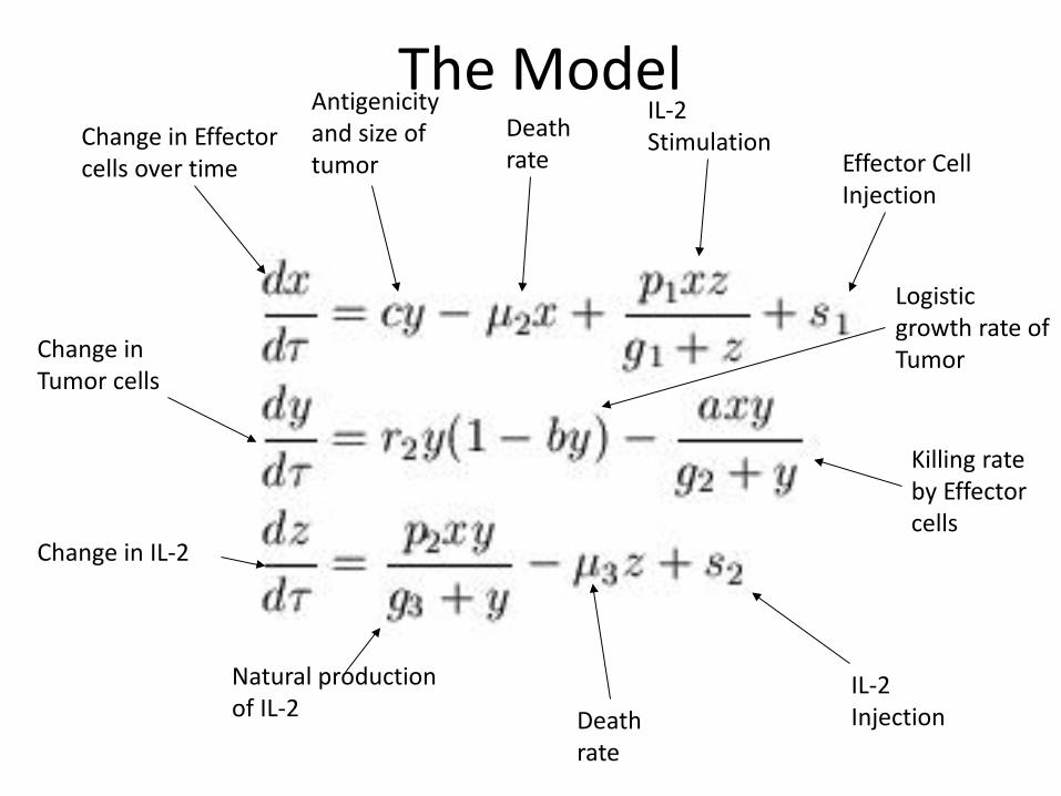
The Model Change in Effector cells over time
Antigenicity and size of tumor
Death rate
Death rate
IL-2 Stimulation
Effector Cell Injection
Change in Tumor cells
Logistic growth rate of Tumor
Killing rate by Effector cells
Change in IL-2
Natural production of IL-2
IL-2 Injection
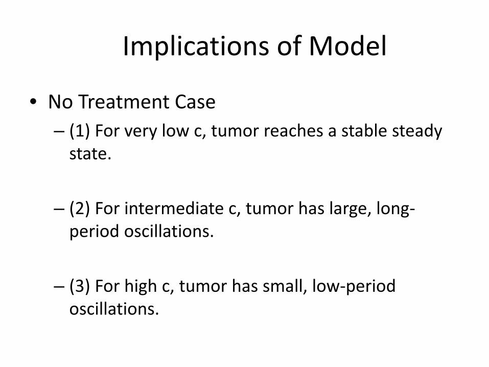
Implications of Model
• No Treatment Case – (1) For very low c, tumor reaches a stable steady
state.
– (2) For intermediate c, tumor has large, long-period oscillations.
– (3) For high c, tumor has small, low-period oscillations.
Reality of IL-2 Therapy
• High-dose IL-2 therapy alone has been shown to cause a variety of side effects. – Generally High Toxicity, e.g. Capillary Leak Syndrome
• Most of these are explainable by a runaway immune system.
• Question: IL-2 therapy does work in some cases; why does the model not predict this? – New models that incorporate “stop of treatment due
to toxicity”
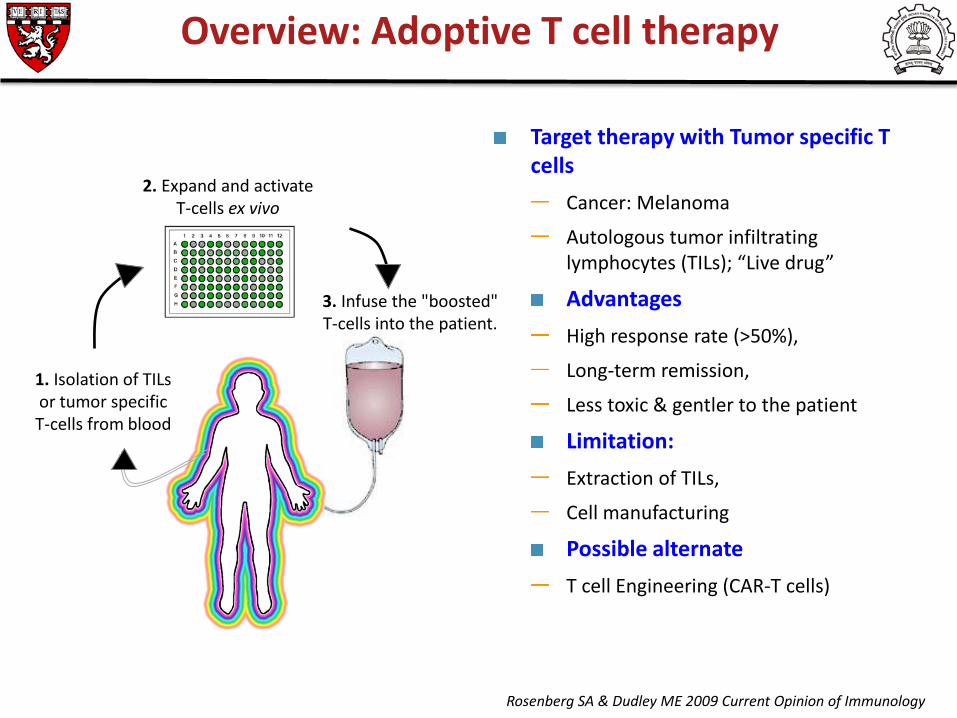
3. Infuse the "boosted" T-cells into the patient.
Overview: Adoptive T cell therapy
1. Isolation of TILs or tumor specific T-cells from blood
2. Expand and activate T-cells ex vivo
Target therapy with Tumor specific T cells
Cancer: Melanoma
Autologous tumor infiltrating lymphocytes (TILs); “Live drug”
Advantages High response rate (>50%),
Long-term remission,
Less toxic & gentler to the patient
Limitation: Extraction of TILs,
Cell manufacturing
Possible alternate T cell Engineering (CAR-T cells)
Rosenberg SA & Dudley ME 2009 Current Opinion of Immunology

Adoptive T cell therapy: CAR-T cells
Antigen specific domain
CAR-T cells (Chimeric antigen receptor-T cells0
T cells transduced with tumor-specific CAR
CAR: Single fusion molecule with antigen specificity plus signaling domain
Three types of CAR: First/second/generations
Based on co-stimulatory receptors
Cancer: Solid tumor & hematological malignancies
Maus M V et al. Blood 2014;123:2625-2635
“Live drug”
Tumor recognition independent of HLA
(no HLA typing needed)
Multiple anti-tumor immuno-modulators can be engineered
Target variety of antigens (protein,
carbohydrate, glycolipid)
Advantages of CAR T cells
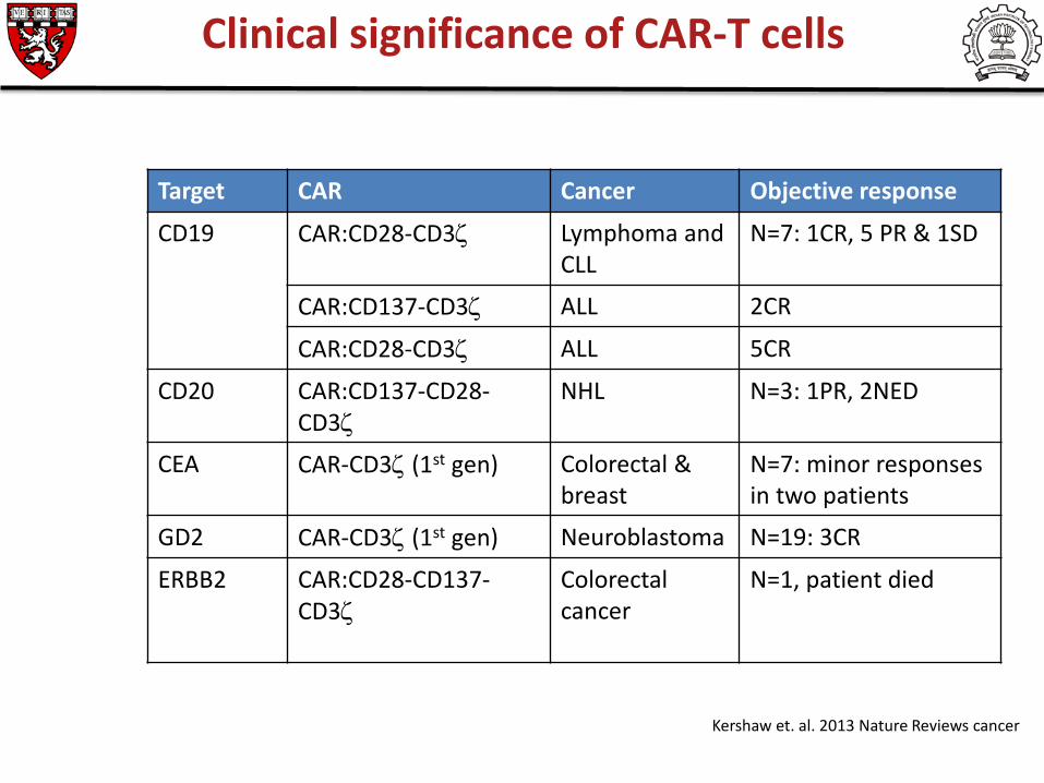
Clinical significance of CAR-T cells
Target CAR Cancer Objective response
CD19 CAR:CD28-CD3ζ Lymphoma and CLL
N=7: 1CR, 5 PR & 1SD
CAR:CD137-CD3ζ ALL 2CR
CAR:CD28-CD3ζ ALL 5CR
CD20 CAR:CD137-CD28-CD3ζ
NHL N=3: 1PR, 2NED
CEA CAR-CD3ζ (1st gen)
Colorectal & breast
N=7: minor responses in two patients
GD2 CAR-CD3ζ (1st gen) Neuroblastoma N=19: 3CR
ERBB2 CAR:CD28-CD137-CD3ζ
Colorectal cancer
N=1, patient died
Kershaw et. al. 2013 Nature Reviews cancer
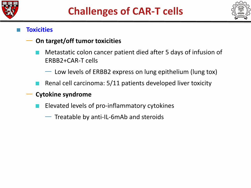
Toxicities
On target/off tumor toxicities
Metastatic colon cancer patient died after 5 days of infusion of ERBB2+CAR-T cells
Low levels of ERBB2 express on lung epithelium (lung tox)
Renal cell carcinoma: 5/11 patients developed liver toxicity
Cytokine syndrome
Elevated levels of pro-inflammatory cytokines
Treatable by anti-IL-6mAb and steroids
Challenges of CAR-T cells
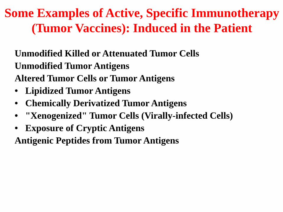
Some Examples of Active, Specific Immunotherapy (Tumor Vaccines): Induced in the Patient
Unmodified Killed or Attenuated Tumor Cells Unmodified Tumor Antigens Altered Tumor Cells or Tumor Antigens • Lipidized Tumor Antigens • Chemically Derivatized Tumor Antigens • "Xenogenized" Tumor Cells (Virally-infected Cells) • Exposure of Cryptic Antigens Antigenic Peptides from Tumor Antigens
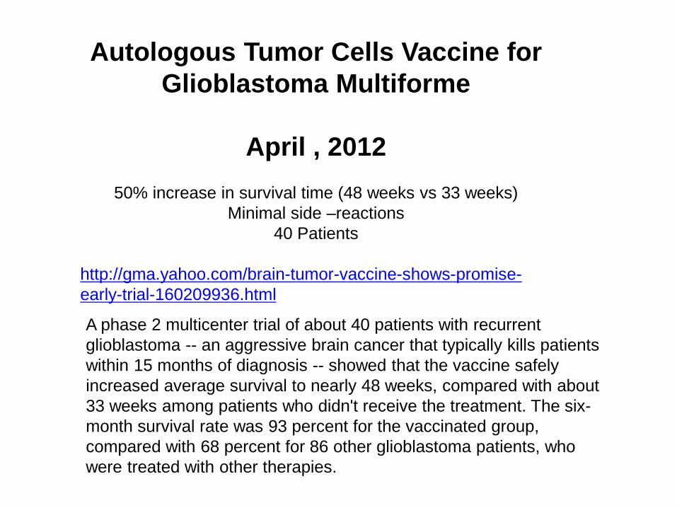
Autologous Tumor Cells Vaccine for Glioblastoma Multiforme
April , 2012
50% increase in survival time (48 weeks vs 33 weeks)
Minimal side –reactions 40 Patients
http://gma.yahoo.com/brain-tumor-vaccine-shows-promise-early-trial-160209936.html
A phase 2 multicenter trial of about 40 patients with recurrent glioblastoma -- an aggressive brain cancer that typically kills patients within 15 months of diagnosis -- showed that the vaccine safely increased average survival to nearly 48 weeks, compared with about 33 weeks among patients who didn't receive the treatment. The six-month survival rate was 93 percent for the vaccinated group, compared with 68 percent for 86 other glioblastoma patients, who were treated with other therapies.

Applications of Monoclonal Antibodies Monoclonal Antibody Diagnosis and Tumor-Imaging • Prostate-specific Antigen (PSA) • Carcino-embryonic Antigen (CEA) • Colon Carcinoma A33 Antigen
Monoclonal Antibody Targeting • Immuno-toxins • Monoclonal antibodies directed to tumor cell
surface markers – Can inhibit the cancer cell function – Can target the cancer cell for destruction by the
immune response
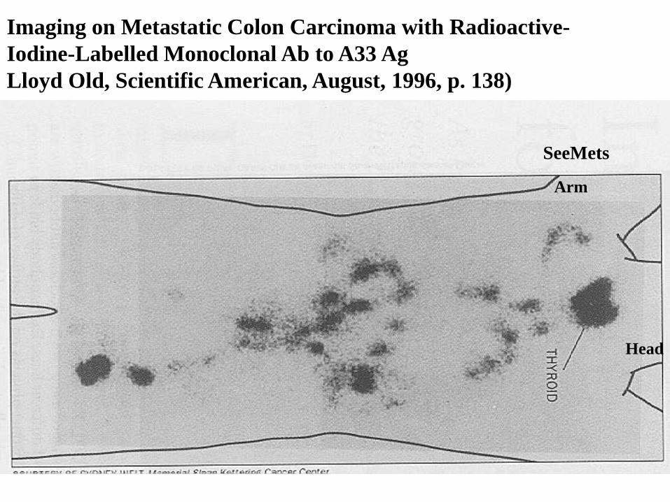
Imaging on Metastatic Colon Carcinoma with Radioactive-Iodine-Labelled Monoclonal Ab to A33 Ag Lloyd Old, Scientific American, August, 1996, p. 138)
SeeMets
Arm
Head

Anti-CD20 Monoclonal Antibodies in Treatment of B-Cell Lymphoma/Leukemia
Rituxan#, Zevalin# (Yttrium 90 Radio-isotope Beta-emitter), and Bexxar* (Iodine-131 Radio-isotope Beta and Gamma Emitter)
# IDEC Pharmaceuticals. *Corixa and Glaxo Smith Kline)

Biotechnology and Clinical Applications of Monoclonals in Cancer Medicine: Carcinoma Therapies
Herceptin: Genentech Anti-HER2/Neu Growth Factor Receptor in Breast Cancer Avastin:
Antibody to Vascular-Endothelial Growth Factor Receptor (Anti-angiogenesis Therapy)
Erbitux
Antibody to Epidermal Growth Factor Receptor
(See page 141, Immunology, 6th Edition)


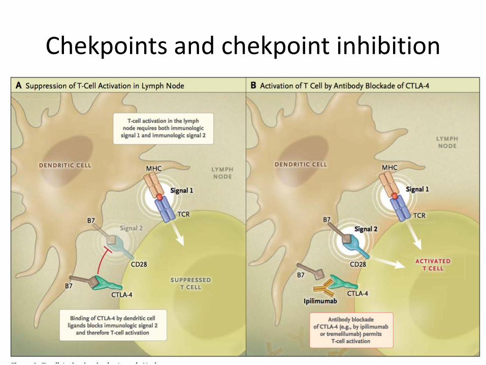
Chekpoints and chekpoint inhibition

Chekpoints and chekpoint inhibition
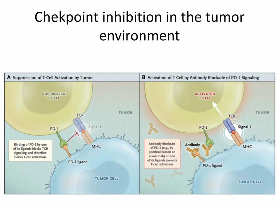
Chekpoint inhibition in the tumor environment

Chekpoint inhibition in the tumor environment

Combination immunotherapy
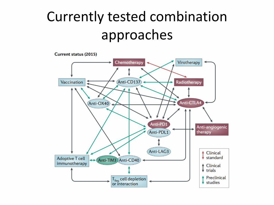
Currently tested combination approaches
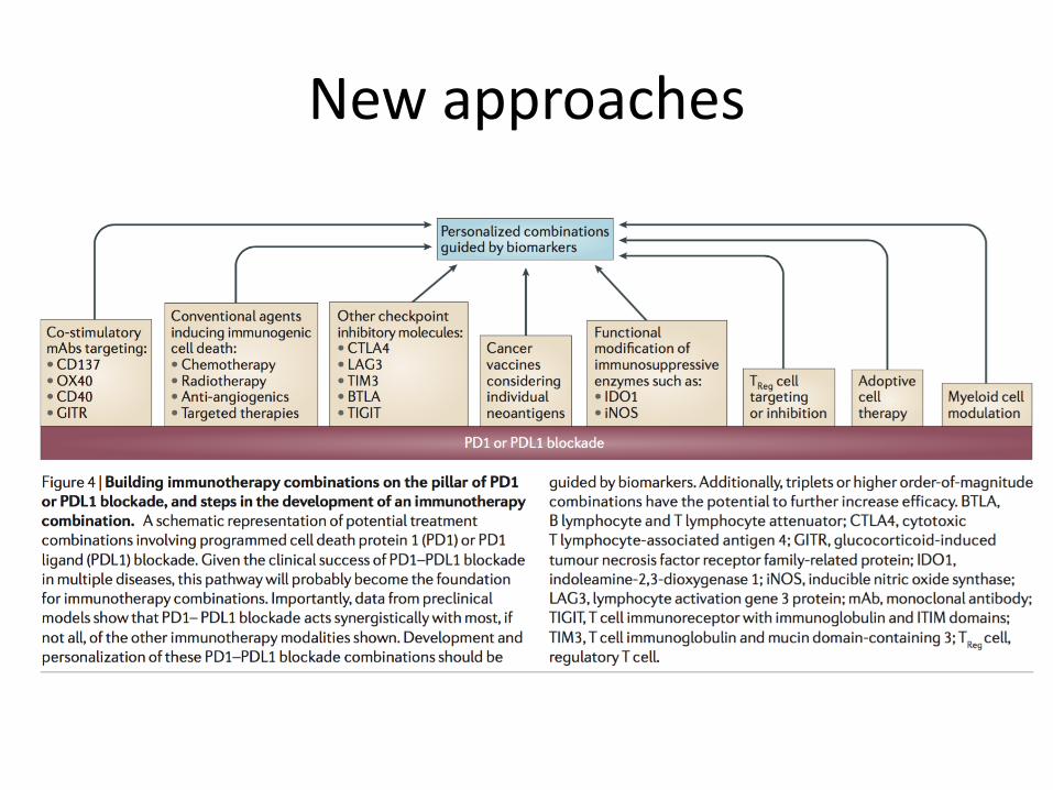
New approaches


