Conformational diversity in prion protein variants influences intermolecular b-sheet formation Seungjoo Lee 1 , Lizamma Antony 2 , Rune Hartmann 1,2 , Karen J Knaus 2 , Krystyna Surewicz 3 , Witold K Surewicz 3 and Vivien C Yee 1,2, * 1 Department of Biochemistry, Case Western Reserve University, Cleveland, OH, USA, 2 Department of Molecular Cardiology, Cleveland Clinic Foundation, Cleveland, OH, USA and 3 Department of Physiology and Biophysics, Case Western Reserve University, Cleveland, OH, USA A conformational transition of normal cellular prion pro- tein (PrP C ) to its pathogenic form (PrP Sc ) is believed to be a central event in the transmission of the devastating neurological diseases known as spongiform encephalo- pathies. The common methionine/valine polymorphism at residue 129 in the PrP influences disease susceptibility and phenotype. We report here seven crystal structures of human PrP variants: three of wild-type (WT) PrP contain- ing V129, and four of the familial variants D178N and F198S, containing either M129 or V129. Comparison of these structures with each other and with previously published WT PrP structures containing M129 revealed that only WT PrPs were found to crystallize as domain- swapped dimers or closed monomers; the four mutant PrPs crystallized as non-swapped dimers. Three of the four mutant PrPs aligned to form intermolecular b-sheets. Several regions of structural variability were identified, and analysis of their conformations provides an explana- tion for the structural features, which can influence the formation and conformation of intermolecular b-sheets involving the M/V129 polymorphic residue. The EMBO Journal (2010) 29, 251–262. doi:10.1038/ emboj.2009.333; Published online 19 November 2009 Subject Categories: proteins; structural biology Keywords: crystal structure; prion proteins; spongiform encephalopathies Introduction Prion diseases, or transmissible spongiform encephalopathies (TSE), are an unusual group of invariably fatal mammalian neurodegenerative diseases (reviewed by Prusiner, 1998; Collinge, 2001; Aguzzi et al, 2008). These rare disorders can be acquired by infection, inheritance via mutations in the gene encoding for the prion protein (PrP), or occur sponta- neously. Typical features of the TSEs are long incubation periods and characteristic brain pathology including spongi- form degeneration, astrogliosis, and accumulation of mis- folded protein deposits. These diseases are intimately associated with conformational conversion of the normal cellular PrP (PrP C ) to a pathogenic form (PrP Sc ). According to the ‘protein-only’ model, PrP Sc itself represents the infec- tious prion agent: it is believed to self-propagate by the mechanism involving binding to PrP C and templating the conversion of the latter protein to the PrP Sc state. The human prion diseases include Creutzfeldt–Jakob dis- ease (CJD), Gerstmann–Stra ¨ussler–Scheinker disease (GSS), fatal familial insomnia (FFI), and kuru. The common methio- nine/valine polymorphism at residue 129 influences disease susceptibility and phenotype. It is striking that the new variant CJD, believed to be transmitted by dietary exposure to bovine spongiform encephalopathy (BSE)-contaminated beef, has to date afflicted only individuals who are homozygous for M129 (Brandel et al, 2009). Also, sporadic CJD cases display a variety of clinicopathological symptoms, which depend on the M/V129 genotype, and a majority of patients are homo- zygous at position 129 (Alpe ´rovitch et al, 1999). Finally, M/ V129 heterozygosity appears to be protective against kuru in the Fore tribe (Cervena ´kova ´ et al, 1998). Thus, the M/V129 polymorphism has an influence in sporadic and transmitted prion diseases. The inherited diseases include GSS, FFI, and some forms of CJD, and are associated with mutations in the PRNP gene. More than 20 disease predisposing point mutations have been reported, with the M/V129 polymorphism also affecting familial prion disease phenotype. For example, the D178N mutation co-segregates with V129 in FFI, but with M129 in CJD (Goldfarb et al, 1992). Other inherited pathogenic mutations also typically co-segregate with only either M129 or V129 (reviewed by Aguzzi et al, 2008). The family of mammalian prion diseases includes scrapie in sheep, BSE in cattle, and chronic wasting disease in elk and deer. Disease transmission between species is usually much less efficient than within the same species, leading to the concept of ‘species barrier’. While these barriers are closely related to differences in PrP primary sequence between the donor and recipient species, another factor contributing to TSE transmissibility is the existence of multiple prion strains even within the same animal species. These distinct strains of the prion agent (leading to distinct disease phenotypes) appear to be associated with different conformational states of the PrP Sc aggregate, although high-resolution insight into these conformational differences is still missing (reviewed by Collinge and Clarke, 2007). The PrP C to PrP Sc transformation involves a conversion from a soluble and predominantly a-helical protein to an aggregated form, which is substantially enriched in b-sheet. Bacterially expressed recombinant PrPs, which can be easily purified, have been more amenable to high-resolution structural studies than the brain-derived proteins; PrP Sc is Received: 10 July 2009; accepted: 22 October 2009; published online: 19 November 2009 *Corresponding author. Department of Biochemistry, Case Western Reserve University, 10900 Euclid Avenue RT500, Cleveland, OH 44106, USA. Tel.: þ 1 216 3681184; Fax: þ 1 216 368 3419; E-mail: [email protected]The EMBO Journal (2010) 29, 251–262 | & 2010 European Molecular Biology Organization | All Rights Reserved 0261-4189/10 www.embojournal.org & 2010 European Molecular Biology Organization The EMBO Journal VOL 29 | NO 1 | 2010 EMBO THE EMBO JOURNAL THE EMBO JOURNAL 251
Transcript
Conformational diversity in prion protein variantsinfluences intermolecular b-sheet formation
and Vivien C Yee1,2,*1Department of Biochemistry, Case Western Reserve University,Cleveland, OH, USA, 2Department of Molecular Cardiology, ClevelandClinic Foundation, Cleveland, OH, USA and 3Department of Physiologyand Biophysics, Case Western Reserve University, Cleveland, OH, USA
A conformational transition of normal cellular prion pro-
tein (PrPC) to its pathogenic form (PrPSc) is believed to be
a central event in the transmission of the devastating
neurological diseases known as spongiform encephalo-
pathies. The common methionine/valine polymorphism
at residue 129 in the PrP influences disease susceptibility
and phenotype. We report here seven crystal structures of
human PrP variants: three of wild-type (WT) PrP contain-
ing V129, and four of the familial variants D178N and
F198S, containing either M129 or V129. Comparison of
these structures with each other and with previously
published WT PrP structures containing M129 revealed
that only WT PrPs were found to crystallize as domain-
swapped dimers or closed monomers; the four mutant
PrPs crystallized as non-swapped dimers. Three of the
four mutant PrPs aligned to form intermolecular b-sheets.
Several regions of structural variability were identified,
and analysis of their conformations provides an explana-
tion for the structural features, which can influence the
formation and conformation of intermolecular b-sheets
involving the M/V129 polymorphic residue.
The EMBO Journal (2010) 29, 251–262. doi:10.1038/
emboj.2009.333; Published online 19 November 2009
Subject Categories: proteins; structural biology
Keywords: crystal structure; prion proteins;
spongiform encephalopathies
Introduction
Prion diseases, or transmissible spongiform encephalopathies
(TSE), are an unusual group of invariably fatal mammalian
neurodegenerative diseases (reviewed by Prusiner, 1998;
Collinge, 2001; Aguzzi et al, 2008). These rare disorders can
be acquired by infection, inheritance via mutations in the
gene encoding for the prion protein (PrP), or occur sponta-
neously. Typical features of the TSEs are long incubation
periods and characteristic brain pathology including spongi-
form degeneration, astrogliosis, and accumulation of mis-
folded protein deposits. These diseases are intimately
associated with conformational conversion of the normal
cellular PrP (PrPC) to a pathogenic form (PrPSc). According
to the ‘protein-only’ model, PrPSc itself represents the infec-
tious prion agent: it is believed to self-propagate by the
mechanism involving binding to PrPC and templating the
conversion of the latter protein to the PrPSc state.
The human prion diseases include Creutzfeldt–Jakob dis-
particularly problematic due to its aggregated nature.
Although recombinant PrPs lack the C-terminal glyco-
phosphatidyinositol (GPI) anchor and N-linked glycosylation
at two sites, both of these post-translational modifications are
not essential for infectivity (Taraboulos et al, 1990; Chesebro
et al, 2005; Tuzi et al, 2008). NMR and circular dichroism
studies of bovine PrPC purified from healthy brains showed
that the thermal stability and three-dimensional structure of
the purified glycoprotein and non-glycosylated recombinant
protein are essentially identical (Hornemann et al, 2004).
NMR structures of various mammalian PrPs revealed a
conserved monomeric protein fold with a highly flexible
N-terminus (residues 23–124) and a globular C-terminal
domain (125–231), which contains a small two-stranded,
anti-parallel b-sheet and three long a-helices (Wuthrich and
Riek, 2001). This predominantly a-helical fold is reiterated in
the crystal structures of a domain-swapped human PrP
(hPrP) (Knaus et al, 2001) and of monomeric hPrP and
ovine PrP (ovPrP) (Eghiaian et al, 2004; Haire et al 2004;
Antonyuk et al, 2009). The published structures all contain
M129. To investigate the structural consequences of the M/
V129 polymorphic residue and understand how it may play a
determinant role in prion disease susceptibility, we have
solved the crystal structures of recombinant wild-type (WT)
hPrP containing V129. We have also determined the crystal
structures of the pathogenic mutants D178N and F198S, with
both M129 and V129, to further probe the conformational
effects of the polymorphic residue and also to investigate the
structural consequences of the disease predisposing muta-
tions themselves.
Results
WT-V129 in three different crystal forms reveals
both dimeric and monomeric structures
Three different WT-V129 crystal forms were obtained: WT-
V129_1, which has space group C2221, is isomorphous with
WT-M129, and was solved at 2.26-A resolution; WT-V129_2
and WT-V129_3 both have space group P212121, but different
unit cell dimensions, and were refined at 3.1 and 1.8-A
resolution, respectively (Table I).
Both WT-V129_1 and WT-V129_2 reveal disulfide-linked,
3D-domain-swapped dimers nearly identical to the WT-M129
structure (Figure 1A and Supplementary Figure S1A, addi-
tional details are provided in the Supplementary data; Knaus
et al, 2001). V129 is well-defined in density located in the first
short b-strand and, consistent with NMR studies (Hosszu
et al, 2004), the M129-to-V129 substitution does not seem to
cause any local conformational changes. In both crystal
forms, the V129 side chain is on the protein surface, exposed
to solvent, and is not involved in any intermolecular inter-
actions. The elucidation of domain-swapped PrP dimers in
two different crystal forms, in which the two halves are
related by different types of symmetry (crystallographic in
WT-V129_1 and non-crystallographic in WT-V129_2), con-
firms that dimerization is a characteristic behaviour of re-
combinant PrP rather than merely a crystallization artefact.
This is also supported by the observation of a small amount
of disulfide-linked dimer in solutions of WT and variant PrP
proteins (Supplementary Figure S1C). WT-V129_3 contains a
single PrP polypeptide in the asymmetric unit (Figure 1B); the
electron density defines a closed hinge-loop conformation
corresponding to a compactly folded, non-swapped monomer
(Supplementary Figure S1B). V129 is a solvent-exposed sur-
face residue, near a crystal packing interface with H155 as its
closest neighbour.
The ordered structures for the three crystal forms (encom-
passing residues A120–Y225 in WT-V129_1 and L125–Q227
in WT-V129_2 and WT-V129_3) appear to be N-terminally
truncated as was seen for WT-M129, crystals of which were
shown to contain N-terminally proteolysed protein (Knaus
et al, 2001). That the N-terminus is susceptible to proteolysis
is not surprising since NMR studies have shown that this
region is flexible to about residue 125 in full-length (23–230)
and truncated (90–230) recombinant PrP. This flexible N-
terminus appears to have little effect on the structure of the
C-terminal domain (Zahn et al, 2000). Furthermore, glycosy-
lated PrP purified from bovine brains (Hornemann et al,
2004) shows a similar ordered structure limited to the
C-terminal region.
By superimposing two WT-V129_3 monomers onto the two
halves of the WT-V129_1 dimer, the resulting steric clashes of
the hinge loops and helices-2 show that a dimer of non-
swapped monomers would require substantial conforma-
tional adjustments (Figure 1C), and confirm that the WT-
V129 dimers are domain-swapped. In addition to the hinge
loop, which changes structure upon helix-3 swapping, the
(R164–S170) loop adjacent to the small b-sheet with M/V129
is conformationally variable. This loop is nearly identical in
both WT-V129_1 and WT-M129 dimers, with characteristic
side-chain hydrogen bonding interactions between Y169 and
D178, and between R164 and E168 (Figure 2A). In one chain
of the WT-V129_2 dimer, R164 forms a salt bridge with E167,
while in the other there is no intra-loop interaction (Figure 2B
and C). The WT-V129_3 monomer loop includes a helical
turn such that D167 swings in to interact with D178
(Figure 2D). In all WT-V129 and WT-M129 crystal structures,
R164 hydrogen bonds to the G126 main chain.
CJD D178N/M129 and FFI D178N/V129 crystal
structures exhibit different conformations and
a non-conserved intermolecular b-sheet
The D178N mutation is associated with two pathologically
distinct inherited prion diseases, CJD and FFI, when the
mutation colocalizes with M129 or V129, respectively
(Goldfarb et al, 1992). Both versions of the mutant have
been crystallized. The CJD D178N/M129 crystal has space
group P43212 and was solved at 1.8-A resolution, while the
FFI D178N/V129 crystal has space group I212121 and yielded
a 2.0-A resolution structure (Table I).
In D178N/M129, there is no continuous density for B35
N-terminal residues, several hinge-loop residues, and for a
few C-terminal residues. Thus, the refined model contains
residues G126–T192 and K194–R228 for chain-A and G126–
T192 and E196–R228 for chain-B. The two chains form a
dimer, which is very similar to those observed in the WT
crystals, except that the D178N/M129 dimer appears to be
composed of two non-swapped, closed monomers. The
hinge-loop density gaps correspond to only 1–3 residues
and indicate that the D178N/M129 dimer is most likely
composed of non-swapped monomers. In contrast, the
D178N/V129 crystal contains only a single chain in the
asymmetric unit, but it combines with a symmetry-related
molecule to form a dimer that is similar to that seen for
Crystal structures of variant prion proteinsS Lee et al
The EMBO Journal VOL 29 | NO 1 | 2010 &2010 European Molecular Biology Organization252
Table
ID
ata
collec
tion
and
refi
nem
ent
stat
isti
cs
Cry
stal
WT-
V129_1
WT-
V129_2
WT-
V129_3
D178N
/M129
D178N
/V129
F198S/M
129
F198S/V
129
Data
collec
tion
Sourc
eA
PS
19B
MA
PS
19ID
APS
19ID
NSLS
X25
APS
19B
MN
SLS
X25
APS
19ID
Wav
elen
gth
(A)
0.9
787
1.0
332
0.9
791
1.1
000
0.9
198
1.1
000
0.9
786
Spac
egr
oup
C222 1
P2 1
2 12 1
P2 1
2 12 1
P4
32 1
2I2
12 1
2 1P4
32 1
2P4
32 1
2U
nit
cell
(A)
a¼
85.3
a¼
28.7
a¼
32.5
a¼
b¼
57.5
a¼
39.2
a¼
b¼
57.0
a¼
b¼
57.6
b¼
86.4
b¼
72.0
b¼
49.1
b¼
57.7
c¼
40.7
c¼
125.9
c¼
56.9
c¼
168.0
c¼
93.3
c¼
167.4
c¼
163.3
Res
olu
tion
(A)
30.0
-2.2
6(2
.38–2.2
6)
50.0
-3.1
(3.2
1–3.1
)30.0
-1.8
(1.8
6–1.8
)50.0
-1.8
(1.8
6–1.8
)50.0
-2.0
(2.0
7–2.0
)50.0
-2.0
(2.0
7–2.0
)50.0
-1.8
5(1
.92–1.8
5)
Rsy
m(%
)8.5
(38.4
)6.6
(27.7
)4.2
(19.8
)5.7
(21.6
)6.7
(47.1
)4.0
(40.5
)5.2
(65.6
)I/s
I15.0
(2.3
)16.4
(4.4
)30.8
(6.8
)36.9
(4.8
)23.2
(3.3
)27.8
(2.5
)41
.0(3
.5)
Com
ple
tenes
s(%
)91
.8(5
9.3
)91
.9(7
3.0
)99.4
(99.8
)95.8
(80.3
)99.5
(99.9
)94.2
(65.1
)98.4
(96.6
)R
edundan
cy5.7
(3.5
)4.3
(4.0
)4.5
(4.5
)10
.1(3
.0)
5.0
(4.7
)8.1
(8.1
)14.6
(14.6
)N
o.
refl
ecti
ons
6679
4728
8871
25
609
7405
19
275
24
056
Refi
nem
ent
Res
olu
tion
(A)
27.3
-2.2
627.9
-3.1
16.1
-1.8
40.1
-1.8
27.4
-2.0
41.9
-2.0
28.8
-1.8
5R-f
acto
r/R
free
(%)
20.3
/27.8
23.0
/29.9
18.5
/24.1
20.6
/23.2
20.5
/26.6
21.6
/24.9
22.6
/26.5
Num
ber
ofato
ms
Pro
tein
/ions/
wat
ers
878/2
/52
170
1/0
/0897/0
/105
1728/2
/217
799/3
/56
1645/2
/172
1654/2
/138
Ave
rage
B-f
act
ors
(A2)
All
pro
tein
46.6
76.2
18.8
25.6
32.9
38.0
34.3
164–170
loop
(A/B
)56.1
78.7
/88.0
17.0
30.1
/36.4
46.6
46.4
/50.8
40.3
/29.9
Ions/
wat
ers
63.3
/57.0
—/—
—/3
3.0
14.8
/34.8
21.5
/39.3
28.6
/47.1
33.5
/45.2
r.m
.s.
dev
iati
ons
Bond
lengt
h(A
)0.0
13
0.0
08
0.0
12
0.0
110.0
12
0.0
110.0
13
Bond
angl
e(d
egre
es)
1.4
31.0
81.3
51.2
41.4
61.2
41.4
1
Ram
ach
an
dra
nplo
t(%
)M
ost
favoure
dre
gion
98.9
91.3
96.8
95.1
91.8
95.9
94.7
Addit
ional
lyal
low
edre
gions
1.1
8.2
3.2
4.9
4.7
4.1
5.3
Dis
allo
wed
regi
ons
0.0
0.0
0.0
0.0
0.0
0.0
0.0
PD
Bac
cess
ion
code
3H
AF
3H
J53H
AK
3H
EQ
3H
JX3H
ES
3H
ER
Val
ues
inpar
enth
eses
are
for
the
hig
hes
t-re
solu
tion
shel
l.
Crystal structures of variant prion proteinsS Lee et al
&2010 European Molecular Biology Organization The EMBO Journal VOL 29 | NO 1 | 2010 253
D178N/M129. The refined D178N/V129 protein model con-
sists of residues G126–T192 and E196–Q223; its small hinge-
loop density gap also suggests a dimer of two non-swapped
monomers. Due to scarcity of crystals, it was not possible to
determine the disulfide-linked dimer content, or the extent of
N-terminal proteolysis, of either of these two mutant crystals.
There is no substantial difference in the overall protein fold
in D178N/M129 or D178N/V129 as compared with the WT
dimers, other than the hinge loops, which are in a partially
disordered closed conformation in the mutants and an
ordered open conformation for the WT dimers (Figure 3A
and C). However, the local environment surrounding the
altered D178N residue shows differences between the M129
and V129 isoforms. In the WT crystal structures, D178 is a
mostly solvent-exposed residue, and in several crystalline
forms the D178 side-chain hydrogen bonds to either Y169
or D167 in the variable (R164–S170) loop. In the two D178N
structures, the N178 residues are in nearly identical confor-
mations as D178 in the WT structures; the local structural
variability is in the adjacent (R164–S170) loop. In D178N/
M129, N178 does not interact with the (R164–S170) loop
(Figure 2E), while in D178N/V129, the N178 side chain
interacts with D167, which in turn forms a salt bridge with
R164 (Figure 2F), corresponding to two very different loop
conformations (3.1 A r.m.s.d. for Ca superimpositions). As is
also the case for the dimeric WT crystal structures, the
average loop B-factors for the M129 and V129 isoforms of
D178N are higher than the corresponding overall average B
for the two proteins (Table I). In accordance with correspond-
ingly higher loop B-factors, the electron density in this region
for D178N/V129 is less clear than for the different loop
conformation in D178N/M129 (Supplementary Figure S2A)
or the similar conformation in WT-V129_3.
An even more striking difference between the D178N and
WT structures, and between the two D178N structures, is in
the environment about the M/V129 polymorphic residue and
the intermolecular packing of dimers. In crystals containing
WT-M129 and WT-V129 dimers, the M129 and V129 side
chains are solvent-exposed and not involved in intermolecu-
lar contacts. In D178N/M129, two dimers related by crystal-
lographic symmetry come together such that the two small
intramolecular b-sheets combine to form continuous inter-
molecular, four-stranded, antiparallel b-sheets at the dimer:-
dimer interface (Figure 3A and B). The N-terminal b-strands
in the middle of the sheet contain M129 side chains that pack
against each other to form part of the ‘sheet interface.’ In
contrast, the presence of V129 in D178N/V129 yields a crystal
with very different packing between dimers, which are
rotated by B90 degrees with respect to each other as com-
pared with their approach in the M129 isoform (Figure 3C).
Due to this rotation, the short b-strands in the neighbouring
molecules do not combine to form a continuous intermole-
cular b-sheet, and the V129 side chains do not interact with
each other but instead are exposed to solvent (Figure 3D).
Figure 1 Crystal structures of WT-V129 and mutant hPrPs. (A) Ribbon diagram of the WT-V129_1 3D-domain-swapped dimer. The twopolypeptide chains are in beige and grey, with the V129 side chains shown as space-filling spheres and the intermolecular disulfide bonds asball-and-stick structures. The two-stranded, antiparallel b-sheet is coloured in orange. (B) Ribbon diagram of the WT-V129_3 monomer (beige).Superimposed is one chain of the WT-V129_1 domain-swapped dimer (grey), to illustrate the differences in hinge region conformation.(C) Non-swapped hPrP dimers cannot adopt the 3D-domain-swapped dimer organization. Superimposed on the WT-V129_1 domain-swappeddimer (beige) are two copies of the non-swapped WT-V129_3 monomers (grey); the helices-2 and hinge loops sterically clash at the dimerinterface. Helices are drawn as cylinders. (D) Asymmetry in the D178N/M129 dimer. Superimposition of two copies of the dimer shows that thetwo halves (green) are rotated B10 degrees with respect to each other. (E) Variations in the bending angles of the hPrP dimer. When one set ofmonomers is superimposed, the second set of monomers is oriented differently in mutant and WT dimers. D178N/M129 (green) and F198S/M129 (purple) dimers have the same bending angle, D178N/V129 (cyan) and F198S/V129 (pink) superimpose well, and the WT-V129_1 dimer(beige) is similar to WT-V129_2 (not shown) and different from both pairs of mutant dimers.
Crystal structures of variant prion proteinsS Lee et al
The EMBO Journal VOL 29 | NO 1 | 2010 &2010 European Molecular Biology Organization254
F198S/M129 and GSS F198S/V129 structures have
similar conformations and intermolecular b-sheets
F198S is a mutation, which colocalizes with the V129 poly-
morphism in GSS (Dlouhy et al, 1992; Hsiao et al, 1992);
colocalization with M129 has not yet been reported. To
provide a full structural investigation of the pathogenic
F198S substitution in the context of both M129 and V129,
both proteins were crystallized. Both crystals have space
group P43212 and similar cell dimensions. The F198S/M129
structure was solved at 2.0-A resolution, while the GSS
F198S/V129 structure was refined to 1.85-A resolution
(Table I).
The refined F198S/M129 model includes residues
L125–T193 and S198–Y226 for chain-A, and G126–V189
and N197–Y226 for chain-B. In F198S/V129, there is contin-
uous density for residues L125–T192 and S198–Y226 in
chain-A, and G126–T190 and S198–Q227 in chain-B. The
two F198S crystals are isomorphous not only with each
other, but also with D178N/M129. It is not surprising then
that both F198S crystals contain dimers similar to those seen
Figure 2 Variable conformation of the (R164–S170) loop in PrP crystal structures. Three different conformations of the (R164–S170) loop,adjacent to the b-strand containing M/V129, are observed. Side chains of selected loop residues are shown as ball-and-stick structures;hydrogen bonds are represented by dashed lines. (A) In the WT-V129_1 dimer loop, Y169 forms a hydrogen bond with D178, while R164interacts with E168 and the main chain of G126. (B, C) The two loops in the WT-V129_2 dimer have similar main-chain conformations, butdifferent hydrogen-bonding interactions. (D) The WT-V129_3 monomer loop has a helical turn and a network of hydrogen bonds formed byD178, D167, R164, and the main chain of G126. (E) The D178N/M129 loop resembles that in the WT-V129_2 chain shown in panel B. (F) TheD178N/V129 loop is similar to that observed in WT–V129_3 shown in panel D. (G) In the monomeric hPrP–Fab structure, the loop resemblesthat in WT-V129_1 except there is no interaction between R164 and G126. (H) In WT ovPrP, the loop is similar to that in WT-V129_1 exceptthere is no interaction between R164 and G126, and there is an additional hydrogen bond between Y128 and D178 that is observed only in theWTand mutant ovPrP crystal structures. (I) Superimposition of the loops from D178N/M129 (green), D178N/V129 (cyan), and V129_1 (beige),highlighting the three distinct conformations.
Crystal structures of variant prion proteinsS Lee et al
&2010 European Molecular Biology Organization The EMBO Journal VOL 29 | NO 1 | 2010 255
in the D178N structures (Figure 3E and Supplementary
Figure S2C). As in the D178N structures, there is discontinuous
hinge-loop density. F198S/M129 crystallization was reprodu-
cible enough to provide adequate samples for Western blot
analysis, which shows a mixture of proteins of different sizes,
but with the major component being intact monomeric
PrP(90–231), while WT crystals contain only the N-terminally
proteolysed protein and are enriched in disulfide-linked dimers
(Supplementary Figure S1C; details in Supplementary data).
This correlates well with the observation of continuous density
for open hinge loops in the swapped WT dimers, and broken
density for flexible closed hinge loops in the mutant dimers.
In the WT-V129_1 swapped dimer and WT-V129_3 closed
monomer, the F198 side chain is buried in a hydrophobic
environment (Supplementary Figure S1A and B). In both
D178N mutant structures, the F198 side chain remains
solvent-inaccessible even with increased flexibility of the
adjacent hinge loop. In contrast, in both F198S mutant
structures the S198 side chain is largely solvent-exposed
(Supplementary Figure S2B). The S198 hinge-loop density
gaps correspond to 4–7 residues as compared with 1–3
residues in the D178N dimers. Thus a structural effect of
the F198S substitution appears to be an increase in
hinge-loop flexibility as a result of replacing a large buried
Figure 3 Crystal structures of hPrP mutants. (A) Packing between two D178N/M129 dimers. An intermolecular antiparallel, four-strandedb-sheet (orange) containing M129 (space-filling spheres) is formed at the dimer:dimer interface; the variant N178 side chains are also shown.(B) A close-up view of the D178N/M129 intermolecular b-sheet containing M129. (C) D178N/V129 dimers do not pack to form intermolecularb-sheets; instead these dimers are rotated B90 degrees with respect to each other. (D) A close-up view of the D178N/V129 b-sheets. The V129side chains do not approach each other and the b-sheets do not interact. (E) F198S/V129 dimers form an intermolecular b-sheet. While theF198S/M129 dimer:dimer interaction (Supplementary Figure S1C) is nearly identical to that for D178N/M129, the F198S/V129 is slightlytwisted in comparison. (F) A close-up view of the F198S/V129 intermolecular b-sheet. The disordered/flexible hinge loop residues arerepresented by dashed lines.
Crystal structures of variant prion proteinsS Lee et al
The EMBO Journal VOL 29 | NO 1 | 2010 &2010 European Molecular Biology Organization256
hydrophobic residue with a small hydrophilic amino acid that
prefers to be exposed to solvent.
The increase in hinge-loop flexibility in the two F198S
isoforms does not interfere with the mutants’ ability to
crystallize as non-covalent dimers. The protein fold and
crystal packing are very similar in both the F198S and
D178N/M129 structures (Figure 3A and E; Supplementary
Figure S2C, Ca r.m.s.d.s of 0.6–1.1 A), including the (R164–
S170) loop (Ca r.m.s.d.s 0.14–0.4 A). In both F198S/M129
and F198S/V129, R164 forms a salt bridge with D167 and
D178 is left to interact with the solvent. These loop interac-
tions and conformations are very similar to those in WT-
V129_2 and D178N/M129 (Figure 2C and E). Also in keeping
with the other dimeric PrP structures, the average loop
B-factor is higher than the overall protein average B for the
F198S/M129 structure; F198S/V129 is the exception since its
loop and overall average B-factors are nearly identical
(Table I).
Unlike the two D178N structures, which showed that the
dimers in the M129 and V129 isoforms participate in drama-
tically different intermolecular interactions, the two F198S
structures reveal a relatively subtle structural effect of the
M/V129 substitution. As for D178N/M129, F198S/M129
dimers come together with M129 side chains packing against
each other at an intermolecular b-sheet interface. A similar
b-sheet is formed between F198S/V129 dimers, except that
this interface is bent by B10 degrees as compared with that in
the M129 isoform. The result is that one face of the b-sheet
becomes more exposed such that the V129 side chains, which
are short and branched as compared with that of M129, do
not pack tightly against each other (Figure 3F).
Discussion
NMR studies of recombinant PrPs have thoroughly character-
ized a conserved monomeric fold presumed to represent PrPC
(Wuthrich and Riek, 2001). The crystal structure of hPrP WT-
M129 revealed an unexpected 3D-domain-swapped dimer
and expanded the understanding of the PrP conformational
repertoire (Knaus et al, 2001). Single-amino-acid substitu-
tions such as the M/V129 isoforms, and pathogenic muta-
tions identified in inherited prion diseases, influence disease
susceptibility and phenotype. Structural comparison of WT
and variant PrPs is an important step in understanding how
variant PrPs may contribute to prion disease mechanisms.
Detailed structural information on hPrP disease variants has
been limited to the NMR structure of the familial CJD-related
Q200K mutant, which was nearly identical to that of WT hPrP
except for minor differences in flexible regions such as both
termini and two loops (Zhang et al, 2000). NMR structures of
non-pathogenic hPrP variants also do not reveal any signifi-
cant structural differences (Calzolai et al, 2000). The ensem-
ble of seven hPrP crystal structures described here, for WT-
V129 and pathogenic D178N and F198S mutants, substan-
tially increases the characterization of PrP conformational
behaviour, and highlights structural details, which may be
relevant to prion disease mechanisms.
PrP dimers exhibit differences in 3D-domain swapping
and dimer organization
Dimers are observed in the crystals of both WT and mutant
PrPs, but vary in several different ways (details in the
Supplementary data). The first is that both WT-M129 and
WT-V129 can crystallize as swapped dimers, while D178N
and F198S mutants crystallize as non-swapped dimers.
Secondly, PrP dimers vary in the orientation of the two
monomers with respect to each other. The two mutant dimers
containing M129 are asymmetric and bent, while the two
mutant dimers containing V129 are symmetric (Figure 1D).
The WT dimers are symmetric irrespective of whether they
contain M129 or V129. When one monomer of each dimer is
superimposed and the orientations of the second monomers
are compared, the WT, M129-containing mutants and V129-
containing mutants are found to exhibit three different
dimer conformations (Figure 1E). This segregation of mutant
dimer organization according to the M/V129 polymorphism
is intriguing since the polymorphic M/V129 residues are
predominantly solvent-exposed in the context of the dimers,
and they are distant from the dimer interface.
The presence of 3D-domain swapping can explain the
significant difference in organization between the WT and
mutant dimers. When two copies of the WT-V129_3 mono-
mer are superimposed onto the two halves of the WT-V129_1
swapped dimer, the C-termini of the two helices-2 and the
adjacent closed hinge loops introduce severe steric clashes
(Figure 1C). Thus, for two monomers to form a non-swapped
dimer, shifts of the helices-2 and the hinge loops are
necessary. Substantial shifts of helix-2 in either one or
both monomers produce asymmetric M129-containing or
for all species is for simplicity). The structure of WT ovPrP
contains symmetry-related monomers that form intermolecu-
lar b-sheets with M129 at the interface (Haire et al, 2004).
Superimposition of the structure of WT ovPrP with the two
hPrP mutants reveals substantial differences in the sheet
interface angle, by up to B40 degrees (Figure 4A). In con-
trast, several ovPrP scrapie-susceptibility variants crystal-
lized as Fab complexes did not exhibit intermolecular
b-sheets, apparently due to potential steric clashes involving
the Fab, which is only B10 A from M129 (Eghiaian et al,
2004). The crystal structure of monomeric WT hPrP bound to
the ICSM 18-Fab also has an intermolecular b-sheet with
M129 (Antonyuk et al, 2009). The hPrP M129/Fab sheet
interface differs substantially from those of ovPrP and the
hPrP mutants, by up to B70 degrees (Figure 4A). In the
ovPrP and hPrP mutant sheets, the M/V129 residues interact
with each other more intimately (Cb–Cb distances of
5.3–5.8 A) than in WT hPrP–Fab (Cb–Cb distance of 6.8 A).
Thus while intermolecular b-sheets containing M/V129 are
observed in five different mammalian PrP structures, there
are differences in the interactions involving M/V129 and a
wide variation in the conformation of the sheet and the angle
between the monomers forming the interface.
Crystal structures of variant prion proteinsS Lee et al
&2010 European Molecular Biology Organization The EMBO Journal VOL 29 | NO 1 | 2010 257
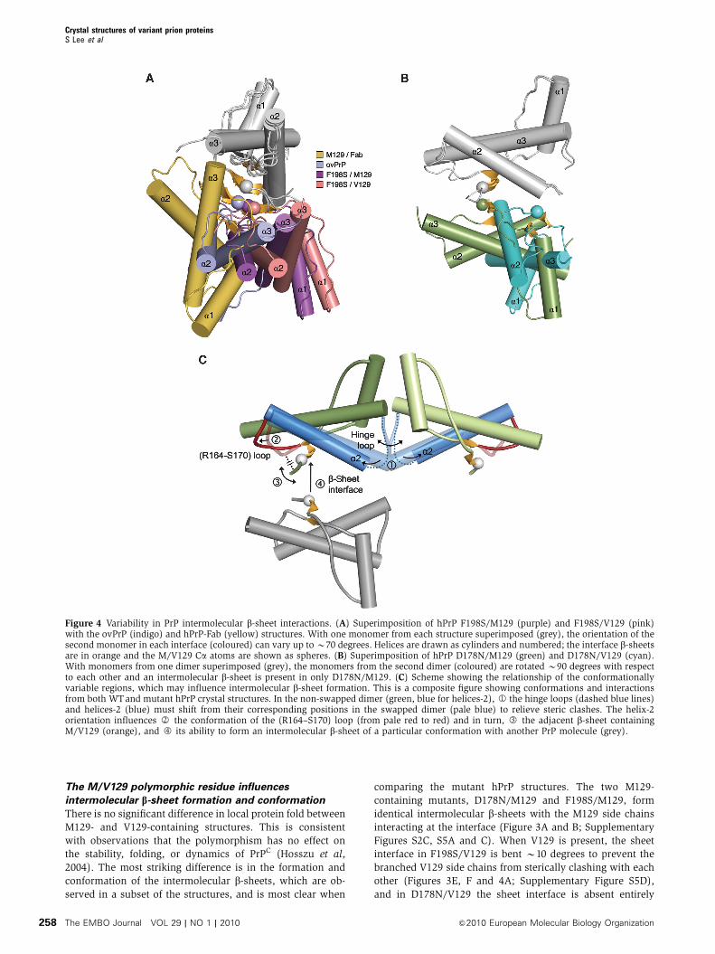
The M/V129 polymorphic residue influences
intermolecular b-sheet formation and conformation
There is no significant difference in local protein fold between
M129- and V129-containing structures. This is consistent
with observations that the polymorphism has no effect on
the stability, folding, or dynamics of PrPC (Hosszu et al,
2004). The most striking difference is in the formation and
conformation of the intermolecular b-sheets, which are ob-
served in a subset of the structures, and is most clear when
comparing the mutant hPrP structures. The two M129-
containing mutants, D178N/M129 and F198S/M129, form
identical intermolecular b-sheets with the M129 side chains
interacting at the interface (Figure 3A and B; Supplementary
Figures S2C, S5A and C). When V129 is present, the sheet
interface in F198S/V129 is bent B10 degrees to prevent the
branched V129 side chains from sterically clashing with each
other (Figures 3E, F and 4A; Supplementary Figure S5D),
and in D178N/V129 the sheet interface is absent entirely
Figure 4 Variability in PrP intermolecular b-sheet interactions. (A) Superimposition of hPrP F198S/M129 (purple) and F198S/V129 (pink)with the ovPrP (indigo) and hPrP-Fab (yellow) structures. With one monomer from each structure superimposed (grey), the orientation of thesecond monomer in each interface (coloured) can vary up to B70 degrees. Helices are drawn as cylinders and numbered; the interface b-sheetsare in orange and the M/V129 Ca atoms are shown as spheres. (B) Superimposition of hPrP D178N/M129 (green) and D178N/V129 (cyan).With monomers from one dimer superimposed (grey), the monomers from the second dimer (coloured) are rotated B90 degrees with respectto each other and an intermolecular b-sheet is present in only D178N/M129. (C) Scheme showing the relationship of the conformationallyvariable regions, which may influence intermolecular b-sheet formation. This is a composite figure showing conformations and interactionsfrom both WTand mutant hPrP crystal structures. In the non-swapped dimer (green, blue for helices-2), J1 the hinge loops (dashed blue lines)and helices-2 (blue) must shift from their corresponding positions in the swapped dimer (pale blue) to relieve steric clashes. The helix-2orientation influences J2 the conformation of the (R164–S170) loop (from pale red to red) and in turn, J3 the adjacent b-sheet containingM/V129 (orange), and J4 its ability to form an intermolecular b-sheet of a particular conformation with another PrP molecule (grey).
Crystal structures of variant prion proteinsS Lee et al
The EMBO Journal VOL 29 | NO 1 | 2010 &2010 European Molecular Biology Organization258
(Figures 3C, D and 4B; Supplementary Figure S5B). One
possible speculation from the comparison of these four
hPrP mutant structures is that M129 is more compatible
with the formation of an intermolecular b-sheet than V129.
In this context, it is interesting to note that both the ovPrP and
the hPrP M129/Fab complex structures, which exhibit b-sheet
interfaces, also contain M129.
In contrast, no sheet interfaces are observed for the hPrP
WT-M129 or WT-V129 dimers, or the WT-V129 monomer
(Supplementary Figure S4). The presence of V129 may
explain the absence of sheet interface in the three WT-V129
crystals. A comparison of the hPrP crystal structures contain-
ing a sheet interface with that of the hPrP WT-M129 dimer
was performed to understand why the interface is not ob-
served for swapped WT dimers. When WT-M129 is super-
imposed on D178N/M129, the different conformation of the
WT-M129 N-terminal residues, V121–G126, would introduce
steric clashes that prevent intermolecular sheet formation.
The WT-M129 N-terminal structure appears to be stabilized
by an (R164–S170) loop conformation whose R164 side chain
is shifted outward towards G126 (Figure 2A), instead of
participating in intra-loop interactions as seen in the mutant
(Figure 2E). Comparison of WT-M129 with M129/Fab shows
a less direct impediment: with a modelled M129/Fab-like
interface, the C-termini of the two WT-M129 halves would
sterically clash. It is interesting to note that these N-terminal
and C-terminal residues, which would interfere with the
formation of ‘mutant-like’ and ‘Fab-complex-like’ sheet inter-
faces in the WT-M129 dimer, flank the opposite sides of the
(R164–S170) loop.
The (R164–S170) loop may act as a conformational
bridge via R164 interactions
The ensemble of PrP crystal structures reveals three distinct
regions of conformational variability. The first is the dimer
interface, where the hinge-loop conformation and flexibility,
and the helix-2 C-terminus orientation are very different
between swapped WT dimers and non-swapped mutant
dimers. The second region is the small b-sheet containing
the M/V129 polymorphic residue, which can form intermo-
lecular sheet interfaces of variable conformation. There ap-
pears to be a relationship between these two regions
since formation of the sheet interface between hPrP dimers
correlates with the non-swapped dimer conformation.
However, there is limited/no direct interaction between the
b-strand containing M/V129 and either helix-2 or the hinge
loop. Instead, a third, structurally variable region, the (R164–
S170) loop located between the first strand and the
N-terminus of helix-2, can potentially allow transmission of
conformational information between the dimer interface and
the b-sheet (Figure 4C).
Specific interactions involving the different (R164–S170)
loop conformations may influence the M/V129-containing
b-strand and its role in intermolecular b-sheet formation
(Figure 2A–I). When the loop conformation is such that the
R164 side-chain hydrogen bonds with G126, the M/V129-
containing b-strand is tethered to the (R164–S170) loop
and does not form an intermolecular b-sheet. This is ob-
served in the WT-M129, the three WT-V129, and the
D178N/V129 crystal structures, which all lack the b-sheet
interface and which have R164 side-chain nitrogen–G126
oxygen (nitrogen for D178N/V129) distances ranging from
2.7 to 3.2 A (Figure 2A–C and F). Conversely, for those hPrP
structures in which the R164 side chain is too far from G126
for hydrogen bonding, the M/V129-containing b-strand is not
directly tethered to the (R164–S170) loop and is able to form
the sheet interface. This is the case for the three hPrP mutants
and the M129/Fab complex, which all exhibit the intermole-
cular b-sheet and in which R164–G126 oxygen distances
range from 4.2 to 12.8 A (Figure 2E and G).
The correlation between the R164–G126 interaction
and absence of intermolecular b-sheet is observed among
all nine hPrP crystal structures. In the WT ovPrP crystal
structure, which exhibits the sheet interface, R164 does not
hydrogen bond to G126 (a distance of 6.4 A to the G126
carbonyl; Figure 2H). Thus, the connection between the
(R164–S170) loop conformation and the sheet interface
holds for both hPrP and ovPrP structures. The Fab in the
three ovPrP variant antibody complex structures sterically
prevent intermolecular b-sheet formation, explaining the
lack of R164–G126 hydrogen bonds (distances of 4.3–4.7 A
to the G126 carbonyl) in these structures, which also lack the
sheet interface.
Conformations of the (R164–S170) loop and b-sheet
interface containing M/V129 may be species-specific
markers
The different (R164–S170) loop conformations seen in the
WT and mutant hPrP structures can be sorted into three
distinct groups: ‘loop-A’ in the WT-M129 and WT-V129_1
swapped dimers (Figure 2A), two variations of ‘loop-B’ in the
WT-V129_2 swapped dimer (Figure 2B and C), and ‘loop-C’
in the WT-V129_3 monomer (Figure 2D). These WT loop
conformations are reiterated in the four mutant hPrP struc-
tures. The three mutants, which exhibit a b-sheet interface,
all have (R164–S170) loop conformations similar to ‘loop-B’
(Figure 2E), while the one mutant, which does not form a
sheet interface, adopts the ‘loop-C’ conformation instead
(Figure 2F). The flexibility of the (R164–S170) loop is well-
documented for many mammalian PrPs. NMR studies show
no observable backbone resonances for hPrP, bovine PrP, or
mouse PrP (mPrP) for residues 166–172 (Riek et al, 1998;
Lopez Garcia et al, 2000; Zahn et al, 2000). However, this
loop region becomes partially stabilized in Syrian hamster
PrP (shPrP) (Liu et al, 1999) and is very well defined in elk
PrP (ePrP) (Gossert et al, 2005).
The (R164–S170) loop is one of two regions in PrP with
most significant inter-species sequence variation; mutations
of residues in/near this loop inhibited PrPSc formation in a
cell culture model, and residues within this region have been
identified as part of the epitope for binding of a hypothetical
‘protein X’ (Telling et al, 1995; Kaneko et al, 1997). In the
designed hPrP S170N mutant (in which the hPrP serine was
replaced with the shPrP asparagine) or mPrP S170N/N174T
mutant (in which mPrP residues 170 and 174 were
replaced with the corresponding residues in ePrP), NMR
data show a switch from a flexible to a partially stabilized
(shPrP-like), and a highly ordered (ePrP-like) loop structure,
respectively (Calzolai et al, 2000; Gossert et al, 2005). Thus,
this loop may represent a conformational marker for species-
specific susceptibility to prion diseases, as well as their
transmissibility barriers. Consistent with this notion, a recent
study demonstrated that transgenic mice expressing the
mPrP variant with loop-ordering mutations S170N/N174T
Crystal structures of variant prion proteinsS Lee et al
&2010 European Molecular Biology Organization The EMBO Journal VOL 29 | NO 1 | 2010 259
spontaneously developed a fully penetrant TSE disease
(Sigurdson et al, 2009).
The crystal structures reveal one (R164–S170) loop inter-
action in ovPrP, which is not observed in hPrP: in all
four ovPrP crystal structures, D178 forms an additional
hydrogen bond with Y128, adjacent to M129 (Figure 2H).
The Y128–D178 interaction is observed in both free ovPrP
and ovPrP–Fab complex structures, whether or not the inter-
molecular b-sheet is present. Similarly, although Y128 is
conserved, the Y128–D178 interaction is absent in all nine
hPrP crystal structures irrespective of the presence or absence
of the sheet interface or bound Fab. The Y128–D178 hydrogen
bond represents a striking species-dependent conformation
near the polymorphic M/V129, and highlights residues
N-terminal to M/V129 as another potential conformational
marker of species-specific disease strains.
Comparison of the hPrP D178N(M/V129) and F198S(M/
V129) crystal structures with those of hPrP-M129/Fab
(Antonyuk et al, 2009) and WT ovPrP (Haire et al, 2004)
reveal very different sheet interface conformations, which are
not only mutant- and polymorphism-dependent, but also
species-dependent (Figure 4A). The conformation of the
(R164–S170) loop and its interactions with residues in or
adjacent to the M/V129-containing b-strand appear to influ-
ence the formation and conformation of the intermolecular
b-sheets. This link between the (R164–S170) loop and sheet
interface conformations suggests that both may play a role in
determining the species-dependent conformational strain of
mammalian prions.
D178N and F198S pathogenic mutant substitutions
influence two highly conformationally variable regions
Mutants associated with familial prion diseases are expected
to be more susceptible to structural transformation to PrPSc.
Studies of recombinant hPrP and mPrP variants show that
pathogenic mutants do not necessarily exhibit decreased
thermodynamic stability relative to the WT proteins
(Swietnicki et al, 1998; Liemann and Glockshuber, 1999).
The ensemble of crystal structures reported here allows the
first detailed comparison of WTwith pathogenic mutants and
reveals that the D178N and F198S substitutions can affect the
conformations of two regions, which are highly variable
conformationally.
The GSS F198S substitution results in the replacement of a
large hydrophobic phenylalanine side chain with a small
hydrophilic serine. The structural effect appears to be in-
creased flexibility of the hinge loop at the dimer interface. It is
possible that this may influence the propensity of the protein
to undergo 3D-domain swapping, a behaviour that is exhib-
ited by other amyloidogenic proteins (reviewed by Bennett
et al, 2006). The increased hinge-loop flexibility may also
affect the mobility of helix-2, which in turn can influence the
conformation of the (R164–S170) loop and the adjacent
M/V129-containing b-strand (Figure 4C).
The structural consequences of the FFI and CJD D178N
substitution are more subtle. The D178N mutation alters a
helix-2 surface residue, which interacts directly with the
(R164–S170) loop in two of the three hPrP conformations
as well as in ovPrP. The ovPrP crystal structures show an
additional D178–Y128 hydrogen bond, which is not present
in any hPrP crystal structure. Since the D178N and loop
conformations in the two mutant structures are similar to
their counterparts in two WT hPrP structures, a possible
effect of the D178N replacement is that it may select
for a single local conformation out of the several WT possi-
bilities. Removal of the negative charge of D178 would be
expected to alter the strength of electrostatic interactions, and
play a role in influencing and/or selecting conformations of
the (R164–S170) loop and the adjacent b-strand containing
M/V129.
A complicated interplay among loop conformation,
3D-domain swapping, helix-2 orientation, and b-sheet
interface may influence prion conversion and strain
The observation of an intermolecular b-sheet containing
M129 was initially made in the crystal structure of mono-
meric ovPrP, and this region was proposed as a possible
initiation point for b-sheet-mediated polymerization (Haire
et al, 2004). Since this interface is small and does not result in
a significant increase in b-sheet structure, it is expected that
substantial refolding of other regions of the protein is still
required in PrPSc conversion. In vitro studies of recombinant
hPrP variants show that D178N/M129 undergoes the a-helix-
to-b-sheet transition and forms amyloid fibrils more quickly
than D178N/V129 or either WT polymorphic forms, and that
the secondary structures of amyloids are slightly different for
the two variant proteins (Apetri et al, 2005). The correlation
of these observations with the presence of the sheet interface
in the D178N/M129 crystal structure and absence of a
corresponding sheet in D178N/V129, WT-M129, and WT-
V129 structures supports the possible role of the sheet inter-
face in initiating extended b-sheet formation. The in vitro
biophysical studies also show that, at least under some
experimental conditions, both polymorphic WT hPrP forms
have similar propensity to form amyloid fibrils (Apetri et al,
2005; Tahiri-Alaoui and James, 2005), consistent with our
observation of similar 3D-domain-swapped dimers and ab-
sence of sheet interfaces in all the free WT-M129 and WT-
V129 hPrP crystal structures.
The crystal structures reported here represent a small
sample of the conformational states that hPrP may adopt as
a consequence of different pathogenic mutations or during
the course of pathogenesis. These seven structures identify
several conformationally variable features, which appear to
be correlated and allow us to speculate on a sequence of
related structural shifts, which may play a role in the patho-
pears to be coupled with conformational changes in the hinge
loop and helix-2 orientation, which in turn can influence the
structure and interactions of the (R164–S170) loop and sub-
sequently the adjacent b-strand containing M/V129. In addi-
tion, the pathogenic D178N and F198S substitutions can
influence the hinge-loop and (R164–S170) loop conforma-
tions, respectively. Overall, the nature of the specific con-
formations in each of these variable regions can combine to
determine whether the M/V129-containing b-strand forms an
intermolecular sheet interface. Finally, the presence of an
M129 or V129 polymorphic residue influences the conforma-
tion of the sheet interface. While the mechanisms of major
conformational changes underlying PrP conversion to the
infectious aggregated form are still largely unknown, identi-
fication of these polymorphism-dependent differences in
intermolecular contact sites may provide a basis for under-
standing the initial events in this complex reaction.
Crystal structures of variant prion proteinsS Lee et al
The EMBO Journal VOL 29 | NO 1 | 2010 &2010 European Molecular Biology Organization260
Materials and methods
Purification and crystallizationWTand variant hPrPs containing residues 90–231 were expressed inEscherichia coli as inclusion bodies, refolded and purified aspreviously described (Zahn et al, 1997; Morillas et al, 1999).Additional details for protein preparation, crystallization, andcryoprotection are provided in the Supplementary data. Briefly, allcrystals were grown by sitting-drop vapour diffusion methods. WT-V129_1 crystals were obtained with 0.1 M Tris (pH 10.0), 3.4 MNaCl, and 5 mM CdCl2 at 201C. WT-V129_2 crystals were obtainedat 41C with 0.1 M succinic acid (pH 8.0) and 15% PEG 6K. WT-V129_3 crystals were grown at 41C from 0.1 M Na/K phosphate(pH 5.2) and 20% PEG 4K. D178N/M129 and F198S/M129 wereboth crystallized at 41C using 0.1 M Tris (pH 8.0), 0.2 M Mg acetate,5% PEG 4K, and 5 mM CdCl2 for D178N/M129, and 0.1 M Tris (pH8.5), 1.0–1.2 M NaCl, 10–12% PEG 8K, and 5 mM CdCl2 for F198S/M129. D178N/V129 and F198S/V129 crystals were both obtained at201C. D178N/V129 crystals were grown using 0.1 M Tris (pH 7.0–8.0), 1.9–2.1 M NaCl, and 5 mM CdCl2, while F198S/V129 crystalswere grown using 0.1 M Tris (pH 8.5), 0.2 M Mg acetate, 8–12%PEG 6 or 8K, and 5–10 mM CdCl2. WT and mutant proteinscrystallized under different conditions; attempts made to crystallizeeach protein under the conditions for the others were unsuccessful.
Data collection and structure determinationCrystals of D178N/M129 and F198S/M129 were grown first and theirstructures were solved by SIRAS to avoid model bias from molecularreplacement calculations (Supplementary Table SI). Heavy-atomderivatization was performed by soaking the crystals in artificialmother liquor containing 5 mM K2PtCl4 for 2 days. Crystals of theother hPrPs were much more difficult to obtain and their structureswere solved by molecular replacement. Native and derivative datawere measured using either synchrotron sources or an in-houseRigaku R-AXIS IV imaging plate detector and CuKa radiation from aRigaku H3R rotating anode X-ray generator equipped with Yalefocusing mirrors (Table I and Supplementary Table SI). All data wereprocessed using HKL (Otwinowski and Minor, 1997). Phasing anddensity modification calculations were performed with SOLVE andRESOLVE (Terwilliger and Berendzen, 1999; Terwilliger 2000), whilemolecular replacement solutions were obtained with EPMR (Kissingeret al, 1999) or MOLREP (Vagin and Teplyakov, 1997), using the hPrPWT-M129 crystal structure (Knaus et al, 2001) with the variant residuetruncated to alanine, and the hinge loop (V189–P198) and disorderedN-terminal region (G90-G124) removed. Cycles of interactive modelbuilding using Coot (Emsley and Cowtan, 2004) and restrainedrefinement using Refmac (Murshudov et al, 1997) were performed.Final refined models were validated using Procheck (Laskowski et al,1993) and MolProbity (Davis et al, 2007). All molecular figures wereprepared using PyMol (DeLano Scientific, San Carlos, CA, USA).
Western blot analysis of crystalsIn cases where there were enough PrP crystals available,Western blots were performed to detect the presence of monomerand/or disulfide-linked 3D-domain-swapped dimer, and N-termin-ally cleaved or intact protein. WT-M129 protein stock solutionand crystals, and F198S/M129 crystals were resolved by 15% SDS–PAGE and analysed by immunoblotting with the monoclonalantibody 7A12 (Li et al, 2000). WT-M129 crystals were obtainedas described previously (Knaus et al, 2001). Five fresh crystals eachof WT-M129 and F198S/M129 were boiled in non-reducing loadingbuffer for 3 min. Each dissolved crystal sample was then dividedinto two aliquots, one of which was boiled for an additional 3 minwith reducing loading buffer containing 6% 2-mercaptoethanol.Crystals of WT-V129 and of the other F198S and D178N mutantswere not abundant enough to be analysed by Western blots at asimilar rate of success.
Accession numbersCoordinates and structure factors for all structures have beendeposited at the Protein Data Bank. The accession codes are givenin Table I.
Supplementary dataSupplementary data are available at The EMBO Journal Online(http://www.embojournal.org).
Acknowledgements
We thank Dr Man-Sun Sy for kindly providing monoclonalantibody 7A12 and Dr David Vanik for providing protein forinitial experiments. Data were measured at Argonne NationalLaboratory, Structural Biology Center at the Advanced PhotonSource, and at beamline X25 of the National Synchrotron LightSource. Argonne is operated by UChicago Argonne, LLC, for the USDepartment of Energy, Office of Biological and EnvironmentalResearch. Financial support for NSLS comes principally from theOffices of Biological and Environmental Research, and of BasicEnergy Sciences of the US Department of Energy, and from theNational Center for Research Resources of the NIH. This work wassupported by a Brain Disorders Award from the McKnightEndowment Fund for Neurosciences, and NIH grant DK075897 toVCY, who is an Established Investigator of the American HeartAssociation.
Conflict of interest
The authors declare that they have no conflict of interest.
References
Aguzzi A, Baumann F, Bremer J (2008) The prion’s elusive reasonfor being. Annu Rev Neurosci 31: 439–477
Alperovitch A, Zerr I, Pocchiari M, Mitrova E, de Pedro Cuesta J,Hegyi I, Collins S, Kretzschmar H, van Dujin C, Will RG (1999)Codon 129 prion protein genotype and sporadic Creutzfeldt–Jakob disease. Lancet 353: 1673–1674
Antonyuk SV, Trevitt CR, Strange RW, Jackson GS, Sangar D,Batchelor M, Cooper S, Fraser C, Jones S, Georgiou T, Khalili-Shirazi A, Clarke AR, Hasnain SS, Collinge J (2009) Crystalstructure of human prion protein bound to a therapeutic anti-body. Proc Natl Acad Sci USA 106: 2554–2558
Apetri AC, Vanik DL, Surewicz WK (2005) Polymorphism at residue129 modulates the conformational conversion of the D178Nvariant of human prion protein 90–231. Biochemistry 44:15880–15888
Bennett MJ, Sawaya MR, Eisenberg D (2006) Deposition diseasesand 3D domain swapping. Structure 14: 811–824
Brandel JP, Heath CA, Head MW, Levavasseur E, Knight R,Laplanche JL, Langeveld JP, Ironside JW, Hauw JJ, MackenzieJ, Alperovitch A, Will RG, Haık S (2009) Variant Creutzfeldt–Jakob disease in France and the United Kingdom: evidence for thesame agent strain. Ann Neurol 65: 249–256
Calzolai L, Lysek DA, Guntert P, von Schroetter C, Riek R, Zahn R,Wuthrich K (2000) NMR structures of three single-residue var-iants of the human prion protein. Proc Natl Acad Sci USA 97:8340–8345
Cervenakova L, Goldfarb LG, Garruto R, Lee HS, Gajdusek DC,Brown P (1998) Phenotype-genotype studies in kuru: implica-tions for new variant Creutzfeldt–Jakob disease. Proc Natl AcadSci USA 95: 13239–13241
Chesebro B, Trifilo M, Race R, Meade-White K, Teng C, LaCasse R,Raymond L, Favara C, Baron G, Priola S, Caughey B,Masliah E, Oldstone M (2005) Anchorless prion protein resultsin infectious amyloid disease without clinical scrapie. Science308: 1435–1439
Collinge J (2001) Prion diseases of humans and animals: theircauses and molecular basis. Annu Rev Neurosci 24: 519–550
Collinge J, Clarke AR (2007) A general model of prion strains andtheir pathogenicity. Science 318: 930–936
Davis IW, Leaver-Fay A, Chen VB, Block JN, Kapral GJ, Wang X,Murray LW, Arendall III WB, Snoeyink J, Richardson JS,Richardson DC (2007) MolProbity: all-atom contacts and struc-ture validation for proteins and nucleic acids. Nucleic Acids Res35: W375–W383
Crystal structures of variant prion proteinsS Lee et al
&2010 European Molecular Biology Organization The EMBO Journal VOL 29 | NO 1 | 2010 261
Dlouhy SR, Hsiao K, Farlow MR, Foroud T, Conneally PM, JohnsonP, Prusiner SB, Hodes ME, Ghetti B (1992) Linkage of the Indianakindred of Gerstmann–Straussler–Scheinker disease to the prionprotein gene. Nat Genet 1: 64–67
Eghiaian F, Grosclaude J, Lesceu S, Debey P, Doublet B, Trequer E,Rezaei H, Knossow M (2004) Insight into the PrPC-PrPSc con-version from the structures of antibody-bound ovine prionscrapie-susceptibility variants. Proc Natl Acad Sci USA 101:10254–10259
Emsley P, Cowtan K (2004) Coot: model-building tools for molecu-lar graphics. Acta Crystallogr D 60: 2126–2132
Goldfarb LG, Petersen RB, Tabaton M, Brown P, LeBlanc AC,Montagna P, Cortelli P, Julien J, Vital C, Pendelbury WW, HaltiaM, Wills PR, Hauw JJ, McKeever PE, Monari L, Schrank B,Swergold GD, Autilio-Gambettie L, Gajdusek DC, Lugaresi Eet al (1992) Fatal familial insomnia and familial Creutzfeldt–Jakob disease: disease phenotype determined by a DNA poly-morphism. Science 258: 806–808
Gossert AD, Bonjour S, Lysek DA, Fiorito F, Wuthrich K (2005)Prion protein NMR structures of elk and mouse/elk hybrids. ProcNatl Acad Sci USA 102: 646–650
Haire LF, Whyte SM, Vasisht N, Gill AC, Verma C, Dodson EJ,Dodson GG, Bayley PM (2004) The crystal structure of theglobular domain of sheep prion protein. J Mol Biol 336: 1175–1183
Hornemann W, Schorn C, Wuthrich K (2004) NMR structure of thebovine prion protein isolated from healthy calf brains. EMBO Rep5: 1159–1164
Hosszu LL, Jackson GS, Trevitt CR, Jones S, Batchelor M, Bhelt D,Prodromidou K, Clarke AR, Waltho JP, Collinge J (2004) Theresidue 129 polymorphism in human prion protein does notconfer susceptibility to Creutzfeldt–Jakob disease by alteringthe structure or global stability of PrPC. J Biol Chem 279:28515–28521
Kaneko K, Zulianello L, Scott M, Cooper CM, Wallace AC,James TL, Cohen FE, Prusiner SB (1997) Evidence for protein Xbinding to a discontinuous epitope on the cellular prion proteinduring scrapie prion propagation. Proc Natl Acad Sci USA 94:10069–10074
Kissinger CR, Gehlhaar DK, Fogel DB (1999) Rapid automatedmolecular replacement by evolutionary search. Acta CrystallogrD 55: 484–491
Knaus KJ, Morillas M, Swietnicki W, Malone M, Surewicz WK, YeeVC (2001) Crystal structure of the human prion protein reveals amechanism for oligomerization. Nat Struct Biol 8: 770–774
Laskowski RA, MacArthur MW, Moss DS, Thornton JM (1993)PROCHECK: a program to check the stereochemical quality ofprotein structures. J Appl Crystallogr 26: 283–291
Li R, Liu T, Wong BS, Pan T, Morillas M, Swietnicki W, O’Rourke K,Gambetti P, Surewicz WK, Sy MS (2000) Identification of anepitope in the C terminus of normal prion protein whose expres-sion is modulated by binding events in the N terminus. J Mol Biol301: 567–573
Liemann S, Glockshuber R (1999) Influence of amino acid substitu-tions related to inherited human prion diseases on the thermo-dynamic stability of the cellular prion protein. Biochemistry 38:3258–3267
Liu H, Farr-Jones S, Ulyanov NB, Llinas M, Marqusee S, Groth D,Cohen FE, Prusiner SB, James TL (1999) Solution structure of
Syrian hamster prion protein rPrP(90–231). Biochemistry 38:5362–5377
Lopez Garcia F, Zahn R, Riek R, Wuthrich K (2000) NMRstructure of the bovine prion protein. Proc Natl Acad Sci USA97: 8334–8339
Morillas M, Swietnicki W, Gambetti P, Surewicz WK (1999)Membrane environment alters the conformational structureof the recombinant human prion protein. J Biol Chem 274:36859–36865
Murshudov GN, Vagin AA, Dodson EJ (1997) Refinement of macro-molecular structures by the maximum-likelihood method. ActaCrystallogr D 53: 240–255
Otwinowski Z, Minor W (1997) Processing of X-ray diffraction datacollected in oscillation mode. Methods in Enzymol 276: 307–325
Prusiner SB (1998) Prions. Proc Natl Acad Sci USA 95: 13363–13383Riek R, Wider G, Billeter M, Hornemann S, Glockshuber R,
Wuthrich K (1998) Prion protein NMR structure and familialhuman spongiform encephalopathies. Proc Natl Acad Sci USA95: 11667–11672
Sigurdson CJ, Nilsson KP, Hornemann S, Heikenwalder M, MancoG, Schwarz P, Ott D, Rulicke T, Liberski PP, Julius C, Falsig J, StitzL, Wuthrich K, Aguzzi A (2009) De novo generation of a trans-missible spongiform encephalopathy by mouse transgenesis. ProcNatl Acad Sci USA 106: 304–309
Swietnicki W, Petersen RB, Gambetti P, Surewicz WK (1998)Familial mutations and the thermodynamic stability of the re-combinant human prion protein. J Biol Chem 273: 31048–31052
Tahiri-Alaoui A, James W (2005) Rapid formation of amyloid froma-monomeric recombinant human PrP in vitro. Protein Sci 14:942–947
Taraboulos A, Rogers M, Borchelt DR, McKinley MP, Scott M,Serban D, Prusiner SB (1990) Acquisition of protease resistanceby prion proteins in scrapie-infected cells does not requireasparagine-linked glycosylation. Proc Natl Acad Sci USA 87:8262–8266
Telling GC, Scott M, Mastrianni J, Gabizon R, Torchia M, Cohen FE,DeArmond SJ, Prusiner SB (1995) Prion propagation in miceexpressing human and chimeric PrP transgenes implicates theinteraction of cellular PrP with another protein. Cell 83: 79–90
Terwilliger TC (2000) Maximum-likelihood density modification.Acta Crystallogr D 56: 965–972
Terwilliger TC, Berendzen J (1999) Automated MAD and MIRstructure solution. Acta Crystallogr D 55: 849–861
Tuzi NL, Cancellotti E, Baybutt H, Blackford L, Bradford B, PlinstonC, Coghill A, Hart P, Piccardo P, Barron RM, Manson JC (2008)Host PrP glycosylation: a major factor determining the outcomeof prion infection. PLoS Biol 6: e100
Vagin A, Teplyakov A (1997) MOLREP: an automated program formolecular replacement. J Appl Crystallogr 30: 1022–1025
Wuthrich K, Riek R (2001) Three-dimensional structures of prionproteins. Adv Protein Chem 57: 55–82
Zahn R, Liu A, Luhrs T, Riek R, von Schroetter C, Lopez Garcia F,Billeter M, Calzolai L, Wider G, Wuthrich K (2000) NMR solutionstructure of the human prion protein. Proc Natl Acad Sci USA 97:145–150
Zahn R, von Schroetter C, Wuthrich K (1997) Human prion proteinsexpressed in Escherichia coli and purified by high-affinity columnrefolding. FEBS Lett 417: 400–404
Zhang Y, Swietnicki W, Zagorski MG, Surewicz WK, Sonnichsen FD(2000) Solution structure of the E200K variant of human prionprotein. Implications for the mechanism of pathogenesis infamilial prion diseases. J Biol Chem 275: 33650–33654
Crystal structures of variant prion proteinsS Lee et al
The EMBO Journal VOL 29 | NO 1 | 2010 &2010 European Molecular Biology Organization262