Page 1
Congenital Anomalies of the Nose
Resident Physician: Sharon Ramos, MD
Faculty Mentor & Discussant: Harold S. Pine, MD, FAAP, FACS
The University of Texas Medical Branch – UTMB Health
Department of Otolaryngology
Grand Rounds Presentation
February 18, 2015
Series Editor: Francis B. Quinn, Jr., MD, FACS -- Archivist: Melinda Stoner Quinn, MSICS
Page 2
Outline
Developmental Errors of the Anterior Neuropore
Embryology
Encephalocele
Nasal Gliomas
Nasal Dermoids
Developmental Errors of Central Midface
Embryology
Nasolacrimal Duct Cyst
Congenital Nasal Pyriform Aperture Stenosis
Cleft lip
Arhinia, Polyrhinia and Proboscis lateralis
Page 3
Developmental errors of the
Anterior Neuropore
Encephaloceles, Nasal gliomas and Dermoids
Page 4
Embryology
Neural tube develops between
the 3rd and 4th week of
gestation
Closure begins in the
midportion of the embryo
before progressing both
anteriorly and posteriorly
The neural tube gives rise to
neural crest cells
Page 5
Embryology
As neural tube closes neural
crest cells migrate
anteriorly and laterally
around the eyes to the
frontonasal process
Nose is formed from the
medial and lateral
prominence and invagination
of the nasal pit
Page 6
Embryology
Normal embryonic anatomy of
nose and anterior skull base
1. Frontal cartilage
2. Fonticulus nasofrontalis
3. Nasal bone
4. Nasal cartilage
5. Prenasal space
6. Nasal capsule
7. Dura
*
3rd-8th week gestation
Page 7
Embryology
A funnel shaped dural projection extends inferiorly and anteriorly through a midline opening anterior to the crista galli of the ethmoid bone. This anterior skull base opening is the foramen cecum
The dural diverticulum extends inferior and posterior to the frontal and nasal bones and superior and anterior to the nasal cartilage (prenasal space) and terminates at the skin of the nasal bridge
With normal regression of the dural diverticulum, the prenasal space is obliterated, the foramen cecum closes and fusion of the fronticulus frontalis occurs forming the nasofrontal suture.
3rd-8th week of
gestation
Page 8
Abnormal Development
Page 9
Encephalocele
Extracranial herniation of meninges and brain tissue
through a defect in the skull.
Meningocele presents similarly without herniation of brain
tissue
Described by location of dehiscence in the skull base
Occipital (75%)
Sincipital (25%)
Basal (~1%)
Encephalocele is an extracranial herniation of cranial contents through a defect in the skull.
When an encepaholocele includes meninges only it is termed meningocele. Encephaloceles
are divided into occipital, sincipital and basal types.
Page 10
Encephalocele
Incidence
North America and Europe
1 in 30,000
Asia
1 in 5,000
No gender predilection or family tendency
Commonly associated with other congenital anomalies
Microcephaly, hydrocephalus, anopthalmia, corpus callosum dysgenesis
Incidence of these lesions vary considerably, ranging from 1 in 30,000 live births in North
America and Europe to 1 in 5,000 live births in Asia. Encephaloceles have no family tendency or
gender predilection. 40% of affected patients have other associated anomalies s
Page 11
Encephalocele
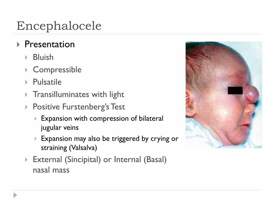
Presentation
Bluish
Compressible
Pulsatile
Transilluminates with light
Positive Furstenberg’s Test
Expansion with compression of bilateral
jugular veins
Expansion may also be triggered by crying or
straining (Valsalva)
External (Sincipital) or Internal (Basal)
nasal mass
Page 12
Encephalocele
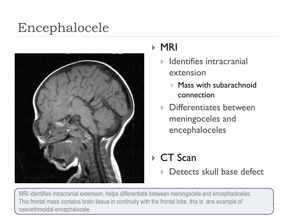
MRI
Identifies intracranial
extension
Mass with subarachnoid
connection
Differentiates between
meningoceles and
encephaloceles
CT Scan
Detects skull base defect
MRI identifies intracranial extension, helps differentiate between meningocele and encephaolceles.
This frontal mass contains brain tissue in continuity with the frontal lobe, this is ans example of
nasoethmoidal encephalocele
Page 13
Encephalocele
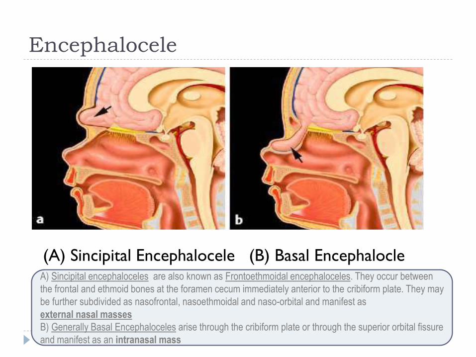
(A) Sincipital Encephalocele (B) Basal Encephalocle A) Sincipital encephaloceles are also known as Frontoethmoidal encephaloceles. They occur between
the frontal and ethmoid bones at the foramen cecum immediately anterior to the cribiform plate. They may
be further subdivided as nasofrontal, nasoethmoidal and naso-orbital and manifest as
external nasal masses
B) Generally Basal Encephaloceles arise through the cribiform plate or through the superior orbital fissure
and manifest as an intranasal mass
Page 15
Nasal Glioma
Heterotopic glial tissue that lacks a patent CSF
communication to subarachnoid space
Also known as
Nasal cerebral heterotopia
Glial heterotopia
Incidence
More common in males (3:2)
No familial tendency
Page 16
Nasal Glioma
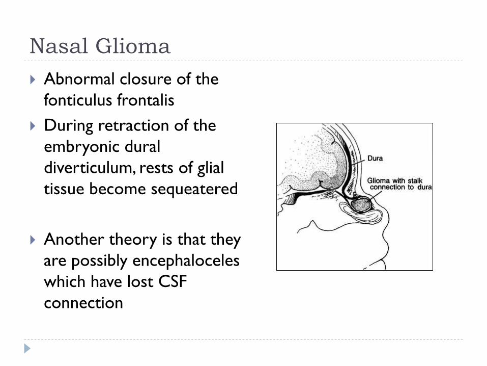
Abnormal closure of the
fonticulus frontalis
During retraction of the
embryonic dural
diverticulum, rests of glial
tissue become sequeatered
Another theory is that they
are possibly encephaloceles
which have lost CSF
connection
Page 17
Nasal Glioma Presentation
Extranasal (60%) Smooth, firm, non-compressible masses, skin telangiectasia
Glabella (most common), nasomaxillary suture line
Intranasal (30%) Polypoid, pale masses
Arise in the lateral nasal wall near the middle turbinate
Nasal septum (rare)
Combined (10%)
Do not transilluminate or enlarge with crying/straining
May be present at birth Grows in proportion with the child
Gliomas manifest as extranasal, intranasal or combined lesions. Extranasal
gliomas are smooth, firm, NONcompressible masses that occur most
commonly at the glabella but may arise at the side of the nose or the
nasomaxillary suture.
Page 18
Nasal Glioma
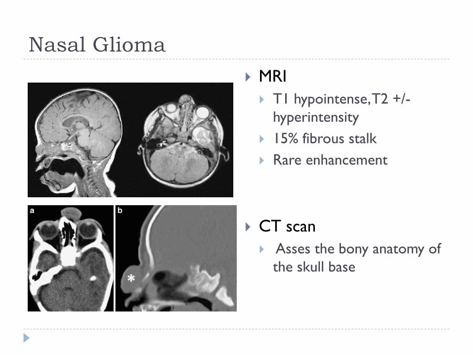
MRI
T1 hypointense, T2 +/-
hyperintensity
15% fibrous stalk
Rare enhancement
CT scan
Asses the bony anatomy of
the skull base
Page 19
Nasal Glioma
Intranasal glioma showing a fibrous stalk and are more commonly seen in those that are
intranasal (35%)
Page 20
Nasal Dermoids Nasal dermoids
1-3% of all dermoids
10-12% of head and neck dermoids. 61% of congenital midline nasal masses in kids
Embryology During development, dura projects through
the foramen cecum and attaches to skin
Separates from the nasal skin and retracts through foramen cecum
If there is a persistent attachment to underling fibrous tissue, nasal capsule or dura, epithelial elements are trapped in the prenasal space
Ectodermal and mesodermal elements Hair follicles, sebaceous glands, sweat glands, keratin,
squamous epithelial lining
Nasal dermoids account for 1-3% of all dermoids and approximatley 10-12% of head and neck dermoids.
Dermoids contain ectodermal and mesodermal embryonic elements. The latter include hair follicles,
sebaceous glands, and sweat glands and keratin debris. from deremoid cysyrs Dermoids lack glial
features of encephaloceles and gliomas
Page 21
Nasal Dermoids
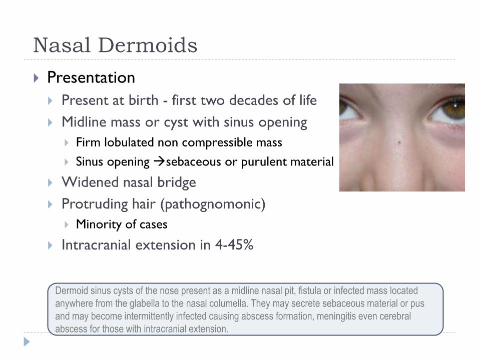
Presentation
Present at birth - first two decades of life
Midline mass or cyst with sinus opening
Firm lobulated non compressible mass
Sinus opening sebaceous or purulent material
Widened nasal bridge
Protruding hair (pathognomonic)
Minority of cases
Intracranial extension in 4-45%
Dermoid sinus cysts of the nose present as a midline nasal pit, fistula or infected mass located
anywhere from the glabella to the nasal columella. They may secrete sebaceous material or pus
and may become intermittently infected causing abscess formation, meningitis even cerebral
abscess for those with intracranial extension.
Page 22
Nasal Dermoids
Up to 50% have a fistula or sinus tract
Tract transverses via the cribiform
plate or foramen cecum
Tract may attach to dura, falx cerebri
or other intracranial structures
Cases with intracranial connection pose
an increased risk for meningitis or
cerebral abscesses
Page 23
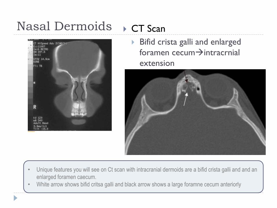
Nasal Dermoids CT Scan
Bifid crista galli and enlarged
foramen cecumintracrnial
extension
• Unique features you will see on Ct scan with intracranial dermoids are a bifid crista galli and and an
enlarged foramen caecum.
• White arrow shows bifid critsa galli and black arrow shows a large foramne cecum anteriorly
Page 24
Nasal Dermoids
MRI
Detects intracranial extension
T1 and T2 hyperintensity
The crista galli in infants is not ossified or contain bone marrow fat, thus a high-
intensity signal on T1-weighted images is suggestive of an intracranial dermoid
Page 25
Treatment of Nasal Gliomas,
Encephaloceles and Dermoid Cysts
Page 26
Surgical Treatment
Direct external excision
Elliptical incision around pit
Lacrimal probe is used to cannulate the tract to
guide dissection
A small diamond bur is used to drill around the
tract through the nasal bones
Nasal bones may be separated along the midline
and retracted laterally for better exposure
Allows access to dermoids that extend to the dura
and/or extending into the crista galli
Medial canthal approach (lynch), external
rhinoplasty, endoscopic resection
• Unique features you will see on Ct scan with intracranial dermoids are a bifid crista galli and and
an enlarged foramen caecum.
• White arrow shows bifid critsa galli and black arrow shows a large foramne cecum anteriorly
Page 27
Surgical Treatment
For Meningioceles, Gliomas limited to nasal cavity
Endoscopic repair with clipping the stalk
Defect is repaired with free mucosal grafts or mucoperichondrial flap
For Encephaloceles, Gliomas and Dermoids with intracranial
extension
Multidisciplinary approach
Transglabellar Subcranial Approach
Frontal craniotomy in combination with external
Rhinoplasty and lateral rhinotomy approach
Page 28
Developmental Errors of the
Central Midface
Nasolacrimal Duct Cyst and CNPAS
Page 29
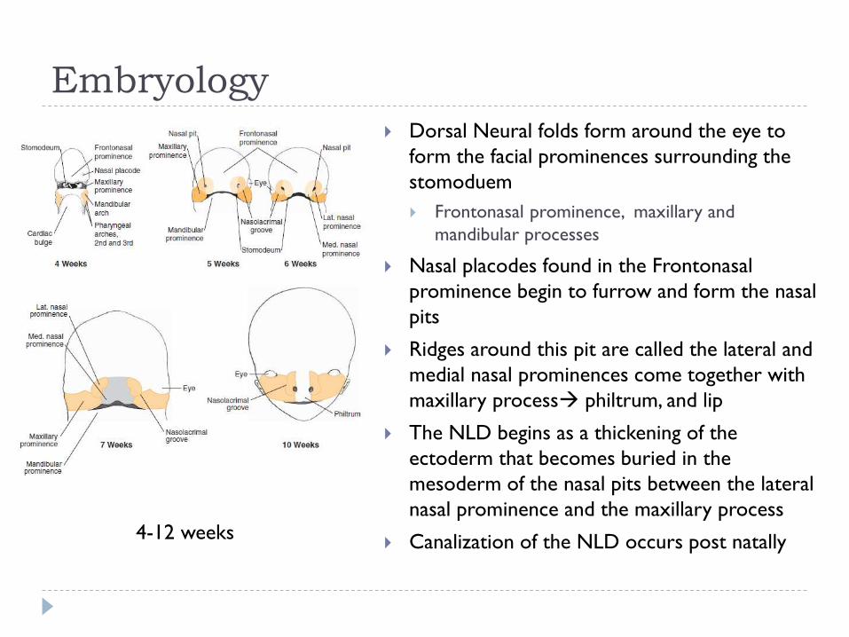
Embryology
Dorsal Neural folds form around the eye to
form the facial prominences surrounding the
stomoduem
Frontonasal prominence, maxillary and
mandibular processes
Nasal placodes found in the Frontonasal
prominence begin to furrow and form the nasal
pits
Ridges around this pit are called the lateral and
medial nasal prominences come together with
maxillary process philtrum, and lip
The NLD begins as a thickening of the
ectoderm that becomes buried in the
mesoderm of the nasal pits between the lateral
nasal prominence and the maxillary process
Canalization of the NLD occurs post natally 4-12 weeks
Page 30
Abnormal Development
Developmental Errors of the central midface
Nasolacrimal duct cyst
Congenital nasal pyriform aperture stenosis
Cleft lip
Most common
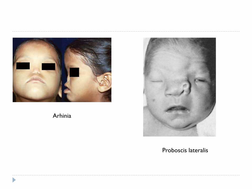
Arhinia
Congenital absence of the external nose and nasal airway
Polyrhinia
Double nose/accessory nostril
Septal, and nasal passage duplication +/-choanal atresia
Proboscis lateralis
Tubular sleeve of skin attached to the inner canthus of the orbit and
ipsilateral heminasal aplasia
Page 31
Arhinia
Proboscis lateralis
Page 32
Nasolacrimal Duct Cyst
Nasolacrimal duct development begins as a thickening of
the ectoderm that becomes buried in the mesoderm of
the nasal pits.
This buried ectoderm canalizes from superior to
inferiorly postnatally.
Failure of the nasolacrimal ectodermal tract to canalize
results in NLDC.
Page 33
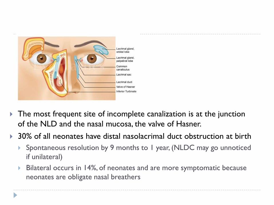
The most frequent site of incomplete canalization is at the junction
of the NLD and the nasal mucosa, the valve of Hasner.
30% of all neonates have distal nasolacrimal duct obstruction at birth
Spontaneous resolution by 9 months to 1 year, (NLDC may go unnoticed
if unilateral)
Bilateral occurs in 14%, of neonates and are more symptomatic because
neonates are obligate nasal breathers
Page 34
Nasolacrimal Duct Cyst
Presentation
Epiphora
Facial swelling
Nasal obstruction
Feeding difficulties
Respiratory distress (if bilateral)
Bluish/red discoloration
inferior to the medial
canthus
Nasal endoscopy will show
a mass below the inferior
turbinate
Page 35
Nasolacrimal Duct Cyst
Diagnosis is made on
physical exam/nasal
endoscopy
Imaging
Not required
Nasolacrimal duct mucocele. Coronal computed
tomographic images a–c show enlargement of the lacrimal
sac, distension of the nasolacrimal duct, and an intranasal
component in the inferior meatus, which corresponds to the
inferior extent of the left-sided mucocele
Page 36
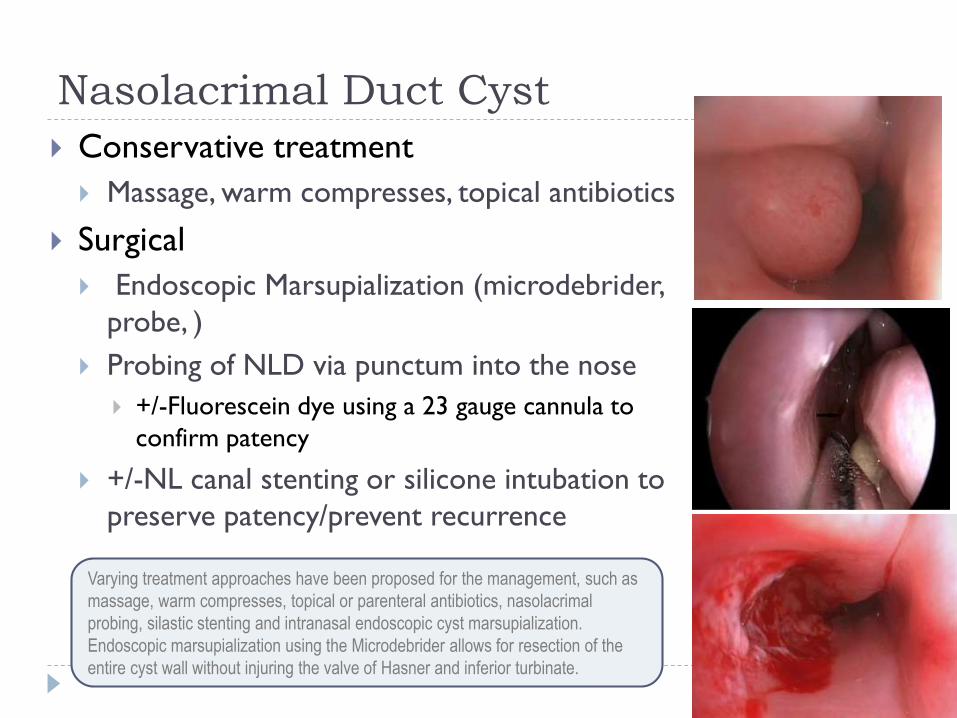
Nasolacrimal Duct Cyst
Conservative treatment
Massage, warm compresses, topical antibiotics
Surgical
Endoscopic Marsupialization (microdebrider,
probe, )
Probing of NLD via punctum into the nose
+/-Fluorescein dye using a 23 gauge cannula to
confirm patency
+/-NL canal stenting or silicone intubation to
preserve patency/prevent recurrence
Varying treatment approaches have been proposed for the management, such as
massage, warm compresses, topical or parenteral antibiotics, nasolacrimal
probing, silastic stenting and intranasal endoscopic cyst marsupialization.
Endoscopic marsupialization using the Microdebrider allows for resection of the
entire cyst wall without injuring the valve of Hasner and inferior turbinate.
Page 37
Congenital Nasal Pyriform Aperture
Stenosis (CNPAS)
Bony overgrowth of the nasal process of the maxilla
Pyriform aperture is the most anterior and narrowest part of the nasal cavity
Any change causing a decrease in this cross-sectional area results in exponential
increase in airway resistance resulting in nasal obstruction
CNPAS may occur in isolation or may manifest as part of holopronsencepahly
sequence
Failure of forebrain to divide into cerebral hemispheres, absence of anterior
pituitary, submucous cleft palate, hypoplastic maxillary sinuses, +/-prominent
mega incisor
Congenital nasal pyriform aperture stenosis (CNPAS) results from bony overgrowth of the nasal process
of the maxilla. The pyriform aperture is a pear-shaped bony inlet comprising the most anterior and
narrowest bony portion of the nasal airway; therefore, any overgrowth causes a decrease in cross-
sectional area with resultant exponential increase in airway resistance and associated obstruction
CNPAS ay occur in isolation or may be part of the holoprosencephaly spectrum of congenital midline
lesions.
Page 38
Congenital Nasal Pyriform Aperture
Stenosis (CNPAS)
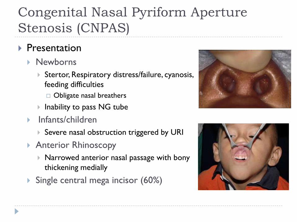
Presentation
Newborns
Stertor, Respiratory distress/failure, cyanosis,
feeding difficulties
Obligate nasal breathers
Inability to pass NG tube
Infants/children
Severe nasal obstruction triggered by URI
Anterior Rhinoscopy
Narrowed anterior nasal passage with bony
thickening medially
Single central mega incisor (60%)
Page 39
Congenital Nasal Pyriform Aperture
Stenosis (CNPAS)
Maxillofacial CT Scan
Confirms diagnosis
Width of pyriform aperture is defined as the
distance between the medial aspects of the
maxilla at the level of the inferior meatus.
<11mm in a full term infant (Belden et al.)
Study by Reeves et al.
suggests that the cutoff should be smaller than 11 mm because the
average pyriform aperture width for the control group in his study
was 10.1 mm and those with CPAS ranged between 5-6mm (avg
5.3mm)
Merea et al.
In his case series which included both premature and full-term
infants patients with CNPAS had a pyriform aperture of < 7 mm
(avg 5.6mm)
Page 40
Congenital Nasal Pyriform Aperture
Stenosis (CNPAS) (cont’d)
Axial CT is typically the imaging method of choice and confirms the
diagnosis. The width of the pyriform aperture is defined as the distance
between the medial aspects of the maxilla at the level of the inferior
meatus.
More recent study by Reeves et al (from MUSC). suggests that the cutoff
should be smaller than 11 mm because the average pyriform aperture
width for the control group (13 patients) was 10.1 mm with an average of
5.3mm in patients with CNPAS.[4] Patients in neonates and this case series
included both premature and full-term infants who had a pyriform aperture
of 7 mm or less with an average pyriform aperture width in line with those
reported by Reeves et al. (5.6 mm in our study vs. 5.3 mm).
Belden in 1999 evaluated CT features of CNPAS in 6 patients.
Page 41
Congenital Nasal Pyriform Aperture
Stenosis (CNPAS)
Treatment
Conservative
Medical treatment
Ciprofloxacin 0.3%/dexamethasone 0.1% drops
Decongestant
Nasal Saline
CPAP
Some authors suggest a width >5mm will respond well to medical
management if no underlying history of respiratory failure
Reeves et al.
All children with nasal obstruction receive a trial of medical management prior
to CT scanning to reduce radiation exposure
Some authors have suggested at a width > 5mm may be predictive of successful treatment
with medical management ciprodex, afrin and nasal saline. Some suggest this treatment in
any child with nasal obstruction and a concern for CNPAS prior to ct scanning to reduce
radiation risk.
Page 42
Congenital Nasal Pyriform Aperture
Stenosis (CNPAS)
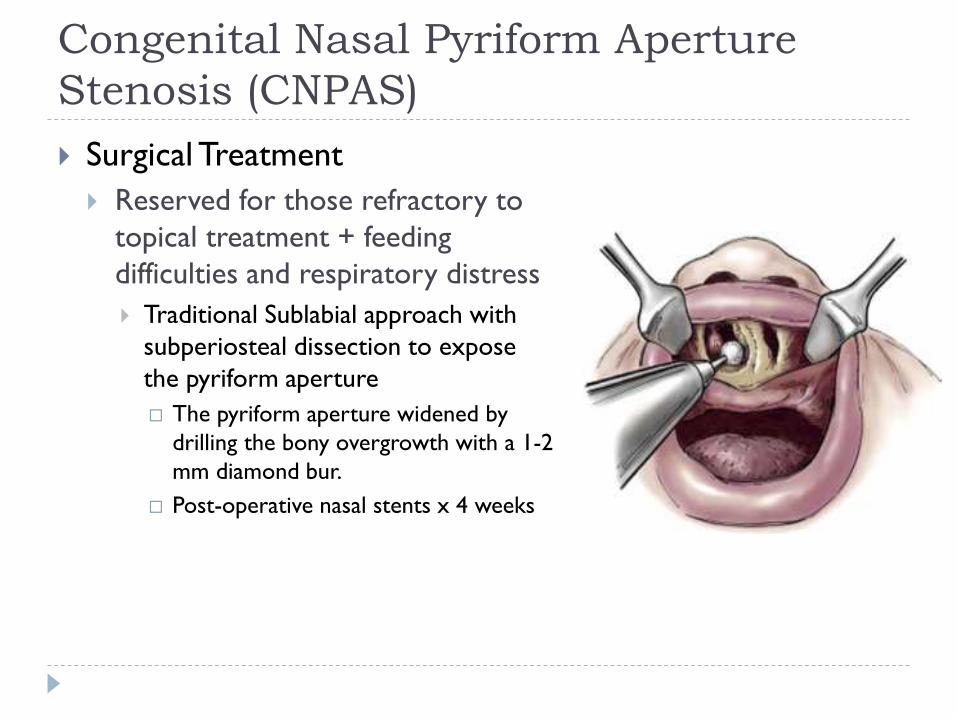
Surgical Treatment
Reserved for those refractory to
topical treatment + feeding
difficulties and respiratory distress
Traditional Sublabial approach with
subperiosteal dissection to expose
the pyriform aperture
The pyriform aperture widened by
drilling the bony overgrowth with a 1-2
mm diamond bur.
Post-operative nasal stents x 4 weeks
Page 43
Congenital Nasal Pyriform Aperture
Stenosis (CNPAS)
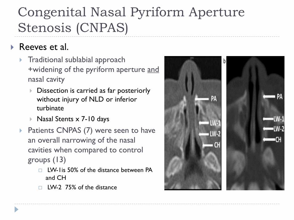
Reeves et al.
Traditional sublabial approach
+widening of the pyriform aperture and
nasal cavity
Dissection is carried as far posteriorly
without injury of NLD or inferior
turbinate
Nasal Stents x 7-10 days
Patients CNPAS (7) were seen to have
an overall narrowing of the nasal
cavities when compared to control
groups (13)
LW-1is 50% of the distance between PA
and CH
LW-2 75% of the distance
Page 44
Congenital Nasal Pyriform Aperture
Stenosis (CNPAS)
Site Mean control (SD) Mean CPAS (SD) p-Values
Pyriform aperture
(PA)10.1 mm (0.30) 5.3 mm (0.08) p < 0.01
LW-1 13.5 mm(0.28) 8.7 mm (0.2) p < 0.01
LW-2 12.5 mm (0.23) 10.1 mm (0.13) p = 0.02
Choana (CH) 11.7 mm (0.19) 10.9 mm (0.24) p = 0.46
Average width of pyriform aperture, lateral nasal wall and choana
when comparing CPAS to control patients
Reeves et al.
Page 45
Congenital Nasal Pyriform Aperture
Stenosis (CNPAS)
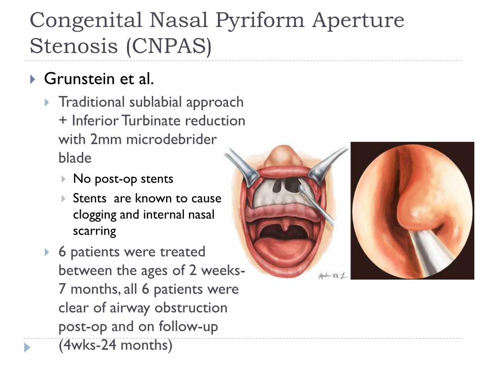
Grunstein et al.
Traditional sublabial approach
+ Inferior Turbinate reduction
with 2mm microdebrider
blade
No post-op stents
Stents are known to cause
clogging and internal nasal
scarring
6 patients were treated
between the ages of 2 weeks-
7 months, all 6 patients were
clear of airway obstruction
post-op and on follow-up
(4wks-24 months)
Page 46
Conclusion
Neonates are obligate nasal breathers
Radiologic findings assist in determining diagnosis and
surgical planning
MRI is preferred to assess for intracranial extension
CT is used to asses the skull base
Page 47
Discussant: Harold S. Pine, MD, FAAP, FACS
An excellent review of pediatric nasal masses. It all looks so nice and
pretty when it’s presented in a Powerpoint presentation but when these
things happen in real life, it’s difficult, it’s hard to figure out what exactly is
going on and the approach is not always so clear cut. When we’re faced
with kids with some sort of intranasal mass that looks a little bit weird
don’t be lured into doing a biopsy without appropriately working the kid
up. Sometimes in the real world situations arise where we get pressure
from other people that don’t know better to do things that we know are
not in the best interests of the patient.
Don’t underestimate a little bit of patience, mother nature and medical
therapy. I have lots of experience with our own NICU here where they’re
very nervous and up in arms and wanting something to be done urgently to
fix the problem, when in fact a few days of Ciprodex nose drops, some
saline and a little bit of time can get a lot of these kids through the trouble
spot. So I certainly agree with how MUSC does it.
Continued next page
Page 48
Discussant: Harold S. Pine, MD, FAAP, FACS
Finally, just a thought about a patient I saw over in Asia where these things
are more common, and I sent out a picture to you of this little kid with a
large nasal mass. It really struck me how dependent we all are here in the
U.S. with all of our teammates. I had the scans and yet looking at the scans
I couldn’t be sure exactly what this was. It was frustrating not to have a
good neuroradologist to help me say this is clearly an encephalocele or this
is clearly a glioma.
In the end my recommendation was that this is probably an encephalocele
or a glioma, and you will probably need a neurosurgeon to help with this.
While that seems like common sense here, in other countries, getting that
cooperation and coordination is not so easy to accomplish, especially when
there are islands of specialty care hospitals. In the end, the ENT guys tried
to take out this nasal mass going just right over the mass and it turned into
a horrible freak show over the ensuing months with what sounded like
infections and CSF leaks. I don’t have any further followup at this point, but
it goes to show you that some thought before you go in and operate can
probably save you some grief down the road.
Page 49
Bibliography 1. Snyderman et al. Endoscopic endonasal surgery for dermoids. Otolaryngol Clin N Am 44(2011) 981-987.
2. Bonne et al. Endoscopic approach for removal of intranasal nasal glial heterotopias. Rhinology 50 (2012)
211-217.
3. Reeves et al. Nasal cavity dimensions in congenital pyriform aperture stenosis. International Journal od
pediatric Otorhinolaryngology. 77(2013) 1830-1832.
4. Merea et al. CPAS: Surgical Approach with combined sublabial bone resection and inferior turbinate
reduction without stents. The laryngoscope. March 2014.
5. Gnagi SH, Schraff SA. Nasal Obstruction in Newborns. Ped Clin N Am 2013
6. Cheng J, Kazahaya K. Management of Pediatric Nasal Dermoids with Intracranial Extension by Direct
Excision. Otol-Head Neck S 2013; 148(4): 694-6.
7. Duncan NO. Combined Approach for Complete Excision of Congenital Nasal Masses in Children. Oper
Tech Head Neck S 1994; 5(1): 18-21. Yuca K, Varsak YK. Thornwaldt’s Cyst. Eur J Gen Med 2012;
9(Suppl 1) 26-29
8. Skinner LJ et al. Radiology Quiz Case. Thornwaldt cyst. Arch Otolaryngol Head Neck Surg 2003; 129
(10):1137-8.
9. Wright C et al. Evaluation of Congenital Midline Nasal Masses. Quinn Online textbook of Otolaryngology.
2006.
10. Wootten and Elluru. Congenital malformations of the nose. Cummings Otolaryngology Head and Neck
Surgery. 5th Ed. Pages 2686-2696.
Page 50
Congenital Anomalies of the Nose
Resident Physician: Sharon Ramos, MD
Faculty Mentor & Discussant: Harold S. Pine, MD, FAAP, FACS
The University of Texas Medical Branch – UTMB Health
Department of Otolaryngology
Grand Rounds Presentation
February 18, 2015
Series Editor: Francis B. Quinn, Jr., MD, FACS -- Archivist: Melinda Stoner Quinn, MSICS


![CASE REPORT Open Access A prominent crista terminalis ...€¦ · finding of a prominent crista terminalis can mimic a right atrial mass, such as a tumor or thrombus [2,3]. Atrial](https://static.documents.pub/doc/80x56/60914c5090def22b9158119d/case-report-open-access-a-prominent-crista-terminalis-finding-of-a-prominent.jpg)


